Mitochondrial Fission, Fusion, and Stress
Mitochondrial Fission, Fusion, and Stress
Mitochondrial Fission, Fusion, and Stress

You also want an ePaper? Increase the reach of your titles
YUMPU automatically turns print PDFs into web optimized ePapers that Google loves.
1062<br />
REVIEW<br />
<strong>Mitochondrial</strong> <strong>Fission</strong>, <strong>Fusion</strong>,<br />
<strong>and</strong> <strong>Stress</strong><br />
Richard J. Youle 1 * <strong>and</strong> Alex<strong>and</strong>er M. van der Bliek 2 *<br />
<strong>Mitochondrial</strong> fission <strong>and</strong> fusion play critical roles in maintaining functional mitochondria when cells<br />
experience metabolic or environmental stresses. <strong>Fusion</strong> helps mitigate stress by mixing the contents<br />
of partially damaged mitochondria as a form of complementation. <strong>Fission</strong> is needed to create new<br />
mitochondria, but it also contributes to quality control by enabling the removal of damaged mitochondria<br />
<strong>and</strong> can facilitate apoptosis during high levels of cellular stress. Disruptions in these processes affect<br />
normal development, <strong>and</strong> they have been implicated in neurodegenerative diseases, such as Parkinson’s.<br />
Mitochondria are double-membrane–<br />
bound subcellular organelles that provide<br />
a host of metabolic functions,<br />
including energy production through oxidative<br />
phosphorylation. <strong>Mitochondrial</strong> morphologies<br />
vary widely among different cell types. Fibroblast<br />
mitochondria, for example, are usually long<br />
filaments (1 to 10 mm inlengthwithafairly<br />
constant diameter of ~700 nm), whereas hepatocyte<br />
mitochondria are more uniformly spheres or<br />
ovoids. When mitochondria are viewed in live<br />
cells, it becomes immediately apparent that their<br />
morphologies are far from static. Their shapes<br />
change continually through the combined actions<br />
of fission, fusion, <strong>and</strong> motility. Rapid fission <strong>and</strong><br />
fusion of mitochondria in cultured fibroblasts<br />
allows for the complete redistribution of mitochondrial<br />
green fluorescent protein (GFP) from<br />
one mitochondrion to all the other mitochondria<br />
of a cell within an hour. The wide range of<br />
mitochondrial lengths observed in different cell<br />
types <strong>and</strong> under different conditions results from<br />
changes in the balance between the rates of mitochondrial<br />
fission <strong>and</strong> fusion. Here, we discuss<br />
how fission <strong>and</strong> fusion contribute to mitochondrial<br />
quality control <strong>and</strong> the responses of mammalian<br />
cells to stress.<br />
<strong>Mitochondrial</strong> <strong>Fusion</strong> <strong>and</strong> <strong>Fission</strong> Proteins<br />
<strong>Mitochondrial</strong> fission <strong>and</strong> fusion processes are<br />
both mediated by large guanosine triphosphatases<br />
(GTPases) in the dynamin family that are well<br />
conserved between yeast, flies, <strong>and</strong> mammals<br />
(1). Their combined actions divide <strong>and</strong> fuse the<br />
two lipid bilayers that surround mitochondria.<br />
The mitochondrial inner membrane, which encloses<br />
the matrix, is folded into cristae that contain<br />
membrane-bound oxidative phosphorylation<br />
enzyme complexes <strong>and</strong> the bulk of the soluble<br />
electron transport proteins such as cytochrome c,<br />
whereas the smooth mitochondrial outer mem-<br />
1 Biochemistry Section, Surgical Neurology Branch, National Institute<br />
of Neurological Disorders <strong>and</strong> Stroke, National Institutes<br />
of Health, Bethesda, MD 20892, USA. 2 Department of Biological<br />
Chemistry, David Geffen School of Medicine at University of<br />
California–Los Angeles, Los Angeles, CA 90095, USA.<br />
*To whom correspondence should be addressed. E-mail: youler@<br />
ninds.nih.gov (R.J.Y.); avan@mednet.ucla.edu (A.M.v.d.B.)<br />
brane encapsulates the inner membrane <strong>and</strong> an<br />
intermembrane space.<br />
<strong>Fission</strong> is mediated by a cytosolic dynamin<br />
family member (Drp1 in worms, flies, <strong>and</strong> mammals<br />
<strong>and</strong> Dnm1 in yeast). Drp1 is recruited from<br />
the cytosol to form spirals around mitochondria<br />
that constrict to sever both inner <strong>and</strong> outer membranes.<br />
Yeast share with mammals this core function<br />
of Drp1 but have distinct accessory proteins.<br />
Mdv1 recruits Dnm1 to mitochondrial fission<br />
sites in yeast, whereas Mid49, Mid51, <strong>and</strong> Mff<br />
recruit Drp1 to mitochondria in mammals (2),<br />
often at sites where mitochondria make contact<br />
with the endoplasmic reticulum (3). <strong>Fusion</strong> between<br />
mitochondrial outer membranes is mediated<br />
by membrane-anchored dynamin family members<br />
named Mfn1 <strong>and</strong> Mfn2 in mammals, whereas<br />
fusion between mitochondrial inner membranes<br />
is mediated by a single dynamin family member<br />
called Opa1 in mammals. <strong>Mitochondrial</strong> fission<br />
<strong>and</strong> fusion machineries are regulated by proteolysis<br />
<strong>and</strong> posttranslational modifications (1).<br />
<strong>Mitochondrial</strong> fission is essential for growing<br />
<strong>and</strong> dividing cells to populate them with adequate<br />
numbers of mitochondria. It has been less clear<br />
why mitochondrial fission <strong>and</strong> fusion are also<br />
needed for nonproliferating cells, but the importance<br />
of these processes is evident from nonproliferating<br />
neurons, which cannot survive<br />
Damaged<br />
without mitochondrial fission, <strong>and</strong> from two human<br />
diseases, dominant optic atrophy <strong>and</strong> Charcot<br />
Marie Tooth disease type 2A, which are caused<br />
by fusion defects. The importance of mitochondrial<br />
fusion for embryogenesis was shown with<br />
Mfn1 <strong>and</strong> Mfn2 knock-out mice, which die in<br />
utero at midgestation because of a placental deficiency,<br />
whereas the Mfn1 Mfn2 double knockout<br />
mice die even earlier in development (4).<br />
Mouse embryo fibroblasts (MEFs) derived from<br />
the double knock-out mice do survive in culture,<br />
despite a complete absence of fusion, but some of<br />
their mitochondria display a reduced mitochondrial<br />
DNA (mtDNA) copy number <strong>and</strong> lose membrane<br />
potential, causing problems with adenosine<br />
triphosphate (ATP) synthesis (5). <strong>Mitochondrial</strong><br />
fusion is therefore not absolutely essential for cell<br />
survival in vitro, but it is required for embryonic<br />
development <strong>and</strong> for cell survival at later stages<br />
in development (4). These differential requirements<br />
for fusion may stem from higher dem<strong>and</strong>s<br />
on oxidative metabolism in different cell types or<br />
on other functions that are indirectly affected by<br />
fusion, such as mitochondrial motility in neurons.<br />
<strong>Fusion</strong> Promotes Complementation<br />
Between Damaged Mitochondria<br />
Mitochondria have their own small circular genomes,<br />
encoding select subunits of ATP synthesis<br />
<strong>and</strong> electron transport proteins that form oxidative<br />
phosphorylation complexes with other subunits<br />
encoded by the nuclear genome, as well as<br />
transfer <strong>and</strong> ribosomal RNAs (tRNAs <strong>and</strong> rRNAs)<br />
needed for their translation. A single somatic cell<br />
can have thous<strong>and</strong>s of copies of these genomes,<br />
whicharegroupedinprotein-richcomplexescalled<br />
nucleoids, with between one <strong>and</strong> eight genome<br />
copies per nucleoid (6). Mutations <strong>and</strong> deletions<br />
that occasionally arise in mitochondrial DNA<br />
yield a heteroplasmic mixture of wild-type <strong>and</strong><br />
mutant mitochondrial genomes within one cell.<br />
Maternal inheritance of these mutations can cause<br />
mitochondrial diseases, such as mitochondrial<br />
encephalomyopathy with lactic acidosis <strong>and</strong><br />
strokelike episodes (MELAS) <strong>and</strong> myoclonus<br />
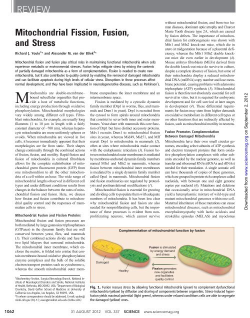
Complementation of mitochondrial function by fusion<br />
Healthy<br />
<strong>Fusion</strong> is stimulated<br />
by energy dem<strong>and</strong><br />
<strong>and</strong> stress<br />
<strong>Fission</strong> generates<br />
new organelles<br />
<strong>and</strong> facilitates<br />
quality control<br />
Fig. 1. <strong>Fusion</strong> rescues stress by allowing functional mitochondria (green) to complement dysfunctional<br />
mitochondria (yellow) by diffusion <strong>and</strong> sharing of components between organelles. <strong>Stress</strong>-induced hyperfusion<br />
yields maximal potential (light green), whereasunderrelaxedconditionscellsareabletosegregate<br />
the damaged (yellow) ones.<br />
31 AUGUST 2012 VOL 337 SCIENCE www.sciencemag.org<br />
on September 2, 2012<br />
www.sciencemag.org<br />
Downloaded from