Quantifying the material and structural determinants of bone strength
Quantifying the material and structural determinants of bone strength
Quantifying the material and structural determinants of bone strength

You also want an ePaper? Increase the reach of your titles
YUMPU automatically turns print PDFs into web optimized ePapers that Google loves.
3<br />
<strong>Quantifying</strong> <strong>the</strong> <strong>material</strong> <strong>and</strong> <strong>structural</strong> <strong>determinants</strong><br />
<strong>of</strong> <strong>bone</strong> <strong>strength</strong><br />
Mary L. Bouxsein, PhD a, *, Ego Seeman, MD b<br />
a Assistant Pr<strong>of</strong>essor <strong>of</strong> Orthopaedic Surgery, Orthopedic Biomechanics Laboratory, Beth Israel Deaconess Medical Center <strong>and</strong><br />
Department <strong>of</strong> Orthopaedic Surgery, Harvard Medical School, RN115, 330 Brookline Ave, Boston, MA 02215, USA<br />
b Pr<strong>of</strong>essor <strong>of</strong> Medicine, Austin Health, University <strong>of</strong> Melbourne, Melbourne, Australia<br />
Keywords:<br />
<strong>bone</strong> <strong>strength</strong><br />
<strong>bone</strong> quality<br />
microarchitecture<br />
quantitative computed tomography<br />
magnetic resonance imaging<br />
finite element analysis<br />
Best Practice & Research Clinical Rheumatology 23 (2009) 741–753<br />
Contents lists available at ScienceDirect<br />
Best Practice & Research Clinical<br />
Rheumatology<br />
journal homepage: www.elsevierhealth.com/berh<br />
The ability <strong>of</strong> a <strong>bone</strong> to resist fracture depends on <strong>the</strong> amount <strong>of</strong><br />
<strong>bone</strong> present, <strong>the</strong> spatial distribution <strong>of</strong> <strong>the</strong> <strong>bone</strong> mass as cortical<br />
<strong>and</strong> trabecular <strong>bone</strong> <strong>and</strong> <strong>the</strong> intrinsic properties <strong>of</strong> <strong>the</strong> <strong>bone</strong><br />
<strong>material</strong>. Whereas low areal <strong>bone</strong> mineral density (aBMD) predicts<br />
fractures, its sensitivity <strong>and</strong> specificity is low, as over 50% <strong>of</strong> fractures<br />
occur in persons without osteoporosis by BMD testing <strong>and</strong><br />
most women with osteoporosis do not sustain a fracture. New<br />
non-invasive imaging techniques, including three-dimensional<br />
(3D) assessments <strong>of</strong> <strong>bone</strong> density <strong>and</strong> geometry, microarchitecture<br />
<strong>and</strong> integrated measurements <strong>of</strong> <strong>bone</strong> <strong>strength</strong> such as finite<br />
element analysis (FEA), provide estimates <strong>of</strong> <strong>bone</strong> <strong>strength</strong> that<br />
can be used to increase <strong>the</strong> sensitivity <strong>and</strong> specificity <strong>of</strong> fracture<br />
risk assessment. Initial observations have shown that <strong>the</strong>se techniques<br />
provide information that will improve our underst<strong>and</strong>ing <strong>of</strong><br />
<strong>the</strong> pathophysiology <strong>of</strong> skeletal fragility <strong>and</strong> suggest that <strong>the</strong>se<br />
techniques are likely to have a role in <strong>the</strong> clinical management <strong>of</strong><br />
individuals at risk for fracture.<br />
Ó 2009 Elsevier Ltd. All rights reserved.<br />
Fractures due to osteoporosis are common, with one in three women <strong>and</strong> one in five men over age<br />
50 predicted to suffer a fracture in <strong>the</strong>ir lifetime [1]. As <strong>the</strong> proportion <strong>of</strong> elderly in <strong>the</strong> population is<br />
growing exponentially, <strong>the</strong> number <strong>of</strong> fractures is expected to double or triple in <strong>the</strong> next 50 years.<br />
While drug <strong>the</strong>rapy is efficacious in reducing <strong>the</strong> risk <strong>of</strong> vertebral fracture, many challenges remain as<br />
few drugs have been shown to reduce <strong>the</strong> risk <strong>of</strong> non-vertebral fractures, <strong>and</strong> usually this risk reduction<br />
* Corresponding author. Tel.: þ1 617 667 2940; Fax: þ1 617 667 7175.<br />
E-mail address: mbouxsei@bidmc.harvard.edu (M.L. Bouxsein).<br />
1521-6942/$ – see front matter Ó 2009 Elsevier Ltd. All rights reserved.<br />
doi:10.1016/j.berh.2009.09.008
742<br />
is no more that 20%, <strong>and</strong> around 40–59% for hip fractures. Moreover, no drugs have been shown to<br />
reduce <strong>the</strong> risk <strong>of</strong> hip fractures in women or men over 75 years <strong>of</strong> age [2]. In addition, access to <strong>and</strong><br />
compliance with osteoporosis <strong>the</strong>rapies are poor [3].<br />
Osteoporosis is defined as ‘‘a disease characterized by low <strong>bone</strong> mass <strong>and</strong> microarchitectural<br />
deterioration <strong>of</strong> <strong>bone</strong> tissue, leading to enhanced <strong>bone</strong> fragility <strong>and</strong> a consequent increase in fracture<br />
risk.’’[4] A fracture occurs when <strong>the</strong> external force applied to a <strong>bone</strong> exceeds its <strong>strength</strong>. For a given<br />
loading condition, <strong>the</strong> ability <strong>of</strong> a <strong>bone</strong> to resist fracture depends on <strong>the</strong> amount <strong>of</strong> <strong>bone</strong>, <strong>the</strong> spatial<br />
distribution <strong>of</strong> <strong>the</strong> <strong>bone</strong> mass <strong>and</strong> <strong>the</strong> intrinsic properties <strong>of</strong> <strong>the</strong> <strong>material</strong>s that comprise <strong>the</strong> <strong>bone</strong> [5].<br />
Presently, diagnosis <strong>of</strong> osteoporosis is based on measurement <strong>of</strong> areal <strong>bone</strong> mineral density (aBMD)<br />
by dual-energy X-ray absorptiometry (DXA). While aBMD is a predictor <strong>of</strong> fracture risk [6], it lacks<br />
sensitivity <strong>and</strong> specificity; most women with osteoporosis do not sustain a fracture <strong>and</strong> over 50% <strong>of</strong><br />
women who sustain a fracture do not have osteoporosis [7–9]. Moreover, changes in BMD following<br />
<strong>the</strong>rapy explain only 4–30% <strong>of</strong> <strong>the</strong> fracture risk reduction [10–13].<br />
These observations have focussed attention on o<strong>the</strong>r factors that influence <strong>bone</strong> <strong>strength</strong> [14,15]<br />
<strong>and</strong> have motivated development <strong>of</strong> new technologies to assess <strong>the</strong>se factors. In this review, <strong>the</strong><br />
general considerations for <strong>the</strong> non-invasive assessment <strong>of</strong> <strong>the</strong> <strong>material</strong> <strong>and</strong> <strong>structural</strong> <strong>determinants</strong> <strong>of</strong><br />
<strong>bone</strong> <strong>strength</strong> are presented <strong>and</strong> developments in this area are critically reviewed. The focus <strong>of</strong><br />
attention here is on technologies that can be applied clinically.<br />
Criteria for evaluating new technologies<br />
Imaging technologies must be accurate <strong>and</strong> precise <strong>and</strong> must have established quality control<br />
procedures, st<strong>and</strong>ardised data acquisition <strong>and</strong> analysis, as well as methods for cross-calibration <strong>of</strong><br />
devices at different clinical centres [16]. Data that help define <strong>the</strong> clinical utility <strong>of</strong> a new imaging<br />
technique include: (1) sex- <strong>and</strong> age-specific reference data; (2) assessment <strong>of</strong> disease severity; (3)<br />
associations with fracture risk in untreated subjects; <strong>and</strong> (4) changes in <strong>the</strong> measurement with<br />
worsening <strong>of</strong> <strong>the</strong> disease or with <strong>the</strong>rapeutic intervention. Ultimately, imaging techniques must have<br />
clinical utility d <strong>the</strong>y must identify individuals at risk for fracture with high sensitivity, assist in<br />
monitoring <strong>of</strong> <strong>the</strong>rapy <strong>and</strong> provide potential surrogates for anti-fracture efficacy.<br />
The techniques should also provide insights into <strong>the</strong> pathogenesis <strong>of</strong> disease. This is a particularly<br />
important concern because some techniques rely on models which simplify <strong>the</strong> structure <strong>of</strong> <strong>bone</strong>. As<br />
such <strong>the</strong>se simplifications may produce misleading notions regarding <strong>the</strong> pathogenesis <strong>of</strong> disease <strong>and</strong><br />
<strong>the</strong> effects <strong>of</strong> treatment. There are many examples <strong>of</strong> this, <strong>and</strong> <strong>the</strong>se limitations are discussed in this<br />
article.<br />
Imaging Techniques<br />
M.L. Bouxsein, E. Seeman / Best Practice & Research Clinical Rheumatology 23 (2009) 741–753<br />
Dual-energy X-ray absorptiometry (DXA) <strong>and</strong> aBMD<br />
Introduced about 25 years ago, dual-energy X-ray absorptiometry (DXA) provides a quantitative<br />
assessment <strong>of</strong> mineralised <strong>bone</strong> mass at <strong>the</strong> axial <strong>and</strong> appendicular skeleton in vivo. This technique<br />
measures <strong>the</strong> attenuation <strong>of</strong> photons <strong>of</strong> two different energies during radiation transmission. Bone<br />
mineral content (BMC, g) <strong>and</strong> aBMD (g cm –2 ) <strong>of</strong> a region <strong>of</strong> interest are obtained. As low aBMD is<br />
a strong risk factor for fractures [17], this technique provided <strong>the</strong> basis for <strong>the</strong> World Health Organization<br />
(WHO)’s guidelines for diagnosis <strong>of</strong> osteoporosis [18]. Advantages to DXA include a low radiation<br />
exposure, excellent precision, low cost, ease <strong>of</strong> use <strong>and</strong> short measurement times. However, <strong>the</strong><br />
measurements are two-dimensional (2D) so larger <strong>bone</strong>s may have higher aBMD than smaller <strong>bone</strong>s<br />
because <strong>of</strong> differences in size [19]; <strong>the</strong> <strong>bone</strong> with <strong>the</strong> lower BMD may not have gained less or lost more<br />
<strong>bone</strong>. DXA also does not distinguish cortical <strong>and</strong> cancellous <strong>bone</strong>. Nor do changes in aBMD provide<br />
information regarding <strong>the</strong> morphological basis <strong>of</strong> that change; aBMD may decrease more in one person<br />
than ano<strong>the</strong>r because resorption from <strong>the</strong> endosteal envelope is greater <strong>and</strong>/or periosteal apposition is<br />
less. Bone loss may occur mainly from trabecular <strong>bone</strong> or from <strong>the</strong> intracortical or endocortical surfaces<br />
or from both. DXA cannot assess 3D geometry or trabecular architecture. Fur<strong>the</strong>rmore, measurements<br />
are subject to artefacts due to degenerative changes such as osteophytes <strong>and</strong> aortic calcification. Thus,
M.L. Bouxsein, E. Seeman / Best Practice & Research Clinical Rheumatology 23 (2009) 741–753 743<br />
although DXA is currently <strong>the</strong> gold st<strong>and</strong>ard for clinical assessment <strong>of</strong> fracture risk, its shortcomings<br />
are increasingly being recognised.<br />
DXA in <strong>the</strong> assessment <strong>of</strong> <strong>bone</strong> structure d hip <strong>structural</strong> analysis (HSA)<br />
Bone geometry, an important determinant <strong>of</strong> <strong>bone</strong> <strong>strength</strong>, has been assessed non-invasively by<br />
several methods including radiogrammetry, automated DXA-based analysis <strong>of</strong> X-ray attenuation<br />
pr<strong>of</strong>iles (termed ‘hip <strong>structural</strong> analysis’, HSA) <strong>and</strong> 3D imaging methods such as computed tomography<br />
or magnetic resonance imaging.<br />
HSA has been widely used because <strong>structural</strong> properties can be estimated from routine DXA scans, <strong>and</strong><br />
<strong>the</strong> s<strong>of</strong>tware needed to compute <strong>the</strong>se variables is now provided by several <strong>of</strong> <strong>the</strong> DXA manufacturers.<br />
HSA uses data from 2D DXA scans to derive measurements <strong>of</strong> <strong>bone</strong> geometry at <strong>the</strong> proximal femur,<br />
including <strong>the</strong> amount <strong>and</strong> distribution <strong>of</strong> <strong>bone</strong> mass, <strong>bone</strong> morphology <strong>and</strong> indices <strong>of</strong> <strong>bone</strong> <strong>strength</strong> such<br />
as section modulus (an index <strong>of</strong> resistance to bending), buckling ratio <strong>and</strong> <strong>bone</strong> area (an index <strong>of</strong><br />
resistance to compression) [20]. However, some <strong>of</strong> <strong>the</strong>se indices are derived under <strong>the</strong> assumption that<br />
<strong>the</strong> <strong>bone</strong> cross section is circular at <strong>the</strong> femoral neck <strong>and</strong> shaft, <strong>and</strong> elliptical at <strong>the</strong> inter-trochanteric<br />
region, that <strong>the</strong> tissue mineral density is constant <strong>and</strong> that cortical <strong>and</strong> trabecular <strong>bone</strong> in <strong>the</strong> cross<br />
section are a constant proportion [21]. Moreover, <strong>the</strong> method is limited to analyses <strong>of</strong> a single plane.<br />
HSA has been used to examine effects <strong>of</strong> anti-resorptive <strong>and</strong> anabolic <strong>the</strong>rapies on femoral<br />
geometry [21–25]. However, anti-resorptive <strong>the</strong>rapies increase tissue mineral density [26–29],<br />
whereas teriparatide may decrease it [30]. An increase in <strong>the</strong> amount <strong>of</strong> mineral per unit <strong>bone</strong> volume<br />
is ‘seen’ by HSA as an increase in <strong>the</strong> cross-sectional area <strong>of</strong> <strong>bone</strong> <strong>material</strong>, leading an investigator to<br />
infer that <strong>the</strong>re has ei<strong>the</strong>r been periosteal apposition <strong>and</strong>/or endocortical apposition produced by<br />
antiresorptive agents; effects that are not produced by this class <strong>of</strong> drug. Thus, interpretation <strong>of</strong> HSA<br />
from studies where mineralisation density changes is difficult <strong>and</strong> suspect.<br />
Several studies have used this method to provide information regarding age-, race- <strong>and</strong> sex-related<br />
differences in femoral geometry that may contribute to hip fracture risk [31–39]; effects <strong>of</strong> physical<br />
activity <strong>and</strong> hormone status on femoral geometry [40,41]; <strong>and</strong> genetic <strong>determinants</strong> <strong>of</strong> femoral<br />
structure [42–44]. Because some <strong>of</strong> <strong>the</strong> assumptions underlying HSA have not been tested across ages<br />
<strong>and</strong> populations, conclusions from <strong>the</strong>se studies should be viewed with caution.<br />
Several prospective studies report that HSA-derived measures <strong>of</strong> femoral geometry are associated<br />
with risk <strong>of</strong> hip fracture [45–48]. However, it is unclear whe<strong>the</strong>r HSA provides information about<br />
fracture risk that is independent <strong>of</strong> BMD. This is likely because femoral BMD <strong>and</strong> HSA-derived <strong>structural</strong><br />
properties are highly correlated because <strong>the</strong> same attenuation pr<strong>of</strong>ile is used to compute <strong>the</strong><br />
measurements. Thus, conclusions regarding independent contributions <strong>of</strong> <strong>bone</strong> density <strong>and</strong> geometry<br />
to femoral fragility are problematic using HSA.<br />
We do not recommend <strong>the</strong> use <strong>of</strong> this technique because <strong>of</strong> <strong>the</strong> uncertainties <strong>and</strong> <strong>the</strong> potentially<br />
misleading notions <strong>of</strong> <strong>bone</strong> physiology that can arise if <strong>the</strong> results are accepted on face value. Moreover,<br />
o<strong>the</strong>r options are available for directly measuring <strong>the</strong> 3D geometry, such as magnetic resonance<br />
imaging (MRI), computed tomography (CT) <strong>and</strong> high-resolution peripheral quantitative computed<br />
tomography (HR-pQCT). These are being used in clinical studies to identify <strong>the</strong> relationships between<br />
geometry, <strong>bone</strong> density <strong>and</strong> fracture risk.<br />
Quantitative computed tomography (QCT)<br />
In QCT, <strong>the</strong> X-ray source <strong>and</strong> detector rotate in synchronised fashion around <strong>the</strong> subject. Algorithms<br />
are used to reconstruct <strong>the</strong> attenuation data into 3D images. Use <strong>of</strong> a <strong>bone</strong> mineral (or hydroxyapatite)<br />
phantom allows calibration <strong>of</strong> <strong>the</strong> data, providing a measurement <strong>of</strong> <strong>bone</strong> density that is independent<br />
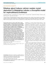
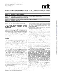
<strong>of</strong> <strong>bone</strong> size. The cortical <strong>and</strong> trabecular compartments are measured separately (Fig. 1). QCT-based<br />
<strong>bone</strong> measurements have been used to evaluate sex-, age- <strong>and</strong> ethnic-related differences in vertebral<br />
<strong>and</strong> femoral geometry <strong>and</strong> density, providing insights into <strong>the</strong> development <strong>of</strong> skeletal fragility [49–<br />
52]. As <strong>the</strong> variance in measures <strong>of</strong> vBMD by QCT are greater than by using DXA, particularly for<br />
cancellous <strong>bone</strong>, changes expressed in percentage terms are greater than changes observed by DXA.<br />
Accelerated decrease in trabecular <strong>bone</strong> is observed with greater decreases in women than men in
744<br />
M.L. Bouxsein, E. Seeman / Best Practice & Research Clinical Rheumatology 23 (2009) 741–753<br />
Fig. 1. Quantitative computed tomography image <strong>of</strong> <strong>the</strong> lumbar spine, showing <strong>bone</strong> density phantom, <strong>and</strong> distinct analysis <strong>of</strong><br />
cortical <strong>and</strong> trabecluar <strong>bone</strong> compartments. Images courtesy <strong>of</strong> Dr. Thomas Lang, UCSF.<br />
midlife [49]. Effects <strong>of</strong> drug <strong>the</strong>rapy on cancellous <strong>and</strong> cortical <strong>bone</strong> have been documented using QCT<br />
[53–58]. There are many cross-sectional studies showing an association between QCT <strong>bone</strong> density <strong>and</strong><br />
fracture risk [59–68] but <strong>the</strong>re is no consensus on whe<strong>the</strong>r QCT performs better than DXA in terms <strong>of</strong><br />
predicting fracture risk. One prospective study <strong>of</strong> risk <strong>of</strong> hip fracture in men used QCT to show that<br />
<strong>bone</strong> density <strong>and</strong> morphology <strong>of</strong> <strong>the</strong> femoral neck were independent predictors <strong>of</strong> hip fracture [69].<br />
St<strong>and</strong>ard QCT generates images with in-plane voxel sizes <strong>of</strong> 300–500 mm <strong>and</strong> slice thickness <strong>of</strong><br />
1–3 mm <strong>and</strong> are <strong>the</strong>refore not adequate to assess trabecular <strong>bone</strong> microarchitecture, as trabecular<br />
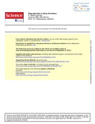
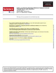
thickness ranges from approximately 100 to 300 mm. High-resolution imaging with multislice spiral CT<br />
scanners (HRCTs) has been used to assess vertebral trabecular architecture (Fig. 2), achieving images<br />
with in-plane voxel size <strong>of</strong> 150–180 mm <strong>and</strong> slice thickness <strong>of</strong> 300–500 mm [70,71]. HRCT provides<br />
superior discrimination <strong>of</strong> vertebral fracture patients compared with BMD [70]. In monitoring changes<br />
in vertebral trabecular architecture following 1 year <strong>of</strong> teriparatide <strong>the</strong>rapy, HRCT provided complementary<br />
information to that derived from densitometry [71].<br />
Advantages to QCT are that it can be employed on st<strong>and</strong>ard clinical scanners with relatively short<br />
imaging times, providing robust assessment <strong>of</strong> geometry <strong>and</strong> volumetric <strong>bone</strong> density in trabecular<br />
<strong>and</strong> cortical compartments at sites most prone to fracture, although even <strong>the</strong> low radiation exposure is<br />
a concern to some. To define <strong>the</strong> clinical utility <strong>of</strong> this technique, additional data are needed on <strong>the</strong><br />
ability <strong>of</strong> QCT-based measures to predict fracture risk prospectively <strong>and</strong> to monitor anti-fracture<br />
efficacy <strong>of</strong> drug interventions.<br />
High-resolution peripheral computed tomography (HR-pQCT)<br />
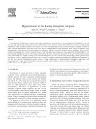
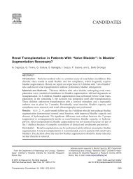
HR-pQCT measures <strong>bone</strong> density <strong>and</strong> trabecular <strong>and</strong> cortical microarchitecture, <strong>the</strong> distal radius<br />
<strong>and</strong> distal tibia with isotropic voxel size <strong>of</strong> w80 mm [72–80] (Fig. 3). This technique has excellent<br />
precision for both density (
M.L. Bouxsein, E. Seeman / Best Practice & Research Clinical Rheumatology 23 (2009) 741–753 745<br />
Fig. 2. Multi-slice, high-resolution computed tomography images <strong>of</strong> <strong>the</strong> 12th thoracic vertebrae, showing image acquisition,<br />
identification <strong>of</strong> <strong>the</strong> region <strong>of</strong> interest, <strong>and</strong> segmentation <strong>of</strong> <strong>the</strong> vertebral trabecular <strong>bone</strong>. Images courtesy <strong>of</strong> Dr. Claus Glüer,<br />
University <strong>of</strong> Kiel.<br />
parameters measured by HR-pQCT in a st<strong>and</strong>ard patient analysis, including <strong>bone</strong> volume ratio,<br />
trabecular number, derived trabecular thickness, derived trabecular separation <strong>and</strong> cortical thickness,<br />
correlated well with measurements made with high-resolution micro-CT (i.e., 20-mm voxel size) [81].<br />
Longitudinal HR-pQCT measurements indicate that whereas substantial cortical <strong>bone</strong> loss begins in<br />
middle life in women, it begins mainly after age 75 in men [78]. By contrast, trabecular <strong>bone</strong> loss begins<br />
early in adulthood in both women <strong>and</strong> men, such that approximately 40% <strong>of</strong> total life time trabecular<br />
<strong>bone</strong> loss occurs before age 50, as compared wi<strong>the</strong> less than 15% for cortical <strong>bone</strong>.<br />
Cross-sectional studies have reported that microarchitecture measurements at <strong>the</strong> distal radius by<br />
HR-pQCT discriminate postmenopausal women with a history <strong>of</strong> fragility fracture from those who have<br />
not suffered a fracture, partly independent <strong>of</strong> BMD [72,75–77,82]. Preliminary reports have shown<br />
treatment-related changes in <strong>bone</strong> architecture as assessed by HR-pQCT, including increased cortical<br />
thickness following 1 year <strong>of</strong> denosumab [83] or strontium ranelate [84] <strong>the</strong>rapy. As denosumab likely<br />
increases tissue mineral density due to its pr<strong>of</strong>ound suppression <strong>of</strong> <strong>bone</strong> resorption [85] <strong>and</strong> strontium<br />
Fig. 3. High-resolution peripheral quantitative computed tomography image (voxel size ¼ 82 mm 3 ) <strong>of</strong> <strong>the</strong> distal radius (left) <strong>and</strong><br />
distal tibia (right). Trabecular <strong>bone</strong> appears white, on a dark background.
746<br />
M.L. Bouxsein, E. Seeman / Best Practice & Research Clinical Rheumatology 23 (2009) 741–753<br />
ranelate is incorporated into <strong>the</strong> mineral substance <strong>of</strong> <strong>bone</strong> [86], it is not known whe<strong>the</strong>r <strong>the</strong>se<br />
observations are true changes in <strong>bone</strong> morphology or artefacts due to altered edge-detection <strong>and</strong>/or<br />
partial volume effects.<br />
Altoge<strong>the</strong>r, HR-pQCT is promising for assessment <strong>of</strong> trabecular <strong>and</strong> cortical architecture in vivo with<br />
high precision. The disadvantages are that it requires specialised scanners, <strong>and</strong> measurements are<br />
limited to peripheral skeletal sites. Fur<strong>the</strong>rmore, motion artefacts are common, requiring re-scanning<br />
<strong>of</strong> many patients.<br />
Magnetic resonance imaging (MRI)<br />
Magnetic resonance (MR) imaging is a non-ionising method that uses a strong magnetic field in<br />
combination with specialised sequences <strong>of</strong> radio-frequency pulses to generate 3D images <strong>of</strong> <strong>bone</strong><br />
structure [87]. Because free hydrogen in water provides <strong>the</strong> ‘signal’ in this type <strong>of</strong> MR imaging <strong>and</strong><br />
since <strong>the</strong> water content <strong>of</strong> <strong>bone</strong> is minimal, <strong>the</strong>re is generally little signal provided by <strong>bone</strong> in<br />
st<strong>and</strong>ard MR imaging. As a result, <strong>the</strong> <strong>bone</strong> structure is assessed indirectly via measurements <strong>of</strong><br />
<strong>the</strong> surrounding marrow <strong>and</strong> o<strong>the</strong>r s<strong>of</strong>t tissues (Fig. 4). Advances in <strong>the</strong> past decade have focussed<br />
on image acquisition <strong>and</strong> analysis techniques to overcome inherent obstacles in MR imaging <strong>of</strong><br />
<strong>bone</strong> [88].<br />
High-resolution MR imaging is performed at peripheral skeletal sites (e.g., distal radius, distal tibia<br />
<strong>and</strong> calcaneus) using clinical MR scanners combined with specially designed coils. In vivo resolutions <strong>of</strong><br />
150–300 mm in plane <strong>and</strong> a slice thickness <strong>of</strong> 300–500 mm have been achieved [89,90]. With this<br />
resolution, it is not possible to produce accurate values for most features <strong>of</strong> trabecular architecture.<br />
None<strong>the</strong>less, <strong>the</strong> ‘apparent’ trabecular properties that are derived from <strong>the</strong>se images correlate with<br />
trabecular architecture obtained with higher-resolution techniques [91,92]. Until recently, evaluation<br />
<strong>of</strong> trabecular <strong>bone</strong> morphology was limited to appendicular sites. However, innovative surface coils<br />
<strong>and</strong> pulse sequences show potential for MR-based assessments <strong>of</strong> trabecular structure in <strong>the</strong> proximal<br />
femur [93–95] (Fig. 5).<br />
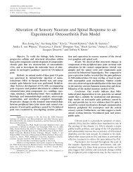
MR-derived trabecular microarchitecture measurements reflect age- <strong>and</strong> disease-specific differences<br />
[96,97], <strong>and</strong> differentiate patients with hip <strong>and</strong> vertebral fractures from controls with <strong>the</strong> best<br />
performance achieved by combinations <strong>of</strong> <strong>structural</strong> parameters <strong>and</strong> BMD [98–103]. There are no<br />
studies demonstrating prospective fracture risk prediction, <strong>and</strong> <strong>the</strong>re are limited data on treatmentrelated<br />
changes [104,105].<br />
Fig. 4. Magnetic resonance image <strong>of</strong> <strong>the</strong> distal tibia (voxel size: 156 156 500 mm). Trabecular <strong>bone</strong> appears dark on a light<br />
background <strong>of</strong> marrow. Image courtesy <strong>of</strong> Dr. Sharmila Majumdar, UCSF.
M.L. Bouxsein, E. Seeman / Best Practice & Research Clinical Rheumatology 23 (2009) 741–753 747<br />
Fig. 5. Magnetic resonance image <strong>of</strong> <strong>the</strong> proximal femur. Image courtesy <strong>of</strong> Dr. Sharmila Majumdar, UCSF.<br />
There are currently no non-invasive techniques available for clinical use that assess <strong>bone</strong> matrix<br />
properties, such as <strong>the</strong> degree <strong>of</strong> mineralisation, collagen content <strong>and</strong>/or <strong>the</strong> degree <strong>of</strong> collagen crosslinking.<br />
Yet, one particularly novel aspect <strong>of</strong> MRI is that it may allow non-invasive assessment <strong>of</strong> <strong>bone</strong><br />
matrix properties in addition to <strong>bone</strong> structure. Solid-state MR imaging uses <strong>the</strong> resonant signals <strong>of</strong> <strong>the</strong><br />
phosphorus ( [31] P) constituent <strong>of</strong> <strong>the</strong> <strong>bone</strong> mineral phase to determine <strong>the</strong> mineral content <strong>of</strong> <strong>bone</strong><br />
[106–110]. Bone tissue mineral density measurements acquired through [31] P solid-state MR<br />
discriminate osteoporotic <strong>and</strong> osteomalacic animal models better than X-ray-based imaging methods<br />
[108]. Wu <strong>and</strong> colleagues[109] report that combined 31 P <strong>and</strong> 1 H water- <strong>and</strong> fat-suppressed solid-state<br />
imaging give MRI <strong>the</strong> unique ability to assess <strong>the</strong> organic <strong>and</strong> inorganic solid-phase densities <strong>of</strong> <strong>bone</strong>,<br />
as well as <strong>the</strong> solid <strong>bone</strong> matrix density (organic þ inorganic). Although 31 P solid-state MRI has been<br />
implemented in vivo with human subjects on a clinical 1.5 T system [111], solid-state MRI is still<br />
a research tool. Solid-state imaging may become an excellent tool for longitudinal <strong>bone</strong> tissue density<br />
evaluations (without consequence <strong>of</strong> radiation exposure), but currently, this method requires excessively<br />
long imaging times (w1 h or more), most studies have used non-clinical scanners with strong<br />
magnetic fields (4.7 T or greater) <strong>and</strong> custom-made coils. Thus, MRI is a non-ionising method that<br />
allows 3D assessment <strong>of</strong> cortical <strong>and</strong> trabecular <strong>bone</strong> structure. Image data can be acquired in any<br />
arbitrary axis with acquisition times <strong>of</strong> 10–15 min [88].<br />
Finite element analysis (FEA) based on QCT- or hr-pQCT images<br />
The finite element (FE) method was first applied to <strong>structural</strong> analysis in <strong>the</strong> 1950s, <strong>and</strong> it has since<br />
been widely used in nearly every engineering <strong>and</strong> engineering-related field. In solid <strong>and</strong> <strong>structural</strong><br />
mechanics (<strong>bone</strong> mechanics included), it is <strong>the</strong> method <strong>of</strong> choice for evaluating <strong>the</strong> how a structure<br />
with complex geometrical shape <strong>and</strong> heterogeneous distribution <strong>of</strong> <strong>material</strong> properties (e.g., a whole<br />
<strong>bone</strong>) behaves when subjected to external loads.<br />
The finite element approach begins by representing <strong>the</strong> object as a collection <strong>of</strong> building blocks, or<br />
elements, each <strong>of</strong> which is defined by reference points, or nodes. The FE method can provide ‘estimates’<br />
<strong>of</strong> quantities that are commonly obtained through mechanical testing (e.g., whole <strong>bone</strong> stiffness <strong>and</strong><br />
failure load), as well as quantities that are difficult, if not impossible, to measure experimentally<br />
(e.g., strain distribution). However, <strong>the</strong> ability <strong>of</strong> <strong>the</strong> finite element solution to approximate <strong>the</strong> actual<br />
biomechanical phenomenon depends on <strong>the</strong> quality <strong>of</strong> <strong>the</strong> input. For example, <strong>the</strong> choice <strong>of</strong> <strong>material</strong>
748<br />
M.L. Bouxsein, E. Seeman / Best Practice & Research Clinical Rheumatology 23 (2009) 741–753<br />
properties <strong>and</strong> loading conditions influence <strong>the</strong> <strong>strength</strong> predictions. None<strong>the</strong>less, this state-<strong>of</strong>-<strong>the</strong>-art<br />
technique is among <strong>the</strong> most promising tools available today for clinical assessment <strong>of</strong> <strong>bone</strong> <strong>strength</strong><br />
<strong>and</strong> fracture risk [112].<br />
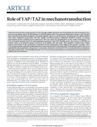
3D-QCT (or HR-pQCT) scans are directly converted, voxel by voxel, into a finite element model,<br />
which accurately represents <strong>the</strong> 3D geometry <strong>and</strong> heterogeneous density distribution <strong>of</strong> <strong>the</strong> <strong>bone</strong> <strong>of</strong><br />
interest (Fig. 6). Forces simulating activities that cause fractures are applied to this model, <strong>and</strong> <strong>the</strong><br />
stiffness <strong>and</strong> <strong>strength</strong> <strong>of</strong> <strong>the</strong> <strong>bone</strong> in response to <strong>the</strong> applied forces are computed. These analyses can<br />
incorporate different in vivo loading conditions, such as compressive <strong>and</strong> bending loading for <strong>the</strong><br />
vertebrae, or stance <strong>and</strong> fall loading for <strong>the</strong> femur. Several studies in human cadaveric femurs,<br />
vertebrae <strong>and</strong> forearms have shown that <strong>bone</strong> <strong>strength</strong> is better predicted by FE analyses than by BMD<br />
alone [113–116].<br />
Over 15 years ago, Faulkner <strong>and</strong> colleagues reported that QCT-based FE analysis <strong>of</strong> vertebral<br />
<strong>strength</strong> discriminated between women with <strong>and</strong> without vertebral fracture, with less overlap<br />
between <strong>the</strong> two groups than observed using QCT [117]. More recently, QCT-based FE analyses have<br />
been shown to discriminate women with vertebral fractures from controls[118] <strong>and</strong> to explore <strong>the</strong><br />
mechanisms underlying increased <strong>bone</strong> <strong>strength</strong> following anabolic <strong>and</strong>/or anti-catabolic treatment[57,119,120,121].<br />
The deleterious effects <strong>of</strong> glucocorticoids on femoral <strong>strength</strong> have also been<br />
explored by in vivo QCT-based FE analyses [122]. In postmenopausal women matched for age, weight<br />
<strong>and</strong> history <strong>of</strong> hormone <strong>the</strong>rapy, FE analyses showed that femoral <strong>strength</strong> in women with a history <strong>of</strong><br />
glucocorticoid use was w15% lower than controls for both fall-loading <strong>and</strong> stance configurations.<br />
The feasibility <strong>of</strong> in vivo micro-FEA using both MR <strong>and</strong> HR-pQCT has also been demonstrated in<br />
clinical studies [78,82,123,124]. High-resolution MR images <strong>of</strong> trabecular <strong>bone</strong> in <strong>the</strong> distal radius were<br />
subjected to FE analysis to determine effects <strong>of</strong> trabecular microarchitecture on <strong>bone</strong> mechanical<br />
properties in normal <strong>and</strong> osteopenic postmenopausal women [123]. MR images <strong>of</strong> trabecular architecture<br />
in <strong>the</strong> calcaneus were also assessed using FE to quantify <strong>the</strong> response to 1 year idoxifene [124].<br />
In both cases, <strong>the</strong> use <strong>of</strong> FE analysis provided additional information regarding <strong>bone</strong> structure <strong>and</strong><br />
<strong>strength</strong> to that available from structure measurements alone. Thus, this approach warrants fur<strong>the</strong>r<br />
investigation. However, because this technique presently requires extensive computational resources<br />
<strong>and</strong> specialised s<strong>of</strong>tware, widespread evaluation will likely be limited. To date, <strong>the</strong>re are few data on<br />
<strong>the</strong> precision <strong>of</strong> <strong>the</strong> technique <strong>and</strong> its ability to reflect disease- <strong>and</strong> age-related changes, <strong>and</strong> no<br />
prospective fracture studies exist. However, cross-sectional studies using HR-pQCT have shown <strong>the</strong><br />
FEA-based <strong>strength</strong> estimates discriminate women with prior history <strong>of</strong> distal radius fracture [76,82].<br />
Improved imaging <strong>and</strong> computational methods have made subject-specific FE analyses more feasible,<br />
<strong>and</strong> fur<strong>the</strong>r technological advances will continue.<br />
Fig. 6. Finite element model <strong>of</strong> <strong>the</strong> proximal femur in a sideways fall configuration. Colors represent plastic strain, showing likely<br />
regions <strong>of</strong> failure. Image courtesy <strong>of</strong> Dr. David Kopperdahl, O.N. Diagnostics.
Conclusions<br />
Novel non-invasive techniques for assessment <strong>of</strong> <strong>the</strong> <strong>structural</strong> <strong>determinants</strong> <strong>of</strong> <strong>bone</strong> <strong>strength</strong> are<br />
available. These techniques quantify <strong>the</strong> macro- <strong>and</strong> microstructure <strong>of</strong> <strong>bone</strong> such as <strong>bone</strong> size, shape,<br />
cortical thickness, cortical density, a surrogate <strong>of</strong> cortical porosity, trabecular number, thickness <strong>and</strong><br />
separation. FE analysis, by combining <strong>bone</strong> geometry with <strong>material</strong> characteristics, provides good<br />
estimates <strong>of</strong> whole <strong>bone</strong> <strong>strength</strong>. Whe<strong>the</strong>r <strong>the</strong>se <strong>strength</strong> <strong>and</strong>/or morphological features singly or<br />
toge<strong>the</strong>r will improve fracture prediction or serve as surrogates <strong>of</strong> anti-fracture efficacy is not known.<br />
Although several studies provide promising data for <strong>the</strong>se technique, <strong>the</strong>se methodologies remain<br />
research tools as most have not been rigorously tested for <strong>the</strong>ir ability to predict fracture risk in<br />
prospective studies <strong>and</strong> to monitor treatment response. Use <strong>of</strong> 3D imaging modalities to assess <strong>the</strong><br />
<strong>determinants</strong> <strong>of</strong> <strong>bone</strong> <strong>strength</strong> is a research area <strong>of</strong> high interest <strong>and</strong> relevance to clinicians. However,<br />
<strong>the</strong>re is a need for additional developments <strong>and</strong> studies to determine <strong>the</strong> clinical utility <strong>of</strong> <strong>the</strong>se<br />
imaging modalities. Future research is aimed at developing techniques that are better suited to assess<br />
those at highest risk <strong>of</strong> fracture, to diagnose patients early in <strong>the</strong> disease process, to identify specific<br />
treatable components <strong>of</strong> skeletal fragility <strong>and</strong> to monitor treatment efficacy. These developments are<br />
needed given <strong>the</strong> inevitable future <strong>of</strong> a growing elderly population combined with shrinking resources<br />
for medical care.<br />
Acknowledgements<br />
We thank Drs. Sharmala Majumdar, Claus Glüer, Thomas Lang <strong>and</strong> David Kopperdahl for generously<br />
providing images.<br />
References<br />
M.L. Bouxsein, E. Seeman / Best Practice & Research Clinical Rheumatology 23 (2009) 741–753 749<br />
[1] U.S. Department <strong>of</strong> Health <strong>and</strong> Human Services. Bone health <strong>and</strong> osteoporosis: a report <strong>of</strong> <strong>the</strong> surgeon general.<br />
Rockville, MD: U.S. Department <strong>of</strong> Health <strong>and</strong> Human Services, Office <strong>of</strong> <strong>the</strong> Surgeon General; 2004.<br />
[2] Rosen CJ. Clinical practice. Postmenopausal osteoporosis. N Engl J Med 2005;353(6):595–603.<br />
[3] Seeman E, Compston J, Adachi J, et al. Non-compliance: <strong>the</strong> Achilles’ heel <strong>of</strong> anti-fracture efficacy. Osteoporos Int<br />
2007;18(6):711–9.<br />
[4] Proceedings <strong>of</strong> a symposium. Consensus Development Conference on Osteoporosis. October 19–20, 1990, Copenhagen,<br />
Denmark. Am J Med 1991;91(5B):1S–68.<br />
[5] Bouxsein M. Biomechanics <strong>of</strong> age-related fractures. In: Marcus R, Feldman D, Nelson D, Rosen C, editors. Osteoporosis.<br />
3rd edn., vol. I. San Diego, CA: Elsevier Academic Press; 2007. p. 601–16.<br />
[6] Johnell O, Kanis JA, Oden A, et al. Predictive value <strong>of</strong> BMD for hip <strong>and</strong> o<strong>the</strong>r fractures. J Bone Miner Res 2005;20(7):<br />
1185–94.<br />
[7] Siris ES, Chen YT, Abbott TA, et al. Bone mineral density thresholds for pharmacological intervention to prevent<br />
fractures. Arch Intern Med 2004;164(10):1108–12.<br />
[8] Wainwright SA, Marshall LM, Ensrud KE, et al. Hip fracture in women without osteoporosis. J Clin Endocrinol Metab<br />
2005;90(5):2787–93.<br />
[9] Schuit SC, van der Klift M, Weel AE, et al. Fracture incidence <strong>and</strong> association with <strong>bone</strong> mineral density in elderly men<br />
<strong>and</strong> women: <strong>the</strong> Rotterdam Study. Bone 2004;34(1):195–202.<br />
[10] Cummings SR. How drugs decrease fracture risk: lessons from trials. J Musculoskelet Neuronal Interact 2002;2(3):<br />
198–200.<br />
[11] Sarkar S, Mitlak BH, Wong M, et al. Relationships between <strong>bone</strong> mineral density <strong>and</strong> incident vertebral fracture risk<br />
with raloxifene <strong>the</strong>rapy. J Bone Miner Res 2002;17(1):1–10.<br />
[12] Delmas P, Seeman E. Changes in <strong>bone</strong> mineral density explain little <strong>of</strong> <strong>the</strong> reduction in vertebral or nonvertebral<br />
fracture risk with anti-resorptive <strong>the</strong>rapy. Bone 2004;34(4):599–604.<br />
[13] Watts NB, Geusens P, Barton IP, Felsenberg D. Relationship between changes in BMD <strong>and</strong> nonvertebral fracture<br />
incidence associated with risedronate: reduction in risk <strong>of</strong> nonvertebral fracture is not related to change in BMD.<br />
J Bone Miner Res 2005;20(12):2097–104.<br />
[14] Bouxsein ML. Bone quality: where do we go from here? Osteoporos Int 2003;14(Suppl. 5):118–27.<br />
*[15] Seeman E, Delmas PD. Bone quality–<strong>the</strong> <strong>material</strong> <strong>and</strong> <strong>structural</strong> basis <strong>of</strong> <strong>bone</strong> <strong>strength</strong> <strong>and</strong> fragility. N Engl J Med<br />
2006;354(21):2250–61.<br />
[16] Engelke K, Gluer CC. Quality <strong>and</strong> performance measures in <strong>bone</strong> densitometry: part 1: errors <strong>and</strong> diagnosis. Osteoporos<br />
Int 2006;17(9):1283–92.<br />
[17] Marshall D, Johnell O, Wedel H. Meta-analysis <strong>of</strong> how well measures <strong>of</strong> <strong>bone</strong> mineral density predict occurrence <strong>of</strong><br />
osteoporotic fractures. BMJ 1996;312(7041):1254–9.<br />
[18] WHO Study Group. Assessment <strong>of</strong> fracture risk <strong>and</strong> its application to screening for postmenopausal osteoporosis.<br />
Geneva: World Health Organization; 1994.<br />
[19] Carter DR, Bouxsein ML, Marcus R. New approaches for interpreting projected <strong>bone</strong> densitometry. J Bone Miner Res<br />
1992;7:137–45.
750<br />
M.L. Bouxsein, E. Seeman / Best Practice & Research Clinical Rheumatology 23 (2009) 741–753<br />
[20] Beck T. Measuring <strong>the</strong> <strong>structural</strong> <strong>strength</strong> <strong>of</strong> <strong>bone</strong>s with dual-energy X-ray absorptiometry: principles, technical<br />
limitations, <strong>and</strong> future possibilities. Osteoporos Int 2003;14(Suppl. 5):81–8.<br />
[21] Uusi-Rasi K, Beck TJ, Semanick LM, et al. Structural effects <strong>of</strong> raloxifene on <strong>the</strong> proximal femur: results from <strong>the</strong><br />
multiple outcomes <strong>of</strong> raloxifene evaluation trial. Osteoporos Int 2006;17(4):575–86.<br />
[22] Greenspan SL, Beck TJ, Resnick NM, et al. Effect <strong>of</strong> hormone replacement, alendronate, or combination <strong>the</strong>rapy on<br />
hip <strong>structural</strong> geometry: a 3-year, double-blind, placebo-controlled clinical trial. J Bone Miner Res 2005;20(9):<br />
1525–32.<br />
[23] Uusi-Rasi K, Semanick LM, Zanchetta JR, et al. Effects <strong>of</strong> teriparatide [rhPTH (1-34)] treatment on <strong>structural</strong> geometry<br />
<strong>of</strong> <strong>the</strong> proximal femur in elderly osteoporotic women. Bone 2005;36(6):948–58.<br />
[24] Bonnick SL, Beck TJ, Cosman F, et al. DXA-based hip <strong>structural</strong> analysis <strong>of</strong> once-weekly bisphosphonate-treated<br />
postmenopausal women with low <strong>bone</strong> mass. Osteoporos Int 2009;20(6):911–21.<br />
[25] Chen Z, Beck TJ, Cauley JA, et al. Hormone <strong>the</strong>rapy improves femur geometry among ethnically diverse postmenopausal<br />
participants in <strong>the</strong> Women’s Health Initiative hormone intervention trials. J Bone Miner Res 2008;23(12):<br />
1935–45.<br />
[26] Boivin GY, Chavassieux PM, Santora AC, et al. Alendronate increases <strong>bone</strong> <strong>strength</strong> by increasing <strong>the</strong> mean degree <strong>of</strong><br />
mineralization <strong>of</strong> <strong>bone</strong> tissue in osteoporotic women. Bone 2000;27(5):687–94.<br />
[27] Roschger P, Rinnerthaler S, Yates J, et al. Alendronate increases degree <strong>and</strong> uniformity <strong>of</strong> mineralization in cancellous<br />
<strong>bone</strong> <strong>and</strong> decreases <strong>the</strong> porosity in cortical <strong>bone</strong> <strong>of</strong> osteoporotic women. Bone 2001;29(2):185–91.<br />
[28] Boivin G, Lips P, Ott SM, et al. Contribution <strong>of</strong> raloxifene <strong>and</strong> calcium <strong>and</strong> vitamin D3 supplementation to <strong>the</strong> increase<br />
<strong>of</strong> <strong>the</strong> degree <strong>of</strong> mineralization <strong>of</strong> <strong>bone</strong> in postmenopausal women. J Clin Endocrinol Metab 2003;88(9):4199–205.<br />
[29] Borah B, Ritman EL, Dufresne TE, et al. The effect <strong>of</strong> risedronate on <strong>bone</strong> mineralization as measured by microcomputed<br />
tomography with synchrotron radiation: correlation to histomorphometric indices <strong>of</strong> turnover. Bone 2005;<br />
37(1):1–9.<br />
[30] Mis<strong>of</strong> BM, Roschger P, Cosman F, et al. Effects <strong>of</strong> intermittent parathyroid hormone administration on <strong>bone</strong> mineralization<br />
density in iliac crest biopsies from patients with osteoporosis: a paired study before <strong>and</strong> after treatment.<br />
J Clin Endocrinol Metab 2003;88(3):1150–6.<br />
[31] Beck TJ, Oreskovic TL, Stone KL, et al. Structural adaptation to changing skeletal load in <strong>the</strong> progression toward hip<br />
fragility: <strong>the</strong> study <strong>of</strong> osteoporotic fractures. J Bone Miner Res 2001;16(6):1108–19.<br />
[32] Beck TJ, Stone KL, Oreskovic TL, et al. Effects <strong>of</strong> current <strong>and</strong> discontinued estrogen replacement <strong>the</strong>rapy on hip<br />
<strong>structural</strong> geometry: <strong>the</strong> study <strong>of</strong> osteoporotic fractures. J Bone Miner Res 2001;16(11):2103–10.<br />
[33] Crabtree N, Lunt M, Holt G, et al. Hip geometry, <strong>bone</strong> mineral distribution, <strong>and</strong> <strong>bone</strong> <strong>strength</strong> in European men <strong>and</strong><br />
women: <strong>the</strong> EPOS study. Bone 2000;27(1):151–9.<br />
[34] Nelson DA, Barondess DA, Hendrix SL, Beck TJ. Cross-sectional geometry, <strong>bone</strong> <strong>strength</strong>, <strong>and</strong> <strong>bone</strong> mass in <strong>the</strong><br />
proximal femur in black <strong>and</strong> white postmenopausal women. J Bone Miner Res 2000;15(10):1992–7.<br />
[35] Looker AC, Beck TJ, Orwoll ES. Does body size account for gender differences in femur <strong>bone</strong> density <strong>and</strong> geometry?<br />
J Bone Miner Res 2001;16(7):1291–9.<br />
[36] Crabtree NJ, Kroger H, Martin A, et al. Improving risk assessment: hip geometry, <strong>bone</strong> mineral distribution <strong>and</strong> <strong>bone</strong><br />
<strong>strength</strong> in hip fracture cases <strong>and</strong> controls. The EPOS study. European Prospective Osteoporosis Study. Osteoporos Int<br />
2002;13(1):48–54.<br />
[37] Cauley JA, Lui LY, Stone KL, et al. Longitudinal study <strong>of</strong> changes in hip <strong>bone</strong> mineral density in Caucasian <strong>and</strong> African-<br />
American women. J Am Geriatr Soc 2005;53(2):183–9.<br />
[38] Kaptoge S, Dalzell N, Loveridge N, et al. Effects <strong>of</strong> gender, anthropometric variables, <strong>and</strong> aging on <strong>the</strong> evolution <strong>of</strong> hip<br />
<strong>strength</strong> in men <strong>and</strong> women aged over 65. Bone 2003;32(5):561–70.<br />
[39] Takada J, Beck TJ, Iba K, Yamashita T. Structural trends in <strong>the</strong> aging proximal femur in Japanese postmenopausal<br />
women. Bone 2007;41(1):97–102.<br />
[40] Kaptoge S, Jakes RW, Dalzell N, et al. Effects <strong>of</strong> physical activity on evolution <strong>of</strong> proximal femur structure in a younger<br />
elderly population. Bone 2007;40(2):506–15.<br />
[41] Kaptoge S, Dalzell N, Folkerd E, et al. Sex hormone status may modulate rate <strong>of</strong> expansion <strong>of</strong> proximal femur diameter<br />
in older women alongside o<strong>the</strong>r skeletal regulators. J Clin Endocrinol Metab 2007;92(1):304–13.<br />
[42] Demissie S, Dupuis J, Cupples LA, et al. Proximal hip geometry is linked to several chromosomal regions: genomewide<br />
linkage results from <strong>the</strong> Framingham Osteoporosis Study. Bone 2007;40(3):743–50.<br />
[43] Karasik D, Dupuis J, Cupples LA, et al. Bivariate linkage study <strong>of</strong> proximal hip geometry <strong>and</strong> body size indices: <strong>the</strong><br />
Framingham study. Calcif Tissue Int 2007;81(3):162–73.<br />
[44] Streeten EA, Beck TJ, O’Connell JR, et al. Autosome-wide linkage analysis <strong>of</strong> hip <strong>structural</strong> phenotypes in <strong>the</strong> Old Order<br />
Amish. Bone 2008;43(3):607–12.<br />
[45] Szulc P, Duboeuf F, Schott AM, et al. Structural <strong>determinants</strong> <strong>of</strong> hip fracture in elderly women: re-analysis <strong>of</strong> <strong>the</strong> data<br />
from <strong>the</strong> EPIDOS study. Osteoporos Int 2006;17(2):231–6.<br />
[46] Ahlborg HG, Nguyen ND, Nguyen TV, et al. Contribution <strong>of</strong> hip <strong>strength</strong> indices to hip fracture risk in elderly men <strong>and</strong><br />
women. J Bone Miner Res 2005;20(10):1820–7.<br />
[47] Rivadeneira F, Zillikens MC, De Laet CE, et al. Femoral neck BMD is a strong predictor <strong>of</strong> hip fracture susceptibility in<br />
elderly men <strong>and</strong> women because it detects cortical <strong>bone</strong> instability: <strong>the</strong> Rotterdam Study. J Bone Miner Res 2007;22(11):<br />
1781–90.<br />
[48] Kaptoge S, Beck TJ, Reeve J, et al. Prediction <strong>of</strong> incident hip fracture risk by femur geometry variables measured by hip<br />
<strong>structural</strong> analysis in <strong>the</strong> study <strong>of</strong> osteoporotic fractures. J Bone Miner Res 2008;23(12):1892–904.<br />
*[49] Riggs BL, Melton Iii 3rd LJ, Robb RA, et al. Population-based study <strong>of</strong> age <strong>and</strong> sex differences in <strong>bone</strong> volumetric<br />
density, size, geometry, <strong>and</strong> structure at different skeletal sites. J Bone Miner Res 2004;19(12):1945–54.<br />
[50] Marshall LM, Lang TF, Lambert LC, et al. Dimensions <strong>and</strong> volumetric BMD <strong>of</strong> <strong>the</strong> proximal femur <strong>and</strong> <strong>the</strong>ir relation to<br />
age among older U.S. men. J Bone Miner Res 2006;21(8):1197–206.<br />
[51] Sigurdsson G, Aspelund T, Chang M, et al. Increasing sex difference in <strong>bone</strong> <strong>strength</strong> in old age: <strong>the</strong> Age, Gene/<br />
Environment Susceptibility-Reykjavik study (AGES-REYKJAVIK). Bone 2006;39(3):644–51.
M.L. Bouxsein, E. Seeman / Best Practice & Research Clinical Rheumatology 23 (2009) 741–753 751<br />
[52] Marshall LM, Zmuda JM, Chan BK, et al. Race <strong>and</strong> ethnic variation in proximal femur structure <strong>and</strong> BMD among older<br />
men. J Bone Miner Res 2008;23(1):121–30.<br />
[53] Black DM, Greenspan SL, Ensrud KE, et al. The effects <strong>of</strong> parathyroid hormone <strong>and</strong> alendronate alone or in combination<br />
in postmenopausal osteoporosis. N Engl J Med 2003;349(13):1207–15.<br />
[54] Finkelstein JS, Hayes A, Hunzelman JL, et al. The effects <strong>of</strong> parathyroid hormone, alendronate, or both in men with<br />
osteoporosis. N Engl J Med 2003;349(13):1216–26.<br />
[55] McClung MR, San Martin J, Miller PD, et al. Opposite <strong>bone</strong> remodeling effects <strong>of</strong> teriparatide <strong>and</strong> alendronate in<br />
increasing <strong>bone</strong> mass. Arch Intern Med 2005;165(15):1762–8.<br />
[56] Sellmeyer DE, Black DM, Palermo L, et al. Hetereogeneity in skeletal response to full-length parathyroid hormone in<br />
<strong>the</strong> treatment <strong>of</strong> osteoporosis. Osteoporos Int 2007;18(7):973–9.<br />
[57] Lewiecki EM, Keaveny TM, Kopperdahl DL, et al. Once-monthly oral ib<strong>and</strong>ronate improves biomechanical <strong>determinants</strong><br />
<strong>of</strong> <strong>bone</strong> <strong>strength</strong> in women with postmenopausal osteoporosis. J Clin Endocrinol Metab 2009;94(1):<br />
171–80.<br />
[58] Miller PD, Delmas PD, Lindsay R, et al. Early responsiveness <strong>of</strong> women with osteoporosis to teriparatide after <strong>the</strong>rapy<br />
with alendronate or risedronate. J Clin Endocrinol Metab 2008;93(10):3785–93.<br />
[59] Duboeuf F, Jergas M, Schott AM, et al. A comparison <strong>of</strong> <strong>bone</strong> densitometry measurements <strong>of</strong> <strong>the</strong> central skeleton in<br />
post-menopausal women with <strong>and</strong> without vertebral fracture. Br J Radiol 1995;68(811):747–53.<br />
[60] Ito M, Ohki M, Hayashi K, et al. Trabecular texture analysis <strong>of</strong> CT images in <strong>the</strong> relationship with spinal fractures.<br />
Radiology 1995;194:55–9.<br />
[61] Grampp S, Genant H, Mathur A, et al. Comparisons <strong>of</strong> noninvasive <strong>bone</strong> mineral measurements in assessing agerelated<br />
loss, fracture discrimination, <strong>and</strong> diagnostic classification. J Bone Miner Res 1997;12:697–711.<br />
[62] Lang TF, Li J, Harris ST, Genant HK. Assessment <strong>of</strong> vertebral <strong>bone</strong> mineral density using volumetric quantitative CT.<br />
J Comput Assist Tomogr 1999;23(1):130–7.<br />
[63] Bergot C, Laval-Jeantet AM, Hutchinson K, et al. A comparison <strong>of</strong> spinal quantitative computed tomography with dual<br />
energy X-ray absorptiometry in European women with vertebral <strong>and</strong> nonvertebral fractures. Calcif Tissue Int 2001;<br />
68(2):74–82.<br />
[64] Kroger H, Lunt M, Reeve J, et al. Bone density reduction in various measurement sites in men <strong>and</strong> women with<br />
osteoporotic fractures <strong>of</strong> spine <strong>and</strong> hip: <strong>the</strong> European quantitation <strong>of</strong> osteoporosis study. Calcif Tissue Int 1999;64(3):<br />
191–9.<br />
[65] Lang TF, Guglielmi G, van Kuijk C, et al. Measurement <strong>of</strong> <strong>bone</strong> mineral density at <strong>the</strong> spine <strong>and</strong> proximal femur by<br />
volumetric quantitative computed tomography <strong>and</strong> dual-energy X-ray absorptiometry in elderly women with <strong>and</strong><br />
without vertebral fractures. Bone 2002;30(1):247–50.<br />
[66] Rehman Q, Lang T, Modin G, Lane NE. Quantitative computed tomography <strong>of</strong> <strong>the</strong> lumbar spine, not dual x-ray<br />
absorptiometry, is an independent predictor <strong>of</strong> prevalent vertebral fractures in postmenopausal women with<br />
osteopenia receiving long-term glucocorticoid <strong>and</strong> hormone-replacement <strong>the</strong>rapy. Arthritis Rheum 2002;46(5):<br />
1292–7.<br />
[67] Clowes JA, Eastell R, Peel NF. The discriminative ability <strong>of</strong> peripheral <strong>and</strong> axial <strong>bone</strong> measurements to identify<br />
proximal femoral, vertebral, distal forearm <strong>and</strong> proximal humeral fractures: a case control study. Osteoporos Int 2005;<br />
16(12):1794–802.<br />
[68] Mackey DC, Eby JG, Harris F, et al. Prediction <strong>of</strong> clinical non-spine fractures in older black <strong>and</strong> white men <strong>and</strong> women<br />
with volumetric BMD <strong>of</strong> <strong>the</strong> spine <strong>and</strong> areal BMD <strong>of</strong> <strong>the</strong> hip: <strong>the</strong> Health, Aging, <strong>and</strong> Body Composition Study*. J Bone<br />
Miner Res 2007;22(12):1862–8.<br />
*[69] Black DM, Bouxsein ML, Marshall LM, et al. Proximal femoral structure <strong>and</strong> <strong>the</strong> prediction <strong>of</strong> hip fracture in men:<br />
a large prospective study using QCT. J Bone Miner Res 2008;23(8):1326–33.<br />
*[70] Ito M, Ikeda K, Nishiguchi M, et al. Multi-detector row CT imaging <strong>of</strong> vertebral microstructure for evaluation <strong>of</strong> fracture<br />
risk. J Bone Miner Res 2005;20(10):1828–36.<br />
[71] Graeff C, Timm W, Nickelsen TN, et al. Monitoring teriparatide-associated changes in vertebral microstructure by highresolution<br />
CT in vivo: results from <strong>the</strong> EUROFORS study. J Bone Miner Res 2007;22(9):1426–33.<br />
*[72] Boutroy S, Bouxsein ML, Munoz F, Delmas PD. In vivo assessment <strong>of</strong> trabecular <strong>bone</strong> microarchitecture by highresolution<br />
peripheral quantitative computed tomography. J Clin Endocrinol Metab 2005;90(12):6508–15.<br />
[73] Khosla S, Riggs BL, Atkinson EJ, et al. Effects <strong>of</strong> sex <strong>and</strong> age on <strong>bone</strong> microstructure at <strong>the</strong> ultradistal radius: a population-based<br />
noninvasive in vivo assessment. J Bone Miner Res 2006;21(1):124–31.<br />
[74] Khosla S, Melton 3rd LJ, Achenbach SJ, et al. Hormonal <strong>and</strong> biochemical <strong>determinants</strong> <strong>of</strong> trabecular microstructure at<br />
<strong>the</strong> ultradistal radius in women <strong>and</strong> men. J Clin Endocrinol Metab 2005;91(3):885–91.<br />
[75] Sornay-Rendu E, Boutroy S, Munoz F, Delmas PD. Alterations <strong>of</strong> cortical <strong>and</strong> trabecular architecture are associated with<br />
fractures in postmenopausal women, partially independent <strong>of</strong> decreased BMD measured by DXA: <strong>the</strong> OFELY study.<br />
J Bone Miner Res 2007;22(3):425–33.<br />
[76] Melton 3rd LJ, Riggs BL, van Len<strong>the</strong> GH, et al. Contribution <strong>of</strong> in vivo <strong>structural</strong> measurements <strong>and</strong> load/<strong>strength</strong> ratios<br />
to <strong>the</strong> determination <strong>of</strong> forearm fracture risk in postmenopausal women. J Bone Miner Res 2007;22(9):1442–8.<br />
[77] Vico L, Zouch M, Amirouche A, et al. High-resolution pQCT analysis at <strong>the</strong> distal radius <strong>and</strong> tibia discriminates patients<br />
with recent wrist <strong>and</strong> femoral neck fractures. J Bone Miner Res 2008;23(11):1741–50.<br />
*[78] Riggs BL, Melton LJ, Robb RA, et al. A population-based assessment <strong>of</strong> rates <strong>of</strong> <strong>bone</strong> loss at multiple skeletal sites:<br />
evidence for substantial trabecular <strong>bone</strong> loss in young adult women <strong>and</strong> men. J Bone Miner Res 2008;23(2):205–14.<br />
*[79] Kazakia GJ, Hyun B, Burghardt AJ, et al. In vivo determination <strong>of</strong> <strong>bone</strong> structure in postmenopausal women:<br />
a comparison <strong>of</strong> HR-pQCT <strong>and</strong> high-field MR imaging. J Bone Miner Res 2008;23(4):463–74.<br />
[80] Dalzell N, Kaptoge S, Morris N, et al. Bone micro-architecture <strong>and</strong> <strong>determinants</strong> <strong>of</strong> <strong>strength</strong> in <strong>the</strong> radius <strong>and</strong> tibia:<br />
age-related changes in a population-based study <strong>of</strong> normal adults measured with high-resolution pQCT. Osteoporos<br />
Int 2009;20(10):1683–94.<br />
[81] MacNeil JA, Boyd SK. Accuracy <strong>of</strong> high-resolution peripheral quantitative computed tomography for measurement <strong>of</strong><br />
<strong>bone</strong> quality. Med Eng Phys 2007;29(10):1096–105.
752<br />
M.L. Bouxsein, E. Seeman / Best Practice & Research Clinical Rheumatology 23 (2009) 741–753<br />
[82] Boutroy S, Van Rietbergen B, Sornay-Rendu E, et al. Finite element analysis based on in vivo HR-pQCT images <strong>of</strong> <strong>the</strong><br />
distal radius is associated with wrist fracture in postmenopausal women. J Bone Miner Res 2008;23(3):392–9.<br />
[83] Seeman E, Delmas PD, Hanley DA, et al. Effects <strong>of</strong> denosumab <strong>and</strong> alendronate on skeletal microarchitecture International<br />
Bone <strong>and</strong> Mineral Society Meeting, Sydney. 2009.<br />
[84] Rizzoli R, Felsenberg D, Laroche M, et al. Strontium ranelate has a more positive influence than alendronate on distal<br />
tibia cortical <strong>and</strong> trabecluar <strong>bone</strong> microstructure in women with postmenopausal osteoporosis. Osteoporos Int 2009;<br />
20:165–6.<br />
[85] Boivin G, Meunier PJ. Changes in <strong>bone</strong> remodeling rate influence <strong>the</strong> degree <strong>of</strong> mineralization <strong>of</strong> <strong>bone</strong>. Connect Tissue<br />
Res 2002;43(2–3):535–7.<br />
[86] Boivin G, Del<strong>of</strong>fre P, Perrat B, et al. Strontium distribution <strong>and</strong> interactions with <strong>bone</strong> mineral in monkey iliac <strong>bone</strong><br />
after strontium salt (S 12911) administration. J Bone Miner Res 1996;11(9):1302–11.<br />
[87] Wehrli FW, Song HK, Saha PK, Wright AC. Quantitative MRI for <strong>the</strong> assessment <strong>of</strong> <strong>bone</strong> structure <strong>and</strong> function. NMR<br />
Biomed 2006;19(7):731–64.<br />
*[88] Wehrli FW. Structural <strong>and</strong> functional assessment <strong>of</strong> trabecular <strong>and</strong> cortical <strong>bone</strong> by micro magnetic resonance<br />
imaging.<br />
J Magn Reson Imaging 2007;25(2):390–409.<br />
[89] Link TM, Majumdar S, Grampp S, et al. Imaging <strong>of</strong> trabecular <strong>bone</strong> structure in osteoporosis. Eur Radiol 1999;9(9):1781–8.<br />
[90] Wehrli FW, Saha PK, Gomberg BR, et al. Role <strong>of</strong> magnetic resonance for assessing structure <strong>and</strong> function <strong>of</strong> trabecular<br />
<strong>bone</strong>. Top Magn Reson Imaging 2002;13(5):335–55.<br />
[91] Majumdar S, Newitt D, Mathur A, et al. Magnetic resonance imaging <strong>of</strong> trabecular <strong>bone</strong> structure in <strong>the</strong> distal radius:<br />
relationship with X-ray tomographic microscopy <strong>and</strong> biomechanics. Osteoporos Int 1996;6(5):376–85.<br />
[92] Link TM, Vieth V, Stehling C, et al. High-resolution MRI vs multislice spiral CT: which technique depicts <strong>the</strong> trabecular<br />
<strong>bone</strong> structure best? Eur Radiol 2003;13(4):663–71.<br />
[93] Krug R, Banerjee S, Han ET, et al. Feasibility <strong>of</strong> in vivo <strong>structural</strong> analysis <strong>of</strong> high-resolution magnetic resonance images<br />
<strong>of</strong> <strong>the</strong> proximal femur. Osteoporos Int 2005;16(11):1307–14.<br />
[94] Banerjee S, Han ET, Krug R, et al. Application <strong>of</strong> refocused steady-state free-precession methods at 1.5 <strong>and</strong> 3 T to in<br />
vivo high-resolution MRI <strong>of</strong> trabecular <strong>bone</strong>: simulations <strong>and</strong> experiments. J Magn Reson Imaging 2005;21(6):818–25.<br />
[95] Blumenfeld J, Studholme C, Carballido-Gamio J, et al. Three-dimensional image registration <strong>of</strong> MR proximal femur<br />
images for <strong>the</strong> analysis <strong>of</strong> trabecular <strong>bone</strong> parameters. Med Phys 2008;35(10):4630–9.<br />
[96] Benito M, Gomberg B, Wehrli FW, et al. Deterioration <strong>of</strong> trabecular architecture in hypogonadal men. J Clin Endocrinol<br />
Metab 2003;88(4):1497–502.<br />
[97] Wehrli FW, Leonard MB, Saha PK, Gomberg BR. Quantitative high-resolution magnetic resonance imaging reveals<br />
<strong>structural</strong> implications <strong>of</strong> renal osteodystrophy on trabecular <strong>and</strong> cortical <strong>bone</strong>. J Magn Reson Imaging 2004;20(1):<br />
83–9.<br />
*[98] Majumdar S, Genant HK, Grampp S, et al. Correlation <strong>of</strong> trabecular <strong>bone</strong> structure with age, <strong>bone</strong> mineral density, <strong>and</strong><br />
osteoporotic status: in vivo studies in <strong>the</strong> distal radius using high resolution magnetic resonance imaging. J Bone<br />
Miner Res 1997;12(1):111–8.<br />
[99] Wehrli FW, Hwang SN, Ma J, et al. Cancellous <strong>bone</strong> volume <strong>and</strong> structure in <strong>the</strong> forearm: noninvasive assessment with<br />
MR microimaging <strong>and</strong> image processing. Radiology 1998;206(2):347–57.<br />
[100] Link TM, Majumdar S, Augat P, et al. In vivo high resolution MRI <strong>of</strong> <strong>the</strong> calcaneus: differences in trabecular structure in<br />
osteoporosis patients. J Bone Miner Res 1998;13(7):1175–82.<br />
[101] Majumdar S, Link TM, Augat P, et al. Trabecular <strong>bone</strong> architecture in <strong>the</strong> distal radius using magnetic resonance<br />
imaging in subjects with fractures <strong>of</strong> <strong>the</strong> proximal femur. Magnetic Resonance Science Center <strong>and</strong> Osteoporosis <strong>and</strong><br />
Arthritis Research Group. Osteoporos Int 1999;10(3):231–9.<br />
*[102] Wehrli FW, Gomberg BR, Saha PK, et al. Digital topological analysis <strong>of</strong> in vivo magnetic resonance microimages <strong>of</strong><br />
trabecular <strong>bone</strong> reveals <strong>structural</strong> implications <strong>of</strong> osteoporosis. J Bone Miner Res 2001;16(8):1520–31.<br />
[103] Link TM, Bauer JS. Imaging <strong>of</strong> trabecular <strong>bone</strong> structure. Semin Musculoskelet Radiol 2002;6(3):253–61.<br />
[104] Benito M, Vasilic B, Wehrli FW, et al. Effect <strong>of</strong> testosterone replacement on trabecular architecture in hypogonadal<br />
men.<br />
J Bone Miner Res 2005;20(10):1785–91.<br />
[105] Chesnut 3rd CH, Majumdar S, Newitt DC, et al. Effects <strong>of</strong> salmon calcitonin on trabecular microarchitecture as<br />
determined by magnetic resonance imaging: results from <strong>the</strong> QUEST study. J Bone Miner Res 2005;20(9):<br />
1548–61.<br />
[106] Wu Y, Chesler DA, Glimcher MJ, et al. Multinuclear solid-state three-dimensional MRI <strong>of</strong> <strong>bone</strong> <strong>and</strong> syn<strong>the</strong>tic calcium<br />
phosphates. Proc Natl Acad Sci U S A 1999;96(4):1574–8.<br />
[107] Wu Y, Ackerman JL, Chesler DA, et al. Density <strong>of</strong> organic matrix <strong>of</strong> native mineralized <strong>bone</strong> measured by water- <strong>and</strong><br />
fat-suppressed proton projection MRI. Magn Reson Med 2003;50(1):59–68.<br />
[108] Anumula S, Magl<strong>and</strong> J, Wehrli SL, et al. Measurement <strong>of</strong> phosphorus content in normal <strong>and</strong> osteomalacic rabbit <strong>bone</strong><br />
by solid-state 3D radial imaging. Magn Reson Med 2006;56(5):946–52.<br />
[109] Wu Y, Dai G, Ackerman JL, et al. Water- <strong>and</strong> fat-suppressed proton projection MRI (WASPI) <strong>of</strong> rat femur <strong>bone</strong>. Magn<br />
Reson Med 2007;57(3):554–67.<br />
[110] Cao H, Ackerman JL, Hrovat MI, et al. Quantitative <strong>bone</strong> matrix density measurement by water- <strong>and</strong> fat-suppressed<br />
proton projection MRI (WASPI) with polymer calibration phantoms. Magn Reson Med 2008;60(6):1433–43.<br />
[111] Robson MD, Gatehouse PD, Bydder GM, Neubauer S. Human imaging <strong>of</strong> phosphorus in cortical <strong>and</strong> trabecular <strong>bone</strong> in<br />
vivo. Magn Reson Med 2004;51(5):888–92.<br />
[112] Stokstad E. Bone quality fills holes in fracture risk. Science 2005;308(5728):1580.<br />
[113] Cody DD, Gross GJ, Hou FJ, et al. Femoral <strong>strength</strong> is better predicted by finite element models than QCT <strong>and</strong> DXA.<br />
J Biomech 1999;32(10):1013–20.<br />
[114] Crawford RP, Cann CE, Keaveny TM. Finite element models predict in vitro vertebral body compressive <strong>strength</strong> better<br />
than quantitative computed tomography. Bone 2003;33(4):744–50.
M.L. Bouxsein, E. Seeman / Best Practice & Research Clinical Rheumatology 23 (2009) 741–753 753<br />
[115] Pistoia W, van Rietbergen B, Lochmuller EM, et al. Estimation <strong>of</strong> distal radius failure load with micro-finite element<br />
analysis models based on three-dimensional peripheral quantitative computed tomography images. Bone 2002;30(6):<br />
842–8.<br />
[116] Pistoia W, van Rietbergen B, Laib A, Ruegsegger P. High-resolution three-dimensional-pQCT images can be an adequate<br />
basis for in-vivo microFE analysis <strong>of</strong> <strong>bone</strong>. J Biomech Eng 2001;123(2):176–83.<br />
[117] Faulkner K, Cann C, Hasedawa B. Effect <strong>of</strong> <strong>bone</strong> distribution on vertebral <strong>strength</strong>: assessment with a patient-specific<br />
nonlinear finite element analysis. Radiology 1991;179:669–74.<br />
[118] Melton 3rd LJ, Riggs BL, Keaveny TM, et al. Structural <strong>determinants</strong> <strong>of</strong> vertebral fracture risk. J Bone Miner Res 2007;<br />
22(12):1885–92.<br />
[119] Keaveny TM, Donley DW, H<strong>of</strong>fmann PF, et al. Effects <strong>of</strong> teriparatide <strong>and</strong> alendronate on vertebral <strong>strength</strong> as assessed<br />
by finite element modeling <strong>of</strong> QCT scans in women with osteoporosis. J Bone Miner Res 2007;22(1):149–57.<br />
*[120] Keaveny TM, H<strong>of</strong>fmann PF, Singh M, et al. Femoral <strong>bone</strong> <strong>strength</strong> <strong>and</strong> its relation to cortical <strong>and</strong> trabecular changes<br />
after treatment with PTH, alendronate, <strong>and</strong> <strong>the</strong>ir combination as assessed by finite element analysis <strong>of</strong> quantitative CT<br />
scans. J Bone Miner Res 2008;23(12):1974–82.<br />
[121] Graeff C, Chevalier Y, Charlebois M, et al. Improvements in vertebral body <strong>strength</strong> under teriparatide treatment<br />
assessed in vivo by finite element analysis: results from <strong>the</strong> EUROFORS study. J Bone Min Res 2009;24(10):1672–80.<br />
[122] Lian KC, Lang TF, Keyak JH, et al. Differences in hip quantitative computed tomography (QCT) measurements <strong>of</strong> <strong>bone</strong><br />
mineral density <strong>and</strong> <strong>bone</strong> <strong>strength</strong> between glucocorticoid-treated <strong>and</strong> glucocorticoid-naive postmenopausal women.<br />
Osteoporos Int 2005;16(6):642–50.<br />
[123] Newitt DC, Majumdar S, van Rietbergen B, et al. In vivo assessment <strong>of</strong> architecture <strong>and</strong> micro-finite element analysis<br />
derived indices <strong>of</strong> mechanical properties <strong>of</strong> trabecular <strong>bone</strong> in <strong>the</strong> radius. Osteoporos Int 2002;13(1):6–17.<br />
[124] van Rietbergen B, Majumdar S, Newitt D, MacDonald B. High-resolution MRI <strong>and</strong> micro-FE for <strong>the</strong> evaluation <strong>of</strong><br />
changes in <strong>bone</strong> mechanical properties during longitudinal clinical trials: application to calcaneal <strong>bone</strong> in postmenopausal<br />
women after one year <strong>of</strong> idoxifene treatment. Clin Biomech (Bristol, Avon) 2002;17(2):81–8.