Quantifying the material and structural determinants of bone strength
Quantifying the material and structural determinants of bone strength
Quantifying the material and structural determinants of bone strength

You also want an ePaper? Increase the reach of your titles
YUMPU automatically turns print PDFs into web optimized ePapers that Google loves.
744<br />
M.L. Bouxsein, E. Seeman / Best Practice & Research Clinical Rheumatology 23 (2009) 741–753<br />
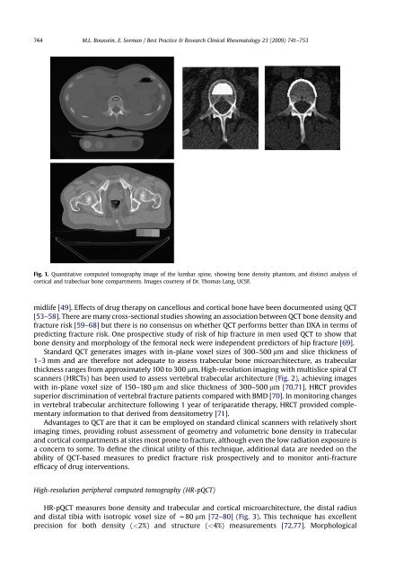
Fig. 1. Quantitative computed tomography image <strong>of</strong> <strong>the</strong> lumbar spine, showing <strong>bone</strong> density phantom, <strong>and</strong> distinct analysis <strong>of</strong><br />
cortical <strong>and</strong> trabecluar <strong>bone</strong> compartments. Images courtesy <strong>of</strong> Dr. Thomas Lang, UCSF.<br />
midlife [49]. Effects <strong>of</strong> drug <strong>the</strong>rapy on cancellous <strong>and</strong> cortical <strong>bone</strong> have been documented using QCT<br />
[53–58]. There are many cross-sectional studies showing an association between QCT <strong>bone</strong> density <strong>and</strong><br />
fracture risk [59–68] but <strong>the</strong>re is no consensus on whe<strong>the</strong>r QCT performs better than DXA in terms <strong>of</strong><br />
predicting fracture risk. One prospective study <strong>of</strong> risk <strong>of</strong> hip fracture in men used QCT to show that<br />
<strong>bone</strong> density <strong>and</strong> morphology <strong>of</strong> <strong>the</strong> femoral neck were independent predictors <strong>of</strong> hip fracture [69].<br />
St<strong>and</strong>ard QCT generates images with in-plane voxel sizes <strong>of</strong> 300–500 mm <strong>and</strong> slice thickness <strong>of</strong><br />
1–3 mm <strong>and</strong> are <strong>the</strong>refore not adequate to assess trabecular <strong>bone</strong> microarchitecture, as trabecular<br />
thickness ranges from approximately 100 to 300 mm. High-resolution imaging with multislice spiral CT<br />
scanners (HRCTs) has been used to assess vertebral trabecular architecture (Fig. 2), achieving images<br />
with in-plane voxel size <strong>of</strong> 150–180 mm <strong>and</strong> slice thickness <strong>of</strong> 300–500 mm [70,71]. HRCT provides<br />
superior discrimination <strong>of</strong> vertebral fracture patients compared with BMD [70]. In monitoring changes<br />
in vertebral trabecular architecture following 1 year <strong>of</strong> teriparatide <strong>the</strong>rapy, HRCT provided complementary<br />
information to that derived from densitometry [71].<br />
Advantages to QCT are that it can be employed on st<strong>and</strong>ard clinical scanners with relatively short<br />
imaging times, providing robust assessment <strong>of</strong> geometry <strong>and</strong> volumetric <strong>bone</strong> density in trabecular<br />
<strong>and</strong> cortical compartments at sites most prone to fracture, although even <strong>the</strong> low radiation exposure is<br />
a concern to some. To define <strong>the</strong> clinical utility <strong>of</strong> this technique, additional data are needed on <strong>the</strong><br />
ability <strong>of</strong> QCT-based measures to predict fracture risk prospectively <strong>and</strong> to monitor anti-fracture<br />
efficacy <strong>of</strong> drug interventions.<br />
High-resolution peripheral computed tomography (HR-pQCT)<br />
HR-pQCT measures <strong>bone</strong> density <strong>and</strong> trabecular <strong>and</strong> cortical microarchitecture, <strong>the</strong> distal radius<br />
<strong>and</strong> distal tibia with isotropic voxel size <strong>of</strong> w80 mm [72–80] (Fig. 3). This technique has excellent<br />
precision for both density (