Quantifying the material and structural determinants of bone strength
Quantifying the material and structural determinants of bone strength
Quantifying the material and structural determinants of bone strength

Create successful ePaper yourself
Turn your PDF publications into a flip-book with our unique Google optimized e-Paper software.
M.L. Bouxsein, E. Seeman / Best Practice & Research Clinical Rheumatology 23 (2009) 741–753 745<br />
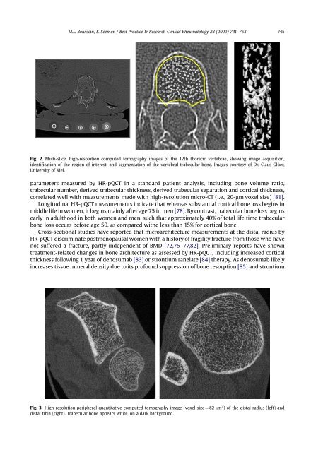
Fig. 2. Multi-slice, high-resolution computed tomography images <strong>of</strong> <strong>the</strong> 12th thoracic vertebrae, showing image acquisition,<br />
identification <strong>of</strong> <strong>the</strong> region <strong>of</strong> interest, <strong>and</strong> segmentation <strong>of</strong> <strong>the</strong> vertebral trabecular <strong>bone</strong>. Images courtesy <strong>of</strong> Dr. Claus Glüer,<br />
University <strong>of</strong> Kiel.<br />
parameters measured by HR-pQCT in a st<strong>and</strong>ard patient analysis, including <strong>bone</strong> volume ratio,<br />
trabecular number, derived trabecular thickness, derived trabecular separation <strong>and</strong> cortical thickness,<br />
correlated well with measurements made with high-resolution micro-CT (i.e., 20-mm voxel size) [81].<br />
Longitudinal HR-pQCT measurements indicate that whereas substantial cortical <strong>bone</strong> loss begins in<br />
middle life in women, it begins mainly after age 75 in men [78]. By contrast, trabecular <strong>bone</strong> loss begins<br />
early in adulthood in both women <strong>and</strong> men, such that approximately 40% <strong>of</strong> total life time trabecular<br />
<strong>bone</strong> loss occurs before age 50, as compared wi<strong>the</strong> less than 15% for cortical <strong>bone</strong>.<br />
Cross-sectional studies have reported that microarchitecture measurements at <strong>the</strong> distal radius by<br />
HR-pQCT discriminate postmenopausal women with a history <strong>of</strong> fragility fracture from those who have<br />
not suffered a fracture, partly independent <strong>of</strong> BMD [72,75–77,82]. Preliminary reports have shown<br />
treatment-related changes in <strong>bone</strong> architecture as assessed by HR-pQCT, including increased cortical<br />
thickness following 1 year <strong>of</strong> denosumab [83] or strontium ranelate [84] <strong>the</strong>rapy. As denosumab likely<br />
increases tissue mineral density due to its pr<strong>of</strong>ound suppression <strong>of</strong> <strong>bone</strong> resorption [85] <strong>and</strong> strontium<br />
Fig. 3. High-resolution peripheral quantitative computed tomography image (voxel size ¼ 82 mm 3 ) <strong>of</strong> <strong>the</strong> distal radius (left) <strong>and</strong><br />
distal tibia (right). Trabecular <strong>bone</strong> appears white, on a dark background.