Quantifying the material and structural determinants of bone strength
Quantifying the material and structural determinants of bone strength
Quantifying the material and structural determinants of bone strength

Create successful ePaper yourself
Turn your PDF publications into a flip-book with our unique Google optimized e-Paper software.
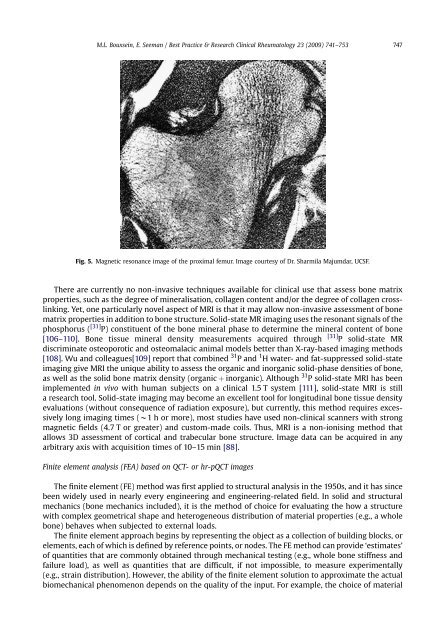
M.L. Bouxsein, E. Seeman / Best Practice & Research Clinical Rheumatology 23 (2009) 741–753 747<br />
Fig. 5. Magnetic resonance image <strong>of</strong> <strong>the</strong> proximal femur. Image courtesy <strong>of</strong> Dr. Sharmila Majumdar, UCSF.<br />
There are currently no non-invasive techniques available for clinical use that assess <strong>bone</strong> matrix<br />
properties, such as <strong>the</strong> degree <strong>of</strong> mineralisation, collagen content <strong>and</strong>/or <strong>the</strong> degree <strong>of</strong> collagen crosslinking.<br />
Yet, one particularly novel aspect <strong>of</strong> MRI is that it may allow non-invasive assessment <strong>of</strong> <strong>bone</strong><br />
matrix properties in addition to <strong>bone</strong> structure. Solid-state MR imaging uses <strong>the</strong> resonant signals <strong>of</strong> <strong>the</strong><br />
phosphorus ( [31] P) constituent <strong>of</strong> <strong>the</strong> <strong>bone</strong> mineral phase to determine <strong>the</strong> mineral content <strong>of</strong> <strong>bone</strong><br />
[106–110]. Bone tissue mineral density measurements acquired through [31] P solid-state MR<br />
discriminate osteoporotic <strong>and</strong> osteomalacic animal models better than X-ray-based imaging methods<br />
[108]. Wu <strong>and</strong> colleagues[109] report that combined 31 P <strong>and</strong> 1 H water- <strong>and</strong> fat-suppressed solid-state<br />
imaging give MRI <strong>the</strong> unique ability to assess <strong>the</strong> organic <strong>and</strong> inorganic solid-phase densities <strong>of</strong> <strong>bone</strong>,<br />
as well as <strong>the</strong> solid <strong>bone</strong> matrix density (organic þ inorganic). Although 31 P solid-state MRI has been<br />
implemented in vivo with human subjects on a clinical 1.5 T system [111], solid-state MRI is still<br />
a research tool. Solid-state imaging may become an excellent tool for longitudinal <strong>bone</strong> tissue density<br />
evaluations (without consequence <strong>of</strong> radiation exposure), but currently, this method requires excessively<br />
long imaging times (w1 h or more), most studies have used non-clinical scanners with strong<br />
magnetic fields (4.7 T or greater) <strong>and</strong> custom-made coils. Thus, MRI is a non-ionising method that<br />
allows 3D assessment <strong>of</strong> cortical <strong>and</strong> trabecular <strong>bone</strong> structure. Image data can be acquired in any<br />
arbitrary axis with acquisition times <strong>of</strong> 10–15 min [88].<br />
Finite element analysis (FEA) based on QCT- or hr-pQCT images<br />
The finite element (FE) method was first applied to <strong>structural</strong> analysis in <strong>the</strong> 1950s, <strong>and</strong> it has since<br />
been widely used in nearly every engineering <strong>and</strong> engineering-related field. In solid <strong>and</strong> <strong>structural</strong><br />
mechanics (<strong>bone</strong> mechanics included), it is <strong>the</strong> method <strong>of</strong> choice for evaluating <strong>the</strong> how a structure<br />
with complex geometrical shape <strong>and</strong> heterogeneous distribution <strong>of</strong> <strong>material</strong> properties (e.g., a whole<br />
<strong>bone</strong>) behaves when subjected to external loads.<br />
The finite element approach begins by representing <strong>the</strong> object as a collection <strong>of</strong> building blocks, or<br />
elements, each <strong>of</strong> which is defined by reference points, or nodes. The FE method can provide ‘estimates’<br />
<strong>of</strong> quantities that are commonly obtained through mechanical testing (e.g., whole <strong>bone</strong> stiffness <strong>and</strong><br />
failure load), as well as quantities that are difficult, if not impossible, to measure experimentally<br />
(e.g., strain distribution). However, <strong>the</strong> ability <strong>of</strong> <strong>the</strong> finite element solution to approximate <strong>the</strong> actual<br />
biomechanical phenomenon depends on <strong>the</strong> quality <strong>of</strong> <strong>the</strong> input. For example, <strong>the</strong> choice <strong>of</strong> <strong>material</strong>