Quantifying the material and structural determinants of bone strength
Quantifying the material and structural determinants of bone strength
Quantifying the material and structural determinants of bone strength

Create successful ePaper yourself
Turn your PDF publications into a flip-book with our unique Google optimized e-Paper software.
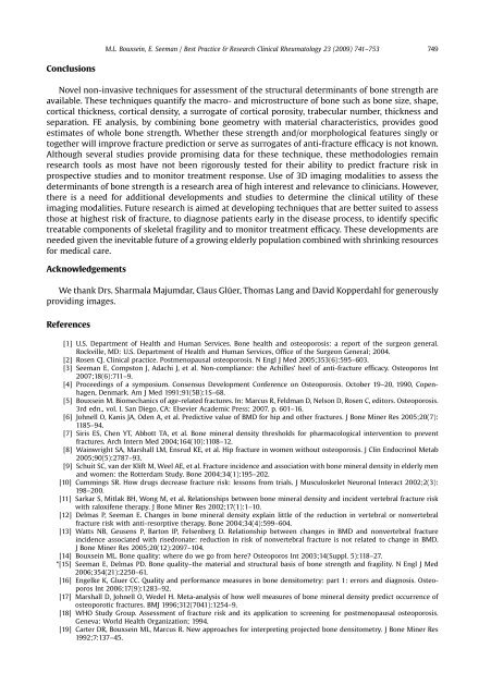
Conclusions<br />
Novel non-invasive techniques for assessment <strong>of</strong> <strong>the</strong> <strong>structural</strong> <strong>determinants</strong> <strong>of</strong> <strong>bone</strong> <strong>strength</strong> are<br />
available. These techniques quantify <strong>the</strong> macro- <strong>and</strong> microstructure <strong>of</strong> <strong>bone</strong> such as <strong>bone</strong> size, shape,<br />
cortical thickness, cortical density, a surrogate <strong>of</strong> cortical porosity, trabecular number, thickness <strong>and</strong><br />
separation. FE analysis, by combining <strong>bone</strong> geometry with <strong>material</strong> characteristics, provides good<br />
estimates <strong>of</strong> whole <strong>bone</strong> <strong>strength</strong>. Whe<strong>the</strong>r <strong>the</strong>se <strong>strength</strong> <strong>and</strong>/or morphological features singly or<br />
toge<strong>the</strong>r will improve fracture prediction or serve as surrogates <strong>of</strong> anti-fracture efficacy is not known.<br />
Although several studies provide promising data for <strong>the</strong>se technique, <strong>the</strong>se methodologies remain<br />
research tools as most have not been rigorously tested for <strong>the</strong>ir ability to predict fracture risk in<br />
prospective studies <strong>and</strong> to monitor treatment response. Use <strong>of</strong> 3D imaging modalities to assess <strong>the</strong><br />
<strong>determinants</strong> <strong>of</strong> <strong>bone</strong> <strong>strength</strong> is a research area <strong>of</strong> high interest <strong>and</strong> relevance to clinicians. However,<br />
<strong>the</strong>re is a need for additional developments <strong>and</strong> studies to determine <strong>the</strong> clinical utility <strong>of</strong> <strong>the</strong>se<br />
imaging modalities. Future research is aimed at developing techniques that are better suited to assess<br />
those at highest risk <strong>of</strong> fracture, to diagnose patients early in <strong>the</strong> disease process, to identify specific<br />
treatable components <strong>of</strong> skeletal fragility <strong>and</strong> to monitor treatment efficacy. These developments are<br />
needed given <strong>the</strong> inevitable future <strong>of</strong> a growing elderly population combined with shrinking resources<br />
for medical care.<br />
Acknowledgements<br />
We thank Drs. Sharmala Majumdar, Claus Glüer, Thomas Lang <strong>and</strong> David Kopperdahl for generously<br />
providing images.<br />
References<br />
M.L. Bouxsein, E. Seeman / Best Practice & Research Clinical Rheumatology 23 (2009) 741–753 749<br />
[1] U.S. Department <strong>of</strong> Health <strong>and</strong> Human Services. Bone health <strong>and</strong> osteoporosis: a report <strong>of</strong> <strong>the</strong> surgeon general.<br />
Rockville, MD: U.S. Department <strong>of</strong> Health <strong>and</strong> Human Services, Office <strong>of</strong> <strong>the</strong> Surgeon General; 2004.<br />
[2] Rosen CJ. Clinical practice. Postmenopausal osteoporosis. N Engl J Med 2005;353(6):595–603.<br />
[3] Seeman E, Compston J, Adachi J, et al. Non-compliance: <strong>the</strong> Achilles’ heel <strong>of</strong> anti-fracture efficacy. Osteoporos Int<br />
2007;18(6):711–9.<br />
[4] Proceedings <strong>of</strong> a symposium. Consensus Development Conference on Osteoporosis. October 19–20, 1990, Copenhagen,<br />
Denmark. Am J Med 1991;91(5B):1S–68.<br />
[5] Bouxsein M. Biomechanics <strong>of</strong> age-related fractures. In: Marcus R, Feldman D, Nelson D, Rosen C, editors. Osteoporosis.<br />
3rd edn., vol. I. San Diego, CA: Elsevier Academic Press; 2007. p. 601–16.<br />
[6] Johnell O, Kanis JA, Oden A, et al. Predictive value <strong>of</strong> BMD for hip <strong>and</strong> o<strong>the</strong>r fractures. J Bone Miner Res 2005;20(7):<br />
1185–94.<br />
[7] Siris ES, Chen YT, Abbott TA, et al. Bone mineral density thresholds for pharmacological intervention to prevent<br />
fractures. Arch Intern Med 2004;164(10):1108–12.<br />
[8] Wainwright SA, Marshall LM, Ensrud KE, et al. Hip fracture in women without osteoporosis. J Clin Endocrinol Metab<br />
2005;90(5):2787–93.<br />
[9] Schuit SC, van der Klift M, Weel AE, et al. Fracture incidence <strong>and</strong> association with <strong>bone</strong> mineral density in elderly men<br />
<strong>and</strong> women: <strong>the</strong> Rotterdam Study. Bone 2004;34(1):195–202.<br />
[10] Cummings SR. How drugs decrease fracture risk: lessons from trials. J Musculoskelet Neuronal Interact 2002;2(3):<br />
198–200.<br />
[11] Sarkar S, Mitlak BH, Wong M, et al. Relationships between <strong>bone</strong> mineral density <strong>and</strong> incident vertebral fracture risk<br />
with raloxifene <strong>the</strong>rapy. J Bone Miner Res 2002;17(1):1–10.<br />
[12] Delmas P, Seeman E. Changes in <strong>bone</strong> mineral density explain little <strong>of</strong> <strong>the</strong> reduction in vertebral or nonvertebral<br />
fracture risk with anti-resorptive <strong>the</strong>rapy. Bone 2004;34(4):599–604.<br />
[13] Watts NB, Geusens P, Barton IP, Felsenberg D. Relationship between changes in BMD <strong>and</strong> nonvertebral fracture<br />
incidence associated with risedronate: reduction in risk <strong>of</strong> nonvertebral fracture is not related to change in BMD.<br />
J Bone Miner Res 2005;20(12):2097–104.<br />
[14] Bouxsein ML. Bone quality: where do we go from here? Osteoporos Int 2003;14(Suppl. 5):118–27.<br />
*[15] Seeman E, Delmas PD. Bone quality–<strong>the</strong> <strong>material</strong> <strong>and</strong> <strong>structural</strong> basis <strong>of</strong> <strong>bone</strong> <strong>strength</strong> <strong>and</strong> fragility. N Engl J Med<br />
2006;354(21):2250–61.<br />
[16] Engelke K, Gluer CC. Quality <strong>and</strong> performance measures in <strong>bone</strong> densitometry: part 1: errors <strong>and</strong> diagnosis. Osteoporos<br />
Int 2006;17(9):1283–92.<br />
[17] Marshall D, Johnell O, Wedel H. Meta-analysis <strong>of</strong> how well measures <strong>of</strong> <strong>bone</strong> mineral density predict occurrence <strong>of</strong><br />
osteoporotic fractures. BMJ 1996;312(7041):1254–9.<br />
[18] WHO Study Group. Assessment <strong>of</strong> fracture risk <strong>and</strong> its application to screening for postmenopausal osteoporosis.<br />
Geneva: World Health Organization; 1994.<br />
[19] Carter DR, Bouxsein ML, Marcus R. New approaches for interpreting projected <strong>bone</strong> densitometry. J Bone Miner Res<br />
1992;7:137–45.