The contribution of Asian researchers to the field of rheumatology
The contribution of Asian researchers to the field of rheumatology
The contribution of Asian researchers to the field of rheumatology

Create successful ePaper yourself
Turn your PDF publications into a flip-book with our unique Google optimized e-Paper software.
eVIewS<br />
Institute <strong>of</strong> Medical<br />
Science, St Marianna<br />
University School <strong>of</strong><br />
Medicine, Kanagawa,<br />
2‑16‑1 Sugao,<br />
Miyamae‑ku, Kawasaki‑<br />
shi, Kanagawa<br />
216‑8512, Japan<br />
(Y. Yamano). Institute<br />
<strong>of</strong> Medical Science,<br />
Tokyo Medical<br />
University, 1‑6‑1<br />
Shinjyuku, Shinjyuku‑ku,<br />
Tokyo 160‑8402, Japan<br />
K. Nishioka).<br />
Correspondence <strong>to</strong>:<br />
K. Nishioka<br />
<strong>to</strong>ranomon‑nishioka@<br />
cpost.plala.or.jp<br />
<strong>The</strong> <strong>contribution</strong> <strong>of</strong> <strong>Asian</strong> <strong>researchers</strong><br />
<strong>to</strong> <strong>the</strong> <strong>field</strong> <strong>of</strong> rheuma<strong>to</strong>logy<br />
Yoshihisa Yamano and Kusuki Nishioka<br />
Abstract | Asia is home <strong>to</strong> more than half <strong>of</strong> <strong>the</strong> world’s population and is a region <strong>of</strong> diverse ethnicity, culture,<br />
microbial endemicity, and economic backgrounds. This diversity is also reflected in <strong>the</strong> heterogeneity among<br />
<strong>Asian</strong> patients with rheumatic diseases in terms <strong>of</strong> clinical manifestations, disease courses, treatment<br />
responses and outcomes, which provides opportunities for <strong>researchers</strong> <strong>to</strong> conduct some unique studies.<br />
Several disease entities, such as Behçet syndrome, Takayasu arteritis, Kawasaki disease, and immunological<br />
disorders associated with human T‑lymphotropic virus type 1 (HTLV‑1), were first observed and defined in<br />
Asia. In addition, <strong>the</strong> region’s <strong>researchers</strong> have been at <strong>the</strong> forefront <strong>of</strong> research in some interesting scientific<br />
<strong>to</strong>pics, which has opened up new research avenues in rheuma<strong>to</strong>logy, such as <strong>the</strong> direct targeting <strong>of</strong> synovial<br />
cells in patients with rheuma<strong>to</strong>id arthritis via activation <strong>of</strong> <strong>the</strong> agonistic Fas pathway, establishment <strong>of</strong> <strong>the</strong> <strong>field</strong><br />
<strong>of</strong> osteoimmunology, <strong>the</strong> discovery <strong>of</strong> regula<strong>to</strong>ry T cells and synoviolin, and <strong>the</strong> development <strong>of</strong> <strong>to</strong>cilizumab, a<br />
humanized monoclonal antibody against interleukin‑6 recep<strong>to</strong>r.<br />
Yamano, Y. & Nishioka, K. Nat. Rev. Rheuma<strong>to</strong>l. 6, 106–111 (2010); doi:10.1038/nrrheum.2009.257<br />
Introduction<br />
Asia is a region <strong>of</strong> diverse ethnicities, cultures, economies,<br />
and microbial endemicity, providing unique opportunities<br />
for <strong>Asian</strong> <strong>researchers</strong> <strong>to</strong> contribute <strong>to</strong> <strong>the</strong> <strong>field</strong> <strong>of</strong> rheuma<strong>to</strong>logy.<br />
For example, <strong>the</strong> Community Oriented Program<br />
for <strong>the</strong> Control <strong>of</strong> Rheumatic Diseases (COPCORD),<br />
sponsored by <strong>the</strong> WHO and International League Against<br />
Rheumatism (WHO-ILAR), has generated extensive data<br />
on <strong>the</strong> epidemiology and charac teristics <strong>of</strong> rheumatic diseases<br />
in Asia. 1 <strong>The</strong>se data revealed that <strong>the</strong> incidence <strong>of</strong><br />
rheumatic fever has markedly increased in Central Asia<br />
during <strong>the</strong> past decade. 2 Several <strong>Asian</strong> countries have<br />
active rheuma<strong>to</strong>logy communities in clinical practice and<br />
academic and research capacities, as well as an active pr<strong>of</strong>essional<br />
society, <strong>the</strong> Asia Pacific League <strong>of</strong> Associations<br />
for Rheuma<strong>to</strong>logy (APLAR).<br />
On <strong>the</strong> basis <strong>of</strong> <strong>the</strong> varied ethnicity and microbial<br />
endemicity in Asia, rheuma<strong>to</strong>logists have identified novel<br />
disease entities endemic <strong>to</strong> <strong>the</strong> region, such as Behçet syndrome,<br />
3 Takayasu arteritis, Kawasaki disease, and immunological<br />
disorders associated with human T-lymphotropic<br />
virus type 1 (HTLV-1). 4–8 Research in<strong>to</strong> <strong>the</strong> pathogenesis <strong>of</strong><br />
<strong>the</strong>se diseases has pr<strong>of</strong>oundly increased our understanding<br />
<strong>of</strong> o<strong>the</strong>r immune-mediated disorders.<br />
Progress in conventional scientific disciplines has<br />
opened up new research avenues in rheuma<strong>to</strong>logy,<br />
which has resulted in various advances, such as <strong>the</strong><br />
establishment <strong>of</strong> <strong>the</strong> new <strong>field</strong> <strong>of</strong> osteoimmunology 9 and<br />
<strong>the</strong> discoveries <strong>of</strong> T-regula<strong>to</strong>ry (T REG ) cells 10,11 and <strong>the</strong><br />
Competing interests<br />
K. Nishioka declares an association with <strong>the</strong> following company:<br />
Argenes. See <strong>the</strong> article online for full details <strong>of</strong> <strong>the</strong> relationship.<br />
Y. Yamano declares no competing interests.<br />
E3 ubiquitin ligase synoviolin. 12 Moreover, large-scale<br />
genetic studies <strong>of</strong> rheumatic diseases have identified polymorphisms<br />
in multiple genes associated with disease susceptibility,<br />
including PADI4 and PTPN22. Notably, disease<br />
suscepti bility conferr ed by genetic fac<strong>to</strong>rs varies between<br />
ethnic groups. 13<br />
Translation <strong>of</strong> labora<strong>to</strong>ry research findings <strong>to</strong> clinical<br />
practice is a challenging prospect for rheuma<strong>to</strong>logists.<br />
In this respect, <strong>Asian</strong> rheuma<strong>to</strong>logists have achieved<br />
consider able success, exemplified by <strong>the</strong> clinical application<br />
<strong>of</strong> <strong>to</strong>cilizumab, a humanized monoclonal antibody<br />
<strong>to</strong> interleukin (IL)-6 recep<strong>to</strong>r (anti-IL-6R), for <strong>the</strong><br />
treatment <strong>of</strong> rheuma<strong>to</strong>id arthritis (RA), systemic juvenile<br />
idiopathic arthritis, Castleman disease, and Crohn<br />
disease. 14 Studies have also highlighted benefits achieved<br />
with agonistic antibodies against Fas recep<strong>to</strong>r (FasR;<br />
also known as CD95, Apo-1 and tumor necrosis fac<strong>to</strong>r<br />
[TNF] recep<strong>to</strong>r superfamily, member 6) in <strong>the</strong> treatment<br />
<strong>of</strong> patients with RA. This Review focuses on new disease<br />
entities and advances in scientific disciplines and treatments<br />
reported from Asia, and outlines <strong>the</strong> <strong>contribution</strong>s<br />
<strong>of</strong> <strong>Asian</strong> <strong>researchers</strong> <strong>to</strong> <strong>the</strong> <strong>field</strong> <strong>of</strong> rheuma<strong>to</strong>logy.<br />
Prevalent diseases in Asia<br />
Behçet syndrome<br />
In 1937, Hulusi Behçet first described patients with<br />
symp <strong>to</strong>ms <strong>of</strong> oral aphtae, genital ulcers, and uveitis. 15<br />
Sub sequently, <strong>the</strong> syndrome was also shown <strong>to</strong> include<br />
articular, gastrointestinal, central nervous system, and<br />
peripheral vascular manifestations. <strong>The</strong> regions with<br />
<strong>the</strong> highest incidence <strong>of</strong> Behçet syndrome are <strong>the</strong> Mediterranean<br />
basin, Middle East, and Far East. <strong>The</strong> reasons for<br />
this peculiar geographic distribution <strong>of</strong> Behçet syndrome<br />
106 | FEBRUARY 2010 | volUmE 6 www.nature.com/nrrheum<br />
© 20 10<br />
Macmillan Publishers Limited. All rights reserved
emain unknown. <strong>The</strong> putative etiological agents associated<br />
with Behçet syndrome may have spread along <strong>the</strong><br />
ancient Silk Routes, which extend from Eastern Europe<br />
<strong>to</strong> Japan. 16 <strong>The</strong> geographical distribution <strong>of</strong> several predisposing<br />
genetic fac<strong>to</strong>rs, such as <strong>the</strong> HLA‑B*51 allele, 16<br />
might also be associated with this regional variation<br />
in incidence. <strong>The</strong> pathogenesis <strong>of</strong> this syndrome also<br />
remains unclear, and several studies have focused on a<br />
potential infectious etiology. <strong>The</strong> acne lesions associated<br />
with Behçet syndrome are frequently colonized by<br />
Staphylococcus aureus, and <strong>to</strong> a lesser extent by Prevotella<br />
spp., 17 suggesting that at least some patients with this syndrome<br />
develop a reactive inflamma<strong>to</strong>ry response <strong>to</strong> infective<br />
agents. Thus, <strong>the</strong> characteristics <strong>of</strong> Behçet syndrome<br />
are different from <strong>the</strong> classic features <strong>of</strong> au<strong>to</strong>immune<br />
disease, and <strong>the</strong>re is an ongoing debate about whe<strong>the</strong>r<br />
Behçet syndrome belongs <strong>to</strong> a newly designated group <strong>of</strong><br />
au<strong>to</strong>inflamma<strong>to</strong>ry diseases. 18<br />
Takayasu arteritis<br />
Takayasu arteritis, first described in 1908 by Miko<strong>to</strong><br />
Takayasu, is an idiopathic chronic inflamma<strong>to</strong>ry disease<br />
that results in granuloma<strong>to</strong>us panarteritis involving <strong>the</strong><br />
large vessels, such as <strong>the</strong> aorta and its major branches.<br />
This disease has a worldwide distribution, with a high<br />
prevalence in people <strong>of</strong> Latin American and <strong>Asian</strong> ancestry.<br />
19 <strong>The</strong> annual incidence <strong>of</strong> Takayasu arteritis in Japan<br />
is estimated <strong>to</strong> be 150 new cases per million people, 20 compared<br />
with 1–3 new cases per million people in <strong>the</strong> uSA<br />
and Europe. 21 Several reports indicate an increased frequency<br />
<strong>of</strong> <strong>the</strong> antigens HLA-Bw52 and HLA-B39.2 among<br />
patients in Japan, suggesting an immunogenetic association.<br />
22 Although <strong>the</strong> pathogenesis <strong>of</strong> Takayasu arteritis<br />
remains unclear, evidence strongly indicates <strong>the</strong> involvement<br />
<strong>of</strong> T cells. Interestingly, immuno his<strong>to</strong>pathologic<br />
examination has shown that <strong>the</strong> cells infiltrating aortic<br />
tissue mainly consist <strong>of</strong> cy<strong>to</strong><strong>to</strong>xic lymphocytes, particularly<br />
γδ T lymphocytes, 23 which might cause vascular<br />
injury by releasing large amounts <strong>of</strong> cy<strong>to</strong>lytic compounds,<br />
such as perforin.<br />
Kawasaki disease<br />
Kawasaki disease, initially described by Tomisaku<br />
Kawasaki in 1967, is one <strong>of</strong> <strong>the</strong> most common forms <strong>of</strong><br />
childhood vasculitides that predominantly affects <strong>the</strong><br />
medium and small arteries. <strong>The</strong> incidence <strong>of</strong> Kawasaki<br />
disease is highest in children residing in East Asia<br />
(annual incidence 140 per 100,000 in Japan, 69 per<br />
100,000 in Taiwan, and 51 per 100,000 in Beijing) 24 and<br />
those <strong>of</strong> <strong>Asian</strong> ancestry residing in o<strong>the</strong>r parts <strong>of</strong> <strong>the</strong><br />
world. In <strong>the</strong> uSA, <strong>the</strong> annual incidence <strong>of</strong> Kawasaki<br />
disease was highest among <strong>Asian</strong>s and Pacific Islanders<br />
(33 per 100,000), and lowest among white people (9 per<br />
100,000). 25 Thus, genetic susceptibility fac<strong>to</strong>rs seem <strong>to</strong><br />
contribute <strong>to</strong> <strong>the</strong> pathogenesis <strong>of</strong> this disorder. Although<br />
<strong>the</strong> precise cause <strong>of</strong> Kawasaki disease remains elusive,<br />
evidence suggests an infectious etiology. For example,<br />
incidence varies seasonally, with increases seen during<br />
<strong>the</strong> winter month <strong>of</strong> January and summer months <strong>of</strong> June<br />
and July, 26 outbreaks have been linked <strong>to</strong> clusters <strong>of</strong> cases<br />
Key points<br />
■ Varied ethnicity, microbial endemicity and heterogeneity among <strong>Asian</strong><br />
patients in presentation and outcomes provide opportunities for some<br />
unique studies<br />
reviews<br />
■ Several diseases, such as Behçet syndrome, Takayasu arteritis, Kawasaki<br />
disease, and immunological disorders associated with human T‑lymphotropic<br />
virus type 1 were first defined in Asia<br />
■ <strong>Asian</strong> research has been at <strong>the</strong> forefront <strong>of</strong> several avenues in rheuma<strong>to</strong>logy,<br />
such as establishment <strong>of</strong> <strong>the</strong> <strong>field</strong> <strong>of</strong> osteoimmunology, and <strong>the</strong> discoveries<br />
<strong>of</strong> regula<strong>to</strong>ry T cells and synoviolin<br />
■ <strong>Asian</strong> scientists have achieved considerable success in <strong>the</strong> translation<br />
<strong>of</strong> labora<strong>to</strong>ry findings <strong>to</strong> clinical practice, exemplified by <strong>the</strong> <strong>the</strong>rapeutic<br />
application <strong>of</strong> <strong>to</strong>cilizumab and agonistic antibodies <strong>to</strong> Fasr<br />
occurring in association with heavy rainfall, and <strong>the</strong> clinical<br />
features <strong>of</strong> Kawasaki disease are similar <strong>to</strong> those <strong>of</strong><br />
o<strong>the</strong>r infectious diseases, such as adenovirus infection<br />
and scarlet fever. However, no single infectious agent has<br />
yet been identified.<br />
Several genetic polymorphisms have been linked <strong>to</strong><br />
Kawasaki disease. Notably, a 2008 study by Onouchi<br />
et al. 27 reported an association between a singlenucleotide<br />
polymorphism in ITPKC and an increased<br />
risk <strong>of</strong> Kawasaki disease in children. 27 ITPKC acts as a<br />
negative regula<strong>to</strong>r <strong>of</strong> T-cell activation, and may contribute<br />
<strong>to</strong> immune hyper-reactivity in this disease. This finding<br />
provides new insights in<strong>to</strong> <strong>the</strong> mechanisms <strong>of</strong> immune<br />
activation in Kawasaki disease, and emphasizes <strong>the</strong> role<br />
<strong>of</strong> activated T cells in its pathogenesis.<br />
hTlv‑1‑associated immunological disorders<br />
HTLV-1 is a retrovirus associated with chronic, persistent<br />
infection <strong>of</strong> human T cells. This virus is endemic in sou<strong>the</strong>rn<br />
Japan, <strong>the</strong> Caribbean, and parts <strong>of</strong> South America,<br />
Africa, <strong>the</strong> Middle East and Melanesia. 28 Studies conducted<br />
in <strong>the</strong>se areas have demonstrated that HTLV-1<br />
infection is associated with several human diseases,<br />
including adult T-cell leukemia, an aggressive mature<br />
T-cell malignancy. 29 HTLV-1 is also associated with nonneoplastic<br />
immunological disorders charac terized by<br />
multiorgan lymphocytic infiltrates, such as myelopathy/<br />
tropical spastic paraparesis, 4 HTLV-1-associated arthropathy<br />
(HAAP), 5 uveitis, 6 bronchoalveolitis, 7 Sjögren<br />
syndrome, and polymyositis. 8 Some patients present<br />
with multiple HTLV-1-associated immuno logical disorders.<br />
A progressive course and persistent inflammation<br />
affecting various organs are frequently seen in patients<br />
with idiopathic au<strong>to</strong>immune disorders. <strong>The</strong>se HTLV-1associated<br />
multiorgan immunological disorders are,<br />
<strong>the</strong>refore, extremely important models for understanding<br />
<strong>the</strong> pathogenic mechanisms <strong>of</strong> o<strong>the</strong>r organ-specific<br />
immune disorders. 30,31<br />
HAAP is characterized by chronic inflamma<strong>to</strong>ry<br />
and proliferative synovitis with lymphoid follicles and<br />
pannus formation in <strong>the</strong> affected joints. Although <strong>the</strong> predominant<br />
viral reservoirs in peripheral blood <strong>of</strong> HTLV-1infected<br />
individuals are CD4 + CD25 + T cells, HTLV-1 has<br />
been reported <strong>to</strong> infect in vitro and in vivo a number<br />
<strong>of</strong> cell types, including synovial cells. In synoviocytes,<br />
NATuRE REVIEWS | rheumATologY VOLuME 6 | FEBRuARY 2010 | 107<br />
© 20 10<br />
Macmillan Publishers Limited. All rights reserved
eviews<br />
HTLV-1 Tax, a transactiva<strong>to</strong>r protein, has been demonstrated<br />
<strong>to</strong> upregulate <strong>the</strong> expression <strong>of</strong> proinflamma<strong>to</strong>ry<br />
cy<strong>to</strong>kines and several oncogenes, such as FOS, JUN and<br />
MYC, which might contribute <strong>to</strong> synovial proliferation. 32<br />
Fur<strong>the</strong>rmore, HTLV-1 Tax transgenic mice develop an<br />
inflamma<strong>to</strong>ry arthropathy resembling human RA. 33 <strong>The</strong>se<br />
findings suggest that HTLV‑1 tax is one <strong>of</strong> <strong>the</strong> exogenous<br />
retrovirus genes responsible for <strong>the</strong> synovial hyperplasia<br />
associated with immune dysregulation in HAAP.<br />
Notable advances in rheuma<strong>to</strong>logy<br />
osteoimmunology<br />
Research indicating that <strong>the</strong> immune and skeletal systems<br />
interact and share a number <strong>of</strong> regula<strong>to</strong>ry molecules,<br />
including cy<strong>to</strong>kines, recep<strong>to</strong>rs, signaling molecules and<br />
transcription fac<strong>to</strong>rs, has led <strong>to</strong> <strong>the</strong> establishment <strong>of</strong> a<br />
new <strong>field</strong>: osteoimmunology. 34 Although <strong>the</strong> inter action<br />
between skeletal and immune systems occurs both in<br />
health and disease, <strong>the</strong> effects <strong>of</strong> disruption <strong>to</strong> this interaction<br />
are exemplified in RA. Bone destruction in RA<br />
is caused by increased osteoclastic activity. <strong>The</strong> osteoclasts<br />
are derived from <strong>the</strong> myeloid cells <strong>of</strong> <strong>the</strong> immune<br />
system, 35 and proinflamma<strong>to</strong>ry cy<strong>to</strong>kines, such as TNF,<br />
seem <strong>to</strong> be crucial for <strong>the</strong> change in <strong>the</strong> activity levels<br />
<strong>of</strong> both bone cell types. TNF increases <strong>the</strong> osteoblastic<br />
expression <strong>of</strong> recep<strong>to</strong>r activa<strong>to</strong>r or nuclear fac<strong>to</strong>r κB<br />
ligand (RANKL; also known as TNF ligand super family,<br />
member 11), 36 which in turn increases <strong>the</strong> local differentiation<br />
<strong>of</strong> bone-resorbing osteoclasts. RANKL is expressed<br />
both by bone-forming osteoblasts and activated T cells,<br />
indicating that osteoclastic bone resorption is influenced<br />
by <strong>the</strong> immune system. 37 Moreover, Takayanagi et al. 38<br />
discovered a crucial counter-regula<strong>to</strong>ry mechanism, by<br />
which activated T cells can inhibit <strong>the</strong> RANKL-induced<br />
maturation and activation <strong>of</strong> osteoclasts. Toge<strong>the</strong>r <strong>the</strong>se<br />
findings indicate <strong>the</strong> crucial role <strong>of</strong> skeletal cells in both<br />
<strong>the</strong> hema<strong>to</strong>poietic and immune systems.<br />
Osteoimmunology encompasses <strong>the</strong> analysis <strong>of</strong><br />
develop mental, homeostatic and pathologic consequences<br />
<strong>of</strong> <strong>the</strong> interactions between <strong>the</strong> immune and<br />
skeletal systems. understanding <strong>the</strong>se complex interactions<br />
should provide new insights in<strong>to</strong> <strong>the</strong> functional<br />
regulation <strong>of</strong> both systems, and also uncover new targets<br />
for <strong>the</strong>rapeutic intervention.<br />
Discovery <strong>of</strong> T‑regula<strong>to</strong>ry cells<br />
<strong>The</strong> precise mechanisms underlying <strong>the</strong> induction <strong>of</strong><br />
uncontrolled immune reactions and <strong>the</strong> induction <strong>of</strong> <strong>the</strong><br />
accelerated immune responses in rheumatic disorders are<br />
not fully unders<strong>to</strong>od. In <strong>the</strong> mid-1990s, Sakaguchi et al. 10<br />
reported <strong>the</strong> concept <strong>of</strong> T-cell-mediated suppression by<br />
showing that a minor population <strong>of</strong> CD4 + T cells, which<br />
coexpress <strong>the</strong> IL-2 recep<strong>to</strong>r α-chain (CD25), is crucial for<br />
<strong>the</strong> control <strong>of</strong> au<strong>to</strong>reactive T cells in vivo. This discovery<br />
<strong>of</strong> a new CD4 + CD25 + T-cell subset, termed T REG cells,<br />
has provided new opportunities and generated increased<br />
interest in elucidating <strong>the</strong>se mechanisms. 10,11 In healthy<br />
individuals, in vitro studies have shown that T REG cells suppress<br />
<strong>the</strong> proliferation <strong>of</strong> and production <strong>of</strong> cy<strong>to</strong>kines by<br />
pathogenic T cells. 11<br />
Although T REG cells are phenotypically similar <strong>to</strong><br />
activated T cells, <strong>the</strong>y can be identified by <strong>the</strong> intracellular<br />
expression <strong>of</strong> <strong>the</strong> transcriptional regula<strong>to</strong>r forkhead<br />
box P3 (FOXP3). 39 This protein is critical for <strong>the</strong><br />
develop ment and function <strong>of</strong> T REG cells in both mice and<br />
humans. Substantial reductions in FOXP3 expression<br />
and/or T REG cell function have been observed in several<br />
human au<strong>to</strong>immune diseases, 12 suggesting that altered<br />
FOXP3 expression and/or T REG function precipitates <strong>the</strong><br />
loss <strong>of</strong> immunologic <strong>to</strong>lerance. A 2009 study by Miyara<br />
et al. 40 demonstrated that human FOXP3 + CD4 + T cells<br />
were composed <strong>of</strong> three phenotypically and functionally<br />
distinct subpopulations: CD45RA + FOXP3 low resting<br />
T REG cells; CD45RA – FOXP3 high activated T REG cells; and<br />
cy<strong>to</strong>kine-secreting CD45RA – FOXP3 low T cells. <strong>The</strong><br />
former two subtypes are suppressive and <strong>the</strong> latter<br />
is non-suppressive. Interestingly, <strong>the</strong> number <strong>of</strong> FOXP3 low<br />
non-suppressive memory T cells was increased in<br />
patients with active systemic lupus ery<strong>the</strong>ma<strong>to</strong>sus 40 and<br />
in those with HTLV-1-associated neuroimmunological<br />
disorders. 41 Fur<strong>the</strong>r investigations <strong>of</strong> <strong>the</strong> mechanism <strong>of</strong><br />
action <strong>of</strong> T REG cells in au<strong>to</strong>immune diseases will help<br />
identify new molecular pathways, which may in turn<br />
provide insight in<strong>to</strong> understanding basic pathogenic<br />
mechanisms <strong>of</strong> immunological disorders.<br />
Discovery <strong>of</strong> synoviolin<br />
<strong>The</strong> pathologic features <strong>of</strong> RA include chronic, systemic<br />
inflammation <strong>of</strong> joints, which is associated with increased<br />
proliferation <strong>of</strong> synovial cells (Figure 1). Studies in Japan<br />
focusing on synovial cells led <strong>to</strong> <strong>the</strong> discovery <strong>of</strong> a novel<br />
molecule that is overexpressed in <strong>the</strong>se cells: synoviolin.<br />
Amano et al. 42 used immunoscreening <strong>to</strong> identify <strong>the</strong> role<br />
<strong>of</strong> synoviolin in <strong>the</strong> process <strong>of</strong> endoplasmic-reticulumassociated<br />
degradation (ERAD)—an ATP-dependent<br />
ubiquitin–proteasome degradation process that eliminates<br />
defective proteins. <strong>The</strong> endoplasmic reticulum<br />
(ER) has an important role in protein folding. When <strong>the</strong><br />
level <strong>of</strong> unfolded proteins in <strong>the</strong> ER exceeds its folding<br />
capacity, <strong>the</strong> burden on <strong>the</strong> ER is reduced by ERAD. 43<br />
A mouse study showed that approximately 30% <strong>of</strong><br />
mice overexpressing synoviolin developed spontaneous<br />
arthropathy as a result <strong>of</strong> reduced apop<strong>to</strong>sis <strong>of</strong> synoviocytes.<br />
Conversely, synoviolin-heterozygous (Syvn1 +/– )<br />
mice showed resistance <strong>to</strong> collagen-induced arthritis<br />
owing <strong>to</strong> and increase in apop<strong>to</strong>sis <strong>of</strong> synovial cells.<br />
Analysis <strong>of</strong> protein expression in cells from synoviolin<br />
knockout mice (Syvn1 –/– ) revealed that synoviolin targets<br />
<strong>the</strong> tumor suppressor p53 for ubiquitination. 44 Thus,<br />
synovi olin is thought <strong>to</strong> function as an antiapop<strong>to</strong>tic<br />
fac<strong>to</strong>r through sequestration <strong>of</strong> tumor suppressor p53,<br />
highlighting its important role in rheuma<strong>to</strong>id synovial<br />
cell hyperplasia.<br />
A 2006 study suggested that elevated levels <strong>of</strong> synovio lin<br />
in peripheral blood were associated with a lack <strong>of</strong> response<br />
<strong>to</strong> infliximab treatment, 45 indicating <strong>the</strong> possible use <strong>of</strong><br />
synoviolin as a predictive marker for response <strong>to</strong> anti-TNF<br />
<strong>the</strong>rapy in patients with RA. Collectively, <strong>the</strong>se findings<br />
indicate <strong>the</strong> importance <strong>of</strong> <strong>the</strong> ubiquitin– proteasome<br />
degrada tion process in <strong>the</strong> pathogenesis <strong>of</strong> RA.<br />
108 | FEBRUARY 2010 | volUmE 6 www.nature.com/nrrheum<br />
© 20 10<br />
Macmillan Publishers Limited. All rights reserved
Increased bone<br />
resorption<br />
Osteoclasts<br />
Bone<br />
Cy<strong>to</strong>kine<br />
production<br />
e.g. IL-6, IL-17<br />
T REG<br />
Proinamma<strong>to</strong>ry<br />
cy<strong>to</strong>kines<br />
T cell<br />
Treatments developed in Asia<br />
humanized monoclonal antibody <strong>to</strong> il‑6 recep<strong>to</strong>r<br />
<strong>The</strong> development <strong>of</strong> anticy<strong>to</strong>kine <strong>the</strong>rapies has led <strong>to</strong><br />
a paradigm shift in <strong>the</strong> <strong>the</strong>rapeutic strategies for rheumatic<br />
diseases. However, many <strong>of</strong> <strong>the</strong>se <strong>the</strong>rapies are<br />
inadequate, as <strong>the</strong>y sometimes induce adverse effects<br />
and do not always elicit a clinically meaningful response.<br />
<strong>The</strong> anti-IL-6R <strong>to</strong>cilizumab was engineered by grafting<br />
<strong>the</strong> complementarity determining regions from<br />
<strong>the</strong> mouse antibody <strong>to</strong> human IL-6R <strong>to</strong> <strong>the</strong> human<br />
IgG1 framework <strong>to</strong> minimize potential immunogenic<br />
responses in human patients. 46 This agent has proven <strong>to</strong><br />
be effective in several immunological disorders, including<br />
RA, systemic juvenile idiopathic arthritis, adult-onset<br />
Still disease, Castleman disease, and Crohn disease. 15<br />
IL-6 is a pleiotropic cy<strong>to</strong>kine that is overexpressed<br />
in <strong>the</strong> synovial tissue and present at high levels in <strong>the</strong><br />
serum and synovial fluid <strong>of</strong> patients with RA. It increases<br />
<strong>the</strong> function <strong>of</strong> neutrophils, T cells, B cells, monocytes,<br />
and osteoclasts, all <strong>of</strong> which are overactivated in<br />
RA, and is also <strong>the</strong> major inducer <strong>of</strong> <strong>the</strong> hepatic acutephase<br />
response. IL-6 exerts its effects by binding <strong>to</strong> its<br />
recep<strong>to</strong>r, IL-6R, which is expressed as a cell surface<br />
recep<strong>to</strong>r and in a circulating soluble form. Tocilizumab<br />
is capable <strong>of</strong> binding both forms <strong>of</strong> IL-6R.<br />
Tocilizumab has already been licensed in Japan for<br />
<strong>the</strong> treatment <strong>of</strong> RA and Castleman disease. Results <strong>of</strong><br />
large studies <strong>of</strong> <strong>to</strong>cilizumab <strong>the</strong>rapy, including two international<br />
phase III clinical trials, have shown that <strong>the</strong> drug<br />
improved signs and symp<strong>to</strong>ms in patients with active RA<br />
when used alone 47 or in combination with methotrexate 48<br />
or DMARDs. 49 Fur<strong>the</strong>rmore, <strong>to</strong>cilizumab is an effective<br />
Activation<br />
Activation<br />
Synovial broblast<br />
Cy<strong>to</strong>kine<br />
production<br />
e.g. IL-1, TNF<br />
B cell<br />
Macrophage<br />
Proliferation<br />
Proliferation<br />
alternative for patients who fail <strong>to</strong> respond <strong>to</strong> anti-TNF<br />
<strong>the</strong>rapy. 50 This drug is not yet available for <strong>the</strong> treatment<br />
<strong>of</strong> RA in <strong>the</strong> uSA, but was approved by <strong>the</strong> European<br />
Medicines Agency (EMEA) in January 2009.<br />
Agonistic antibody <strong>to</strong> Fasr<br />
Apop<strong>to</strong>sis is essential for normal development and<br />
homeostasis maintenance in multicellular organisms. 51<br />
Several stimuli, such as ligation <strong>of</strong> FasR, TNF recep<strong>to</strong>rs<br />
and o<strong>the</strong>r death recep<strong>to</strong>rs by <strong>the</strong>ir respective ligands or<br />
agonistic antibodies, have been reported <strong>to</strong> induce apop<strong>to</strong>sis<br />
in various mammalian cell systems. 52,53 FasR is a cell<br />
surface death recep<strong>to</strong>r that transduces apop<strong>to</strong>tic signals<br />
in cells when activated by binding with Fas ligand (FasL;<br />
also known as CD178 and TNF superfamily, member 6)<br />
or agonistic monoclonal antibody <strong>to</strong> FasR. 52,53<br />
RA is a chronic inflamma<strong>to</strong>ry disease associated with<br />
persistent synovial inflammation, proliferation <strong>of</strong> synovial<br />
fibroblasts, macrophages and lymphocytes, and<br />
destruction <strong>of</strong> <strong>the</strong> adjacent bone and cartilage (Figure 1).<br />
<strong>The</strong> mechanisms underlying <strong>the</strong> chronicity <strong>of</strong> RA<br />
inflammation remain elusive; however, increased proliferation<br />
or insufficient apop<strong>to</strong>sis, or both, might contribute<br />
<strong>to</strong> <strong>the</strong> increased numbers <strong>of</strong> synovial fibroblasts<br />
and chronic inflamma<strong>to</strong>ry cells present in RA-affected<br />
joints. Researchers have, <strong>the</strong>refore, examined RA-affected<br />
synovial tissue <strong>to</strong> characterize <strong>the</strong> process <strong>of</strong> apop <strong>to</strong>sis<br />
in this setting. 54 Apop<strong>to</strong>tic cells are rarely observed in<br />
RA-affected tissues in vivo, but in vitro studies have shown<br />
that synoviocytes, synovial T cells and macro phages<br />
express high levels <strong>of</strong> FasR and/or FasL, and are highly<br />
susceptible <strong>to</strong> Fas-induced apop<strong>to</strong>sis. This discrepancy<br />
NATuRE REVIEWS | rheumATologY VOLuME 6 | FEBRuARY 2010 | 109<br />
FasR<br />
Fas-mediated<br />
apop<strong>to</strong>sis<br />
Anti-Fas<br />
antibody<br />
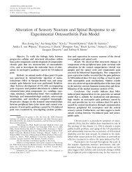
Figure 1 | A schematic representation <strong>of</strong> <strong>the</strong> pathogenesis <strong>of</strong> rheuma<strong>to</strong>id arthritis. T and B cells are attracted <strong>to</strong> synovial<br />
tissue, where <strong>the</strong>y undergo activation and proliferation. <strong>The</strong>se cells secrete proinflamma<strong>to</strong>ry cy<strong>to</strong>kines, including IL‑6 and<br />
IL‑17, which activate synovial cells, such as fibroblasts and macrophages, and cause fur<strong>the</strong>r migration <strong>of</strong> T and B cells <strong>to</strong><br />
<strong>the</strong> joint. <strong>The</strong> activated macrophages secrete cy<strong>to</strong>kines, such as TNF and IL‑1, which contribute <strong>to</strong> cartilage and bone<br />
destruction induced by osteoclasts. <strong>The</strong> proliferated synovial fibroblasts, macrophages and lymphocytes are resistant <strong>to</strong><br />
apop<strong>to</strong>sis, but sensitive <strong>to</strong> Fas‑mediated cell death. Intra‑articular injection <strong>of</strong> agonistic anti‑Fasr antibodies might,<br />
<strong>the</strong>refore, reduce synovial hyperproliferation in rA‑affected joints. Abbreviations: IL, interleukin; TNF, tumor necrosis fac<strong>to</strong>r.<br />
© 20 10<br />
Macmillan Publishers Limited. All rights reserved<br />
reviews
eviews<br />
1. Zeng, Q. Y. et al. rheumatic diseases in China.<br />
Arthritis Res. <strong>The</strong>r. 10, r17 (2008).<br />
2. Omurzakova, N. A. et al. High incidence <strong>of</strong><br />
rheumatic fever and rheumatic heart disease in<br />
<strong>the</strong> republics <strong>of</strong> Central Asia. Int. J. Rheumatic<br />
Dis. 12, 79–83 (2009).<br />
3. Yazici, H., Fresko, I. & Yurdakul, S. Behcet’s<br />
syndrome: disease manifestations,<br />
management, and advances in treatment. Nat.<br />
Clin. Pract. Rheuma<strong>to</strong>l. 3, 148–155 (2007).<br />
4. Osame, M. et al. HTLV‑I associated myelopathy, a<br />
new clinical entity. Lancet 1, 1031–1032 (1986).<br />
5. Nishioka, K. et al. Chronic inflamma<strong>to</strong>ry<br />
arthropathy associated with HTLV‑I. Lancet 1,<br />
441 (1989).<br />
6. Mochizuki, M. et al. Uveitis associated with<br />
human T‑cell lymphotropic virus type I. Am. J.<br />
Ophthalmol. 114, 123–129 (1992).<br />
7. Maruyama, I., Mori, S., Kawabata, M. &<br />
Osame, M. Bronchopneumonopathy in HTLV‑1<br />
associated myelopathy (HAM) and non‑HAM<br />
HTLV‑1 carriers [Japanese]. Nihon Kyobu Shikkan<br />
Gakkai Zasshi 30, 775–779 (1992).<br />
8. Nakagawa, M. et al. HTLV‑I‑associated<br />
myelopathy: analysis <strong>of</strong> 213 patients based on<br />
clinical features and labora<strong>to</strong>ry findings.<br />
J. Neurovirol. 1, 50–61 (1995).<br />
9. Takayanagi, H. Osteoimmunology: shared<br />
mechanisms and crosstalk between <strong>the</strong> immune<br />
between <strong>the</strong> in vivo and in vitro results can be attributed<br />
<strong>to</strong> <strong>the</strong> presence <strong>of</strong> multiple antiapop<strong>to</strong>tic processes and/or<br />
phenomena in <strong>the</strong> rheuma<strong>to</strong>id syno vium. For instance,<br />
invading T cells have been found <strong>to</strong> possess defective FasL<br />
expression, which possibly accounts for <strong>the</strong> ineffec tive<br />
clearance <strong>of</strong> FasR-expressing cells. 55 In addition, cy<strong>to</strong>kines<br />
derived from synoviocytes and stromal cells, including<br />
TNF and IL-1, protect synovi ocytes involved in RA from<br />
Fas-induced apop<strong>to</strong>sis. 56<br />
<strong>The</strong>se fac<strong>to</strong>rs might account for <strong>the</strong> capability <strong>of</strong><br />
T cells involved in RA <strong>to</strong> escape apop<strong>to</strong>sis via close<br />
interactions with synoviocytes. 57 Moreover, synovial<br />
macrophages and fibroblasts in RA-affected joints<br />
upregulate endo genous inhibi<strong>to</strong>rs <strong>of</strong> <strong>the</strong> Fas pathway,<br />
including c-FLIP. 58 Thus, multiple pathways—both intracellular<br />
and extracellular—impair Fas-induced apop<strong>to</strong>sis<br />
in RA-affected joints. <strong>The</strong> above observations strongly<br />
suggest that modulation <strong>of</strong> <strong>the</strong> Fas pathway in vivo could<br />
be a useful <strong>the</strong>rapeutic strategy.<br />
More-direct evidence for <strong>the</strong> involvement <strong>of</strong> <strong>the</strong> Fas<br />
pathway in arthritis was obtained in mouse studies—<br />
many <strong>of</strong> which were conducted in Japan—in which <strong>the</strong><br />
agonistic apop<strong>to</strong>sis-inducing antibodies <strong>to</strong> FasR were<br />
shown <strong>to</strong> be efficacious in treating arthritis. 59 Similar<br />
findings have also been confirmed in humanized experimental<br />
models. 60 Thus, current evidence suggests that<br />
activation <strong>of</strong> <strong>the</strong> agonistic Fas pathway can be considered<br />
as a potentially useful treatment strategy for <strong>the</strong> treatment<br />
<strong>of</strong> RA and o<strong>the</strong>r inflamma<strong>to</strong>ry arthritides. Indeed, clinical<br />
trials <strong>of</strong> antibody <strong>to</strong> FasR, administered via intra-articular<br />
injection, in patients with RA are currently underway.<br />
Preliminary results <strong>of</strong> a Belgian phase I clinical trial indicate<br />
that this <strong>the</strong>rapy is safe and effective, even at very low<br />
antibody concentrations. 61 In Japan, a multicenter, phase I<br />
trial in more than 140 patients is in progress, with detailed<br />
results expected soon.<br />
Conclusions<br />
Some unique immunological disease entities, <strong>the</strong> study <strong>of</strong><br />
which have influenced our understanding <strong>of</strong> <strong>the</strong> concepts<br />
involved in o<strong>the</strong>r rheumatic disorders, have been discovered<br />
in <strong>the</strong> <strong>Asian</strong> region. Research on <strong>the</strong> pathogenesis<br />
<strong>of</strong> <strong>the</strong>se diseases has greatly increased <strong>the</strong> understanding<br />
<strong>of</strong> immune-mediated disorders. Fur<strong>the</strong>rmore, novel<br />
discoveries made by <strong>Asian</strong> re searchers in conventional<br />
scientific disciplines, such as <strong>the</strong> discovery <strong>of</strong> T REG cells<br />
and <strong>the</strong> antiapop<strong>to</strong>tic molecule synoviolin, and <strong>the</strong><br />
development <strong>of</strong> <strong>the</strong> new discipline <strong>of</strong> osteoimmunology,<br />
have opened up new research avenues in rheuma<strong>to</strong>logy.<br />
<strong>Asian</strong> rheuma<strong>to</strong>logists have also success fully applied <strong>the</strong><br />
findings <strong>of</strong> labora<strong>to</strong>ry research <strong>to</strong> clinical practice and<br />
developed new strategies, such as <strong>to</strong>cilizumab and antibodies<br />
<strong>to</strong> FasR. Clinical trials are currently underway <strong>to</strong><br />
confirm <strong>the</strong> safety and efficacy <strong>of</strong> <strong>the</strong>se <strong>the</strong>rapies.<br />
Review criteria<br />
and bone systems. Nat. Rev. Immunol. 7,<br />
292–304 (2007).<br />
10. Sakaguchi, S., Sakaguchi, N., Asano, M., I<strong>to</strong>h, M.<br />
& Toda, M. Immunologic self‑<strong>to</strong>lerance<br />
maintained by activated T cells expressing IL‑2<br />
recep<strong>to</strong>r alpha‑chains (CD25). Breakdown <strong>of</strong> a<br />
single mechanism <strong>of</strong> self‑<strong>to</strong>lerance causes<br />
various au<strong>to</strong>immune diseases. J. Immunol. 155,<br />
1151–1164 (1995).<br />
11. Sakaguchi, S., Yamaguchi, T., Nomura, T. &<br />
Ono, M. regula<strong>to</strong>ry T cells and immune<br />
<strong>to</strong>lerance. Cell 133, 775–787 (2008).<br />
12. Yagishita, N., Yamasaki, S., Nishioka, K. &<br />
Nakajima, T. Synoviolin, protein folding and <strong>the</strong><br />
maintenance <strong>of</strong> joint homeostasis. Nat. Clin.<br />
Pract. Rheuma<strong>to</strong>l. 4, 91–97 (2008).<br />
13. Yamada, r. & Yamamo<strong>to</strong>, K. Mechanisms <strong>of</strong><br />
disease: genetics <strong>of</strong> rheuma<strong>to</strong>id arthritis—<br />
ethnic differences in disease‑associated genes.<br />
Nat. Clin. Pract. Rheuma<strong>to</strong>l. 3, 644–650 (2007).<br />
14. Nishimo<strong>to</strong>, N. & Kishimo<strong>to</strong>, T. Interleukin 6:<br />
from bench <strong>to</strong> bedside. Nat. Clin. Pract.<br />
Rheuma<strong>to</strong>l. 2, 619–626 (2006).<br />
15. Behçet, H. Uber rezidivierende, aphthose, dürch<br />
ein Virus verursachte Geshwure am Munde, am<br />
Auge und an den Genitalien. Derma<strong>to</strong>logische<br />
Wochenschrift 36, 1152–1157 (1937).<br />
16. Verity, D. H., Marr, J. e., Ohno, S., wallace, G. r. &<br />
Stanford, M. r. Behçet’s disease, <strong>the</strong> Silk road<br />
Data for this review were obtained by searching <strong>the</strong><br />
PubMed database for english and Japanese‑language<br />
papers, published from 1960 onwards, using <strong>the</strong><br />
following search terms: “Asia”, “rheuma<strong>to</strong>logy”,<br />
“HTLV”, “rheuma<strong>to</strong>id arthritis”, “au<strong>to</strong>immune disease”,<br />
“Behçet”, “Takayasu’s arteritis”, “Kawasaki disease”,<br />
“osteoimmunology”, “Treg”, “Foxp3”, “synoviolin”,<br />
“ethnic”, “IL‑6”, “Fas”, “FasL”, “apop<strong>to</strong>sis” and<br />
“cy<strong>to</strong>kine”. Additional studies were identified from <strong>the</strong><br />
abstracts <strong>of</strong> <strong>the</strong> American College <strong>of</strong> rheuma<strong>to</strong>logy<br />
Scientific Meetings.<br />
and HLA‑B51: his<strong>to</strong>rical and geographical<br />
perspectives. Tissue Antigens 54, 213–220<br />
(1999).<br />
17. Hatemi, G. et al. <strong>The</strong> pustular skin lesions in<br />
Behcet’s syndrome are not sterile. Ann. Rheum.<br />
Dis. 63, 1450–1452 (2004).<br />
18. Gul, A. Behcet’s disease as an au<strong>to</strong>inflamma<strong>to</strong>ry<br />
disorder. Curr. Drug Targets Inflamm. Allergy 4,<br />
81–83 (2005).<br />
19. Numano, F. <strong>The</strong> s<strong>to</strong>ry <strong>of</strong> Takayasu arteritis.<br />
Rheuma<strong>to</strong>logy (Oxford) 41, 103–106 (2002).<br />
20. Koide, K. Takayasu arteritis in Japan. Heart<br />
Vessels Suppl. 7, 48–54 (1992).<br />
21. Arend, w. P. et al. <strong>The</strong> American College <strong>of</strong><br />
rheuma<strong>to</strong>logy 1990 criteria for <strong>the</strong><br />
classification <strong>of</strong> Takayasu arteritis. Arthritis<br />
Rheum. 33, 1129–1134 (1990).<br />
22. Kimura, A., Kitamura, H., Date, Y. & Numano, F.<br />
Comprehensive analysis <strong>of</strong> HLA genes in<br />
Takayasu arteritis in Japan. Int. J. Cardiol. 54<br />
(Suppl.), S61–S69 (1996).<br />
23. Seko, Y. et al. Perforin‑secreting killer cell<br />
infiltration and expression <strong>of</strong> a 65‑kD heat‑shock<br />
protein in aortic tissue <strong>of</strong> patients with<br />
Takayasu’s arteritis. J. Clin. Invest. 93, 750–758<br />
(1994).<br />
24. Huang, w. C. et al. epidemiologic features <strong>of</strong><br />
Kawasaki disease in Taiwan, 2003–2006.<br />
Pediatrics 123, e401–e405 (2009).<br />
110 | FEBRUARY 2010 | volUmE 6 www.nature.com/nrrheum<br />
© 20 10<br />
Macmillan Publishers Limited. All rights reserved
25. Holman, r. C., Curns, A. T., Belay, e. D.,<br />
Steiner, C. A. & Schonberger, L. B. Kawasaki<br />
syndrome hospitalizations in <strong>the</strong> United States,<br />
1997 and 2000. Pediatrics 112, 495–501<br />
(2003).<br />
26. Burns, J. C. et al. Seasonality and temporal<br />
clustering <strong>of</strong> Kawasaki syndrome. Epidemiology<br />
16, 220–225 (2005).<br />
27. Onouchi, Y. et al. ITPKC functional polymorphism<br />
associated with Kawasaki disease susceptibility<br />
and formation <strong>of</strong> coronary artery aneurysms.<br />
Nat. Genet. 40, 35–42 (2008).<br />
28. Birmann, B. M. et al. Population differences in<br />
immune marker pr<strong>of</strong>iles associated with human<br />
T‑lymphotropic virus type I infection in Japan and<br />
Jamaica. Int. J. Cancer 124, 614–621 (2009).<br />
29. Uchiyama, T., Yodoi, J., Sagawa, K., Takatsuki, K.<br />
& Uchino, H. Adult T‑cell leukemia: clinical and<br />
hema<strong>to</strong>logic features <strong>of</strong> 16 cases. Blood 50,<br />
481–492 (1977).<br />
30. Nishioka, K., Sumida, T. & Hasunuma, T. Human<br />
T lymphotropic virus type I in arthropathy and<br />
au<strong>to</strong>immune disorders. Arthritis Rheum. 39,<br />
1410–1418 (1996).<br />
31. Yamano, Y. et al. Virus‑induced dysfunction <strong>of</strong><br />
CD4 + CD25 + T cells in patients with<br />
HTLV‑I‑associated neuroimmunological disease.<br />
J. Clin. Invest. 115, 1361–1368 (2005).<br />
32. Nakajima, T. et al. Overgrowth <strong>of</strong> human synovial<br />
cells driven by <strong>the</strong> human T cell leukemia virus<br />
type I tax gene. J. Clin. Invest. 92, 186–193<br />
(1993).<br />
33. Iwakura, Y. et al. Induction <strong>of</strong> inflamma<strong>to</strong>ry<br />
arthropathy resembling rheuma<strong>to</strong>id arthritis in<br />
mice transgenic for HTLV‑I. Science 253,<br />
1026–1028 (1991).<br />
34. Takayanagi, H. Mechanistic insight in<strong>to</strong><br />
osteoclast differentiation in osteoimmunology.<br />
J. Mol. Med. 83, 170–179 (2005).<br />
35. Sa<strong>to</strong>, K. & Takayanagi, H. Osteoclasts,<br />
rheuma<strong>to</strong>id arthritis, and osteoimmunology. Curr.<br />
Opin. Rheuma<strong>to</strong>l. 18, 419–426 (2006).<br />
36. Asagiri, M. & Takayanagi, H. <strong>The</strong> molecular<br />
understanding <strong>of</strong> osteoclast differentiation.<br />
Bone 40, 251–264 (2007).<br />
37. Kong, Y. Y. et al. Activated T cells regulate bone<br />
loss and joint destruction in adjuvant arthritis<br />
through osteoprotegerin ligand. Nature 402,<br />
304–309 (1999).<br />
38. Takayanagi, H. et al. T‑cell‑mediated regulation <strong>of</strong><br />
osteoclas<strong>to</strong>genesis by signaling crosstalk<br />
between rANKL and IFN‑gamma. Nature 408,<br />
600–605 (2000).<br />
39. Hori, S., Nomura, T. & Sakaguchi, S. Control <strong>of</strong><br />
regula<strong>to</strong>ry T cell development by <strong>the</strong><br />
transcription fac<strong>to</strong>r Foxp3. Science 299,<br />
1057–1061 (2003).<br />
40. Miyara, M. et al. Functional delineation and<br />
differentiation dynamics <strong>of</strong> human CD4 + T cells<br />
expressing <strong>the</strong> FoxP3 transcription fac<strong>to</strong>r.<br />
Immunity 30, 899–911 (2009).<br />
41. Yamano, Y. et al. Abnormally high levels <strong>of</strong> virus‑<br />
infected IFN‑γ + CCr4 + CD4 + CD25 + T cells in a<br />
retrovirus‑associated neuroinflamma<strong>to</strong>ry<br />
disorder. PLoS One 4, e6517 (2009).<br />
42. Amano. T. et al. Synoviolin/Hrd1, an e3 ubiquitin<br />
ligase, as a novel pathogenic fac<strong>to</strong>r for<br />
arthropathy. Genes Dev. 17, 2436–2449 (2003).<br />
43. Schroder, M. & Kaufman, r. J. <strong>The</strong> mammalian<br />
unfolded protein response. Annu. Rev. Biochem.<br />
74, 739–789 (2005).<br />
44. Yamasaki, S. et al. Cy<strong>to</strong>plasmic destruction <strong>of</strong><br />
p53 by <strong>the</strong> endoplasmic reticulum‑resident<br />
ubiquitin ligase ‘Synoviolin’. EMBO J. 26,<br />
113–122 (2007).<br />
45. Toh, M. L. et al. Overexpression <strong>of</strong> synoviolin in<br />
peripheral blood and synoviocytes from<br />
rheuma<strong>to</strong>id arthritis patients and continued<br />
elevation in nonresponders <strong>to</strong> infliximab<br />
treatment. Arthritis Rheum. 54, 2109–2118<br />
(2006).<br />
46. Sa<strong>to</strong>, K. et al. reshaping a human antibody <strong>to</strong><br />
inhibit <strong>the</strong> interleukin 6‑dependent tumor cell<br />
growth. Cancer Res. 53, 851–856 (1993).<br />
47. Nishimo<strong>to</strong>, N. et al. Study <strong>of</strong> active controlled<br />
<strong>to</strong>cilizumab mono<strong>the</strong>rapy for rheuma<strong>to</strong>id<br />
arthritis patients with an inadequate response<br />
<strong>to</strong> methotrexate (SATOrI): significant reduction<br />
in disease activity and serum vascular<br />
endo<strong>the</strong>lial growth fac<strong>to</strong>r by IL‑6 recep<strong>to</strong>r<br />
inhibition <strong>the</strong>rapy. Mod. Rheuma<strong>to</strong>l. 19, 12–19<br />
(2009).<br />
48. Smolen, J. S. et al. effect <strong>of</strong> interleukin‑6<br />
recep<strong>to</strong>r inhibition with <strong>to</strong>cilizumab in patients<br />
with rheuma<strong>to</strong>id arthritis (OPTION study):<br />
a double‑blind, placebo‑controlled, randomised<br />
trial. Lancet 371, 987–997 (2008).<br />
49. Genovese, M. C. et al. Interleukin‑6 recep<strong>to</strong>r<br />
inhibition with <strong>to</strong>cilizumab reduces disease<br />
activity in rheuma<strong>to</strong>id arthritis with inadequate<br />
response <strong>to</strong> disease‑modifying antirheumatic<br />
drugs: <strong>the</strong> <strong>to</strong>cilizumab in combination with<br />
traditional disease‑modifying antirheumatic drug<br />
<strong>the</strong>rapy study. Arthritis Rheum. 58, 2968–2980<br />
(2008).<br />
50. emery, P. et al. IL‑6 recep<strong>to</strong>r inhibition with<br />
<strong>to</strong>cilizmab improves treatment outcomes in<br />
patients with rheuma<strong>to</strong>id arthritis refrac<strong>to</strong>ry <strong>to</strong><br />
anti‑tumor necrosis fac<strong>to</strong>r biological: results<br />
from a 24‑week multicentre randomized placebo‑<br />
controlled trial. Ann. Rheum. Dis. 67,<br />
1516–1523 (2008).<br />
51. raff, M. C. Social controls on cell survival and<br />
cell death. Nature 356, 397–400 (1992).<br />
reviews<br />
52. Yonehara, S., Ishii, A. & Yonehara, M. A cell‑<br />
killing monoclonal antibody (anti‑Fas) <strong>to</strong> a cell<br />
surface antigen co‑downregulated with <strong>the</strong><br />
recep<strong>to</strong>r <strong>of</strong> tumor necrosis fac<strong>to</strong>r. J. Exp. Med.<br />
169, 1747–1756 (1989).<br />
53. Nagata, S. Apop<strong>to</strong>sis by death fac<strong>to</strong>r. Cell 88,<br />
355–365 (1997).<br />
54. Nishioka, K., Hasunuma, T., Ka<strong>to</strong>, T., Sumida, T.<br />
& Kobata, T. Apop<strong>to</strong>sis in rheuma<strong>to</strong>id arthritis:<br />
a novel pathway in <strong>the</strong> regulation <strong>of</strong> synovial<br />
tissue. Arthritis Rheum. 41, 1–9 (1998).<br />
55. Cantwell, M. J., Hua, T., Zvaifler, N. J. &<br />
Kipps, T. J. Deficient Fas ligand expression by<br />
synovial lymphocytes from patients with<br />
rheuma<strong>to</strong>id arthritis. Arthritis Rheum. 40,<br />
1644–1652 (1997).<br />
56. Kobayashi, T. et al. Differential regulation <strong>of</strong> Fas‑<br />
mediated apop<strong>to</strong>sis <strong>of</strong> rheuma<strong>to</strong>id synoviocytes<br />
by tumor necrosis fac<strong>to</strong>r alpha and basic<br />
fibroblast growth fac<strong>to</strong>r is associated with <strong>the</strong><br />
expression <strong>of</strong> apop<strong>to</strong>sis‑related molecules.<br />
Arthritis Rheum. 43, 1106–1114 (2000).<br />
57. Salmon, M. et al. Inhibition <strong>of</strong> T cell apop<strong>to</strong>sis in<br />
<strong>the</strong> rheuma<strong>to</strong>id synovium. J. Clin. Invest. 99,<br />
439–446 (1997).<br />
58. Perlman, H. et al. rheuma<strong>to</strong>id arthritis synovial<br />
macrophages express <strong>the</strong> Fas‑associated death<br />
domain‑like interleukin‑1β‑converting enzyme‑<br />
inhibi<strong>to</strong>ry protein and are refrac<strong>to</strong>ry <strong>to</strong> Fas‑<br />
mediated apop<strong>to</strong>sis. Arthritis Rheum. 44, 21–30<br />
(2001).<br />
59. Fujisawa, K. et al. <strong>The</strong>rapeutic effect <strong>of</strong> <strong>the</strong> anti‑<br />
Fas antibody on arthritis in HTLV‑1 tax<br />
transgenic mice. J. Clin. Invest. 98, 271–278<br />
(1996).<br />
60. Matsuno, H. et al. Antirheumatic effects <strong>of</strong><br />
humanized anti‑Fas monoclonal antibody in<br />
human rheuma<strong>to</strong>id arthritis/SCID mouse<br />
chimera. J. Rheuma<strong>to</strong>l. 29, 1609–1614 (2002).<br />
61. Appelboom, T. et al. Preliminary results <strong>of</strong> a<br />
phase I clinical trial <strong>of</strong> intra‑articular<br />
administration <strong>of</strong> ArG098, a novel anti‑Fas IgM<br />
Mab, in rA [presentation number 421].<br />
Presented at <strong>the</strong> ACr/ArHP Scientific Meeting,<br />
Philadelphia, PA (18 Oc<strong>to</strong>ber 2009).<br />
Acknowledgments<br />
This work was partially supported by a Grant‑in‑Aid for<br />
Scientific research from <strong>the</strong> Japan Society for <strong>the</strong><br />
Promotion <strong>of</strong> Science, <strong>The</strong> Ministry <strong>of</strong> education,<br />
Culture, Sports, Science and Technology, Japanese<br />
Ministry <strong>of</strong> Health, Labor, and welfare, National<br />
Institute <strong>of</strong> Biomedical Innovation, Uehara Memorial<br />
Foundation, Nagao Takeshi Nanbyo Foundation,<br />
Kanagawa Nanbyo Foundation, Mishima Kaiun<br />
Memorial Foundation, Takeda Science Foundation,<br />
and ITSUU Labora<strong>to</strong>ry research Foundation.<br />
NATuRE REVIEWS | rheumATologY VOLuME 6 | FEBRuARY 2010 | 111<br />
© 20 10<br />
Macmillan Publishers Limited. All rights reserved