Quantifying the material and structural determinants of bone strength
Quantifying the material and structural determinants of bone strength
Quantifying the material and structural determinants of bone strength

Create successful ePaper yourself
Turn your PDF publications into a flip-book with our unique Google optimized e-Paper software.
748<br />
M.L. Bouxsein, E. Seeman / Best Practice & Research Clinical Rheumatology 23 (2009) 741–753<br />
properties <strong>and</strong> loading conditions influence <strong>the</strong> <strong>strength</strong> predictions. None<strong>the</strong>less, this state-<strong>of</strong>-<strong>the</strong>-art<br />
technique is among <strong>the</strong> most promising tools available today for clinical assessment <strong>of</strong> <strong>bone</strong> <strong>strength</strong><br />
<strong>and</strong> fracture risk [112].<br />
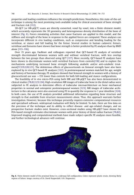
3D-QCT (or HR-pQCT) scans are directly converted, voxel by voxel, into a finite element model,<br />
which accurately represents <strong>the</strong> 3D geometry <strong>and</strong> heterogeneous density distribution <strong>of</strong> <strong>the</strong> <strong>bone</strong> <strong>of</strong><br />
interest (Fig. 6). Forces simulating activities that cause fractures are applied to this model, <strong>and</strong> <strong>the</strong><br />
stiffness <strong>and</strong> <strong>strength</strong> <strong>of</strong> <strong>the</strong> <strong>bone</strong> in response to <strong>the</strong> applied forces are computed. These analyses can<br />
incorporate different in vivo loading conditions, such as compressive <strong>and</strong> bending loading for <strong>the</strong><br />
vertebrae, or stance <strong>and</strong> fall loading for <strong>the</strong> femur. Several studies in human cadaveric femurs,<br />
vertebrae <strong>and</strong> forearms have shown that <strong>bone</strong> <strong>strength</strong> is better predicted by FE analyses than by BMD<br />
alone [113–116].<br />
Over 15 years ago, Faulkner <strong>and</strong> colleagues reported that QCT-based FE analysis <strong>of</strong> vertebral<br />
<strong>strength</strong> discriminated between women with <strong>and</strong> without vertebral fracture, with less overlap<br />
between <strong>the</strong> two groups than observed using QCT [117]. More recently, QCT-based FE analyses have<br />
been shown to discriminate women with vertebral fractures from controls[118] <strong>and</strong> to explore <strong>the</strong><br />
mechanisms underlying increased <strong>bone</strong> <strong>strength</strong> following anabolic <strong>and</strong>/or anti-catabolic treatment[57,119,120,121].<br />
The deleterious effects <strong>of</strong> glucocorticoids on femoral <strong>strength</strong> have also been<br />
explored by in vivo QCT-based FE analyses [122]. In postmenopausal women matched for age, weight<br />
<strong>and</strong> history <strong>of</strong> hormone <strong>the</strong>rapy, FE analyses showed that femoral <strong>strength</strong> in women with a history <strong>of</strong><br />
glucocorticoid use was w15% lower than controls for both fall-loading <strong>and</strong> stance configurations.<br />
The feasibility <strong>of</strong> in vivo micro-FEA using both MR <strong>and</strong> HR-pQCT has also been demonstrated in<br />
clinical studies [78,82,123,124]. High-resolution MR images <strong>of</strong> trabecular <strong>bone</strong> in <strong>the</strong> distal radius were<br />
subjected to FE analysis to determine effects <strong>of</strong> trabecular microarchitecture on <strong>bone</strong> mechanical<br />
properties in normal <strong>and</strong> osteopenic postmenopausal women [123]. MR images <strong>of</strong> trabecular architecture<br />
in <strong>the</strong> calcaneus were also assessed using FE to quantify <strong>the</strong> response to 1 year idoxifene [124].<br />
In both cases, <strong>the</strong> use <strong>of</strong> FE analysis provided additional information regarding <strong>bone</strong> structure <strong>and</strong><br />
<strong>strength</strong> to that available from structure measurements alone. Thus, this approach warrants fur<strong>the</strong>r<br />
investigation. However, because this technique presently requires extensive computational resources<br />
<strong>and</strong> specialised s<strong>of</strong>tware, widespread evaluation will likely be limited. To date, <strong>the</strong>re are few data on<br />
<strong>the</strong> precision <strong>of</strong> <strong>the</strong> technique <strong>and</strong> its ability to reflect disease- <strong>and</strong> age-related changes, <strong>and</strong> no<br />
prospective fracture studies exist. However, cross-sectional studies using HR-pQCT have shown <strong>the</strong><br />
FEA-based <strong>strength</strong> estimates discriminate women with prior history <strong>of</strong> distal radius fracture [76,82].<br />
Improved imaging <strong>and</strong> computational methods have made subject-specific FE analyses more feasible,<br />
<strong>and</strong> fur<strong>the</strong>r technological advances will continue.<br />
Fig. 6. Finite element model <strong>of</strong> <strong>the</strong> proximal femur in a sideways fall configuration. Colors represent plastic strain, showing likely<br />
regions <strong>of</strong> failure. Image courtesy <strong>of</strong> Dr. David Kopperdahl, O.N. Diagnostics.