Diagnosis of arrhythmogenic right ventricular cardiomyopathy ...
Diagnosis of arrhythmogenic right ventricular cardiomyopathy ...
Diagnosis of arrhythmogenic right ventricular cardiomyopathy ...

You also want an ePaper? Increase the reach of your titles
YUMPU automatically turns print PDFs into web optimized ePapers that Google loves.
808<br />
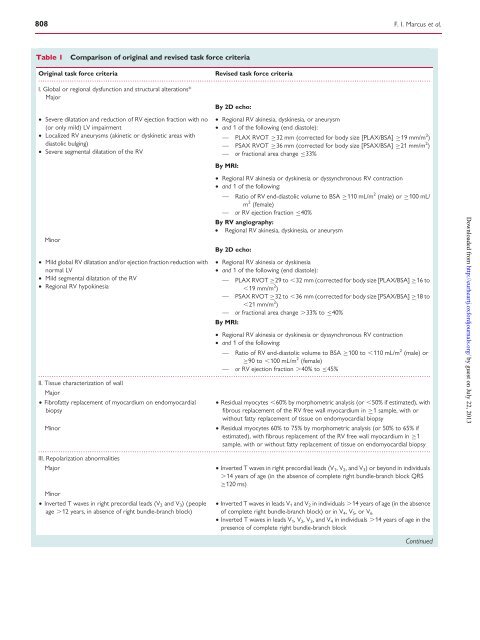
Table 1 Comparison <strong>of</strong> original and revised task force criteria<br />
Original task force criteria Revised task force criteria<br />
...............................................................................................................................................................................<br />
I. Global or regional dysfunction and structural alterations*<br />
Major<br />
† Severe dilatation and reduction <strong>of</strong> RV ejection fraction with no<br />
(or only mild) LV impairment<br />
† Localized RV aneurysms (akinetic or dyskinetic areas with<br />
diastolic bulging)<br />
† Severe segmental dilatation <strong>of</strong> the RV<br />
Minor<br />
† Mild global RV dilatation and/or ejection fraction reduction with<br />
normal LV<br />
† Mild segmental dilatation <strong>of</strong> the RV<br />
† Regional RV hypokinesia<br />
By 2D echo:<br />
† Regional RV akinesia, dyskinesia, or aneurysm<br />
† and 1 <strong>of</strong> the following (end diastole):<br />
— PLAX RVOT 32 mm (corrected for body size [PLAX/BSA] 19 mm/m 2 )<br />
— PSAX RVOT 36 mm (corrected for body size [PSAX/BSA] 21 mm/m 2 )<br />
— or fractional area change 33%<br />
By MRI:<br />
† Regional RV akinesia or dyskinesia or dyssynchronous RV contraction<br />
† and 1 <strong>of</strong> the following:<br />
— Ratio <strong>of</strong> RV end-diastolic volume to BSA 110 mL/m 2 m<br />
(male) or 100 mL/<br />
2 (female)<br />
— or RV ejection fraction 40%<br />
By RV angiography:<br />
† Regional RV akinesia, dyskinesia, or aneurysm<br />
By 2D echo:<br />
F. I. Marcus et al.<br />
† Regional RV akinesia or dyskinesia<br />
† and 1 <strong>of</strong> the following (end diastole):<br />
— PLAX RVOT 29 to ,32 mm (corrected for body size [PLAX/BSA] 16 to<br />
,19 mm/m 2 )<br />
— PSAX RVOT 32 to ,36 mm (corrected for body size [PSAX/BSA] 18 to<br />
,21 mm/m 2 —<br />
)<br />
or fractional area change .33% to 40%<br />
By MRI:<br />
† Regional RV akinesia or dyskinesia or dyssynchronous RV contraction<br />
† and 1 <strong>of</strong> the following:<br />
— Ratio <strong>of</strong> RV end-diastolic volume to BSA 100 to ,110 mL/m<br />
...............................................................................................................................................................................<br />
...............................................................................................................................................................................<br />
2 90 to ,100 mL/m<br />
(male) or<br />
2 (female)<br />
— or RV ejection fraction .40% to 45%<br />
II. Tissue characterization <strong>of</strong> wall<br />
Major<br />
† Fibr<strong>of</strong>atty replacement <strong>of</strong> myocardium on endomyocardial † Residual myocytes ,60% by morphometric analysis (or ,50% if estimated), with<br />
biopsy<br />
fibrous replacement <strong>of</strong> the RV free wall myocardium in 1 sample, with or<br />
Minor<br />
without fatty replacement <strong>of</strong> tissue on endomyocardial biopsy<br />
† Residual myocytes 60% to 75% by morphometric analysis (or 50% to 65% if<br />
estimated), with fibrous replacement <strong>of</strong> the RV free wall myocardium in 1<br />
sample, with or without fatty replacement <strong>of</strong> tissue on endomyocardial biopsy<br />
III. Repolarization abnormalities<br />
Major<br />
Minor<br />
† Inverted T waves in <strong>right</strong> precordial leads (V1,V2, and V3) or beyond in individuals<br />
.14 years <strong>of</strong> age (in the absence <strong>of</strong> complete <strong>right</strong> bundle-branch block QRS<br />
120 ms)<br />
† Inverted T waves in <strong>right</strong> precordial leads (V2 and V3) (people † Inverted T waves in leads V1 and V2 in individuals .14 years <strong>of</strong> age (in the absence<br />
age .12 years, in absence <strong>of</strong> <strong>right</strong> bundle-branch block) <strong>of</strong> complete <strong>right</strong> bundle-branch block) or in V4,V5,orV6 † Inverted T waves in leads V1, V2, V3, and V4 in individuals .14 years <strong>of</strong> age in the<br />
presence <strong>of</strong> complete <strong>right</strong> bundle-branch block<br />
Continued<br />
Downloaded from<br />
http://eurheartj.oxfordjournals.org/ by guest on July 22, 2013