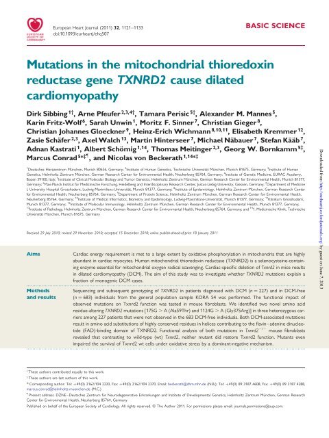
Mutations in the mitochondrial thioredoxin reductase gene TXNRD2 ...
Mutations in the mitochondrial thioredoxin reductase gene TXNRD2 ...
Mutations in the mitochondrial thioredoxin reductase gene TXNRD2 ...

Create successful ePaper yourself
Turn your PDF publications into a flip-book with our unique Google optimized e-Paper software.
European Heart Journal (2011) 32, 1121–1133<br />
doi:10.1093/eurheartj/ehq507<br />
<strong>Mutations</strong> <strong>in</strong> <strong>the</strong> <strong>mitochondrial</strong> thioredox<strong>in</strong><br />
<strong>reductase</strong> <strong>gene</strong> <strong>TXNRD2</strong> cause dilated<br />
cardiomyopathy<br />
BASIC SCIENCE<br />
Dirk Sibb<strong>in</strong>g 1† , Arne Pfeufer 2,3,4† , Tamara Perisic 5† , Alexander M. Mannes 5 ,<br />
Kar<strong>in</strong> Fritz-Wolf 6 , Sarah Unw<strong>in</strong> 1 , Moritz F. S<strong>in</strong>ner 7 , Christian Gieger 8 ,<br />
Christian Johannes Gloeckner 9 , He<strong>in</strong>z-Erich Wichmann 8,10,11 , Elisabeth Kremmer 12 ,<br />
Zasie Schäfer 2,3 , Axel Walch 13 , Mart<strong>in</strong> H<strong>in</strong>terseer 7 , Michael Näbauer 7 , Stefan Kääb 7 ,<br />
Adnan Kastrati 1 , Albert Schömig 1,14 , Thomas Meit<strong>in</strong>ger 2,3 , Georg W. Bornkamm 5‡ ,<br />
Marcus Conrad 5 * ‡} , and Nicolas von Beckerath 1,14 * ‡<br />
1 2 3<br />
Deutsches Herzzentrum München, Munich 80636, Germany; Institute of Human Genetics, Technische Universität München, Munich 81675, Germany; Institute of Human<br />
Genetics, Helmholtz Zentrum München, German Research Center for Environmental Health, Neuherberg 85764, Germany; 4 Institute of Genetic Medic<strong>in</strong>e, EURAC Academy,<br />
Bozen 39100, Italy; 5 Institute of Cl<strong>in</strong>ical Molecular Biology and Tumor Genetics, Helmholtz Zentrum München, German Research Center for Environmental Health, Munich 81377,<br />
Germany; 6 Max-Planck Institut für Mediz<strong>in</strong>ische Forschung, Heidelberg and Interdiscipl<strong>in</strong>ary Research Center, Justus-Liebig-University, Giessen, Germany; 7 Department of Medic<strong>in</strong>e<br />
I, University Hospital Grosshadern, Ludwig-Maximilians-Universität, Munich 81377, Germany; 8 Institute of Epidemiology, Helmholtz Zentrum München, German Research Center<br />
for Environmental Health, Neuherberg 85764, Germany; 9 Department of Prote<strong>in</strong> Science, Helmholtz Zentrum München, German Research Center for Environmental Health,<br />
Neuherberg 85764, Germany; 10 Institute of Medical Informatics, Biometry and Epidemiology, Ludwig-Maximilians-Universität, Munich 81377, Germany; 11 Kl<strong>in</strong>ikum Grosshadern,<br />
Munich 81377, Germany; 12 Institute of Molecular Immunology, Helmholtz Zentrum München, German Research Center for Environmental Health, Munich 81377, Germany;<br />
13 14<br />
Institute of Pathology, Helmholtz Zentrum München, German Research Center for Environmental Health, Neuherberg 85764, Germany; and 1. Mediz<strong>in</strong>ische Kl<strong>in</strong>ik, Technische<br />
Universität München, Munich 81675, Germany<br />
Received 29 July 2010; revised 29 November 2010; accepted 15 December 2010; onl<strong>in</strong>e publish-ahead-of-pr<strong>in</strong>t 18 January 2011<br />
Aims Cardiac energy requirement is met to a large extent by oxidative phosphorylation <strong>in</strong> mitochondria that are highly<br />
abundant <strong>in</strong> cardiac myocytes. Human <strong>mitochondrial</strong> thioredox<strong>in</strong> <strong>reductase</strong> (<strong>TXNRD2</strong>) is a selenocyste<strong>in</strong>e-conta<strong>in</strong><strong>in</strong>g<br />
enzyme essential for <strong>mitochondrial</strong> oxygen radical scaveng<strong>in</strong>g. Cardiac-specific deletion of Txnrd2 <strong>in</strong> mice results<br />
<strong>in</strong> dilated cardiomyopathy (DCM). The aim of this study was to <strong>in</strong>vestigate whe<strong>the</strong>r <strong>TXNRD2</strong> mutations expla<strong>in</strong> a<br />
fraction of monogenic DCM cases.<br />
.....................................................................................................................................................................................<br />
Methods Sequenc<strong>in</strong>g and subsequent genotyp<strong>in</strong>g of <strong>TXNRD2</strong> <strong>in</strong> patients diagnosed with DCM (n ¼ 227) and <strong>in</strong> DCM-free<br />
and results (n ¼ 683) <strong>in</strong>dividuals from <strong>the</strong> <strong>gene</strong>ral population sample KORA S4 was performed. The functional impact of<br />
observed mutations on Txnrd2 function was tested <strong>in</strong> mouse fibroblasts. We identified two novel am<strong>in</strong>o acid<br />
residue-alter<strong>in</strong>g <strong>TXNRD2</strong> mutations [175G . A (Ala59Thr) and 1124G . A (Gly375Arg)] <strong>in</strong> three heterozygous carriers<br />
among 227 patients that were not observed <strong>in</strong> <strong>the</strong> 683 DCM-free <strong>in</strong>dividuals. Both DCM-associated mutations<br />
result <strong>in</strong> am<strong>in</strong>o acid substitutions of highly conserved residues <strong>in</strong> helices contribut<strong>in</strong>g to <strong>the</strong> flav<strong>in</strong>–aden<strong>in</strong>e d<strong>in</strong>ucleotide<br />
(FAD)-b<strong>in</strong>d<strong>in</strong>g doma<strong>in</strong> of <strong>TXNRD2</strong>. Functional analysis of both mutations <strong>in</strong> Txnrd2<br />
.....................................................................................................................................................................................<br />
2/2 mouse fibroblasts<br />
revealed that contrast<strong>in</strong>g to wild-type (wt) Txnrd2, nei<strong>the</strong>r mutant did restore Txnrd2 function. Mutants even<br />
impaired <strong>the</strong> survival of Txnrd2 wt cells under oxidative stress by a dom<strong>in</strong>ant-negative mechanism.<br />
† These authors contributed equally to this work.<br />
‡ These authors are last authors of this work.<br />
* Correspond<strong>in</strong>g author. Tel: +49(0) 2162/104 2220, Fax: +49(0) 2162/104 2370, Email: beckerath@dhm.mhn.de (N.B.); Tel: +49(0) 89 3187 4608, Fax: +49(0) 89 3187 4288,<br />
marcus.conrad@helmholtz-muenchen.de (M.C.)<br />
}<br />
Present address. DZNE–Deutsches Zentrum für Neurode<strong>gene</strong>rative Erkrankungen and Institute of Developmental Genetics, Helmholtz Zentrum München, German Research<br />
Center for Environmental Health, Neuherberg 85764, Germany<br />
Published on behalf of <strong>the</strong> European Society of Cardiology. All rights reserved. & The Author 2011. For permissions please email: journals.permissions@oup.com.<br />
Downloaded from<br />
http://eurheartj.oxfordjournals.org/ by guest on June 7, 2013
1122<br />
Conclusion For <strong>the</strong> first time, we describe mutations <strong>in</strong> DCM patients <strong>in</strong> a <strong>gene</strong> <strong>in</strong>volved <strong>in</strong> <strong>the</strong> regulation of cellular redox state.<br />
<strong>TXNRD2</strong> mutations may expla<strong>in</strong> a fraction of human DCM disease burden.<br />
-----------------------------------------------------------------------------------------------------------------------------------------------------------<br />
Keywords Dilated cardiomyopathy † Genetics † <strong>TXNRD2</strong><br />
Introduction<br />
Dilated cardiomyopathy (DCM) is a frequent cause of congestive<br />
heart failure and is <strong>the</strong> most common diagnosis <strong>in</strong> patients undergo<strong>in</strong>g<br />
heart transplantation. Accord<strong>in</strong>g to systematic studies, famil-<br />
1 – 3<br />
ial transmission is observed <strong>in</strong> about 20–30% of DCM cases.<br />
Rare mutations <strong>in</strong> more than 20 different disease <strong>gene</strong>s, most of<br />
<strong>the</strong>m encod<strong>in</strong>g for structural prote<strong>in</strong>s of cardiomyocytes, have<br />
been identified. 4 – 7 <strong>Mutations</strong> <strong>in</strong> known disease <strong>gene</strong>s, though,<br />
expla<strong>in</strong> ,20% of <strong>in</strong>herited cases. 8<br />
Thioredox<strong>in</strong> <strong>reductase</strong>s are essential components of <strong>the</strong> thioredox<strong>in</strong><br />
system 9 and are <strong>the</strong>refore crucial for <strong>the</strong> control of cellular<br />
redox balance. They are selenocyste<strong>in</strong>e (Sec)-conta<strong>in</strong><strong>in</strong>g, homodimeric<br />
flavoenzymes that ma<strong>in</strong>ta<strong>in</strong> thioredox<strong>in</strong>s, small prote<strong>in</strong>s that<br />
catalyse redox reactions, <strong>in</strong> <strong>the</strong>ir reduced state us<strong>in</strong>g <strong>the</strong> reduc<strong>in</strong>g<br />
power of NADPH. 10 Three mammalian thioredox<strong>in</strong> <strong>reductase</strong>s<br />
exist; a cytosolic (TXNRD1), a <strong>mitochondrial</strong> (<strong>TXNRD2</strong>), and a<br />
testis-specific thioredox<strong>in</strong> <strong>reductase</strong> (TXNRD3). The Sec-residue,<br />
encoded by a UGA codon, is <strong>the</strong> penultimate am<strong>in</strong>o acid of <strong>the</strong> Cterm<strong>in</strong>al<br />
catalytic centre of all thioredox<strong>in</strong> <strong>reductase</strong>s and is essential<br />
for enzyme activity. The premature term<strong>in</strong>ation of prote<strong>in</strong> syn<strong>the</strong>sis<br />
at <strong>the</strong> UGA codon is prevented by a stem-loop-like<br />
structure, <strong>the</strong> selenocyste<strong>in</strong>e <strong>in</strong>sertion sequence (SECIS) element<br />
located <strong>in</strong> <strong>the</strong> 3 ′ UTR. 11,12 Cardiac energy requirement is met to<br />
a large extent by oxidative phosphorylation <strong>in</strong> mitochondria that<br />
are highly abundant <strong>in</strong> cardiac myocytes. 13 The <strong>mitochondrial</strong><br />
thioredox<strong>in</strong> <strong>reductase</strong> (<strong>TXNRD2</strong>), along with <strong>mitochondrial</strong> thioredox<strong>in</strong><br />
and peroxiredox<strong>in</strong>s III and V, is of paramount importance<br />
for <strong>mitochondrial</strong> scaveng<strong>in</strong>g of reactive oxygen species (ROS). 9,10<br />
It is well established that excessive ROS causes oxidative stress<br />
and cell death. 14 On <strong>the</strong> o<strong>the</strong>r hand, compell<strong>in</strong>g evidence has<br />
established a role for ROS as modulators of <strong>in</strong>tracellular signall<strong>in</strong>g<br />
cascades. 15 Beyond provid<strong>in</strong>g protection aga<strong>in</strong>st ROS, thioredox<strong>in</strong>s<br />
are known to <strong>in</strong>hibit or activate apoptotic signall<strong>in</strong>g molecules like<br />
apoptosis signal-regulat<strong>in</strong>g k<strong>in</strong>ase 1 and Ras or transcription factors<br />
like NF-kB. 16 We showed that glutathione (GSH) peroxidase 4,<br />
along with GSH, senses and translates oxidative stress <strong>in</strong>to a dist<strong>in</strong>ct<br />
cell death signall<strong>in</strong>g cascade <strong>in</strong>volv<strong>in</strong>g <strong>the</strong> activation of 12/<br />
15-lipoxygenase and apoptosis-<strong>in</strong>duc<strong>in</strong>g factor. 17<br />
Recently, we <strong>gene</strong>rated and characterized transgenic mice deficient<br />
for Txnrd2. 18 Ubiquitous <strong>in</strong>activation resulted <strong>in</strong> embryonic death of<br />
anaemic embryos exhibit<strong>in</strong>g marked th<strong>in</strong>n<strong>in</strong>g of <strong>the</strong> ventricular heart<br />
walls. Analysis of fibroblasts isolated from Txnrd2 2/2 embryos<br />
revealed a critical role for Txnrd2 <strong>in</strong> <strong>the</strong> removal of toxic ROS<br />
species. Heart-specific <strong>in</strong>activation of Txnrd2 resulted <strong>in</strong> a phenotype<br />
rem<strong>in</strong>iscent of human DCM with dilatation of heart chambers and th<strong>in</strong>n<strong>in</strong>g<br />
of ventricular walls and death shortly after birth. 18 This study set<br />
out to search for and characterize rare <strong>TXNRD2</strong> mutations associated<br />
with DCM <strong>in</strong> humans.<br />
Methods<br />
DCM patients<br />
From January 1996 to July 2004, we collected blood from 227 consecutive<br />
patients with DCM at three participat<strong>in</strong>g centres <strong>in</strong><br />
Germany: Deutsches Herzzentrum München, 1. Mediz<strong>in</strong>ische Kl<strong>in</strong>ik,<br />
Kl<strong>in</strong>ikum rechts der Isar, München, and Zentrum für Innere Mediz<strong>in</strong>,<br />
Kl<strong>in</strong>ikum Garmisch-Partenkirchen. Cardiac ca<strong>the</strong>terization and echocardiography<br />
was performed <strong>in</strong> all patients. Patients’ charts were<br />
used as <strong>the</strong> ma<strong>in</strong> source for cl<strong>in</strong>ical <strong>in</strong>formation. Diagnosis of DCM<br />
was based on <strong>the</strong> ‘Guidel<strong>in</strong>es for <strong>the</strong> study of familial dilated cardiomyopathies’.<br />
19 Patients were <strong>in</strong>cluded if <strong>the</strong>y had an ejection fraction<br />
of <strong>the</strong> left ventricle (LV) of ,45% and an LV end-diastolic diameter<br />
of .117% of <strong>the</strong> predicted value corrected for age and body<br />
surface area accord<strong>in</strong>g to <strong>the</strong> equation of Henry et al. 20 Patients<br />
with coronary heart disease (.50% stenosis of at least one coronary<br />
artery or a major branch), a history of severe systemic arterial hypertension<br />
(arterial blood pressure .160/100 mmHg documented at<br />
repeated measurements), myocarditis (suspected or confirmed), persistent<br />
high-rate supraventricular arrhythmias, systemic disease, pericardial<br />
disease, congenital heart disease, or cor pulmonale were<br />
excluded. All patients had given written <strong>in</strong>formed consent for participation<br />
<strong>in</strong> <strong>the</strong> study. The <strong>in</strong>vestigation conforms to <strong>the</strong> pr<strong>in</strong>ciples outl<strong>in</strong>ed<br />
<strong>in</strong> <strong>the</strong> Declaration of Hels<strong>in</strong>ki and was approved by <strong>the</strong><br />
<strong>in</strong>stitutional Ethics Committee.<br />
General population control sample<br />
D. Sibb<strong>in</strong>g et al.<br />
Between 1999 and 2001, we conducted an epidemiological survey of<br />
<strong>the</strong> <strong>gene</strong>ral population liv<strong>in</strong>g <strong>in</strong> or near <strong>the</strong> city of Augsburg,<br />
Germany (KORA S4). 21 This was <strong>the</strong> fourth <strong>in</strong> a series of populationbased<br />
surveys orig<strong>in</strong>at<strong>in</strong>g from our participation <strong>in</strong> <strong>the</strong> World Health<br />
Organisation (WHO) Mult<strong>in</strong>ational MONItor<strong>in</strong>g of trends and determ<strong>in</strong>ants<br />
<strong>in</strong> CArdiovascular disease (MONICA) project. The study<br />
population consisted of residents of German nationality born<br />
between 1 July 1925 and 30 June 1975 and identified through <strong>the</strong> registration<br />
office. A sample of 6640 subjects was drawn with 10 strata of<br />
equal size accord<strong>in</strong>g to gender and age. Follow<strong>in</strong>g a pilot study of 100<br />
<strong>in</strong>dividuals, 4261 <strong>in</strong>dividuals (66.8%) agreed to participate <strong>in</strong> <strong>the</strong> survey,<br />
which were ethnic Germans with very few exceptions (.99.5%).<br />
Dur<strong>in</strong>g 2002 and 2003, we re<strong>in</strong>vestigated a subsurvey of 880<br />
persons specifically for cardiovascular diseases. Seven hundred and<br />
two <strong>in</strong>dividuals from that subsurvey were selected as population-based<br />
controls. In n<strong>in</strong>eteen (2.7%) <strong>in</strong>dividuals, an ejection fraction of <strong>the</strong> LV<br />
of ,45% was determ<strong>in</strong>ed by echocardiography or symptoms/signs of<br />
heart failure were observed. The rema<strong>in</strong><strong>in</strong>g 683 <strong>in</strong>dividuals were used<br />
as a DCM and congestive heart failure-free control sample. Blood<br />
samples were drawn after <strong>in</strong>formed consent had been obta<strong>in</strong>ed.<br />
DNA extraction and DNA sequenc<strong>in</strong>g<br />
Peripheral venous whole blood samples were collected us<strong>in</strong>g EDTAvials<br />
(Sarstedt, Numbrecht, Germany). DNA was extracted from<br />
Downloaded from<br />
http://eurheartj.oxfordjournals.org/ by guest on June 7, 2013
<strong>TXNRD2</strong> and dilated cardiomyopathy 1123<br />
<strong>the</strong>se samples us<strong>in</strong>g <strong>the</strong> commercially available QIAamp DNA Blood<br />
M<strong>in</strong>i Kit (Qiagen, Hilden, Germany).<br />
A randomly selected subset of 96 patients of <strong>the</strong> entire DCM study<br />
population was subjected to capillary Sanger sequenc<strong>in</strong>g. Primers,<br />
flank<strong>in</strong>g <strong>the</strong> 17 cod<strong>in</strong>g exons plus <strong>the</strong> 3 ′ SECIS (exon 18) element of<br />
<strong>TXNRD2</strong>, were designed us<strong>in</strong>g <strong>the</strong> ExonPrimer software based on<br />
Primer3 (http://primer3.sourceforge.net/) accord<strong>in</strong>g to <strong>the</strong> human<br />
genome build hg17. Amplifications were conducted follow<strong>in</strong>g standard<br />
polymerase cha<strong>in</strong> reaction (PCR) protocol procedures. PCR products<br />
were purified with both Exonuclease I and Shrimp alkal<strong>in</strong>e phosphatase<br />
(Fermentas, St Leon-Rot, Germany) to digest <strong>the</strong> rema<strong>in</strong><strong>in</strong>g primers<br />
and to reduce <strong>the</strong> rema<strong>in</strong><strong>in</strong>g dNTPs. Subsequent cycle sequenc<strong>in</strong>g<br />
was performed <strong>in</strong>clud<strong>in</strong>g <strong>the</strong> respective forward and reverse primers<br />
and BigDye-Term<strong>in</strong>ator 3.1 (Qiagen). Thereafter, products were<br />
cleaned by DyeEx (Qiagen). Both forward and reverse strands were<br />
sequenced for all 18 <strong>TXNRD2</strong> exons <strong>in</strong> <strong>the</strong> population of 96 patients.<br />
Sequenc<strong>in</strong>g was performed with an ABI 3730 Automated Sequencer<br />
(Applied Biosystems, Foster City, CA, USA) accord<strong>in</strong>g to <strong>the</strong> manufacturer’s<br />
recommendations. Results of sequenc<strong>in</strong>g were analysed with<br />
Mutation Surveyor v3.00 (Soft<strong>gene</strong>tics, State College, PA, USA).<br />
Matrix-assisted laser desorption/<br />
ionization-time-of-flight mass spectrometry<br />
genotyp<strong>in</strong>g<br />
All synonymous and non-synonymous exonic sequence variants identified<br />
by direct sequenc<strong>in</strong>g <strong>in</strong> <strong>the</strong> population of 96 DCM patients were<br />
subsequently genotyped <strong>in</strong> all DCM patients (n ¼ 227) and <strong>in</strong> <strong>the</strong><br />
KORA controls (n ¼ 683) us<strong>in</strong>g matrix-assisted laser desorption/<br />
ionization-time-of-flight mass spectrometry (MALDI-TOF, Autoflex<br />
HT, Sequenom, San Diego, CA, USA) <strong>in</strong> a 384-well format us<strong>in</strong>g <strong>the</strong><br />
standard protocols supplied by <strong>the</strong> manufacturer as described previously.<br />
22 Genotype-analyses were performed with <strong>the</strong> Sequenom<br />
MassARRAY Typer Analyzer software v3.4.0.6 (Sequenom).<br />
Clon<strong>in</strong>g of full-length wild-type Txnrd2,<br />
<strong>gene</strong>ration of both Txnrd2 mutants, and<br />
clon<strong>in</strong>g <strong>in</strong>to <strong>the</strong> lentiviral expression system<br />
Vector pGEM-Teasy-Txnrd2 was a k<strong>in</strong>d gift from Dr Antonio Miranda-<br />
Vizuete (Universidad Pablo de Olavide, Sevilla, Spa<strong>in</strong>). The vector conta<strong>in</strong>ed<br />
most parts of <strong>the</strong> 5 ′ region of mur<strong>in</strong>e Txnrd2 cDNA <strong>in</strong>clud<strong>in</strong>g<br />
<strong>the</strong> <strong>mitochondrial</strong> leader sequence; however, it lacked <strong>the</strong> 3 ′ UTR<br />
<strong>in</strong>clud<strong>in</strong>g <strong>the</strong> SECIS element. The miss<strong>in</strong>g sequence was amplified by<br />
PCR from mouse liver cDNA 18 with <strong>the</strong> primers Oligo-SECIS-BglII-for<br />
(5 ′ -TCTCAGAGATCTGAGAAGATGTGGATGGAAC-3 ′ ) and Oligo-<br />
SECIS-rev (5 ′ -GTTTGAACCCCTGGCATTTCTAGAGCACT-3 ′ ). The<br />
result<strong>in</strong>g 160 bp PCR product was purified, cloned <strong>in</strong>to pDrive via PaeI<br />
and BglII (Qiagen) and sequenced. The 3 ′ region of Txnrd2 was cloned<br />
via PaeI and BglII <strong>in</strong> pGEM-T-Easy-Txnrd2 (4702 bp), yield<strong>in</strong>g<br />
pGEM-T-Easy-Txnrd2 [<strong>in</strong> <strong>the</strong> follow<strong>in</strong>g referred to as wild-type (wt)].<br />
Site-directed PCR muta<strong>gene</strong>sis us<strong>in</strong>g pGEM-T-Easy-Txnrd2 as a template<br />
was performed to <strong>gene</strong>rate <strong>the</strong> Txnrd2-A59T and Txnrd2-G375R<br />
mutant forms. Codons were chosen accord<strong>in</strong>g to <strong>the</strong> highest mur<strong>in</strong>e<br />
codon usage of <strong>the</strong> respective am<strong>in</strong>o acid. The Ala codon (GCC) at<br />
position 175 of Txnrd2 <strong>gene</strong> was replaced with Thr (ACC) us<strong>in</strong>g <strong>the</strong><br />
primer pair Oligo-Txnrd2-A59T-for (5 ′ -AGCGGGAATCGATTATAA<br />
AGAT-3 ′ )/Oligo-Txnrd2-A59T-rev (5 ′ -GCCAGAAGCTTTCTTGCCT<br />
TGATAGC-3 ′ ); <strong>the</strong> Gly codon (GGG) at position 375 was mutated<br />
to Arg (AGG) with <strong>the</strong> primer pair Oligo-Txnrd2-G375R-for<br />
5 ′ -GCTATCAAGGCAAGAAAGCTTCTGGC-3 ′ )/Oligo-Txnrd2-G375Rrev<br />
(5 ′ -GCCAGAAGCTTTCTTGCCTTGATAGC-3 ′ ). <strong>Mutations</strong><br />
were verified by sequenc<strong>in</strong>g. All three forms were additionally<br />
tagged by a novel N-term<strong>in</strong>al TAPe (tandem aff<strong>in</strong>ity purification tag<br />
enhanced) 23 for detection of <strong>the</strong> tagged prote<strong>in</strong>s with a FLAG-specific<br />
antibody <strong>in</strong> immunocytochemistry.<br />
The lentiviral expression vector 442L1 17 was used for efficient <strong>gene</strong><br />
transfer and expression of wt Txnrd2 and <strong>the</strong> two mutant variants <strong>in</strong><br />
mouse embryonic fibroblasts (MEFs). XhoI and EcoRI were used<br />
for digestion of 442L1 (7870 bp) and pcDNA3-NTAPe-Txnrd2<br />
(1992 bp). Two shorter fragments carry<strong>in</strong>g <strong>the</strong> <strong>mitochondrial</strong><br />
NTAPe version of Txnrd2 were transferred <strong>in</strong>to <strong>the</strong> backbone of<br />
442L1, yield<strong>in</strong>g <strong>the</strong> different NTAPe <strong>mitochondrial</strong> Txnrd2 forms. In<br />
any of <strong>the</strong>se vectors, <strong>the</strong> VENUS nuclear membrane anchor prote<strong>in</strong><br />
<strong>in</strong> <strong>the</strong> bicistronic expression cassette was replaced by <strong>the</strong> puromyc<strong>in</strong><br />
acetyltransferase <strong>gene</strong> from <strong>the</strong> plasmid 442L1-NTAPe-Txnrd1 us<strong>in</strong>g<br />
BsrGI and SnaBI. This allowed stable expression of <strong>the</strong> various forms<br />
under puromyc<strong>in</strong> selection.<br />
Isolation, ma<strong>in</strong>tenance of mouse embryonic<br />
fibroblasts, and stable expression of <strong>the</strong><br />
different Txnrd2 forms <strong>in</strong> mouse embryonic<br />
fibroblasts<br />
Mouse embryonic fibroblasts were isolated from E12.5 Txnrd2 2/2 and<br />
Txnrd2 +/+ embryos and cultured <strong>in</strong> DMEM supplemented with 10%<br />
FCS, 1% glutam<strong>in</strong>e, 50 U/mL penicill<strong>in</strong> G, and 50 mg/mL streptomyc<strong>in</strong><br />
as described. For stable expression of wt Txnrd2, Txnrd2-A59T, and<br />
Txnrd2-G375R <strong>in</strong> wt and knockout cells <strong>in</strong> a bicistronic manner, a third<strong>gene</strong>ration<br />
lentiviral expression system was utilized as described. 17<br />
Transduced cells were selected with puromyc<strong>in</strong> (1 mg/mL) for at<br />
least 3 weeks prior to <strong>the</strong> start of <strong>the</strong> experiment.<br />
Immunoblott<strong>in</strong>g and production of an<br />
antibody directed aga<strong>in</strong>st mur<strong>in</strong>e Txnrd2<br />
Cell lysis, prote<strong>in</strong> determ<strong>in</strong>ation, SDS–PAGE, and prote<strong>in</strong> transfer<br />
were performed as described. 17 A peptide sequence next to <strong>the</strong> Cterm<strong>in</strong>us<br />
of Txnrd2 (VKLHISKRSGLEPTVTG) lack<strong>in</strong>g <strong>the</strong> three Cterm<strong>in</strong>al<br />
am<strong>in</strong>o acids Cys, Sec, and Gly was used to raise monoclonal<br />
antibodies aga<strong>in</strong>st Txnrd2 <strong>in</strong> rats. The peptide was obta<strong>in</strong>ed from<br />
Peptide Specialty Laboratories (Heidelberg, Germany) and was<br />
coupled to ovalbum<strong>in</strong> (OVA) or bov<strong>in</strong>e serum album<strong>in</strong> (BSA) at <strong>the</strong><br />
C-term<strong>in</strong>us (peptide-OVA/KLH).<br />
Immunocytochemistry and confocal<br />
microscopy<br />
Mouse embryonic fibroblasts were seeded onto cover slips <strong>in</strong> six-well<br />
cell culture dishes at ≏45 000 cells per well, cultured for 24 h <strong>in</strong> standard<br />
DMEM and <strong>the</strong>n fixed for 15 m<strong>in</strong> <strong>in</strong> 2% PFA solution. Cells were<br />
washed twice with PBS and <strong>the</strong>n permeabilized by treatment with<br />
0.15% Nonidet P-40 (NP-40) <strong>in</strong> PBS for 3 m<strong>in</strong>. Unspecific antibody<br />
b<strong>in</strong>d<strong>in</strong>g was m<strong>in</strong>imized by wash<strong>in</strong>g three times <strong>in</strong> PBS+ [PBS conta<strong>in</strong><strong>in</strong>g<br />
1% (w/v) BSA, 0.15% (w/v) glyc<strong>in</strong>e]. After <strong>in</strong>cubation of cells over night<br />
with <strong>the</strong> primary antibody (FLAG; Sigma-Aldrich, Deisenhofen,<br />
Germany; F1804 and Prx III; LabFrontier; LP-PA0030) at 48C <strong>in</strong> a<br />
humidified chamber, cells were washed with PBS, treated twice with<br />
PBS/NP-40 (0.15%), and transferred to PBS+. Cells were <strong>the</strong>n <strong>in</strong>cubated<br />
with fluorophore-labelled secondary antibodies (anti-Mouse-<br />
Alexa Fluor 568; A-10037 and anti-Rabbit-Alexa Fluor 488 A-21441;<br />
Invitrogen, Karlsruhe, Germany) <strong>in</strong> <strong>the</strong> dark for 45 m<strong>in</strong>. Cells were<br />
<strong>the</strong>n washed twice <strong>in</strong> PBS/NP-40 (0.15%) and twice <strong>in</strong> PBS. F<strong>in</strong>ally,<br />
DAPI solution (1:10 000 <strong>in</strong> PBS) was briefly added, and cells were<br />
washed twice <strong>in</strong> PBS and mounted <strong>in</strong> mount<strong>in</strong>g medium (Dako,<br />
Glostrup, Denmark). Cover slips were sealed with nail polish to<br />
prevent dry<strong>in</strong>g and <strong>the</strong>y were kept overnight at 48C <strong>in</strong> <strong>the</strong> dark.<br />
Downloaded from<br />
http://eurheartj.oxfordjournals.org/ by guest on June 7, 2013
1124<br />
Confocal microscopy was performed with a Leica DM IRBE microscope<br />
(500 nm excitation and 550 nm emission), a Leica TCS SP2<br />
scanner, and Leica Confocal software (Leica Microsystems GmbH,<br />
Wetzlar, Germany). Pictures were processed by Adobe Photoshop<br />
CS3. Process<strong>in</strong>g <strong>in</strong>cluded <strong>the</strong> removal of unspecific background and<br />
addition of false colours (blue for DAPI, green for Alexa488, and red<br />
for Alexa568) as well as <strong>the</strong> assembly of an overlay of a representative<br />
optical section from each cell. Scale bars <strong>in</strong>cluded represent 10 mm.<br />
Treatment of mouse embryonic fibroblasts<br />
with L-buthion<strong>in</strong>e sulfoxim<strong>in</strong>e and<br />
determ<strong>in</strong>ation of cell viability<br />
Cells were treated with <strong>in</strong>creas<strong>in</strong>g concentrations of L-buthion<strong>in</strong>e sulfoxim<strong>in</strong>e<br />
(BSO; Sigma-Aldrich). The cell number was determ<strong>in</strong>ed 48 h<br />
later by trypan blue exclusion as described previously. 18<br />
Transmission electron microscopy<br />
Cells were harvested and fixed <strong>in</strong> 2.5% glutaraldehyde <strong>in</strong> 0.1 M sodium<br />
cacodylate buffer (pH 7.4; Electron Microscopy Sciences, Hatfield, PA,<br />
USA) and embedded <strong>in</strong> epoxy res<strong>in</strong> (epon 812; Electron Microscopy<br />
Sciences). Ultrath<strong>in</strong> sections were exam<strong>in</strong>ed with an EM 10 CR transmission<br />
electron microscope (Carl Zeiss, Inc.). For image acquisition, a<br />
MegaView III camera system (Olympus) was used.<br />
Statistical analyses<br />
Statistical analysis of patients and controls was performed with <strong>the</strong><br />
STATA SE 8.0 Statistics/Data analysis package (StataCorp LP, TX,<br />
USA). Genotypes were compared between patients and controls<br />
with two-sided x 2 test. Statistical analysis of <strong>the</strong> cellular studies was<br />
performed us<strong>in</strong>g SigmaStat 3.1 (Systat Software GmbH, Erkrath,<br />
Germany). With<strong>in</strong> each panel, wt and knockout cells express<strong>in</strong>g<br />
empty vector (mock), wt Txnrd2, and <strong>the</strong> different mutants were compared<br />
us<strong>in</strong>g all pairwise multiple comparison procedures (Holm–Sidak<br />
method). P-values ,0.05 were considered as significant.<br />
Homology modell<strong>in</strong>g<br />
We modelled <strong>the</strong> structure of human <strong>TXNRD2</strong> <strong>in</strong>clud<strong>in</strong>g FAD and<br />
NADPH accord<strong>in</strong>g to <strong>the</strong> crystal structure of human TXNRD1<br />
(monomers C and D, 22) 24 and mur<strong>in</strong>e Txnrd2, 25 us<strong>in</strong>g <strong>the</strong> Swiss-<br />
Model automated comparative prote<strong>in</strong> modell<strong>in</strong>g server (http://<br />
swissmodel.expasy.org/).<br />
Results<br />
Study populations<br />
To identify disease-associated <strong>TXNRD2</strong> mutations, we studied a<br />
cohort of 227 DCM patients and 683 <strong>in</strong>dividuals from a <strong>gene</strong>ral<br />
population sample. The basel<strong>in</strong>e characteristics of patients (n ¼<br />
227) and <strong>the</strong> <strong>gene</strong>ral population sample (n ¼ 683) are shown <strong>in</strong><br />
Table 1. The mean (+SD) ejection fraction <strong>in</strong> DCM patients was<br />
26.9 + 9.5 vs. 65.5 + 11.9% <strong>in</strong> <strong>the</strong> <strong>gene</strong>ral population sample.<br />
Sequenc<strong>in</strong>g of all exons and <strong>the</strong> SECIS region of <strong>TXNRD2</strong> <strong>in</strong> a<br />
subset of <strong>the</strong> DCM patients (n ¼ 96) yielded seven synonymous<br />
and seven non-synonymous <strong>TXNRD2</strong> variants (Table 2). Genotype<br />
distributions of non-synonymous variants <strong>in</strong> <strong>the</strong> DCM patients and<br />
<strong>the</strong> <strong>gene</strong>ral population sample were not significantly different<br />
(Table 3).<br />
D. Sibb<strong>in</strong>g et al.<br />
Table 1 Cl<strong>in</strong>ical characteristics of dilated<br />
cardiomyopathy patients and control <strong>in</strong>dividuals from<br />
<strong>the</strong> <strong>gene</strong>ral population sample<br />
DCM patients Controls<br />
(n 5 227) (n 5 683)<br />
................................................................................<br />
Age (years) 59.7 + 12.8 57.4 + 12.4<br />
Women 50 (22.0) 342 (50.8)<br />
EF (%) 26.9 + 9.5 65.5 + 11.9<br />
LVEDD (mm) 67.3 + 8.3 47.5 + 6.4<br />
Family history for DCM<br />
(+/2/unknown)<br />
34/68/125<br />
Mitral regurgitation 180 (79)<br />
Left bundle branch block 54 (23.8)<br />
Atrioventricular block 27 (11.9)<br />
Previous pace maker<br />
implantation<br />
17 (7.5)<br />
Previous ICD implantation 21 (9.3)<br />
Previous heart<br />
transplantation<br />
13 (5.7)<br />
Data are presented as mean + standard deviation or number of patients (%).<br />
DCM, dilated cardiomyopathy; EF, ejection fraction; LVEDD, left ventricular<br />
end-diastolic diameter; ICD, implantable cardioverter defibrillator.<br />
Table 2 Genotype distributions of synonymous and<br />
non-synonymous exonic <strong>TXNRD2</strong> variants derived from<br />
forward and reverse sequenc<strong>in</strong>g of 96 of <strong>the</strong> 227 dilated<br />
cardiomyopathy patients<br />
Nucleotide Exon Genotype distribution<br />
................................................................................<br />
175G . A 3 GG 95 AG 01 AA 00<br />
177C . T 3 CC 20 CT 57 TT 19<br />
196G . T 3 GG 41 GT 46 TT 09<br />
762C . T 10 CC 94 CT 02 TT 00<br />
816C . T 11 CC 94 CT 02 TT 00<br />
858G . C 11 GG 95 GC 01 CC 00<br />
895A . C 11 AA 56 AC 38 CC 02<br />
933C . T 11 CC 95 CT 01 TT 00<br />
1077C . T 12 CC 95 CT 01 TT 00<br />
1101G . A 13 GG 95 AG 01 AA 00<br />
1109C . T 13 CC 56 CT 37 TT 03<br />
1124G . A 13 GG 95 AG 01 AA 00<br />
1150G . A 13 GG 92 AG 04 AA 00<br />
1206G . A 14 GG 65 AG 26 AA 05<br />
Detected variants <strong>in</strong> n ¼ 96 DCM patients with nucleotide and exon position<br />
with<strong>in</strong> <strong>the</strong> <strong>TXNRD2</strong> <strong>gene</strong>. Genotype distributions of <strong>the</strong> detected variants showed<br />
no significant deviation from <strong>the</strong> Hardy–We<strong>in</strong>berg equilibrium (P . 0.05 for all<br />
variants). Genotypes are highlighted <strong>in</strong> bold letters <strong>in</strong> <strong>the</strong> table.<br />
<strong>Mutations</strong> <strong>in</strong> dilated cardiomyopathy<br />
cases<br />
We identified two novel am<strong>in</strong>o acid residue-alter<strong>in</strong>g <strong>TXNRD2</strong><br />
mutations [175G . A (Ala59Thr ¼ A59T) and 1124G . A<br />
Downloaded from<br />
http://eurheartj.oxfordjournals.org/ by guest on June 7, 2013
<strong>TXNRD2</strong> and dilated cardiomyopathy 1125<br />
Table 3 Genotype distributions of non-synonymous <strong>TXNRD2</strong> variants <strong>in</strong> dilated cardiomyopathy patients and controls<br />
Nucleotide Am<strong>in</strong>o acid Genotype distributions<br />
...........................................................................................<br />
P-value<br />
DCM patients, n 5 227 (CR) Controls, n 5 683 (CR)<br />
...............................................................................................................................................................................<br />
175G . A Ala59Thr GG 222 AG 2 AA 0 (98.7%) GG 674 AG 0 AA 0 (98.6%) n.d.<br />
1124G . A Gly375Arg GG 216 AG 1 AA 0 (95.6%) GG 670 AG 0 AA 0 (98.1%) n.d.<br />
196G . T Ala66Ser 1<br />
GG 101 GT 100 TT 26 (100%) GG 282 GT 314 TT 76 (98.4%) 0.64<br />
858G . C Arg286Ser GG 224 GC 2 CC 0 (99.6%) GG 677 GC 2 CC 0 (99.4%) 0.26<br />
895A . C Arg299Ser 2<br />
AA 149 AC 70 CC 8 (100%) AA 450 AC 200 CC 26 (99.0%) 0.88<br />
1109C . T Ile370Thr 3<br />
CC 135 CT 81 TT 11 (100%) CC 353 CT 233 TT 34 (90.8%) 0.49<br />
1150G . A Gly384Ser GG 216 GA 6 AA 0 (97.8%) GG 543 GA 5 AA 0 (80.2%) 0.06<br />
Detected am<strong>in</strong>o acid exchang<strong>in</strong>g variants <strong>in</strong> <strong>the</strong> entire DCM population (n ¼ 227) and n ¼ 683 controls from a <strong>gene</strong>ral population sample (KORA S4). CR, call rate of MALDI-TOF<br />
genotyp<strong>in</strong>g. n.d., not determ<strong>in</strong>ed. Genotype distributions of <strong>the</strong> detected variants showed no significant deviation from <strong>the</strong> Hardy–We<strong>in</strong>berg equilibrium <strong>in</strong> <strong>the</strong> controls (P . 0.05<br />
for all variants). RefSNP accession IDs (rs numbers) of previously described variants: 1 rs5748469, 2 rs5992495, 3 rs2073752. Genotypes are highlighted <strong>in</strong> bold letters <strong>in</strong> <strong>the</strong> table.<br />
Figure 1 Identification of dilated cardiomyopathy-associated <strong>TXNRD2</strong> mutations (marked with arrows). (A) Sequence electropherogram<br />
from Patient 2, who was a heterozygous carrier of <strong>the</strong> 175G . A (Ala59Thr, A59T) mutation. He was also a heterozygous carrier of <strong>the</strong> synonymous<br />
variant 177C . T (Ala59Ala) (asterisks). (B) Sequence electropherogram from Patient 3, who was a heterozygous carrier of <strong>the</strong><br />
1123G . A (Gly375Arg, G375R) mutation.<br />
(Gly375Arg ¼ G375R)] <strong>in</strong> three heterozygous carriers of <strong>the</strong> 227<br />
patients (3/227 ¼ 1.3%). Both A59T and G375R were not<br />
observed <strong>in</strong> <strong>the</strong> <strong>gene</strong>ral population KORA S4 sample. The first<br />
mutation (A59T) results <strong>in</strong> a substitution of alan<strong>in</strong>e by threon<strong>in</strong>e<br />
at residue 59 of <strong>the</strong> major splice isoform (isoform 1) of <strong>TXNRD2</strong><br />
and was identified twice (2/227 ¼ 0.88%) <strong>in</strong> <strong>the</strong> 227 DCM patients.<br />
Figure 1A shows a sequence electropherogram of one of <strong>the</strong> A59T<br />
mutation carriers. Both patients carry<strong>in</strong>g this mutation were not<br />
know<strong>in</strong>gly related. The second mutation (G375R) results <strong>in</strong> a substitution<br />
of glyc<strong>in</strong>e by arg<strong>in</strong><strong>in</strong>e at residue 375 of <strong>the</strong> major splice<br />
isoform (isoform 1) of <strong>TXNRD2</strong> and was found <strong>in</strong> one of <strong>the</strong><br />
227 DCM patients (1/227 ¼ 0.44%). Figure 1B shows a sequence<br />
electropherogram of <strong>the</strong> G375R mutation carrier. Cl<strong>in</strong>ical characteristics<br />
of <strong>the</strong> three patients with novel <strong>TXNRD2</strong> mutations that<br />
were only found <strong>in</strong> DCM patients are displayed <strong>in</strong> Table 4. We<br />
obta<strong>in</strong>ed all available <strong>in</strong>formation about <strong>the</strong> three <strong>in</strong>dex patients<br />
and <strong>the</strong>ir families by contact<strong>in</strong>g <strong>the</strong> patients and <strong>the</strong>ir relatives by<br />
phone and by schedul<strong>in</strong>g visits for patients and relatives <strong>in</strong> our outpatient<br />
cl<strong>in</strong>ic. In addition, <strong>gene</strong>tic analyses <strong>in</strong> search of <strong>the</strong> mutation<br />
carried by <strong>the</strong> <strong>in</strong>dex case <strong>in</strong> relatives were performed whenever<br />
possible: Patient 1 (A59T mutation carrier) was childless and died<br />
at <strong>the</strong> age of 68. The mo<strong>the</strong>r of Patient 1 died at <strong>the</strong> age of 75<br />
due to congestive heart failure. The fa<strong>the</strong>r had a negative history<br />
for cardiovascular disease and died <strong>in</strong> World War II. The only<br />
sister of Patient 1 had atrial fibrillation and died of sudden<br />
cardiac death at <strong>the</strong> age of 69. No family members were available<br />
for cl<strong>in</strong>ical assessment and <strong>gene</strong>tic analyses. Patient 2 (A59T<br />
mutation carrier) had an entirely negative family history of DCM<br />
by hearsay. The patient was childless, had no sibl<strong>in</strong>gs, and died at<br />
<strong>the</strong> age of 65. No family members were available for cl<strong>in</strong>ical assessment<br />
and <strong>gene</strong>tic analyses. Patient 3 (G375R mutation carrier) was<br />
childless as well and also had a negative family history for DCM. He<br />
died at <strong>the</strong> age of 83. Two half-sisters and two daughters of <strong>the</strong><br />
half-sisters were available for cl<strong>in</strong>ical assessment <strong>in</strong>clud<strong>in</strong>g physical<br />
exam<strong>in</strong>ation, electrocardiogram, and ultrasound echocardiography.<br />
Cl<strong>in</strong>ical assessment, <strong>in</strong> particular LV size and function, was normal<br />
<strong>in</strong> all four relatives. Genetic analyses showed that <strong>the</strong> four relatives<br />
do not carry <strong>the</strong> G375R mutation. The discovery of two heterozygous<br />
mutations <strong>in</strong> three DCM patients raised <strong>the</strong> questions<br />
whe<strong>the</strong>r <strong>the</strong> mutations were functionally silent or important, i.e.<br />
abolish<strong>in</strong>g or impair<strong>in</strong>g <strong>the</strong> function of <strong>the</strong> enzyme. In case <strong>the</strong> variants<br />
were functionally important, <strong>the</strong> question would have to be<br />
Downloaded from<br />
http://eurheartj.oxfordjournals.org/ by guest on June 7, 2013
1126<br />
Table 4 Characteristics of <strong>the</strong> three patients carry<strong>in</strong>g dilated cardiomyopathy-associated <strong>TXNRD2</strong> mutations<br />
Patient <strong>TXNRD2</strong> mutation Family history LVEDD EF (%) Coronary ECG Outcome<br />
(gender)<br />
of DCM (mm)<br />
angiography<br />
...............................................................................................................................................................................<br />
Patient 1 (male) 175G . A (Ala59Thr) Positive 64 18 Normal Left bundle branch block Died at <strong>the</strong> age of 68<br />
Patient 2 (male) 175G . A (Ala59Thr) Negative 66 25 Normal AV block 1st degree Died at <strong>the</strong> age of 65<br />
Patient 3 (male) 1124G . A (Gly375Arg) Negative 68 27 Normal Right bundle branch<br />
block<br />
Died at <strong>the</strong> age of 83<br />
DCM, dilated cardiomyopathy; EF, ejection fraction; LVEDD, left ventricular end-diastolic diameter; ECG, electrocardiogram.<br />
answered, whe<strong>the</strong>r <strong>the</strong> mutations act <strong>in</strong> a dom<strong>in</strong>ant-negative<br />
fashion, thus impair<strong>in</strong>g <strong>the</strong> function of <strong>the</strong> heterozygous wt allele.<br />
The prote<strong>in</strong> structure of <strong>TXNRD2</strong> and<br />
<strong>the</strong> FAD-b<strong>in</strong>d<strong>in</strong>g doma<strong>in</strong><br />
To ga<strong>in</strong> better <strong>in</strong>sight <strong>in</strong>to a possible adverse role of <strong>the</strong> mutations<br />
for <strong>TXNRD2</strong> function, we followed two complementary<br />
approaches: structural modell<strong>in</strong>g of <strong>TXNRD2</strong> and <strong>in</strong>tegrated analysis<br />
of <strong>the</strong> evolutionary conservation of FAD-b<strong>in</strong>d<strong>in</strong>g doma<strong>in</strong>s <strong>in</strong><br />
thioredox<strong>in</strong> <strong>reductase</strong>s and GSH <strong>reductase</strong>s (GR), which are evolutionary<br />
highly related enzymes. Although <strong>the</strong> crystal structure of<br />
mur<strong>in</strong>e Txnrd2 as well as rat and human Txnrd1 have been<br />
resolved, 24 – 26 <strong>the</strong> molecular nature of <strong>the</strong> FAD-b<strong>in</strong>d<strong>in</strong>g doma<strong>in</strong><br />
has rema<strong>in</strong>ed poorly def<strong>in</strong>ed <strong>in</strong> thioredox<strong>in</strong> <strong>reductase</strong>s. The FADb<strong>in</strong>d<strong>in</strong>g<br />
doma<strong>in</strong> is essential for enzyme function s<strong>in</strong>ce <strong>the</strong> reduc<strong>in</strong>g<br />
equivalents from NADPH are first transferred to FAD, from where<br />
<strong>the</strong>y are passed on to <strong>the</strong> N-term<strong>in</strong>al redox-reactive centre with<strong>in</strong><br />
<strong>the</strong> same molecule and eventually to <strong>the</strong> Sec-conta<strong>in</strong><strong>in</strong>g C-term<strong>in</strong>al<br />
catalytic site of <strong>the</strong> second monomer. 27 Close <strong>in</strong>spection of <strong>the</strong><br />
structure revealed that <strong>the</strong> human <strong>TXNRD2</strong>-b<strong>in</strong>d<strong>in</strong>g pocket for<br />
FAD is formed by <strong>the</strong> N-term<strong>in</strong>al parts of four helices (h1: 49–<br />
61, h2: 92–106, h4: 228–240, and h6: 368–382), <strong>the</strong> N-term<strong>in</strong>ally<br />
adjacent am<strong>in</strong>o acids, and eight non-contiguous, non-helical short<br />
stretches of am<strong>in</strong>o acids. The mutations A59T and G375R are<br />
located <strong>in</strong> helices 1 and 6, respectively, that both contribute to<br />
<strong>the</strong> formation of <strong>the</strong> FAD-b<strong>in</strong>d<strong>in</strong>g pocket (Figure 2A).<br />
As FAD b<strong>in</strong>d<strong>in</strong>g is common to thioredox<strong>in</strong> <strong>reductase</strong>s and GR,<br />
we reasoned that am<strong>in</strong>o acids conserved among both types of<br />
enzymes would participate <strong>in</strong> FAD b<strong>in</strong>d<strong>in</strong>g. To address this, we<br />
made two sequence alignments, one compar<strong>in</strong>g human thioredox<strong>in</strong><br />
<strong>reductase</strong> 2 with human GR (Figure 2B), and a second that<br />
<strong>in</strong>cluded 36 thioredox<strong>in</strong> <strong>reductase</strong>s (<strong>in</strong>clud<strong>in</strong>g 13 thioredox<strong>in</strong><br />
<strong>reductase</strong> 2 <strong>gene</strong>s) and 10 GR <strong>gene</strong>s (see Supplementary material<br />
onl<strong>in</strong>e, Figure S1 and Table S1). Am<strong>in</strong>o acids shared among all or<br />
almost all enzymes across a large number of species were<br />
def<strong>in</strong>ed and marked <strong>in</strong> <strong>the</strong> alignment of <strong>TXNRD2</strong> and GR. The<br />
overlay of <strong>the</strong> structural and evolutionary analysis clearly demonstrated<br />
that <strong>the</strong> regions participat<strong>in</strong>g <strong>in</strong> FAD b<strong>in</strong>d<strong>in</strong>g are evolutionary<br />
highly conserved. We <strong>the</strong>n compared <strong>the</strong> FAD-b<strong>in</strong>d<strong>in</strong>g doma<strong>in</strong>,<br />
which had been previously determ<strong>in</strong>ed for GR, 28 with <strong>the</strong> structural<br />
<strong>in</strong>formation obta<strong>in</strong>ed from <strong>the</strong> analysis of <strong>the</strong> modelled<br />
<strong>TXNRD2</strong> structure. Notably, <strong>the</strong> FAD-b<strong>in</strong>d<strong>in</strong>g doma<strong>in</strong> of GR as<br />
def<strong>in</strong>ed by Schulz et al. 28 is virtually identical to that of <strong>TXNRD2</strong><br />
as def<strong>in</strong>ed by <strong>in</strong>spection of <strong>the</strong> <strong>TXNRD2</strong> structure (Figure 2B).<br />
The four helices as well as <strong>the</strong> non-contiguous, non-helical<br />
stretches of am<strong>in</strong>o acids are highly conserved <strong>in</strong> evolution. In<br />
GR, a fifth helix participates <strong>in</strong> <strong>the</strong> formation of <strong>the</strong> FAD-b<strong>in</strong>d<strong>in</strong>g<br />
pocket. This helix is somewhat disturbed <strong>in</strong> <strong>TXNRD2</strong> (am<strong>in</strong>o<br />
acids 208–212), but <strong>the</strong> correspond<strong>in</strong>g conserved residues contribute<br />
to <strong>the</strong> b<strong>in</strong>d<strong>in</strong>g pocket <strong>in</strong> a similar manner.<br />
Implications of <strong>the</strong> mutations for<br />
<strong>TXNRD2</strong> prote<strong>in</strong> structure<br />
D. Sibb<strong>in</strong>g et al.<br />
G375R is located <strong>in</strong> <strong>the</strong> middle of helix 6. Not only is arg<strong>in</strong><strong>in</strong>e<br />
much larger than glyc<strong>in</strong>e, it is also a polar am<strong>in</strong>o acid. Our<br />
model <strong>in</strong>dicates that <strong>the</strong> charged side cha<strong>in</strong> po<strong>in</strong>ts <strong>in</strong>to <strong>the</strong> core<br />
of <strong>the</strong> enzyme (Figure 2A). This makes it likely that <strong>the</strong> bulky<br />
charged side cha<strong>in</strong> disrupts <strong>the</strong> hydrophobic <strong>in</strong>teraction with <strong>the</strong><br />
neighbour<strong>in</strong>g helix 1 and thus destroys <strong>the</strong> FAD-b<strong>in</strong>d<strong>in</strong>g pocket.<br />
A59T (Figure 2A) is located at <strong>the</strong> end of helix 1 and is thus not<br />
directly <strong>in</strong>volved <strong>in</strong> FAD b<strong>in</strong>d<strong>in</strong>g. Our sequence alignments (see<br />
Supplementary material onl<strong>in</strong>e, Figure S1) <strong>in</strong>clud<strong>in</strong>g 46 enzymes<br />
revealed only alan<strong>in</strong>e and val<strong>in</strong>e at this position. Two explanations<br />
may be given that are not mutually exclusive. First, an unpolar<br />
am<strong>in</strong>o acid at this position may be required <strong>in</strong> this region. In our<br />
model, <strong>the</strong> side cha<strong>in</strong> of threon<strong>in</strong>e causes clashes with A55,<br />
located on helix1 or with V65 <strong>in</strong> <strong>the</strong> adjacent b-sheet<br />
(Figure 2A). Proper backfold<strong>in</strong>g of this b-sheet towards FAD is<br />
required for hydrogen bond<strong>in</strong>g of D69 to <strong>the</strong> ribose of FAD (as<br />
shown for E50 <strong>in</strong> GR 28 ) and for br<strong>in</strong>g<strong>in</strong>g helix 2 <strong>in</strong>to <strong>the</strong> proper<br />
position relative to <strong>the</strong> o<strong>the</strong>r helices form<strong>in</strong>g <strong>the</strong> FAD-b<strong>in</strong>d<strong>in</strong>g<br />
pocket. Fur<strong>the</strong>rmore, <strong>the</strong> polar threon<strong>in</strong>e may disturb <strong>the</strong> movement<br />
of <strong>the</strong> second redox centre, which is located on <strong>the</strong> flexible<br />
C-term<strong>in</strong>al part of <strong>the</strong> second subunit.<br />
In addition, we took a close look at <strong>the</strong> position of <strong>the</strong> five nonsynonymous<br />
variants that were identified <strong>in</strong> patients as well as <strong>in</strong> controls.<br />
Four of <strong>the</strong>m (A66S, R286S, S299R, and G384S) are located<br />
more <strong>in</strong> peripheral regions of <strong>TXNRD2</strong> fac<strong>in</strong>g <strong>the</strong> solvent and are<br />
nei<strong>the</strong>r <strong>in</strong>volved <strong>in</strong> FAD/NADPH b<strong>in</strong>d<strong>in</strong>g nor enzymatic function<br />
(Figure 2A). The fifth non-synonymous variant I370T is located at<br />
<strong>the</strong> beg<strong>in</strong>n<strong>in</strong>g of helix 6, but it po<strong>in</strong>ts away from FAD and is thus<br />
not <strong>in</strong>volved <strong>in</strong> FAD b<strong>in</strong>d<strong>in</strong>g. It po<strong>in</strong>ts <strong>in</strong>to a pocket formed by residues<br />
P365, L367, M390, Y392, and V395 of one subunit and V494’ of<br />
<strong>the</strong> o<strong>the</strong>r. All am<strong>in</strong>o acids of this pocket are strictly conserved <strong>in</strong><br />
mouse Txnrd2, which harbours threon<strong>in</strong>e at this position. This<br />
shows that threon<strong>in</strong>e is tolerated. Moreover, am<strong>in</strong>o acid sequence<br />
alignment of orthologous <strong>TXNRD2</strong> sequences revealed that 8 of<br />
Downloaded from<br />
http://eurheartj.oxfordjournals.org/ by guest on June 7, 2013
<strong>TXNRD2</strong> and dilated cardiomyopathy 1127<br />
Figure 2 The identified mutations localize to highly conserved am<strong>in</strong>o acid residues <strong>in</strong> <strong>the</strong> FAD-b<strong>in</strong>d<strong>in</strong>g doma<strong>in</strong> of <strong>TXNRD2</strong>. (A) Overview of<br />
one <strong>TXNRD2</strong> molecule (grey). The surfaces of FAD and NADP are shown <strong>in</strong> yellow and blue, respectively. Am<strong>in</strong>o acid variants [red (observed<br />
<strong>in</strong> patients); blue and green (observed <strong>in</strong> patients and controls, not tolerated or tolerated accord<strong>in</strong>g to SIFT analysis, respectively)] and relevant<br />
am<strong>in</strong>o acids (grey) are shown as a ball and stick model. N- and C-term<strong>in</strong>i are marked by N (grey) and C (orange). Molecular graphics images<br />
were produced us<strong>in</strong>g <strong>the</strong> UCSF Chimera package. (B) Alignment between human thioredox<strong>in</strong> <strong>reductase</strong> 2 and human glutathione <strong>reductase</strong>.<br />
The alignment was performed us<strong>in</strong>g NCBI Blast. The number<strong>in</strong>g of am<strong>in</strong>o acids is taken from <strong>the</strong> prote<strong>in</strong> structures of mouse Txnrd2 (1ZDL)<br />
and human glutathione <strong>reductase</strong> (3DJG). Am<strong>in</strong>o acids shown <strong>in</strong> bold are conserved to more than 80% <strong>in</strong> 46 FAD- and NADPH-b<strong>in</strong>d<strong>in</strong>g oxido<strong>reductase</strong>s<br />
(36 thioredox<strong>in</strong> <strong>reductase</strong>s and 10 glutathione <strong>reductase</strong>s) across a large number of species (see also Supplementary material onl<strong>in</strong>e,<br />
Figure S1). Helices <strong>in</strong> <strong>TXNRD2</strong> are underl<strong>in</strong>ed <strong>in</strong> green and helices <strong>in</strong> glutathione <strong>reductase</strong>s <strong>in</strong> yellow. Am<strong>in</strong>o acids <strong>in</strong> close contact with FAD as<br />
revealed by analysis of <strong>the</strong> modelled h<strong>TXNRD2</strong> structure are shown <strong>in</strong> magenta. Correspond<strong>in</strong>g am<strong>in</strong>o acids form<strong>in</strong>g <strong>the</strong> FAD-b<strong>in</strong>d<strong>in</strong>g doma<strong>in</strong><br />
of human glutathione <strong>reductase</strong> 28 are underl<strong>in</strong>ed <strong>in</strong> cyan. The b<strong>in</strong>d<strong>in</strong>g pocket for FAD is formed by four conserved helices, am<strong>in</strong>o acids Nterm<strong>in</strong>ally<br />
adjacent to <strong>the</strong>se helices, and eight non-helical, non-contiguous stretches of highly conserved am<strong>in</strong>o acids. In glutathione <strong>reductase</strong>s,<br />
a fifth helix participates <strong>in</strong> <strong>the</strong> formation of <strong>the</strong> FAD-b<strong>in</strong>d<strong>in</strong>g pocket. This helix is somewhat disturbed <strong>in</strong> <strong>TXNRD2</strong> (am<strong>in</strong>o acids 208–212), but<br />
<strong>the</strong> correspond<strong>in</strong>g conserved residues contribute to <strong>the</strong> b<strong>in</strong>d<strong>in</strong>g pocket <strong>in</strong> a similar manner. The mutations G375R and A59T are shown as red<br />
letters and <strong>the</strong> non-synonymous variants observed <strong>in</strong> patients as well as <strong>in</strong> controls <strong>in</strong> blue letters. Note that mutations G375 and A59T are<br />
located <strong>in</strong> helices contribut<strong>in</strong>g to <strong>the</strong> formation of <strong>the</strong> FAD-b<strong>in</strong>d<strong>in</strong>g pocket. Of <strong>the</strong> non-synonymous variants, only I370T is located <strong>in</strong> a helix<br />
contribut<strong>in</strong>g to FAD b<strong>in</strong>d<strong>in</strong>g, but threon<strong>in</strong>e at this position is structurally tolerated and represents <strong>the</strong> evolutionary more ancient allele (see also<br />
Supplementary material onl<strong>in</strong>e, Figure S1, Figure S2, and Table S1).<br />
11 species harbour threon<strong>in</strong>e at this position, provid<strong>in</strong>g evidence<br />
that threon<strong>in</strong>e is <strong>the</strong> evolutionary older variant.<br />
Taken toge<strong>the</strong>r, G375 and A59 are highly conserved across a<br />
wide range of species, whereas <strong>the</strong> five o<strong>the</strong>r non-synonymous<br />
variants are conserved to a much lower degree <strong>in</strong> evolution and<br />
can be predicted not to <strong>in</strong>terfere with FAD b<strong>in</strong>d<strong>in</strong>g (for a<br />
summary, see Supplementary material onl<strong>in</strong>e, Table S2).<br />
Both mutations abolish <strong>the</strong> function<br />
of Txnrd2<br />
We reasoned that if <strong>the</strong> mutations were functionally silent and did<br />
not impact on Txnrd2 function, <strong>the</strong>y would be able to rescue <strong>the</strong><br />
phenotype of Txnrd2 2/2 cells <strong>in</strong> a manner similar to <strong>the</strong> wt Txnrd2<br />
<strong>gene</strong>. To address this question experimentally, we cloned mouse<br />
wt Txnrd2 and <strong>the</strong> two mutants Txnrd2-A59T and Txnrd2-G375R<br />
<strong>in</strong>to a bicistronic lentiviral vector and expressed <strong>the</strong>m stably <strong>in</strong><br />
primary Txnrd2 2/2 MEFs. Immunoblott<strong>in</strong>g of cellular lysates with a<br />
Txnrd2-specific antibody showed that all three variants were<br />
expressed <strong>in</strong> knockout MEFs, albeit, to vary<strong>in</strong>g extent that was<br />
highly reproducible (Figure 3A). Double immunocytochemical sta<strong>in</strong><strong>in</strong>g<br />
of <strong>the</strong>se cells with a FLAG-specific antibody for <strong>the</strong> reconstituted<br />
wt Txnrd2 and a peroxiredox<strong>in</strong> III (Prx III)-specific antibody for mitochondria<br />
29 revealed <strong>the</strong> expected <strong>mitochondrial</strong> localization of wt<br />
Txnrd2 <strong>in</strong> Txnrd2 2/2 MEFs (Figure 3B). Likewise, localization of <strong>the</strong><br />
Downloaded from<br />
http://eurheartj.oxfordjournals.org/ by guest on June 7, 2013
1128<br />
A59T mutant was <strong>mitochondrial</strong> <strong>in</strong> Txnrd2 2/2 MEFs as shown by<br />
co-sta<strong>in</strong><strong>in</strong>g of Prx III and <strong>the</strong> TAPe-tagged mutant (Figure 3B). The<br />
G375R mutant, however, was barely detectable immunocytochemically<br />
<strong>in</strong> accordance with <strong>the</strong> data obta<strong>in</strong>ed by immunoblott<strong>in</strong>g<br />
(compare Figure 3A), render<strong>in</strong>g unequivocal assignment to a dist<strong>in</strong>ct<br />
cellular compartment difficult. In Txnrd2 2/2 cells, this mutant also<br />
appeared to be localized solely or predom<strong>in</strong>antly <strong>in</strong> mitochondria<br />
(Figure 3B). Next, we studied <strong>the</strong> function of both Txnrd2 mutants<br />
D. Sibb<strong>in</strong>g et al.<br />
<strong>in</strong> Txnrd2 2/2 MEFs. To this end, we used an assay that is based on<br />
<strong>the</strong> fact that Txnrd2 is <strong>in</strong>dispensible for cell survival of fibroblasts<br />
when <strong>the</strong> cells are depleted of GSH by treatment with <strong>the</strong><br />
g-glutamyl-cyste<strong>in</strong>e-syn<strong>the</strong>tase <strong>in</strong>hibitor BSO. 18 As illustrated <strong>in</strong><br />
Figure 3C, reconstitution of wt Txnrd2 <strong>in</strong> Txnrd2 2/2 cells fully<br />
rescued cell death <strong>in</strong>duced by GSH depletion. In contrast, cells<br />
express<strong>in</strong>g <strong>the</strong> mutants A59T and G375R died at very low BSO concentrations<br />
similarly to mock-transduced Txnrd2 2/2 cells<br />
Downloaded from<br />
http://eurheartj.oxfordjournals.org/ by guest on June 7, 2013
<strong>TXNRD2</strong> and dilated cardiomyopathy 1129<br />
(Figure 3C). We conclude from this experiment that both mutations<br />
abolish <strong>the</strong> function of Txnrd2.<br />
Both mutants impair <strong>the</strong> function of<br />
endogenous wild-type Txnrd2<br />
S<strong>in</strong>ce Txnrd2 is enzymatically active as a homodimer and s<strong>in</strong>ce all<br />
three patients were heterozygous for <strong>the</strong> mutations, it was important<br />
to see whe<strong>the</strong>r <strong>the</strong> mutant allele also impacts on <strong>the</strong> function<br />
of <strong>the</strong> wt enzyme allele by a dom<strong>in</strong>ant-negative mechanism. To<br />
address this question, <strong>the</strong> lentiviral vectors encod<strong>in</strong>g wt as well<br />
as <strong>the</strong> A59T and G375R Txnrd2 mutants were transduced <strong>in</strong>to<br />
Txnrd2 +/+ cells. Subcellular localization and cell survival <strong>in</strong> <strong>the</strong><br />
presence of BSO were monitored <strong>in</strong> <strong>the</strong> same manner as<br />
described above for lentivirally transduced Txnrd2 2/2 cells.<br />
Although wt Txnrd2 and <strong>the</strong> A59T mutant were expressed at<br />
high level <strong>in</strong> Txnrd2 +/+ cells, <strong>the</strong> mutant G375R was barely detectable<br />
by western blott<strong>in</strong>g (Figure 4A) as well as immunofluorescent<br />
sta<strong>in</strong><strong>in</strong>g (Figure 4B). Like <strong>in</strong> Txnrd2 2/2 cells, wt Txnrd2 and <strong>the</strong><br />
A59T mutant localized to mitochondria <strong>in</strong> Txnrd2 +/+ cells,<br />
whereas <strong>the</strong> low expression of mutant G375R <strong>in</strong> Txnrd2 +/+ cells<br />
precluded any conclusion regard<strong>in</strong>g its subcellular localization<br />
(Figure 4B). Although mock-transduced and wt Txnrd2-transduced<br />
cells were resistant to GSH depletion over a wide range of BSO<br />
concentrations, cell survival of A59T- and G375R-transduced<br />
Txnrd2 +/+ cells was significantly impaired <strong>in</strong> <strong>the</strong> presence of<br />
<strong>in</strong>creas<strong>in</strong>g BSO concentrations (Figure 4C). The fact that both<br />
mutants had a similar impact on cell survival despite different<br />
levels of expression suggests that mutant G375R is more toxic<br />
and that a non-toxic threshold of expression is selected for<br />
upon retroviral transduction. Notably, <strong>the</strong> <strong>in</strong>troduction of <strong>the</strong><br />
mutant alleles <strong>in</strong>to Txnrd2 +/2 cells would have been closer to<br />
<strong>the</strong> <strong>in</strong> vivo situation <strong>in</strong> <strong>the</strong> patients. But, as it is impossible to<br />
adjust <strong>the</strong> expression level to that of <strong>the</strong> endogenous Txnrd2<br />
alleles <strong>in</strong> <strong>the</strong> patients <strong>in</strong> vivo (which has been unknown anyway),<br />
it was of lesser importance whe<strong>the</strong>r one or two copies of <strong>the</strong><br />
wt allele have resided <strong>in</strong> <strong>the</strong> cells. Apparently, it is more difficult<br />
to detect a dom<strong>in</strong>ant-negative effect <strong>in</strong> cells with two wt Txnrd2<br />
copies left than <strong>in</strong> cells with one copy. Yet, <strong>the</strong> dom<strong>in</strong>ant-negative<br />
effect was already visible <strong>in</strong> an unequivocal manner <strong>in</strong> cells harbour<strong>in</strong>g<br />
two wt alleles preclud<strong>in</strong>g <strong>the</strong> necessity of fur<strong>the</strong>r breed<strong>in</strong>g<br />
and of establish<strong>in</strong>g cell l<strong>in</strong>es heterozygous for Txnrd2.<br />
Ultrastructural analysis of cells express<strong>in</strong>g<br />
<strong>the</strong> Txnrd2 mutants A59T and G375R<br />
We <strong>the</strong>n performed ultrastructural analyses to address <strong>the</strong> questions<br />
(i) whe<strong>the</strong>r morphological differences <strong>in</strong> mitochondria can<br />
be observed <strong>in</strong> knockout when compared with wt fibroblasts, similarly<br />
to those observed <strong>in</strong> heart-specific Txnrd2 2/2 cardiomyocytes<br />
ex vivo, 18 and (ii) whe<strong>the</strong>r <strong>the</strong> add-back of wt Txnrd2 or of<br />
<strong>the</strong> different mutants would affect <strong>mitochondrial</strong> morphology<br />
(Figure 5). We found that <strong>the</strong> size of mitochondria varied to a<br />
greater extent <strong>in</strong> knockout (Figure 5B) when compared with wt<br />
mitochondria (Figure 5A). Ultrastructural changes, rang<strong>in</strong>g from<br />
swell<strong>in</strong>g and loss of cristae to deterioration of matrix and <strong>mitochondrial</strong><br />
membranes, were sometimes detectable <strong>in</strong> knockout<br />
mitochondria (Figure 5B). The add-back of wt Txnrd2 and of <strong>the</strong><br />
different mutants, however, did not <strong>in</strong>duce major changes <strong>in</strong> <strong>the</strong><br />
overall morphology of mitochondria (Figure 5A and B). From this,<br />
we conclude that <strong>the</strong> functional parameters described <strong>in</strong><br />
Figures 3 and 4 are more sensitive and more readily quantifiable<br />
than ultrastructural parameters.<br />
Discussion<br />
For <strong>the</strong> first time, we describe rare mutations <strong>in</strong> human <strong>TXNRD2</strong> <strong>in</strong><br />
patients with DCM. Both identified mutations result <strong>in</strong> am<strong>in</strong>o acid<br />
substitutions <strong>in</strong> <strong>the</strong> highly conserved and functionally essential<br />
FAD-b<strong>in</strong>d<strong>in</strong>g doma<strong>in</strong> of <strong>the</strong> enzyme. A major <strong>in</strong>fluence of<br />
mutations located <strong>in</strong> FAD-b<strong>in</strong>d<strong>in</strong>g doma<strong>in</strong>s on enzyme activity of<br />
30 – 32<br />
oxido<strong>reductase</strong>s has been repeatedly demonstrated.<br />
Figure 3 Expression of wild-type (wt) Txnrd2 and <strong>the</strong> novel mutations (A59T and G375R) <strong>in</strong> Txnrd2 2/2 cells. Immunoblott<strong>in</strong>g us<strong>in</strong>g a<br />
Txnrd2-specific antibody (A) and confocal microscopy of immunocytochemical sta<strong>in</strong><strong>in</strong>g of transduced cells with an anti-Prx III-specific antibody<br />
(Alexa488) for mitochondria, an anti-FLAG-specific antibody (Alexa568) for reconstituted wt Txnrd2 and <strong>the</strong> A59T and G375R mutants and a<br />
nuclear countersta<strong>in</strong> with DAPI (B) revealed similar expression of wt Txnrd2 and A59T <strong>in</strong> both wild-type and knockout cells, whereas <strong>the</strong><br />
G375R mutant was only weakly expressed <strong>in</strong>Txnrd2 2/2 cells. (B) Wild-type Txnrd2 and <strong>the</strong> A59T mutant predom<strong>in</strong>antly localized to mitochondria<br />
[co-localization (yellow) of Prx III (green) and TAPe-tagged prote<strong>in</strong> (red)] <strong>in</strong> knockout cells as shown <strong>in</strong> <strong>the</strong> false colour merge. The G375R<br />
mutant was only weakly expressed [compare (A)], but appeared also to localize to mitochondria. Shown are optical sections acquired by confocal<br />
microscopy of transduced cells sta<strong>in</strong>ed with a nuclear DAPI countersta<strong>in</strong><strong>in</strong>g (blue), an anti-Prx III-specific antibody (Alexa488 shown <strong>in</strong><br />
green) and an anti-FLAG-specific antibody for TAPe-tagged Txnrd2 (Alexa568 shown <strong>in</strong> red). Co-localization is <strong>in</strong>dicated by a yellow colour<br />
aris<strong>in</strong>g from Prx III (green) and TAPe-tagged Txnrd2 (red) sta<strong>in</strong><strong>in</strong>g of knockout and wild-type cells as shown <strong>in</strong> <strong>the</strong> false colour merge. Mock ¼<br />
empty virus-transduced cells. Scale bars, 10 mm. (C) The novel mutations did not restore <strong>the</strong> function of Txnrd2 <strong>in</strong> Txnrd2 2/2 fibroblasts under<br />
oxidative stress. Both transduced mutants A59T and G375R failed to rescue cell death of Txnrd2 2/2 cells under glutathione depletion <strong>in</strong>duced<br />
by <strong>in</strong>creas<strong>in</strong>g L-buthion<strong>in</strong>e sulfoxim<strong>in</strong>e concentrations. Txnrd2 2/2 cells transduced with <strong>the</strong> mutant forms were even more sensitive to glutathione<br />
depletion than mock-transduced Txnrd2 2/2 cells. Cell numbers were determ<strong>in</strong>ed by trypan blue exclusion 48 h after treatment with <strong>the</strong><br />
<strong>in</strong>dicated L-buthion<strong>in</strong>e sulfoxim<strong>in</strong>e concentrations. Shown are results of one out of three <strong>in</strong>dependent experiments with similar results. Cell<br />
numbers are presented as relative to <strong>the</strong> number of mock-transfected Txnrd2 +/+ cells <strong>in</strong> <strong>the</strong> absence of L-buthion<strong>in</strong>e sulfoxim<strong>in</strong>e. Bars represent<br />
means and error bars standard deviations derived from cell count<strong>in</strong>g of three different wells for each condition. P-values ,0.001<br />
were considered as highly significant and are marked with two asterisks. Values between mock-transfected Txnrd2 +/+ cells (black bars) and<br />
Txnrd2 2/2 cells (empty bars) treated with different concentrations of L-buthion<strong>in</strong>e sulfoxim<strong>in</strong>e were always highly significant (not shown for<br />
a simpler illustration).<br />
Downloaded from<br />
http://eurheartj.oxfordjournals.org/ by guest on June 7, 2013
1130<br />
D. Sibb<strong>in</strong>g et al.<br />
Figure 4 Expression of wild-type (wt) Txnrd2 and <strong>the</strong> novel mutations (A59T and G375R) <strong>in</strong> Txnrd2 +/+ cells. Immunoblott<strong>in</strong>g us<strong>in</strong>g a<br />
Txnrd2-specific antibody (A) and confocal microscopy of immunocytochemical sta<strong>in</strong><strong>in</strong>g of transduced cells with an anti-Prx III-specific antibody<br />
(Alexa488) for mitochondria, an anti-FLAG-specific antibody (Alexa568) for reconstituted wild-type (wt) Txnrd2 and <strong>the</strong> A59T and G375R<br />
mutants and a nuclear countersta<strong>in</strong> with DAPI (B) revealed comparable expression of wt Txnrd2 and A59T <strong>in</strong> wild-type cells, whereas <strong>the</strong><br />
G375R mutant was barely detectable <strong>in</strong> Txnrd2 +/+ cells. (B) Wild-type Txnrd2 and <strong>the</strong> A59T mutant predom<strong>in</strong>antly localized to mitochondria<br />
<strong>in</strong> Txnrd2 +/+ cells as shown <strong>in</strong> <strong>the</strong> false colour merge. The low expression of mutant G375R <strong>in</strong> Txnrd2 +/+ cells, however, precluded any conclusion<br />
regard<strong>in</strong>g its subcellular localization (for details regard<strong>in</strong>g sta<strong>in</strong><strong>in</strong>g and illustration, see Figure 3 and <strong>the</strong> Methods section). Mock ¼ empty virustransduced<br />
cells. Scale bars, 10 mm. (C) Cell survival of Txnrd2 +/+ cells transduced with A59T or G375R was impaired <strong>in</strong> response to <strong>in</strong>creas<strong>in</strong>g<br />
L-buthion<strong>in</strong>e sulfoxim<strong>in</strong>e concentrations, <strong>in</strong>dicat<strong>in</strong>g that both forms harbour a dom<strong>in</strong>ant-negative function. The fact that both mutants had a<br />
similar impact on cell survival despite different levels of expression suggests that mutant G375R is more toxic and that a non-toxic threshold of<br />
expression is selected for upon retroviral transduction Bars represent means and error bars standard deviations derived from cell count<strong>in</strong>g of<br />
three different wells for each condition. P-values ,0.05 were considered as significant and are marked with an asterisk, and P-values ,0.001<br />
were considered as highly significant and are marked with two asterisks (for experimental details and statistical analysis, see <strong>the</strong> legend of Figure 3).<br />
Downloaded from<br />
http://eurheartj.oxfordjournals.org/ by guest on June 7, 2013
<strong>TXNRD2</strong> and dilated cardiomyopathy 1131<br />
Figure 5 Ultrastructural analysis of Txnrd2 +/+ and Txnrd2 2/2 cells express<strong>in</strong>g wt Txnrd2 and <strong>the</strong> different Txnrd2 mutants. (A) Empty virustransduced<br />
(mock), wt Txnrd2-, and A59T- and G375R-transduced Txnrd2 +/+ mouse embryonic fibroblasts did not reveal major differences <strong>in</strong><br />
<strong>mitochondrial</strong> structures. (B) Although <strong>the</strong> sub<strong>mitochondrial</strong> structures (cristae) were less dist<strong>in</strong>ct <strong>in</strong> Txnrd2 2/2 mouse embryonic fibroblasts<br />
compared with wild-type cells, <strong>the</strong> add-back of <strong>the</strong> different Txnrd2 variants did not grossly change <strong>the</strong> overall <strong>mitochondrial</strong> structure. The<br />
scale bar represents 500 nm.<br />
Functional analysis of <strong>the</strong> two identified mutants reconstructed <strong>in</strong><br />
mur<strong>in</strong>e fibroblasts revealed that both forms do not rescue<br />
Txnrd2 2/2 cells from cell death <strong>in</strong>duced by GSH depletion and<br />
that <strong>the</strong>y exerted a dom<strong>in</strong>ant-negative effect when expressed <strong>in</strong><br />
Txnrd2 +/+ cells.<br />
Regard<strong>in</strong>g a possible causative role of <strong>the</strong> mutations for DCM,<br />
two questions had to be answered: first, are <strong>the</strong> mutations rare<br />
polymorphisms that are functionally silent or do <strong>the</strong>y abolish or<br />
impair <strong>the</strong> function of Txnrd2? And second, as <strong>the</strong> mutations<br />
were heterozygous <strong>in</strong> all three patients, do <strong>the</strong>y exert a dom<strong>in</strong>ant-<br />
negative function on <strong>the</strong> wt enzyme? Both questions raise fundamental<br />
issues regard<strong>in</strong>g <strong>the</strong> enzymatic function of Txnrd2 that is<br />
shared among different cell types and tissues and should <strong>the</strong>refore<br />
be <strong>in</strong>dependent of <strong>the</strong> cellular model system. Apparently, <strong>the</strong> first<br />
question could only be addressed <strong>in</strong> Txnrd2 2/2 cells, thus preclud<strong>in</strong>g<br />
<strong>the</strong> use of cardiomyocytes. We reasoned that if <strong>the</strong> mutant<br />
alleles could rescue <strong>the</strong> Txnrd2 2/2 phenotype <strong>in</strong> a manner<br />
similar to <strong>the</strong> wt allele, <strong>the</strong> mutations would be functionally<br />
silent. Conversely, a functional significance of <strong>the</strong> mutations<br />
could be disclosed if <strong>the</strong> mutant alleles were not able to rescue<br />
Downloaded from<br />
http://eurheartj.oxfordjournals.org/ by guest on June 7, 2013
1132<br />
<strong>the</strong> phenotype of Txnrd2 2/2 cells as <strong>the</strong> wt Txnrd2 <strong>gene</strong> does. The<br />
phenotype of Txnrd2 2/2 cells is def<strong>in</strong>ed as high sensitivity to GSH<br />
depletion, a phenotype that is highly sensitive and well quantifiable.<br />
The necessity to assess <strong>the</strong> function of Txnrd2 quantitatively <strong>in</strong> a<br />
homo<strong>gene</strong>ous population of cells precluded <strong>the</strong> use of primary<br />
cells. Given <strong>the</strong>se experimental constra<strong>in</strong>ts, we considered <strong>the</strong><br />
question whe<strong>the</strong>r conclusions drawn from fibroblasts might be<br />
irrelevant or relevant for cardiomyocytes. At least two po<strong>in</strong>ts<br />
suggest that a phenotype observed <strong>in</strong> fibroblasts will also be relevant<br />
for cardiomyocytes. First, cardiomyocytes are extremely<br />
dependent on energy production by mitochondria, probably<br />
more so than fibroblasts. F<strong>in</strong>d<strong>in</strong>gs made <strong>in</strong> fibroblasts are <strong>the</strong>refore<br />
likely to impact also on cardiomyocytes. Secondly, fibroblasts<br />
express Txnrd1 as well as Txnrd2, whereas cardiomyocytes only<br />
show low expression of Txnrd1 33 and depend on Txnrd2 for<br />
reduction of thioredox<strong>in</strong>. 18 If <strong>the</strong>re is some redundancy between<br />
<strong>the</strong> thioredox<strong>in</strong>-1 and thioredox<strong>in</strong>-2 system, which is suggested<br />
by <strong>the</strong> phenotype of mouse knockouts <strong>in</strong> both systems, cardiomyocytes<br />
should be functionally affected by mutations at least as<br />
severely as fibroblasts.<br />
We did not reconstruct or functionally test any of <strong>the</strong> five nonsynonymous<br />
variants identified <strong>in</strong> both patients and controls to<br />
rule out a major pathological effect by <strong>the</strong>m. Structural considerations<br />
comb<strong>in</strong>ed with evolutionary studies on 46 aligned FADb<strong>in</strong>d<strong>in</strong>g<br />
oxido<strong>reductase</strong>s (Figure 2A and B; see Supplementary<br />
material onl<strong>in</strong>e, Figure S1) <strong>in</strong>clud<strong>in</strong>g 13 Txnrd2 <strong>gene</strong>s revealed<br />
that A59 and G375 are located <strong>in</strong> two helices contribut<strong>in</strong>g to<br />
<strong>the</strong> formation of <strong>the</strong> FAD-b<strong>in</strong>d<strong>in</strong>g pocket. The mutations A59T<br />
and G375R are predicted not to be tolerated (SIFT; see Supplementary<br />
material onl<strong>in</strong>e, Table S3) and to disrupt <strong>the</strong> formation<br />
of <strong>the</strong> FAD-b<strong>in</strong>d<strong>in</strong>g pocket. G375 is conserved <strong>in</strong> all FAD-b<strong>in</strong>d<strong>in</strong>g<br />
oxido<strong>reductase</strong>s across evolution that we have studied. In all but<br />
two oxido<strong>reductase</strong> <strong>gene</strong>s, A59 is conserved and only two Drosophila<br />
thioredox<strong>in</strong> <strong>reductase</strong>s harbour val<strong>in</strong>e as ano<strong>the</strong>r hydrophobic<br />
am<strong>in</strong>o acid at this position. Both mutations A59T and<br />
G375R stand out <strong>in</strong> that <strong>the</strong>y are unique across all <strong>in</strong> silico analyses<br />
performed which discrim<strong>in</strong>ates <strong>the</strong>m from <strong>the</strong> o<strong>the</strong>r five variants<br />
and supports <strong>the</strong>ir causality (see Supplementary material onl<strong>in</strong>e,<br />
Table S3). None of <strong>the</strong> five non-synonymous variants observed<br />
<strong>in</strong> patients and controls are conserved <strong>in</strong> evolution. Four of<br />
<strong>the</strong>m are located at <strong>the</strong> surface of <strong>the</strong> molecule and are thus irrelevant<br />
for NADPH/FAD b<strong>in</strong>d<strong>in</strong>g and enzymatic activity. The fifth<br />
I370T is located <strong>in</strong> <strong>the</strong> same helix as G375R, but <strong>the</strong> side cha<strong>in</strong><br />
po<strong>in</strong>ts away from FAD. Most importantly, T370 is present at this<br />
position <strong>in</strong> mouse Txnrd2 used for modell<strong>in</strong>g human <strong>TXNRD2</strong>,<br />
and sequence alignments have fur<strong>the</strong>rmore provided conclusive<br />
evidence that threon<strong>in</strong>e at this position is <strong>the</strong> evolutionary<br />
ancient allele.<br />
In summary, two mechanisms are likely to account for <strong>the</strong><br />
observed phenotype <strong>in</strong> <strong>the</strong> human DCM patients: first, <strong>in</strong>creased<br />
oxidative stress due to reduced expression of fully functional<br />
<strong>TXNRD2</strong>. Even though heterozygosity of Txnrd2 (Txnrd2 +/2 )or<br />
of <strong>mitochondrial</strong> thioredox<strong>in</strong> (Txn2 +/2 ) has no gross effect on<br />
mouse development or maturation, 18,34 <strong>gene</strong> dosage may contribute<br />
to <strong>the</strong> capacity to cope with oxidative stress under challenge.<br />
Secondly, <strong>in</strong>creased oxidative stress due to a dom<strong>in</strong>ant-negative<br />
effect of <strong>the</strong> toxic mutants impair<strong>in</strong>g <strong>the</strong> function of <strong>the</strong> wt<br />
prote<strong>in</strong> that is plausible because thioredox<strong>in</strong> <strong>reductase</strong>s act as<br />
homodimers. 27 All three patients were heterozygous carriers of<br />
<strong>the</strong> two <strong>TXNRD2</strong> mutations. Although hav<strong>in</strong>g severely impaired<br />
LV function when diagnosed, <strong>the</strong>y died at ra<strong>the</strong>r advanced age.<br />
This suggests that heterozygous carriage of <strong>the</strong> mutations has<br />
resulted <strong>in</strong> a moderate phenotype and accumulation of cardiac<br />
tissue damage dur<strong>in</strong>g life that has long been compensated.<br />
Similar to previous observations for o<strong>the</strong>r already established<br />
DCM caus<strong>in</strong>g mutations <strong>in</strong> different <strong>gene</strong>s, 4 – 7 <strong>TXNRD2</strong> mutations<br />
described here expla<strong>in</strong> only a small fraction of overall disease<br />
burden. As <strong>TXNRD2</strong> is a Sec-conta<strong>in</strong><strong>in</strong>g enzyme, <strong>the</strong> presence<br />
of selenium is essential for proper enzyme function. In this<br />
context, <strong>TXNRD2</strong> <strong>in</strong> addition to GPX1 may represent <strong>the</strong> l<strong>in</strong>k<br />
between selenium deficiency and Keshan’s disease, an endemic cardiomyopathy<br />
prevalent <strong>in</strong> Ch<strong>in</strong>a. 18,35,36<br />
Limitations<br />
The follow<strong>in</strong>g limitations of our study must be acknowledged: due<br />
to <strong>the</strong> fact that mostly rare mutations <strong>in</strong> more than 20 different<br />
disease <strong>gene</strong>s have been described <strong>in</strong> <strong>in</strong>herited cases of DCM,<br />
we are unable to fully exclude <strong>the</strong> presence of already known<br />
mutations <strong>in</strong> our study cohort. In addition, due to <strong>the</strong>ir advanced<br />
age <strong>in</strong> none of <strong>the</strong> three patients with novel am<strong>in</strong>o acid<br />
residue-alter<strong>in</strong>g <strong>TXNRD2</strong> mutations, parents were available for<br />
study. Thus, <strong>the</strong> question of an <strong>in</strong>herited vs. a de novo mutation<br />
and cosegregation with<strong>in</strong> <strong>the</strong> family cannot be resolved <strong>in</strong> ei<strong>the</strong>r<br />
of <strong>the</strong>m. The fact that we observed only three <strong>TXNRD2</strong> mutation<br />
carry<strong>in</strong>g patients precludes to reliably describ<strong>in</strong>g a <strong>TXNRD2</strong><br />
mutant DCM subphenotype. The answer to this question will<br />
have to await <strong>the</strong> identification of fur<strong>the</strong>r <strong>TXNRD2</strong> mutation carry<strong>in</strong>g<br />
patients <strong>in</strong> o<strong>the</strong>r cohorts and <strong>the</strong> present study may provide<br />
<strong>the</strong> rationale for fur<strong>the</strong>r <strong>in</strong>vestigations. Experimental limitations of<br />
our study <strong>in</strong>clude <strong>the</strong> fact that cardiomyocytes would have been<br />
<strong>the</strong> optimal model system. Yet, <strong>the</strong>re are experimental constra<strong>in</strong>ts<br />
regard<strong>in</strong>g <strong>the</strong> use of cardiomyocytes and <strong>the</strong>refore we had to<br />
choose <strong>the</strong> MEFs as <strong>the</strong> experimental system.<br />
Conclusions<br />
Our data underl<strong>in</strong>e <strong>the</strong> essential role of <strong>TXNRD2</strong> for <strong>mitochondrial</strong><br />
redox homeostasis <strong>in</strong> cardiomyocytes and normal heart function<br />
18 and po<strong>in</strong>t to a novel pathophysiological mechanism for heart<br />
failure: failure of <strong>the</strong> cellular mechanisms that counterbalance <strong>the</strong><br />
production of ROS. For <strong>the</strong> first time, we describe mutations <strong>in</strong><br />
DCM patients <strong>in</strong> a <strong>gene</strong> <strong>in</strong>volved <strong>in</strong> <strong>the</strong> regulation of cellular<br />
redox state. <strong>TXNRD2</strong> mutations may expla<strong>in</strong> a fraction of<br />
human DCM disease burden and fur<strong>the</strong>r studies are needed to<br />
corroborate <strong>the</strong> present results.<br />
Supplementary material<br />
Supplementary material is available at European Heart Journal<br />
onl<strong>in</strong>e.<br />
Acknowledgements<br />
D. Sibb<strong>in</strong>g et al.<br />
We thank Dr Antonio Miranda-Vizuete (Universidad Pablo de<br />
Olavide, Sevilla, Spa<strong>in</strong>) for provid<strong>in</strong>g <strong>the</strong> plasmid encod<strong>in</strong>g <strong>the</strong><br />
Downloaded from<br />
http://eurheartj.oxfordjournals.org/ by guest on June 7, 2013
<strong>TXNRD2</strong> and dilated cardiomyopathy 1133<br />
mur<strong>in</strong>e Txnrd2 cDNA and Luise Jennen for excellent technical<br />
assistance. We are most grateful to Stefan Rahlfs for his suggestion<br />
to <strong>in</strong>clude glutathione <strong>reductase</strong>s <strong>in</strong> <strong>the</strong> def<strong>in</strong>ition of <strong>the</strong> FADb<strong>in</strong>d<strong>in</strong>g<br />
doma<strong>in</strong> <strong>in</strong> <strong>TXNRD2</strong>.<br />
Fund<strong>in</strong>g<br />
This work has been supported by a grant from <strong>the</strong> Technische Universität<br />
München (KKF 69-04) to N.B. and A.P. as well as by grants from<br />
<strong>the</strong> German Research Foundation (DFG) Priority Programme 1087<br />
‘Selenoprote<strong>in</strong>s’ to G.W.B. and M.C. The KORA research platform<br />
was <strong>in</strong>itiated and f<strong>in</strong>anced by <strong>the</strong> Helmholtz Center Munich, German<br />
Research Center for Environmental Health, which is funded by <strong>the</strong><br />
German Federal M<strong>in</strong>istry of Education and Research and by <strong>the</strong><br />
State of Bavaria. The work of KORA is supported by <strong>the</strong> German<br />
Federal M<strong>in</strong>istry of Education and Research (BMBF) <strong>in</strong> <strong>the</strong> context of<br />
<strong>the</strong> German National Genome Research Network (NGFN-2 and<br />
NGFN-plus). Our research was also supported with<strong>in</strong> <strong>the</strong> Munich<br />
Center of Health Sciences (MC Health) as part of LMU<strong>in</strong>novativ.<br />
Additional fund<strong>in</strong>g was obta<strong>in</strong>ed by A.P. from <strong>the</strong> German National<br />
Genome Research Network NGFN 01GR0803 and <strong>the</strong> BMBF<br />
German Federal M<strong>in</strong>istry of Research BMBF 01EZ0874, T.M. German<br />
National Genome Research Network NGFN and S.K. from German<br />
National Genome Research Network NGFN BMBF 01GS0838;<br />
Leducq Foundation 07-CVD 03, LMU Excellence Initiative. The sponsors<br />
had no role <strong>in</strong> <strong>the</strong> design and conduct of <strong>the</strong> study; collection,<br />
management, analysis, and <strong>in</strong>terpretation of <strong>the</strong> data; or preparation,<br />
review, or approval of <strong>the</strong> manuscript.<br />
Conflict of <strong>in</strong>terest: none declared.<br />
References<br />
1. Towb<strong>in</strong> JA, Bowles NE. The fail<strong>in</strong>g heart. Nature 2002;415:227–233.<br />
2. Grunig E, Tasman JA, Kucherer H, Franz W, Kubler W, Katus HA. Frequency and<br />
phenotypes of familial dilated cardiomyopathy. J Am Coll Cardiol 1998;31:186–194.<br />
3. Michels VV, Moll PP, Miller FA, Tajik AJ, Chu JS, Driscoll DJ, Burnett JC,<br />
Rodeheffer RJ, Chesebro JH, Tazelaar HD. The frequency of familial dilated cardiomyopathy<br />
<strong>in</strong> a series of patients with idiopathic dilated cardiomyopathy. N Engl<br />
J Med 1992;326:77–82.<br />
4. Mestroni L, Rocco C, Gregori D, S<strong>in</strong>agra G, Di Lenarda A, Miocic S, Vatta M,<br />
P<strong>in</strong>amonti B, Muntoni F, Caforio AL, McKenna WJ, Falaschi A, Giacca M,<br />
Camer<strong>in</strong>i F. Familial dilated cardiomyopathy: evidence for <strong>gene</strong>tic and phenotypic<br />
hetero<strong>gene</strong>ity. Heart Muscle Disease Study Group. J Am Coll Cardiol 1999;34:<br />
181–190.<br />
5. Liew CC, Dzau VJ. Molecular <strong>gene</strong>tics and genomics of heart failure. Nat Rev<br />
Genet 2004;5:811–825.<br />
6. Ahmad F, Seidman JG, Seidman CE. The <strong>gene</strong>tic basis for cardiac remodel<strong>in</strong>g. Annu<br />
Rev Genomics Hum Genet 2005;6:185–216.<br />
7. Hershberger RE, Cowan J, Morales A, Siegfried JD. Progress with <strong>gene</strong>tic cardiomyopathies:<br />
screen<strong>in</strong>g, counsel<strong>in</strong>g, and test<strong>in</strong>g <strong>in</strong> dilated, hypertrophic, and<br />
arrhythmogenic right ventricular dysplasia/cardiomyopathy. Circ Heart Fail 2009;<br />
2:253–261.<br />
8. Osterziel KJ, Perrot A. Dilated cardiomyopathy: more <strong>gene</strong>s means more phenotypes.<br />
Eur Heart J 2005;26:751–754.<br />
9. Arner ES, Holmgren A. Physiological functions of thioredox<strong>in</strong> and thioredox<strong>in</strong><br />
<strong>reductase</strong>. Eur J Biochem 2000;267:6102–6109.<br />
10. Berndt C, Lillig CH, Holmgren A. Thiol-based mechanisms of <strong>the</strong> thioredox<strong>in</strong> and<br />
glutaredox<strong>in</strong> systems: implications for diseases <strong>in</strong> <strong>the</strong> cardiovascular system. Am J<br />
Physiol Heart Circ Physiol 2007;292:H1227–H1236.<br />
11. Gasdaska JR, Harney JW, Gasdaska PY, Powis G, Berry MJ. Regulation of human<br />
thioredox<strong>in</strong> <strong>reductase</strong> expression and activity by 3 ′ -untranslated region selenocyste<strong>in</strong>e<br />
<strong>in</strong>sertion sequence and mRNA <strong>in</strong>stability elements. J Biol Chem 1999;<br />
274:25379–25385.<br />
12. Gasdaska PY, Berggren MM, Berry MJ, Powis G. Clon<strong>in</strong>g, sequenc<strong>in</strong>g and functional<br />
expression of a novel human thioredox<strong>in</strong> <strong>reductase</strong>. FEBS Lett 1999;442:<br />
105–111.<br />
13. Giordano FJ. Oxygen, oxidative stress, hypoxia, and heart failure. J Cl<strong>in</strong> Invest 2005;<br />
115:500–508.<br />
14. Nordberg J, Arner ES. Reactive oxygen species, antioxidants, and <strong>the</strong> mammalian<br />
thioredox<strong>in</strong> system. Free Radic Biol Med 2001;31:1287–1312.<br />
15. Veal EA, Day AM, Morgan BA. Hydrogen peroxide sens<strong>in</strong>g and signal<strong>in</strong>g. Mol Cell<br />
2007;26:1–14.<br />
16. Hansen JM, Zhang H, Jones DP. Mitochondrial thioredox<strong>in</strong>-2 has a key role <strong>in</strong><br />
determ<strong>in</strong><strong>in</strong>g tumor necrosis factor-alpha-<strong>in</strong>duced reactive oxygen species <strong>gene</strong>ration,<br />
NF-kappaB activation, and apoptosis. Toxicol Sci 2006;91:643–650.<br />
17. Seiler A, Schneider M, Forster H, Roth S, Wirth EK, Culmsee C, Plesnila N,<br />
Kremmer E, Radmark O, Wurst W, Bornkamm GW, Schweizer U, Conrad M.<br />
Glutathione peroxidase 4 senses and translates oxidative stress <strong>in</strong>to 12/<br />
15-lipoxygenase dependent- and AIF-mediated cell death. Cell Metab 2008;8:<br />
237–248.<br />
18. Conrad M, Jakupoglu C, Moreno SG, Lippl S, Banjac A, Schneider M, Beck H,<br />
Hatzopoulos AK, Just U, S<strong>in</strong>owatz F, Schmahl W, Chien KR, Wurst W,<br />
Bornkamm GW, Brielmeier M. Essential role for <strong>mitochondrial</strong> thioredox<strong>in</strong><br />
<strong>reductase</strong> <strong>in</strong> hematopoiesis, heart development, and heart function. Mol Cell<br />
Biol 2004;24:9414–9423.<br />
19. Mestroni L, Maisch B, McKenna WJ, Schwartz K, Charron P, Rocco C, Tesson F,<br />
Richter A, Wilke A, Komajda M. Guidel<strong>in</strong>es for <strong>the</strong> study of familial dilated cardiomyopathies.<br />
Collaborative Research Group of <strong>the</strong> European Human and Capital<br />
Mobility Project on Familial Dilated Cardiomyopathy. Eur Heart J 1999;20:<br />
93–102.<br />
20. Henry WL, Gard<strong>in</strong> JM, Ware JH. Echocardiographic measurements <strong>in</strong> normal subjects<br />
from <strong>in</strong>fancy to old age. Circulation 1980;62:1054–1061.<br />
21. Wichmann HE, Gieger C, Illig T. KORA-gen–resource for population <strong>gene</strong>tics,<br />
controls and a broad spectrum of disease phenotypes. Gesundheitswesen 2005;<br />
67(Suppl. 1):S26–S30.<br />
22. Pfeufer A, Jalilzadeh S, Perz S, Mueller JC, H<strong>in</strong>terseer M, Illig T, Akyol M, Huth C,<br />
Schopfer-Wendels A, Kuch B, Ste<strong>in</strong>beck G, Holle R, Nabauer M, Wichmann HE,<br />
Meit<strong>in</strong>ger T, Kaab S. Common variants <strong>in</strong> myocardial ion channel <strong>gene</strong>s modify<br />
<strong>the</strong> QT <strong>in</strong>terval <strong>in</strong> <strong>the</strong> <strong>gene</strong>ral population: results from <strong>the</strong> KORA study. Circ<br />
Res 2005;96:693–701.<br />
23. Gloeckner CJ, Boldt K, Schumacher A, Roepman R, Ueff<strong>in</strong>g M. A novel tandem<br />
aff<strong>in</strong>ity purification strategy for <strong>the</strong> efficient isolation and characterisation of<br />
native prote<strong>in</strong> complexes. Proteomics 2007;7:4228–4234.<br />
24. Fritz-Wolf K, Urig S, Becker K. The structure of human thioredox<strong>in</strong> <strong>reductase</strong> 1<br />
provides <strong>in</strong>sights <strong>in</strong>to C-term<strong>in</strong>al rearrangements dur<strong>in</strong>g catalysis. J Mol Biol 2007;<br />
370:116–127.<br />
25. Biterova EI, Turanov AA, Gladyshev VN, Barycki JJ. Crystal structures of oxidized<br />
and reduced <strong>mitochondrial</strong> thioredox<strong>in</strong> <strong>reductase</strong> provide molecular details of<br />
<strong>the</strong> reaction mechanism. Proc Natl Acad Sci USA 2005;102:15018–15023.<br />
26. Sandalova T, Zhong L, L<strong>in</strong>dqvist Y, Holmgren A, Schneider G. Three-dimensional<br />
structure of a mammalian thioredox<strong>in</strong> <strong>reductase</strong>: implications for mechanism and<br />
evolution of a selenocyste<strong>in</strong>e-dependent enzyme. Proc Natl Acad Sci USA 2001;98:<br />
9533–9538.<br />
27. Zhong L, Arner ES, Holmgren A. Structure and mechanism of mammalian thioredox<strong>in</strong><br />
<strong>reductase</strong>: <strong>the</strong> active site is a redox-active selenolthiol/selenenylsulfide<br />
formed from <strong>the</strong> conserved cyste<strong>in</strong>e-selenocyste<strong>in</strong>e sequence. Proc Natl Acad<br />
Sci USA 2000;97:5854–5859.<br />
28. Schulz GE, Schirmer RH, Pai EF. FAD-b<strong>in</strong>d<strong>in</strong>g site of glutathione <strong>reductase</strong>. J Mol<br />
Biol 1982;160:287–308.<br />
29. Kang SW, Chae HZ, Seo MS, Kim K, Ba<strong>in</strong>es IC, Rhee SG. Mammalian peroxiredox<strong>in</strong><br />
isoforms can reduce hydrogen peroxide <strong>gene</strong>rated <strong>in</strong> response to<br />
growth factors and tumor necrosis factor-alpha. J Biol Chem 1998;273:<br />
6297–6302.<br />
30. Dias-Gunasekara S, van Lith M, Williams JA, Kataky R, Benham AM. <strong>Mutations</strong> <strong>in</strong><br />
<strong>the</strong> FAD b<strong>in</strong>d<strong>in</strong>g doma<strong>in</strong> cause stress-<strong>in</strong>duced misoxidation of <strong>the</strong> endoplasmic<br />
reticulum oxido<strong>reductase</strong> Ero1beta. J Biol Chem 2006;281:25018–25025.<br />
31. Marohnic CC, Panda SP, Martasek P, Masters BS. Dim<strong>in</strong>ished FAD b<strong>in</strong>d<strong>in</strong>g <strong>in</strong> <strong>the</strong><br />
Y459H and V492E Antley-Bixler syndrome mutants of human cytochrome P450<br />
<strong>reductase</strong>. J Biol Chem 2006;281:35975–35982.<br />
32. Pandey AV, Kempna P, Hofer G, Mullis PE, Fluck CE. Modulation of human<br />
CYP19A1 activity by mutant NADPH P450 oxido<strong>reductase</strong>. Mol Endocr<strong>in</strong>ol<br />
2007;21:2579–2595.<br />
33. Jakupoglu C, Przemeck GK, Schneider M, Moreno SG, Mayr N, Hatzopoulos AK,<br />
de Angelis MH, Wurst W, Bornkamm GW, Brielmeier M, Conrad M. Cytoplasmic<br />
thioredox<strong>in</strong> <strong>reductase</strong> is essential for embryo<strong>gene</strong>sis but dispensable for cardiac<br />
development. Mol Cell Biol 2005;25:1980–1988.<br />
34. Perez VI, Lew CM, Cortez LA, Webb CR, Rodriguez M, Liu Y, Qi W, Li Y,<br />
Chaudhuri A, Van Remmen H, Richardson A, Ikeno Y. Thioredox<strong>in</strong> 2 haplo<strong>in</strong>sufficiency<br />
<strong>in</strong> mice results <strong>in</strong> impaired <strong>mitochondrial</strong> function and <strong>in</strong>creased oxidative<br />
stress. Free Radic Biol Med 2008;44:882–892.<br />
35. Moghadaszadeh B, Beggs AH. Selenoprote<strong>in</strong>s and <strong>the</strong>ir impact on human health<br />
through diverse physiological pathways. Physiology (Be<strong>the</strong>sda) 2006;21:307–315.<br />
36. Lu J, Holmgren A. Selenoprote<strong>in</strong>s. J Biol Chem 2009;284:723–727.<br />
Downloaded from<br />
http://eurheartj.oxfordjournals.org/ by guest on June 7, 2013