Diagnosis of arrhythmogenic right ventricular cardiomyopathy ...
Diagnosis of arrhythmogenic right ventricular cardiomyopathy ...
Diagnosis of arrhythmogenic right ventricular cardiomyopathy ...

You also want an ePaper? Increase the reach of your titles
YUMPU automatically turns print PDFs into web optimized ePapers that Google loves.
<strong>Diagnosis</strong> <strong>of</strong> <strong>arrhythmogenic</strong> <strong>right</strong> <strong>ventricular</strong> <strong>cardiomyopathy</strong>/dysplasia 811<br />
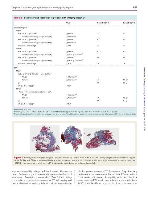
Table 2 Sensitivity and specificity <strong>of</strong> proposed RV imaging criteria*<br />
Value Sensitivity, % Specificity, %<br />
...............................................................................................................................................................................<br />
Echocardiogram<br />
Major<br />
PLAX RVOT (diastole) 32 mm 75 95<br />
Corrected for body size (PLAX/BSA) 19 mm/m 2<br />
PSAX RVOT (diastole) 36 mm 62 95<br />
Corrected for body size (PSAX/BSA) 21 mm/m 2<br />
Fractional area change 33% 55 95<br />
Minor<br />
PLAX RVOT (diastole) 29 mm 87 87<br />
Corrected for body size (PLAX/BSA) 16 to 18 mm/m 2i<br />
PSAX RVOT (diastole) 32 mm 80 80<br />
Corrected for body size (PSAX/BSA) 18 to 20 mm/m 2<br />
Fractional area change 40% 76 76<br />
...............................................................................................................................................................................<br />
MRI †<br />
Major<br />
Ratio <strong>of</strong> RV end-diastolic volume to BSA<br />
Males 110 mL/m 2<br />
Females 100 mL/m 2<br />
improved the capability to image the RV with reproducible measurements<br />
<strong>of</strong> volume and systolic function, which permits classification <strong>of</strong><br />
severity and differentiation from normality 47 (Table 2). Previous diagnostic<br />
reliance on subjective assessment <strong>of</strong> RV wall thinning, wall<br />
motion abnormalities, and fatty infiltration <strong>of</strong> the myocardium by<br />
76 90 F<br />
or 68 98 C<br />
RV ejection fraction 40%<br />
Minor<br />
Ratio <strong>of</strong> RV end-diastolic volume to BSA<br />
Males 100 mL/m 2<br />
Females 90 mL/m 2<br />
79 85 F<br />
or 89 97 C<br />
RV ejection fraction 45%<br />
Abbreviations as in Table 1.<br />
* All the major and minor criteria listed in this table are in addition to the requirement that regional wall motion abnormalities must also be present.<br />
† The sensitivity and specificity for males and females are the same as listed if, in addition to the stated wall motion criteria, there is either abnormal RV size or function or both.<br />
Figure 2 Endomyocardial biopsy findings in a proband affected by a diffuse form <strong>of</strong> ARVC/D. All 3 biopsy samples are from different regions<br />
<strong>of</strong> the RV free wall. There is extensive fibr<strong>of</strong>atty tissue replacement with myocardial atrophy, which is a major criterion (i.e. residual myocytes<br />
,60% by morphometric analysis or ,50% if estimated). Contributed by C. Basso, Padua, Italy.<br />
MRI has proven problematic. 48,49 Recognition <strong>of</strong> significant fatty<br />
involvement without concomitant fibrosis <strong>of</strong> the RV in normal individuals<br />
renders this unique MRI capability <strong>of</strong> limited value. Late<br />
enhancement on MRI permits myocardial tissue characterization in<br />
the LV. It can be difficult to be certain <strong>of</strong> late enhancement for<br />
Downloaded from<br />
http://eurheartj.oxfordjournals.org/ by guest on July 22, 2013