Sulcoflex ®Pseudophakic Supplementary IOLs - Iogen
Sulcoflex ®Pseudophakic Supplementary IOLs - Iogen
Sulcoflex ®Pseudophakic Supplementary IOLs - Iogen

Create successful ePaper yourself
Turn your PDF publications into a flip-book with our unique Google optimized e-Paper software.
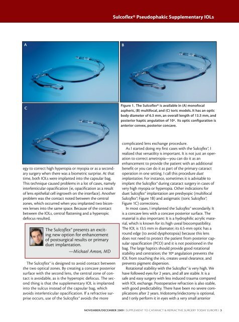
A<br />
C<br />
egy to correct high hyperopia or myopia or as a secondary<br />
surgery when there was a biometric surprise. At that<br />
time, both <strong>IOLs</strong> were implanted into the capsular bag.<br />
This technique caused problems in a lot of cases, namely<br />
interlenticular opacification (ie, opacification as a result<br />
of lens epithelial cell ingrowth on the interface). Another<br />
problem was the contact noted between the central<br />
zones, which occurred when you implanted two biconvex<br />
lenses into the same space. Because of the contact<br />
between the <strong>IOLs</strong>, central flattening and a hyperopic<br />
defocus resulted.<br />
The <strong>Sulcoflex</strong>® presents an exciting<br />
new option for enhancement<br />
of postsurgical results or primary<br />
duet implantation.<br />
—Michael Amon, MD<br />
The <strong>Sulcoflex</strong>® is designed to avoid contact between<br />
the two optical zones. By creating a concave posterior<br />
surface with the second lens, the central zone of contact<br />
is avoidable, as is the hyperopic defocus. The second<br />
thing is that the supplementary IOL is implanted<br />
into the sulcus instead of the capsular bag, which<br />
avoids interlenticular opacification. If a refractive surprise<br />
occurs, use of the <strong>Sulcoflex</strong>® avoids the more<br />
<strong>Sulcoflex</strong>® Pseudophakic <strong>Supplementary</strong> <strong>IOLs</strong><br />
B<br />
Figure 1. The <strong>Sulcoflex</strong>® is available in (A) monofocal<br />
aspheric, (B) multifocal, and (C) toric models. It has an optic<br />
body diameter of 6.5 mm, an overall length of 13.5 mm, and<br />
posterior haptic angulation of 10º. Its optic configuration is<br />
anterior convex, posterior concave.<br />
complicated lens exchange procedure.<br />
As I started doing my first cases with the <strong>Sulcoflex</strong>®, I<br />
realized that versatility is important. It is not just an operation<br />
to correct ametropia—you can do it as an<br />
enhancement to provide the patient with an additional<br />
benefit or you can do it as part of the primary cataract<br />
operation in one setting. I call this procedure duet<br />
implantation. For instance, sometimes it is advisable to<br />
implant the <strong>Sulcoflex</strong>® during cataract surgery in cases of<br />
very high myopia or hyperopia. Other indications for<br />
duet <strong>Sulcoflex</strong>® implantation are presbyopic (multifocal<br />
<strong>Sulcoflex</strong>®; Figure 1B) and astigmatic (toric <strong>Sulcoflex</strong>®;<br />
Figure 1C) corrections.<br />
In most cases, I implanted the <strong>Sulcoflex</strong>® secondarily. It<br />
is a concave lens with a concave posterior surface. The<br />
material is also important: It is a hydrophilic acrylic material,<br />
which is known for its high uveal biocompatibility.<br />
The IOL is 13.5 mm in diamater; its 6.5-mm optic has a<br />
round edge (to avoid dysphotopsia) because this lens<br />
does not need to protect the patient from posterior capsular<br />
opacification (PCO) and it is not positioned in the<br />
bag. The large haptics should provide good rotational<br />
stability and centration; the 10º angulation prevents the<br />
IOL from touching the iris, creates uveal clearance, and<br />
prevents pigment dispersion.<br />
Rotational stability with the <strong>Sulcoflex</strong>® is very high. We<br />
have followed eyes for 2 years, and all are stable. It is a<br />
safe and easy surgery with less induced trauma compared<br />
with IOL exchange. Postoperative refraction is also stable,<br />
with good predictability. There have been no severe complications<br />
after 2 years. Iridotomy/iridectomy is optional,<br />
and I only perform it in eyes with a very small anterior<br />
NOVEMBER/DECEMBER 2009 I SUPPLEMENT TO CATARACT & REFRACTIVE SURGERY TODAY EUROPE I 3