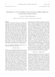
EFFECT OF AN OILY CALCIUM HYDROXIDE SUSPENSION ...
EFFECT OF AN OILY CALCIUM HYDROXIDE SUSPENSION ...
EFFECT OF AN OILY CALCIUM HYDROXIDE SUSPENSION ...

Create successful ePaper yourself
Turn your PDF publications into a flip-book with our unique Google optimized e-Paper software.
268<br />
Abstract<br />
The efficacy of an oily calcium hydroxide suspension<br />
(Osteoinductal ® ) as an adjunct to periodontal regenerative<br />
therapy has been demonstrated in recent clinical<br />
and histological studies. However, little is known<br />
about the molecular mechanisms in vitro, particularly,<br />
about the effect of oily calcium hydroxide paste on<br />
periodontal ligament (PDL) cells. Therefore the aim of<br />
the present study was to analyze the mitogenic response<br />
of cultured PDL cells to Osteoinductal ® in<br />
comparison to calcium hydroxide and enamel matrix<br />
derivative (EMD) in vitro. Human periodontal ligament<br />
cells were derived from a third molar extracted<br />
for orthodontic reasons and incubated in the presence<br />
of Osteoinductal ® , calcium hydroxide, EMD, phosphate-buffered<br />
saline plus 10% glycerol (PBS) or standard<br />
culture medium for 15 and 60 minutes. The mitogenic<br />
response of the PDL cells was determined by<br />
Western Blot with antibodies specific for extracellular<br />
signal-related kinase (ERK)1/2 as well as the activated,<br />
tyrosine-phosphorylated form of ERK1/2 (p-ERK).<br />
Relative phosphorylation of ERK1/2 was normalized<br />
to total ERK1/2 levels by densitometry. Cell proliferation<br />
was measured after 1, 3 and 8 days using a<br />
Neubauer haemocytometer to determine the total cell<br />
number. Results demonstrated that the mitogenic response<br />
to Osteoinductal ® , calcium hydroxide and<br />
enamel matrix derivative was associated with the activation<br />
of ERK1/2 and was more pronounced as compared<br />
to PBS and standard culture medium treated<br />
cells. Although Osteoinductal ® and calcium hydroxide<br />
activated mitosis and caused phosphorylation of<br />
ERK1/2 in PDL cells, its effects were less pronounced<br />
as compared to EMD. Furthermore EMD<br />
exhibited the highest proliferative response in comparison<br />
to Osteoinductal ® , calcium hydroxide and the<br />
negative control after one, three and eight days. In<br />
conclusion, our data indicate that Osteoinductal ® enhances<br />
the mitogenic response of human PDL cells by<br />
activation of ERK1/2 and increases cell proliferation,<br />
however, it is inferior in comparison to EMD.<br />
Key words: Oily calcium hydroxide suspension, periodontal<br />
ligament cells, cell proliferation, cell culture,<br />
ERK1/2, periodontal regeneration<br />
EU RO PE <strong>AN</strong> JOUR NAL <strong>OF</strong> MED I CAL RE SEARCH June 27, 2007<br />
Eur J Med Res (2007) 12: 268-272 © I. Holzapfel Publishers 2007<br />
<strong>EFFECT</strong> <strong>OF</strong> <strong>AN</strong> <strong>OILY</strong> <strong>CALCIUM</strong> <strong>HYDROXIDE</strong> <strong>SUSPENSION</strong><br />
(OSTEOINDUCTAL ®) ON HUM<strong>AN</strong> PERIODONTAL FIBROBLASTS.<br />
<strong>AN</strong> IN VITRO STUDY<br />
A. Kasaj 1, B. Willershausen 1, N. Jewszyk 1, M. Schmidt 2<br />
1 Department of Conservative Dentistry and Periodontology, Johannes Gutenberg University, Mainz, Germany<br />
2 Institute for Biochemistry II, Goethe University Medical School, Frankfurt/Main, Germany<br />
INTRODUCTION<br />
The aim of periodontal therapy is to control infection<br />
and to repair lost tooth supporting structures, including<br />
the periodontal ligament (PDL), root cementum<br />
and alveolar bone. Several treatment modalities, including<br />
bone grafts, application of growth factors and<br />
guided tissue regeneration have been proposed to restore<br />
lost periodontal attachment in intrabony defects<br />
(Karring et al. 1993, Sculean et al. 2000). Although<br />
treatment of lost periodontal tissue according to the<br />
principles of guided tissue regeneration (Nyman et al.<br />
1982) and the use of different grafting materials (Bartold<br />
et al. 2000, Trombelli et al. 2002) has gained increasing<br />
acceptance, true periodontal regeneration has<br />
not reached its ideal objective so far. Recent studies<br />
have demonstrated that an oily calcium hydroxide suspension<br />
(Osteoinductal®), applied to the root surface<br />
in conjunction with surgical periodontal therapy, may<br />
promote periodontal regeneration (Stratul et al. 2006).<br />
The major components of the oily calcium hydroxide<br />
formulation are calcium hydroxide, liquid and solid<br />
carbohydrate chains and fatty acids esterified with<br />
glycerol. The oily part consists of oleum pedum of<br />
porcine origin and vaselinum album. Stratul et al. (2006)<br />
compared the effects of Osteoinductal ® in deep intrabony<br />
defects versus access flap surgery and found significant<br />
higher clinical attachment level (CAL) gains in<br />
sites treated with Osteoinductal ® compared to the access<br />
flap surgery alone. Schwarz et al. (2006) histologically<br />
demonstrated that Osteoinductal ® may favour<br />
periodontal regeneration through the formation of<br />
new bone, periodontal ligament and cementum with<br />
inserting collagen fibers in acute-type intrabony defects<br />
in dogs. In addition, findings from histological studies<br />
in animals and humans showed that the use of an oily<br />
calcium hydroxide paste leads to an accelerated bone<br />
regeneration (Lazzerini 2001, Ito et al. 2002).<br />
Although the efficacy of Osteoinductal ® as an adjunct<br />
to periodontal regenerative therapy has been<br />
demonstrated in recent clinical studies (Stratul &<br />
Sculean 2004, Stratul et al. 2006), little is known about<br />
the molecular mechanisms, particularly the effect of<br />
oily calcium hydroxide paste, on PDL cells in vitro.<br />
Since the periodontal ligament is regarded as the
June 27, 2007 EUROPE<strong>AN</strong> JOURNAL <strong>OF</strong> MEDICAL RESEARCH<br />
269<br />
source of cementoblasts and osteoblasts and since<br />
only the tissue originating from the PDL possesses the<br />
ability to form a new connective tissue attachment,<br />
cells from the PDL are thought to play a key role in<br />
periodontal regeneration. Therefore the behaviour of<br />
these cells might be influenced by oily calcium hydroxide.<br />
The cellular response to various environmental<br />
stimuli such as growth factors or different biomaterials<br />
and bone substitutes is regulated through transmembrane<br />
molecules that initiate a great variety of<br />
signal transduction cascades. One major player in<br />
this context is the extracellular signal-related kinase<br />
(ERK) 1/2.<br />
The aim of the present study was to evaluate the<br />
mitogenic effect of Osteoinductal ® in cultured human<br />
periodontal ligament cells by measurement of ERK1/2<br />
activation. In addition, the results of Osteoinductal ®<br />
on PDL cells were compared with those of pure calcium<br />
hydroxide as well as enamel matrix derivative<br />
(EMD).<br />
MATERIAL <strong>AN</strong>D METHODS<br />
REAGENTS<br />
The oily calcium hydroxide suspension (Osteoinductal®,<br />
Munich, Germany; 10 mg), a calcium hydroxide<br />
pharmaceutical containing Neatsfoot Oil 1024 of<br />
porcine origin, was dissolved in 100 ml phosphate<br />
buffered saline (PBS) containing 10 % glycerol by addition<br />
of two drops concentrated HCl and subsequent<br />
boiling at 100° C for 5 minutes. The Emdogain®<br />
(Enamel Matrix Derivative - EMD) was supplied by<br />
Straumann, Switzerland. For study purposes 5 ml of<br />
lyophilised porcine enamel matrix protein (EMD),<br />
containing 80 % amelogenin, small amounts of residual<br />
water, calcium phosphate and acetic acid, were dissolved<br />
in 1.5 ml PBS containing 10 % glycerol by addition<br />
of two drops concentrated HCl and subsequent<br />
boiling at 100° C for 5 minutes. Furthermore, 1 g of<br />
calcium hydroxide was dissolved in 1 ml hydrochloric<br />
acid and diluted with 89 ml water. Subsequently, 10%<br />
glycerol were added to yield a solution of about 135<br />
mM. The pH of each solution was adjusted to 7.5. It<br />
should be mentioned that HCl was used to solubilise<br />
the calcium hydroxide component and glycerol to solubilise<br />
the lipid components of each pharmaceutical.<br />
Control cultures consisted of similarly plated cells in<br />
media containing phosphate-buffered saline plus 10 %<br />
glycerol (PBS) or standard culture medium alone.<br />
CELL CULTURES <strong>AN</strong>D MEDIA<br />
The human periodontal ligament cells (PDL) were<br />
obtained from healthy human periodontal tissues isolated<br />
from a third molar extracted for orthodontic reasons<br />
from a 23 year-old female patient without any<br />
known medical disorders. Under sterile conditions, the<br />
PDL tissue fragments were mechanically removed by<br />
scraping the middle third of the root surface with a<br />
sharp blade. Tissue explants were maintained in Dulbecco`s<br />
modified Eagle`s minimum essential medium<br />
(DMEM, Invitrogen) containing 10 % Fetal Bovine<br />
Serum (FBS, PAA), 1 % penicillin/streptomycin (In-<br />
vitrogen), and 1 % fungizone (Invitrogen). Cultures<br />
were incubated in a humidified atmosphere (95 %) of<br />
5 % CO2 . Within three weeks the PDL explants<br />
formed primary colonies. Tissue culture medium was<br />
changed every two days until confluence was reached<br />
(approximately 7x 10⁴ cells/cm2) and cells were passaged<br />
at a 1 : 2 split ratio following trypsinization with<br />
0.05 % trypsin (Invitrogen). Cell cultures were tested<br />
regularly to be free of mycoplasma by PCR. Cells were<br />
used for experiments between passage four and nine.<br />
WESTERN BLOTTING<br />
For detection of proteins in Western Blots monoclonal<br />
mouse anti-p-ERK (E-4) and polyclonal rabbit anti-<br />
ERK1/2 (C-14) antibodies have been purchased from<br />
Santa Cruz Biotechnology. Cells were incubated in the<br />
presence of standard culture medium, phosphatebuffered<br />
saline (PBS) plus glycerol, calcium hydroxide,<br />
oily calcium hydroxide suspension (Osteoinductal®)<br />
or enamel matrix derivative (EMD) in standard culture<br />
medium for 15 and 60 minutes. Protein samples were<br />
analyzed by SDS-PAGE using an XCell SureLock<br />
Mini-Cell (Invitrogen) in combination with precast<br />
NuPAGE 4-12 % or 10 % Bis-Tris gels (1 mm) at 200<br />
volts according to the manufacturers guidelines. Following<br />
electrophoresis, proteins were blotted to a<br />
PVDF membrane and were incubated for at least one<br />
hour in blocking buffer (5 % BSA and 1 % Tween-20<br />
in tris-buffered saline (TBS). Membranes were incubated<br />
over night with appropriate dilutions of primary<br />
antibody in blocking buffer. The next day membranes<br />
were washed and incubated for one hour with alkalinephosphatase<br />
conjugated secondary antibody solution<br />
in blocking buffer (Sigma; dilutions: anti-mouse antibody<br />
1 : 3.000 and anti-rabbit antibody 1 : 5.000). After<br />
additional washing steps, antibody complexes were<br />
visualized on film by Immun-Star AP substrate (Bio-<br />
Rad). Western Blot results were quantified by densitometry<br />
using NIH’s ImageJ software.<br />
PROLIFERATION <strong>AN</strong>D CELL VITALITY<br />
Proliferation rate analysis was carried out over an eight<br />
day period to provide information on cell proliferation<br />
and cell toxity. PDL cells were plated on 35 mm petri<br />
dishes at 5 x 10 3 cells per dish in standard culture<br />
medium and incubated for 24 h under standard conditions.<br />
Subsequently, enamel matrix derivative, oily calcium<br />
hydroxide suspension and calcium hydroxide<br />
were added to a final concentration of about 1 mM<br />
calcium hydroxide. Control cultures consisted of<br />
equally plated cells in standard culture medium plus<br />
PBS containing 10% glycerol. Each experiment was<br />
performed in triplicate and for each experimental<br />
group three dishes were plated for 8 days. Cells were<br />
harvested, mixed with trypan blue dye and counted<br />
by microscopy using a Neubauer Haemocytometer<br />
(Hausser Scientific).<br />
STATISTICAL <strong>AN</strong>ALYSIS<br />
Statistical analysis has been performed using the<br />
Wilcoxon matched-pairs signed ranks test. Differences
270 EUROPE<strong>AN</strong> JOURNAL <strong>OF</strong> MEDICAL RESEARCH<br />
June 27, 2007<br />
a<br />
b<br />
Fig. 1. a: ERK1/2, b: Tyrosine-phosphorylated ERK1/2. Response of extracellular signal-regulated kinase (ERK) 1/2 to Osteoinductal<br />
® , calcium hydroxide and enamel matrix derivative after 15 and 60 minutes. Total PDL lysates were blotted for<br />
ERK1/2 levels (a) and phosphorylation as well as activation of ERK1/2 was detected with a specific p-ERK antibody (b).<br />
a<br />
b<br />
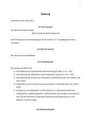
Fig. 2. a: Relative activation of ERK1/2 after 15 minutes, 2b:<br />
Relative activation of ERK1/2 after 60 minutes. Relative activation<br />
of ERK in PDL cells after 15 (a) and 60 minutes (b)<br />
was determined by densitometry using NIH’s ImageJ software.<br />
Clearly, after 60 minutes ERK1/2 displayed enhanced<br />
phosphorylation upon stimulation with Osteoinductal®, calcium<br />
hydroxide and EMD.<br />
were considered significant at a P-value < 0.05.<br />
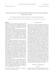
The mitogenic response of PDL cells towards stimulation<br />
with EMD, Osteoinductal ® and calcium hydroxide<br />
was determined by measurement of the phosphorylation<br />
level of ERK1/2 using a p-ERK-specific<br />
antibody. Cells were harvested, seeded in 6 Well plates<br />
and were grown under standard culture conditions for<br />
24 hours. Subsequently, PDL cells were starved in<br />
serum free medium over night and eventually stimulated<br />
with a 1 : 100 dilution of EMD, Osteoinductal ®<br />
and calcium hydroxide in serum-free medium for various<br />
times. This yielded an approximate concentration<br />
of 1 mM calcium hydroxide in each sample. Finally,<br />
cells were frozen in liquid nitrogen to stop any<br />
reaction towards different stimuli and were lysed in<br />
lysis buffer. Aliquots were boiled in Laemmli buffer<br />
and underwent SDS-PAGE and immunoblotting<br />
using antibodies specific for ERK1/2 and p-ERK<br />
(Fig. 1 a, b).<br />
The result was quantified and the relative phosphorylation<br />
of ERK1/2 determined using densitometry<br />
(Fig. 2 a, b). The relative intensity of p-ERK bands<br />
was set into relation to the total ERK1/2 levels and<br />
expressed as induction of a certain stimulants as compared<br />
to non-treated control (empty). PBS containing<br />
10 % glycerol (PBS) served as a second control in order<br />
to evaluate the effect of changing the culture conditions<br />
by adding small amounts of EMD, Osteoinductal<br />
® and calcium hydroxide solutions. We expected<br />
that the glycerol, which was added to the solutions to<br />
solubilise the lipid components of each pharmaceutical,<br />
could cause some type of reaction within PDL<br />
cells. Indeed, a small increase in relative ERK1/2<br />
phosphorylation in PBS as well as EMD, Osteoinductal<br />
® and calcium hydroxide solutions treated cells was<br />
observed after 15 minutes of treatment (Fig. 2a).<br />
However, after 60 minutes the stimulating effect of<br />
PBS plus glycerol vanished and revealed that EMD,<br />
Osteoinductal ® and calcium hydroxide significantly increased<br />
ERK1/2 phosphorylation as compared to<br />
controls (Fig. 2b). It was concluded that EMD, Osteoinductal<br />
® and calcium hydroxide induce activation<br />
of ERK1/2 in PDL cells. Therefore enamel matrix derivative,<br />
oily calcium hydroxide suspension and calcium<br />
hydroxide itself acted mitogenic towards PDL<br />
cells as compared to controls. Despite Osteoinductal ®<br />
and calcium hydroxide increased the phosphorylation<br />
of ERK1/2 in PDL cells after 60 minutes, the effect<br />
was more pronounced in EMD-treated cells in the<br />
long run.<br />
As we detected this mitogenic effect of calcium hydroxide<br />
solutions it was decided to study cell proliferation<br />
of PDL cells in a long term fashion. Therefore<br />
cells were incubated for 1, 3 and 8 days with EMD, Osteoinductal<br />
® and calcium hydroxide in standard culture<br />
medium with PBS plus 10 % glycerol in medium as a<br />
control. At the respective days, cells were harvested,<br />
stained with Trypan blue dye and counted in a<br />
Neubauer chamber. EMD, Osteoinductal ® and calcium<br />
hydroxide affected cell proliferation, with a small
June 27, 2007 EUROPE<strong>AN</strong> JOURNAL <strong>OF</strong> MEDICAL RESEARCH<br />
271<br />
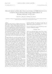
Fig. 3: Cell proliferation assay. Effect of Osteoinductal ® , calcium<br />
hydroxide and enamel matrix derivative at various time<br />
points on proliferation of periodontal ligament fibroblasts (1,<br />
3 and 8 days). Osteoinductal ® , calcium hydroxide and enamel<br />
matrix derivative increased cell proliferation and consequently,<br />
total cell numbers as compared to control.<br />
increase after day 1 and a marked enhancement after<br />
days 3 and 8 (Fig. 3). This data suggest that after 8 days<br />
the difference in cell numbers between EMD and Osteoinductal<br />
® as well as calcium hydroxide is remarkable<br />
in magnitude, and the amount of cells using EMD<br />
was much higher as compared to the application of<br />
Osteoinductal®, calcium hydroxide and control solutions.<br />
However, no statistically significant difference in<br />
cell proliferation between the groups (p > 0.05) could<br />
be noticed. Furthermore, no significant difference between<br />
the oily calcium hydroxide suspension (Osteoinductal®)<br />
and the pure calcium hydroxide was noted.<br />
DISCUSSION<br />
Data from histological and controlled clinical studies<br />
demonstrated that grafting procedures may result in<br />
“periodontal regeneration”. However, a predictable reconstitution<br />
of lost periodontal structures is still difficult<br />
to obtain. In this context the recruitment of PDL<br />
cells, which have the capacity to differentiate into cells<br />
of cementogenic, osteogenic and fibroblastic lineage,<br />
their proliferation as well as their colonization of the<br />
wound area are of great importance to the success of<br />
regenerative periodontal therapy (Melcher 1976). According<br />
to Stratul et al. (2006) regeneration of lost periodontal<br />
structures can be obtained by an oily calcium<br />
hydroxide suspension, commercially known as Osteoinductal®.<br />
In a further investigation, an improved<br />
early wound healing following the topical subgingival<br />
application of Osteoinductal ® after non-surgical periodontal<br />
therapy was observed (Kasaj et al. 2006). The<br />
long-acting antibacterial and anti-inflammatory properties<br />
of calcium hydroxide were demonstrated in several<br />
experimental studies (Cvek et al. 1976, Staehle &<br />
Pioch 1989). While aqueous solutions of calcium hydroxide<br />
cause a rapid increase of the pH up to 12-13<br />
in living tissues, from the oily calcium hydroxide suspension<br />
a stable, long-lasting pH gradient of 7 – 11 is<br />
formed within the tissue without causing irritation<br />
(Dietz et al. 1998). Thus, the oily suspension produces<br />
a long-term, mild alkaline enviroment, because only<br />
the calcium hydroxide at the interface between the liq-<br />
uid/oily phase is released. As a result, a significant<br />
pain relief and supression of inflammation can be observed<br />
(Dietz et al. 1998). Further studies described<br />
the anti-inflammatory and analgetic properties when<br />
used in cases of wounded or surgically exposed bone<br />
surfaces (Filippi et al. 2000, Filippi 2001). Although<br />
Osteoinductal ® is used in current clinical treatment,<br />
its biological characteristics and properties are still<br />
poorly characterized. It is well recognized that human<br />
PDL cells can serve as a proper model for the analysis<br />
of cell bioactivity in vitro (Hoang et al. 1997, Lekic et<br />
al. 1997). For this reason, we investigated the mitogenic<br />
effects on PDL cells cultured with Osteoinductal®,<br />
pure calcium hydroxide and enamel matrix derivative.<br />
The results of the present study demonstrate the<br />
mitogenic effect of an oily calciumhydroxid suspension<br />
on PDL cells. The amount of phosphorylated<br />
ERK1/2 in PDL cells increased within 60 minutes following<br />
exposure to the oily calcium hydroxide suspension.<br />
The response was greater as compared to the<br />
control cells. The activation of ERK1/2 by Osteoinductal<br />
® occurred just after 60 minutes, therefore a<br />
possible induced formation and release of secondary<br />
products, such as transforming growth factor (TGF)-ß<br />
cannot be excluded. Furthermore, the poor solubility<br />
of the calcium hydroxide suspension might be one of<br />
the reasons why the effects of Osteoinductal ® on<br />
ERK1/2 and on cell proliferation were similar to the<br />
pure calcium hydroxide and smaller than those of<br />
EMD. Maybe, the components of Osteoinductal ®<br />
were more sensitive to the harsh treatment that had to<br />
be applied to solubilise it for this study as compared to<br />
EMD. A third possibility is that the glycerol that had<br />
been added to solubilise the lipid component Neatsfoot<br />
Oil 1024 of Osteoinductal ® might have sequestered<br />
it from the contact with PDL cells or was<br />
not sufficient enough to solubilise all oil. In either<br />
case, the effect of Osteoinductal ® was underrepresented<br />
as compared to EMD. Its protein components<br />
were much easier to solubilise. Thus, a possible beneficial<br />
effect of Osteoinductal ® due to its oily component<br />
in comparison to pure calcium hydroxide cannot<br />
be excluded at present.<br />
EMD has been studied extensively in previous studies<br />
in vitro and has been demonstrated to promote cellular<br />
functions required for the biological process of<br />
periodontal regeneration, such as proliferation, adherence,<br />
protein synthesis and differentiation (Gestrelius<br />
et al. 1997, van der Pauw et al. 2000, Lyngstadaas et al.<br />
2001). According to the findings of our study the<br />
most pronounced mitogenic effect on PDL cells was<br />
mediated by enamel matrix derivative. In the present<br />
study, human PDL cells exhibited a greater proliferative<br />
response to EMD when compared to Osteoinductal<br />
® or pure calcium hydroxide, however the difference<br />
was not statistically significant. These observations<br />
are in agreement with a study by Chong et al.<br />
(2006), which showed that EMD and amelogenin<br />
alone had no significant effect on PDL cell proliferation<br />
or migration. Potentially, the contradictory effects<br />
of EMD in vitro in published studies can be explained<br />
by different phenotypes of PDL fibroblasts that can<br />
be isolated and may respond differently to a regenera-
272 EUROPE<strong>AN</strong> JOURNAL <strong>OF</strong> MEDICAL RESEARCH<br />
June 27, 2007<br />
tive treatment (Chong et al. 2006). Therefore a possible<br />
impact of this appearance also cannot be excluded<br />
in our study.<br />
In conclusion, the addition of Osteoinductal ® to<br />
cell culture media resulted in an enhanced mitogenic<br />
response mediated by an increased activation of<br />
ERK1/2 and an increased cell proliferation rate similar<br />
to that of pure calcium hydroxide. However, the effect<br />
measured with Osteoinductal ® was inferior in<br />
comparison to EMD. Nevertheless, the current data<br />
justifies a hypothesis of cellular changes after application<br />
of Osteoinductal ® . Further studies are required<br />
to unravel the exact mechanisms by which Osteo -<br />
inductal ® influences cell function with the goal to optimize<br />
Osteoinductal ® treatment in periodontal regenerative<br />
therapy.<br />
Acknowledgement: The authors wish to acknowledge Ivan Dikic<br />
for his invaluable assistance in preparing the manuscript.<br />
REFERENCES<br />
1. Bartold PM, McCulloch CAG, Narayanan AS, Pitaru S<br />
(2000) Tissue engineering: A new paradigm for periodontal<br />
regeneration based on molecular and cell biology. Periodontol<br />
2000 24: 253-269<br />
2. Chong CH, Carnes DL, Moritz AJ, Oates T, Ryu OH,<br />
Simmer J, Cochran DL (2006) Human periodontal fibroblast<br />
response to enamel matrix derivative, amelogenin,<br />
and platelet-derived growth factor-BB. J Periodontol<br />
77: 1242-1252<br />
3. Cvek M, Hollender L, Nord CE (1976) Treatment of<br />
nonvital permanent incisors with calcium hydroxide. VI.<br />
A clinical, microbiological, and radiological evaluation of<br />
treatment in one setting of teeth with mature and imature<br />
root. Odontol Revy 27: 93-108<br />
4. Dietz G, Bartolmes P, Dietz GH jr, Roecher W (1998)<br />
Calcium hydroxide and bone regeneration. Odontological<br />
aspects of induced osteogenesis in experiment and clinical<br />
practice. Byblos, Munich<br />
5. Filippi A, Irnich G, Kirschner H, Pohl Y (2000) Lokale<br />
Beeinflussbarkeit der Wundheilung nach Osteotomie dritter<br />
Molaren. Quintessenz 51: 847-856<br />
6. Fillipi A (2001) Wundheilung und Heilungsstörungen<br />
nach Entfernung dritter Molaren. Schweiz Monatsschr<br />
Zahnmed 7: 847-856<br />
7. Gestrelius S, Andersson C, Lidström D, Hammarström L,<br />
Sommerman M (1997) In vitro studies on periodontal ligament<br />
cells and enamel matrix derivative. J Clin Periodontol<br />
24: 685-692<br />
8. Hoang AM, Chen D, Oates TW, Jiang C, Harris SE,<br />
Cochran DL (1997) Development and characterization of<br />
a transformed human periodontal ligament cell line. J Periodontol<br />
68: 1054-1062<br />
9. Ito T, Shibukawa Y, Amano H, Kawai H, Yamada S<br />
(2002) Effect of calcium hydroxide paste on bone formation.<br />
J Dent Res 81:128 (abstract)<br />
10. Karring T, Lindhe J, Cortellini P (1993) Development of<br />
the biological concept of guided tissue regeneration – animal<br />
and human studies. Periodontol 2000 1: 26-35<br />
11. Kasaj A, Willershausen B, Berakdar M, Tekyatan H,<br />
Sculean A (2006) Effect of an oily calcium hydroxide suspension<br />
on early wound healing after nonsurgical periodontal<br />
therapy. Clin Oral Invest 10: 72-76<br />
12. Lazzerini L (2001) Uso di una sospensione oleaosa di<br />
idrossido di calcio nella rigenerazione ossea. PhD Thesis,<br />
University of Pisa<br />
13. Lekic PC, Pender N, McCulloch CAG (1997) Is fibroblast<br />
heterogeneity relevant to the health, diseases and treatment<br />
of periodontal tissue? Crit Rev Oral Biol Med 8:<br />
253-268<br />
14. Lyngstadaas S, Lundberg E, Ekdahl H, Andersson C,<br />
Gestrelius S (2001) Autocrine growth factors in human<br />
periodontal ligament cells cultured on enamel matrix derivative.<br />
J Clin Periodontol 28: 181-188<br />
15. Melcher A (1976) On the repair potential of periodontal<br />
tissues. J Periodontol 47: 256-260<br />
16. Nyman S, Gottlow J, Karring T, Lindhe J (1982) The regenerative<br />
potential of the periodontal ligament. An experimental<br />
study in the monkey. J Clin Periodontol 2:<br />
257-265<br />
17. Schwarz F, Stratul SI, Herten M, Beck B, Becker J,<br />
Sculean A (2006) Effect of an oily calcium hydroxide suspension<br />
(Osteoinductal) on healing of intrabony periodontal<br />
defects. A pilot study in dogs. Clin Oral Invest<br />
10: 29-34<br />
18. Sculean A, Chiantella GC, Windisch P, Donos N (2000)<br />
Clinical and histologic evaluation of treatment of intrabony<br />
defects with an enamel matrix protein derivative<br />
(Emdogain). Int J Periodontics Restorative Dent 23: 47-<br />
55<br />
19. Staehle HJ, Pioch Th (1989) Antimikrobielle Wirksamkeit<br />
und alkalisierender Effekt verschiedener Calciumhydroxidpräparate.<br />
Dtsch Zahnärztl Z 44: 344-348<br />
20. Stratul SI, Schwarz F, Becker J, Willershausen B, Sculean<br />
A (2006) Healing of intrabony defects following treatment<br />
with an oily calcium hydroxide suspension (Osteoinductal).<br />
A controlled clinical study. Clin Oral Invest<br />
10: 55-60<br />
21. Trombelli L, Heitz-Mayfield LJ, Needleman I, Moles D,<br />
Scabbia A (2002) A systematic review of graft materials<br />
and biological agents for periodontal intraosseous defects.<br />
J Clin Periodontol 29 Suppl 3: 117-135<br />
22. van der Pauw M, van den Boss T, Everts V, Beertsen W<br />
(2000) Enamel matrix-derived protein stimulates attachment<br />
of periodontal ligament fibroblasts and enhances alkaline<br />
phosphatase activity and transforming growth factor<br />
beta 1 release of periodontal ligament and gingival fibroblasts.<br />
J Periodontol 71: 31-43<br />
Received: October 12, 2006 / Accepted: January 31, 2007<br />
Address for correspondence:<br />
Prof. Dr. B. Willershausen<br />
Department for Operative Dentistry,<br />
Johannes-Gutenberg University Mainz<br />
D-55131 Mainz, Germany