fecal elastase 1 and vitamin d3 in patients with osteoporotic bone ...
fecal elastase 1 and vitamin d3 in patients with osteoporotic bone ...
fecal elastase 1 and vitamin d3 in patients with osteoporotic bone ...

Create successful ePaper yourself
Turn your PDF publications into a flip-book with our unique Google optimized e-Paper software.
68<br />
Abstract<br />
Background <strong>and</strong> Aims: The aim of the present study<br />
was to clarify if <strong>patients</strong> <strong>with</strong> <strong>osteoporotic</strong> <strong>bone</strong> fractures<br />
have exocr<strong>in</strong>e pancreatic <strong>in</strong>sufficiency, especially<br />
reduced <strong>fecal</strong> <strong>elastase</strong> 1, connected <strong>with</strong> lowered<br />
serum levels of <strong>vitam<strong>in</strong></strong> D 3 that could be relevant for<br />
predom<strong>in</strong>ant osteoporosis.<br />
Methods: Between October 1999 <strong>and</strong> September 2001,<br />
we <strong>in</strong>vestigated on 167 <strong>patients</strong> <strong>with</strong> an average age of<br />
approx. 69 years suffer<strong>in</strong>g from typical <strong>osteoporotic</strong><br />
<strong>bone</strong> fractures, as well as 20 healthy controls <strong>with</strong> an<br />
average age of 53 years. A st<strong>and</strong>ardized osteodensitometry<br />
via dual energy X-ray absorptiometry (DEXA)<br />
was performed <strong>in</strong> all participants. Levels of PTH,<br />
1,25(OH) 2Vitam<strong>in</strong> D 3, 25(OH)Vitam<strong>in</strong> D 3, calcium<br />
<strong>and</strong> phosphate <strong>in</strong> serum, <strong>elastase</strong> 1 <strong>in</strong> feces as well as<br />
the body mass <strong>in</strong>dex were determ<strong>in</strong>ed <strong>in</strong> all <strong>patients</strong><br />
<strong>and</strong> controls.<br />
Results: In <strong>patients</strong> 25(OH)D 3 was more than 60%<br />
<strong>and</strong> 1,25(OH) 2D 3 was more than 53% decreased compared<br />
to controls. Fecal <strong>elastase</strong> 1 was lower than the<br />
lowest reference of 200 µg/g feces <strong>in</strong> more than 34%<br />
of the <strong>patients</strong> <strong>and</strong> it was more than 65% reduced <strong>in</strong><br />
comparison to healthy controls (<strong>fecal</strong> <strong>elastase</strong> 1 <strong>patients</strong>:<br />
240.7 ± 96.3 µg/g; controls 694.9 ± 138.6<br />
µg/g). Separation of the <strong>patients</strong> <strong>in</strong> accordance <strong>with</strong><br />
the <strong>elastase</strong> 1 contend <strong>in</strong> feces <strong>in</strong>to four groups (below<br />
100 µg/g, between 100 <strong>and</strong> 200 µg/g, between 201<br />
<strong>and</strong> 300 µg/g <strong>and</strong> above 300 µg/g) resulted <strong>in</strong> significant<br />
variations for 25(OH)D 3, 1,25(OH) 2D 3, calcium<br />
<strong>and</strong> PTH between these groups (p < 0.01). Furthermore<br />
25(OH)D 3, 1,25(OH) 2D 3, calcium <strong>and</strong> PTH correlated<br />
significantly <strong>with</strong> <strong>elastase</strong> 1 <strong>in</strong> feces (p < 0.01)<br />
the way, that lower <strong>fecal</strong> <strong>elastase</strong> 1 was connected <strong>with</strong><br />
lower levels of the other parameters. BMI shows no<br />
relevant differences <strong>with</strong><strong>in</strong> the <strong>patients</strong> or between <strong>patients</strong><br />
<strong>and</strong> controls.<br />
Conclusion: Exocr<strong>in</strong>e pancreatic <strong>in</strong>sufficiency, especially<br />
lowered <strong>fecal</strong> <strong>elastase</strong> 1, may be much more frequent<br />
<strong>in</strong> <strong>patients</strong> <strong>with</strong> <strong>osteoporotic</strong> <strong>bone</strong> fractures<br />
than suggested so far. Lowered exocr<strong>in</strong>e pancreatic<br />
function <strong>with</strong> lowered <strong>fecal</strong> <strong>elastase</strong> 1 seems to be relevant<br />
as a reason for reduced levels of circulat<strong>in</strong>g <strong>vitam<strong>in</strong></strong><br />
D 3 metabolites be<strong>in</strong>g an appropriate additional<br />
cause for predom<strong>in</strong>ant osteoporosis.<br />
Key words: <strong>fecal</strong> <strong>elastase</strong> 1, <strong>vitam<strong>in</strong></strong> D 3, osteoporosis,<br />
<strong>bone</strong>-fracture, <strong>bone</strong> metabolism.<br />
EU RO PE AN JOUR NAL OF MED I CAL RE SEARCH February 25, 2008<br />
Eur J Med Res (2008) 13: 68-72 © I. Holzapfel Publishers 2008<br />
FECAL ELASTASE 1 AND VITAMIN D 3 IN PATIENTS WITH OSTEOPOROTIC<br />
BONE FRACTURES<br />
S. T. W. Mann, V. Mann, H. Stracke, U. Lange, H. U. Klör, P. Hardt, J. Teichmann<br />
Department of Internal Medic<strong>in</strong>e, Medical Cl<strong>in</strong>ic III <strong>and</strong> Polycl<strong>in</strong>ic of the Justus-Liebig-University Giessen, Germany<br />
INTRODUCTION<br />
Fracture risk is a function of trauma susta<strong>in</strong>ed (e.g. <strong>in</strong><br />
falls) <strong>and</strong> <strong>bone</strong> strength (which depends on both, the<br />
quantity of <strong>bone</strong> <strong>and</strong> its architecture). Osteoporotic<br />
<strong>bone</strong> fractures, commonly of the hip, sp<strong>in</strong>e or forearm,<br />
are typically susta<strong>in</strong>ed <strong>with</strong> little or no antecedent<br />
trauma. The comb<strong>in</strong>ed lifetime risk for hip, forearm<br />
<strong>and</strong> vertebral fractures com<strong>in</strong>g to cl<strong>in</strong>ical attention is<br />
around 40% [1]. Thus Osteoporosis <strong>and</strong> its consequences<br />
as <strong>osteoporotic</strong> <strong>bone</strong> fractures is a major public<br />
health problem <strong>with</strong> serious consequences <strong>in</strong> terms<br />
of mortality, morbidity <strong>and</strong> economic costs [2-6]. Beside<br />
other reasons for osteoporosis <strong>vitam<strong>in</strong></strong> D 3 ga<strong>in</strong>s a<br />
special relevance. The basic importance of <strong>vitam<strong>in</strong></strong> D 3<br />
for calcium homeostasis, <strong>bone</strong> m<strong>in</strong>eralisation, osteoblastic<br />
differentiation, <strong>and</strong> <strong>bone</strong> matrix snythesis is<br />
still irrefutable [7]. Apart from the consequences of an<br />
extreme <strong>vitam<strong>in</strong></strong> D 3-deficiency, such as rickets (<strong>in</strong>fants),<br />
osteomalacia (adults) or fibrotic changes (osteitis fibrosa<br />
Reckl<strong>in</strong>gshausen), Scharla et al. <strong>and</strong> Chapuy et al.<br />
[8, 9] have already described the extensive consequences<br />
of a sub-cl<strong>in</strong>ical deficiency <strong>with</strong> values <strong>with</strong><strong>in</strong><br />
the "normal" references. Vitam<strong>in</strong> D 3 <strong>and</strong> Calcium have<br />
been shown <strong>in</strong> some r<strong>and</strong>omised cl<strong>in</strong>ical trials to reduce<br />
hip fractures <strong>and</strong> other non vertebral fractures <strong>in</strong><br />
men <strong>and</strong> woman as much as over 40% [10-12]. Poskitt<br />
et al. [13] reported, that a depletion of <strong>vitam<strong>in</strong></strong> D storage<br />
is ma<strong>in</strong>ly caused by a reduced exposition to the sun,<br />
but altogether serum levels of lipid soluble <strong>vitam<strong>in</strong></strong> D 3<br />
depends on photosynthesis <strong>in</strong> the sk<strong>in</strong> as well as on direct<br />
<strong>in</strong>test<strong>in</strong>al resorption. In our further studies we<br />
could demonstrate a connection between reduced exocr<strong>in</strong>e<br />
pancreatic function, especially reduced <strong>fecal</strong> <strong>elastase</strong><br />
1, lowered levels of <strong>vitam<strong>in</strong></strong> D 3 <strong>and</strong> loss of skeletal<br />
mass <strong>in</strong> <strong>patients</strong> <strong>with</strong> chronic pancreatitis [14, 15]. The<br />
aim of the present study is to clarify, if <strong>patients</strong> <strong>with</strong><br />
<strong>osteoporotic</strong> <strong>bone</strong> fractures <strong>and</strong> <strong>with</strong>out chronic pancreatitis<br />
although have reduced <strong>fecal</strong> <strong>elastase</strong> 1 connected<br />
<strong>with</strong> reduced serum levels of <strong>vitam<strong>in</strong></strong> D 3, that<br />
could be relevant for predom<strong>in</strong>ant osteoporosis.<br />
MATERIAL AND METHODS<br />
PATIENTS<br />
Between October 1999 <strong>and</strong> September 2001 we <strong>in</strong>vestigated<br />
on <strong>patients</strong> <strong>with</strong> typical <strong>osteoporotic</strong> <strong>bone</strong>
February 25, 2008 EUROPEAN JOURNAL OF MEDICAL RESEARCH<br />
69<br />
fractures such as forearm, hip <strong>and</strong> sp<strong>in</strong>e. A st<strong>and</strong>ardized<br />
osteodensitometry via dual energy X-ray absorptiometry<br />
(DEXA) was performed on all <strong>patients</strong>. If<br />
DEXA revealed T-scores lower than –2,5 SD (accord<strong>in</strong>gly<br />
to actual WHO-def<strong>in</strong>ition [16] this means severe<br />
osteoporosis) <strong>patients</strong> matched our criteria <strong>and</strong> f<strong>in</strong>ally<br />
167 could be <strong>in</strong>cluded <strong>in</strong> our study. Exclusion criteria<br />
were: ages under 40 or over 86 years; steatorrhea; pancreatic-biliary<br />
obstructions; actual <strong>and</strong> relevant alcohol<br />
consumption; medication <strong>with</strong> <strong>in</strong>fluence on osteological<br />
<strong>and</strong>/or endocr<strong>in</strong>e parameters (hepar<strong>in</strong>, ketoconazol,<br />
glucocorticoids, thiacide-diuretics, psychopharmacological<br />
agents, carbamazep<strong>in</strong>); chronic or severe<br />
concommitant diseases.<br />
CONTROLS<br />
Twenty healthy persons between 40 <strong>and</strong> 60 years of<br />
age served as controls.<br />
BIOCHEMICAL MEASUREMENTS<br />
Blood samples were taken from all participants once at<br />
a fixed time <strong>in</strong> the mor<strong>in</strong><strong>in</strong>g. The specific study parameters<br />
were parathormone ("INTACT PTH"-kit from<br />
Nichols Institute Diagnostics, San Juan Capistrano,<br />
California; double-sided immuno-radiometric assay),<br />
1,25(OH) 2 Vitam<strong>in</strong> D 3 (“1,25(OH) 2 Vitam<strong>in</strong> D”-kit<br />
from Immun Diagnostik, Bensheim, Germany; competitive<br />
radio receptor assay), 25(OH)Vitam<strong>in</strong> D 3<br />
(“25(OH) Vitam<strong>in</strong> D”-kit from Immun Diagnostik,<br />
Bensheim, Germany; competitive prote<strong>in</strong>-b<strong>in</strong>d<strong>in</strong>g-assay),<br />
calcium <strong>and</strong> phosphate from serum as well as<br />
pancreatic <strong>elastase</strong> 1 ("Pankreatic Elastase 1"-kit from<br />
ScheBo Biotech, Giessen, Germany; double-sided enzyme-immuno<br />
assay) from feces of <strong>patients</strong> <strong>and</strong> con-<br />
trols. The body mass <strong>in</strong>dex (BMI) was also determ<strong>in</strong>ed<br />
<strong>in</strong> all.<br />
OSTEODENSITOMERTY<br />
St<strong>and</strong>ardized osteodensitometry via dual energy X-ray<br />
absorptiometry (DEXA) was carried out <strong>in</strong> all participants.<br />
A Lunar DPX densitometer (LUNAR Radiation<br />
Corporation, Madison, Wiscons<strong>in</strong>) was used for measurement<br />
of BMD. The three scan regions <strong>in</strong>cluded<br />
lumbar vertebra 2 to 4 ap <strong>and</strong> lateral as well as Ward`s<br />
triangle <strong>in</strong> the neck of the left femur. The results were<br />
determ<strong>in</strong>ed as T-score of a normal reference collective<br />
of young healthy persons of approx. 30 years of age,<br />
therefore, at a time of "peak <strong>bone</strong> mass".<br />
STATISTICAL ANALYSIS<br />
Results are presented by mean values <strong>and</strong> st<strong>and</strong>ard deviation.<br />
The follow<strong>in</strong>g methods were applied for statistical<br />
analysis: a s<strong>in</strong>gle factor variance analysis, the<br />
Scheffé-Test, the non-parametric Kurskal-Wallis-Test<br />
<strong>with</strong> subsequent Dunn-Test as well as the t-Test for<br />
<strong>in</strong>dependent r<strong>and</strong>om samples <strong>with</strong> <strong>and</strong> <strong>with</strong>out the<br />
Welche´s correction. The Pearons´s correlation coefficient<br />
<strong>and</strong> also the non-parametric Spearman correlation<br />
coefficient were applied for f<strong>in</strong>d<strong>in</strong>g any connections<br />
[17, 18].<br />
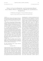
RESULTS<br />
All over <strong>in</strong> <strong>patients</strong> <strong>with</strong> <strong>osteoporotic</strong> <strong>bone</strong> fractures<br />
25(OH)D 3 was more than 60% <strong>and</strong> 1,25(OH) 2D 3 was<br />
more than 53% decreased compared to controls<br />
(Table 1). Fecal <strong>elastase</strong> 1 was lower than the lowest<br />
reference of 200 µg/g feces <strong>in</strong> more than 34% of the<br />
Table 1. 25(OH)D 3, 1,25(OH) 2D 3, Calcium, PTH <strong>and</strong> BMI (means ± st<strong>and</strong>ard deviation) <strong>in</strong> <strong>patients</strong> <strong>with</strong> <strong>osteoporotic</strong> <strong>bone</strong><br />
fractures <strong>and</strong> controls. p < 0.05 <strong>in</strong>dicates a significant difference between <strong>patients</strong> <strong>with</strong> different <strong>fecal</strong> <strong>elastase</strong> 1 ranges.<br />
Patients<br />
Fecal <strong>elastase</strong> 1 ranges (mg/g)<br />
Parameters Controls 300 p Total<br />
(N=20) (N=7) (N=50) (N=41) (N=69) (N=167)<br />
Age<br />
(years) 52.6 ± 6.4 73.4 ± 8.6 70.4 ± 7.4 72.1 ± 8.3 64.9 ± 11.2 P = 0.24 68.7 ± 9.2<br />
25(OH)D 3<br />
(nmol/l) 69.5 ± 13.5 13.2 ± 3.7 21.6 ± 8.7 25.7 ± 5.3 34.3 ± 7.7 p < 0.01 27.5 ± 7.2<br />
(N=126)<br />
1,25(OH) 2D 3<br />
(pg/ml) 67.5 ± 4.3 22.3 ± 16.9 26.1 ± 12.4 32.3 ± 10.8 36.2 ± 12.4 p < 0.01 31.6 ± 12.2<br />
(N=97)<br />
Calcium<br />
(mmol/l) 2.4 ± 0.15 2.23 ± 0.17 2.25 ± 0.16 2.27 ± 0.15 2.35 ± 0.14 p < 0.01 2.30 ± 0.15<br />
PTH<br />
(pg/ml) 37.8 ± 4.8 21.8 ± 4.0 28.6 ± 14.5 35.2 ± 14.6 42.0 ± 12.5 p < 0.01 35.5 ± 13.3<br />
(N=140)<br />
BMI<br />
(kg/m2) 25.2 ± 1.5 25.0 ± 1.4 25.7 ± 1.8 25.4 ± 1.6 25.0 ± 1.4 P = 0.141 25.3 ± 1.6
70 EUROPEAN JOURNAL OF MEDICAL RESEARCH<br />
February 25, 2008<br />
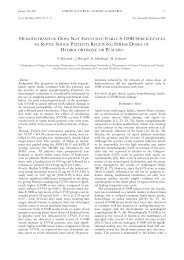
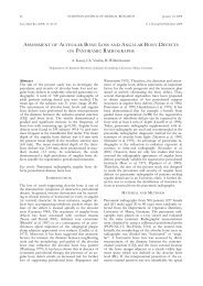
25(OH)D 3 (pg/ml)<br />
Fig. 1. 25(OH)D 3 <strong>with</strong><strong>in</strong> the different ranges of <strong>fecal</strong> <strong>elastase</strong><br />
1.<br />
p < 0.01<br />
r = 0.697<br />
Fig. 3. 25(OH)D 3 <strong>in</strong> <strong>patients</strong> <strong>with</strong> <strong>osteoporotic</strong> <strong>bone</strong> fractures<br />
depend<strong>in</strong>g on <strong>fecal</strong> <strong>elastase</strong> 1.<br />
<strong>patients</strong> <strong>and</strong> it was more than 65% reduced <strong>in</strong> comparison<br />
to healthy controls (<strong>fecal</strong> <strong>elastase</strong> 1 <strong>patients</strong>: 240.7<br />
± 96.3 µg/g; controls 694.9 ± 138.6 µg/g). Separation<br />
of the <strong>patients</strong> <strong>in</strong> accordance <strong>with</strong> the <strong>elastase</strong> 1 contend<br />
<strong>in</strong> feces <strong>in</strong>to four groups (below 100 µg/g, between<br />
100 <strong>and</strong> 200 µg/g, between 201 <strong>and</strong> 300 µg/g<br />
<strong>and</strong> above 300 µg/g) resulted <strong>in</strong> significant variations<br />
for 25(OH)D 3, 1,25(OH) 2D 3, calcium <strong>and</strong> PTH between<br />
these groups (p < 0.01; Table 1; Figures 1-2).<br />
25(OH)D 3 decreases significantly from group <strong>with</strong> <strong>fecal</strong><br />
<strong>elastase</strong> 1 of above 300 µg/g to all other groups (p<br />
< 0.01) <strong>and</strong> also from group <strong>with</strong> between 201 <strong>and</strong><br />
300µg/g to group <strong>with</strong> below 100 µg/g (p < 0.01). For<br />
1,25(OH) 2D 3 there is a significant decrease between<br />
group <strong>with</strong> above 300µg/g to group <strong>with</strong> between 100<br />
<strong>and</strong> 200 µg/g (p = 0.013). Calcium <strong>and</strong> PTH was<br />
markedly decreased from group <strong>with</strong> <strong>fecal</strong> <strong>elastase</strong> 1 of<br />
above 300 µg/g to group <strong>with</strong> between 100 <strong>and</strong> 200<br />
µg/g (calcium p = 0.006; PTH p = 0.001) as well as to<br />
1,25(OH) 2 D 3 (ng/ml)<br />
Fig. 2. 1,25(OH) 2D 3. <strong>with</strong><strong>in</strong> the different ranges of <strong>fecal</strong> <strong>elastase</strong><br />
1.<br />
Fig. 4. 1,25(OH) 2D 3 <strong>in</strong> <strong>patients</strong> <strong>with</strong> <strong>osteoporotic</strong> <strong>bone</strong> fractures<br />
depend<strong>in</strong>g on <strong>fecal</strong> <strong>elastase</strong> 1.<br />
group <strong>with</strong> below 100 µg/g (p = 0.021) only for PTH.<br />
Furthermore the parameters 25(OH)D 3, 1,25(OH) 2D 3,<br />
calcium <strong>and</strong> PTH correlated significantly <strong>with</strong> <strong>elastase</strong><br />
1 <strong>in</strong> feces of <strong>patients</strong> (p < 0.01; Table 2; Figures 3-4).<br />
Lower <strong>fecal</strong> <strong>elastase</strong> 1 therefore is connected <strong>with</strong> lower<br />
<strong>vitam<strong>in</strong></strong> D 3 together <strong>with</strong> lowered PTH <strong>and</strong> calcium<br />
levels. BMI shows no relevant differences <strong>with</strong><strong>in</strong> the<br />
<strong>patients</strong> or between <strong>patients</strong> <strong>and</strong> controls (Table 1).<br />
DISCUSSION<br />
p < 0.01<br />
r = 0.361<br />
Until now there are no studies deal<strong>in</strong>g <strong>with</strong> the l<strong>in</strong>k<br />
between reduced circulat<strong>in</strong>g <strong>vitam<strong>in</strong></strong> D 3 metabolites<br />
<strong>and</strong> lowered <strong>fecal</strong> <strong>elastase</strong> 1 <strong>in</strong> <strong>patients</strong> <strong>with</strong> <strong>osteoporotic</strong><br />
<strong>bone</strong> fractures. In the present study <strong>fecal</strong> <strong>elastase</strong><br />
1 <strong>in</strong> <strong>patients</strong> <strong>with</strong> <strong>osteoporotic</strong> <strong>bone</strong> fractures is<br />
lower than the lowest reference of 200 µg/g feces <strong>in</strong><br />
more than 34% of the <strong>patients</strong> <strong>and</strong> it is more than<br />
65% reduced <strong>in</strong> comparison to healthy controls. Re-
February 25, 2008 EUROPEAN JOURNAL OF MEDICAL RESEARCH<br />
71<br />
Table 2. Correlation between <strong>fecal</strong> <strong>elastase</strong> 1 <strong>and</strong> 25(OH)D 3, 1,25(OH) 2D 3, Calcium, PTH <strong>and</strong> BMI. p < 0.05 <strong>in</strong>dicates a significant<br />
correlation.<br />
Parameter 25(OH)D 3 1,25(OH) 2D 3 Calcium PTH BMI<br />
Fecal <strong>elastase</strong> 1<br />
Correlation Pearson 0.620 0.300 0.256 0.423 -0.114<br />
p p < 0.01 p < 0.01 p < 0.01 p < 0.01 p= 0.141<br />
N 126 97 167 140 167<br />
Fecal <strong>elastase</strong> 1<br />
Correlation Spearman 0.697 0.361 0.236 0.490 -0.137<br />
p p < 0.01 p < 0.01 p < 0.01 p < 0.01 p=0.079<br />
N 126 97 167 140 167<br />
duced <strong>fecal</strong> <strong>elastase</strong> 1 is connected <strong>with</strong> lowered <strong>vitam<strong>in</strong></strong><br />
D 3 <strong>and</strong> this could be demonstrated by comparison<br />
between patient groups <strong>with</strong> different severance<br />
grades of <strong>fecal</strong> <strong>elastase</strong> 1 deficiency as well as by direct<br />
correlation. In our further studies we could demonstrate<br />
a similar connection between <strong>fecal</strong> <strong>elastase</strong> 1, <strong>vitam<strong>in</strong></strong><br />
D 3 <strong>and</strong> BMD [14, 15], but no one of the <strong>patients</strong><br />
had <strong>osteoporotic</strong> <strong>bone</strong> fractures <strong>and</strong> all had<br />
chronic pancreatitis. Nevertheless our present results<br />
are consistant <strong>with</strong> the observations we made at our<br />
<strong>patients</strong> <strong>with</strong> exocr<strong>in</strong>e <strong>in</strong>sufficiency caused by chronic<br />
pancreatitis. The fact of reduced BMD <strong>in</strong> our pancreatic<br />
<strong>patients</strong> <strong>with</strong> this way reduced <strong>vitam<strong>in</strong></strong> D 3 serum<br />
levels allows the conclusion, that osteoporosis <strong>in</strong> our<br />
<strong>patients</strong> <strong>with</strong> <strong>osteoporotic</strong> <strong>bone</strong> fractures could be additional<br />
caused by <strong>vitam<strong>in</strong></strong> D deficiency <strong>in</strong> consequence<br />
of occult exocr<strong>in</strong>e pancreatic <strong>in</strong>sufficiency.<br />
Poskitt et al. [13] reported, that a depletion of <strong>vitam<strong>in</strong></strong><br />
D storage is ma<strong>in</strong>ly caused by a reduced exposition to<br />
the sun, but altogether serum levels of lipid soluble <strong>vitam<strong>in</strong></strong><br />
D 3 depends on photosynthesis <strong>in</strong> the sk<strong>in</strong> as<br />
well as on direct <strong>in</strong>test<strong>in</strong>al resorption. S<strong>in</strong>ce only 40%<br />
of experimentally adm<strong>in</strong>istrered, radio-actively labeled<br />
<strong>vitam<strong>in</strong></strong> D 3 is absorbed by the <strong>in</strong>test<strong>in</strong>es of <strong>patients</strong><br />
<strong>with</strong> pancreatic <strong>in</strong>sufficiency [19], contrary to 80-90%<br />
<strong>in</strong> healthy persons, exocr<strong>in</strong>e pancreatic function ga<strong>in</strong>s<br />
<strong>in</strong> significance <strong>and</strong> supports our own results <strong>with</strong> correspond<strong>in</strong>g<br />
evaluation of <strong>fecal</strong> <strong>elastase</strong> 1. It is conceivable<br />
that <strong>fecal</strong> <strong>elastase</strong> 1 plays an <strong>in</strong>dependent role<br />
<strong>with</strong> regard to <strong>vitam<strong>in</strong></strong> D 3 supply <strong>in</strong> the organism.<br />
Upon pass<strong>in</strong>g through the <strong>in</strong>test<strong>in</strong>es, <strong>elastase</strong> 1 complexes<br />
<strong>with</strong> neutral steroids [20]. S<strong>in</strong>ce <strong>vitam<strong>in</strong></strong> D 3 is<br />
also a sterol molecule, there is a hypothetical mechanism<br />
by which reduced <strong>vitam<strong>in</strong></strong> D 3 absorption at reduced<br />
<strong>fecal</strong> <strong>elastase</strong> 1 could be l<strong>in</strong>ked. In the present<br />
study it is evident, that <strong>vitam<strong>in</strong></strong> D 3 serum levels are<br />
more than 53% respectively more than 60% reduced<br />
<strong>in</strong> <strong>patients</strong> <strong>in</strong> comparison to controls. Therefore, as<br />
described by Scharla et al. <strong>and</strong> Chapuy et al. [8, 9],<br />
even low normal serum concentrations of <strong>vitam<strong>in</strong></strong> D<br />
can lead to osteopenia due to <strong>in</strong>creased <strong>bone</strong> loss. The<br />
prevalence of exocr<strong>in</strong>e pancreatic <strong>in</strong>sufficiency or lowered<br />
<strong>fecal</strong> <strong>elastase</strong> 1 <strong>in</strong> <strong>patients</strong> <strong>with</strong> <strong>osteoporotic</strong> <strong>bone</strong><br />
fractures is jet unknown, because, to our knowledge,<br />
no data were published until now. But results of the<br />
present study make it highly probable that previous<br />
unknown exocr<strong>in</strong>e pancreatic <strong>in</strong>sufficiency, especially<br />
lowered <strong>fecal</strong> <strong>elastase</strong> 1, <strong>in</strong> <strong>patients</strong> <strong>with</strong> <strong>osteoporotic</strong><br />
<strong>bone</strong> fracture is much more prevalent than suggested<br />
so far.<br />
Even when other authors describe that BMI is, of<br />
all anthropometric factors, the strongest predictor of<br />
BMD [21, 22], our data do not support the relevance<br />
of BMI because no one of the <strong>patients</strong> had a low BMI<br />
but all had <strong>osteoporotic</strong> <strong>bone</strong> fracture.<br />
REFERENCES<br />
1. Kanis JA. Diagnosis of osteoporosis <strong>and</strong> assessment of<br />
fracture risk. Lancet. 2002;359(9321):1929-36.<br />
2. Center JR, Nguyen TV, Schneider D, Sambrook PN, Eisman<br />
JA. Mortality after all major types of <strong>osteoporotic</strong><br />
fracture <strong>in</strong> men <strong>and</strong> women: an observational study.<br />
Lancet. 1999;353(9156):878-82.<br />
3. Hasserius R, Karlsson MK, Nilsson BE, Redlund-Johnell<br />
I, Johnell O. Prevalent vertebral deformities predict <strong>in</strong>creased<br />
mortality <strong>and</strong> <strong>in</strong>creased fracture rate <strong>in</strong> both men<br />
<strong>and</strong> women: a 10-year population-based study of 598 <strong>in</strong>dividuals<br />
from the Swedish cohort <strong>in</strong> the European Vertebral<br />
Osteoporosis Study. Osteoporos Int. 2003;14(1):<br />
61-8.<br />
4. R<strong>and</strong>ell AG, Nguyen TV, Bhalerao N, Silverman SL,<br />
Sambrook PN, Eisman JA. Deterioration <strong>in</strong> quality of life<br />
follow<strong>in</strong>g hip fracture: a prospective study. Osteoporos<br />
Int. 2000;11(5):460-6.<br />
5. Scaf-Klomp W, van Sonderen E, S<strong>and</strong>erman R, Ormel J,<br />
Kempen GI. Recovery of physical function after limb <strong>in</strong>juries<br />
<strong>in</strong> <strong>in</strong>dependent older people liv<strong>in</strong>g at home. Age<br />
Age<strong>in</strong>g. 2001;30(3):213-9.<br />
6. Nguyen T, Sambrook P, Kelly P, et al. Prediction of <strong>osteoporotic</strong><br />
fractures by postural <strong>in</strong>stability <strong>and</strong> <strong>bone</strong> density.<br />
Bmj. 1993;307(6912):1111-5.<br />
7. Schmidt-Gayk H, Thomas L, Stracke H. M<strong>in</strong>eralhaushalt<br />
und Nebenschilddrüse. In: Thomas L, ed. Labor und Diagnose.<br />
4 ed. Marburg: Die Mediz<strong>in</strong>ische Verlagsgesell -<br />
schaft; 1992:342-79.<br />
8. Scharla SH, Scheidt-Nave C, Leidig G, et al. Lower serum<br />
25-hydroxy<strong>vitam<strong>in</strong></strong> D is associated <strong>with</strong> <strong>in</strong>creased <strong>bone</strong><br />
resorption markers <strong>and</strong> lower <strong>bone</strong> density at the proximal<br />
femur <strong>in</strong> normal females: a population-based study.<br />
Exp Cl<strong>in</strong> Endocr<strong>in</strong>ol Diabetes. 1996;104(3):289-92.<br />
9. Chapuy MC, Chapuy P, Thomas JL, Hazard MC, Meunier<br />
PJ. Biochemical effects of calcium <strong>and</strong> <strong>vitam<strong>in</strong></strong> D supplementation<br />
<strong>in</strong> elderly, <strong>in</strong>stitutionalized, <strong>vitam<strong>in</strong></strong> D-deficient<br />
<strong>patients</strong>. Rev Rhum Engl Ed. 1996;63(2):135-40.<br />
10. Chapuy MC, Arlot ME, Duboeuf F, et al. Vitam<strong>in</strong> D 3 <strong>and</strong><br />
calcium to prevent hip fractures <strong>in</strong> the elderly women. N<br />
Engl J Med. 1992;327(23):1637-42.<br />
11. Dawson-Hughes B, Harris SS, Krall EA, Dallal GE. Effect<br />
of calcium <strong>and</strong> <strong>vitam<strong>in</strong></strong> D supplementation on <strong>bone</strong>
72 EUROPEAN JOURNAL OF MEDICAL RESEARCH<br />
February 25, 2008<br />
density <strong>in</strong> men <strong>and</strong> women 65 years of age or older. N<br />
Engl J Med. 1997;337(10):670-6.<br />
12. Gillespie WJ, Avenell A, Henry DA, O'Connell DL,<br />
Robertson J. Vitam<strong>in</strong> D <strong>and</strong> <strong>vitam<strong>in</strong></strong> D analogues for prevent<strong>in</strong>g<br />
fractures associated <strong>with</strong> <strong>in</strong>volutional <strong>and</strong> postmenopausal<br />
osteoporosis. Cochrane Database Syst Rev.<br />
2001(1):CD000227.<br />
13. Poskitt EM, Cole TJ, Lawson DE. Diet, sunlight, <strong>and</strong> 25hydroxy<br />
<strong>vitam<strong>in</strong></strong> D <strong>in</strong> healthy children <strong>and</strong> adults. Br Med<br />
J. 1979;1(6158):221-3.<br />
14. Mann ST, Stracke H, Lange U, Klor HU, Teichmann J.<br />
Vitam<strong>in</strong> D 3 <strong>in</strong> <strong>patients</strong> <strong>with</strong> various grades of chronic<br />
pancreatitis, accord<strong>in</strong>g to morphological <strong>and</strong> functional<br />
criteria of the pancreas. Dig Dis Sci. 2003;48(3):533-8.<br />
15. Mann ST, Stracke H, Lange U, Klor HU, Teichmann J.<br />
Alterations of <strong>bone</strong> m<strong>in</strong>eral density <strong>and</strong> <strong>bone</strong> metabolism<br />
<strong>in</strong> <strong>patients</strong> <strong>with</strong> various grades of chronic pancreatitis.<br />
Metabolism. 2003;52(5):579-85.<br />
16. Kanis JA. Assessment of fracture risk <strong>and</strong> its application<br />
to screen<strong>in</strong>g for postmenopausal osteoporosis: synopsis<br />
of a WHO report. WHO Study Group. Osteoporos Int.<br />
1994;4(6):368-81.<br />
17. Dufner J, Jensen U, Schumacher E. Statistik mit SAS. 1<br />
ed Stuttgart: Teubner-Verlag; 1992.<br />
18. Sachs L. Angew<strong>and</strong>te Statistik. 7 ed Berl<strong>in</strong>: Spr<strong>in</strong>ger-Verlag;<br />
1997.<br />
19. Krawatt EL, Maner EB, Davies M. Absorption of <strong>vitam<strong>in</strong></strong><br />
D <strong>and</strong> 25-OH <strong>vitam<strong>in</strong></strong> D <strong>in</strong> <strong>patients</strong> <strong>with</strong> <strong>in</strong>test<strong>in</strong>al malabsorption.<br />
In: Norman AW, Schaefer K, Herrath DV, al. e,<br />
eds. Vitam<strong>in</strong> D, Basic Research <strong>and</strong> its Cl<strong>in</strong>ical Application.<br />
1 ed. Berl<strong>in</strong> - New York: Gruyter-Verlag; 1979:975-8.<br />
20. Sziegoleit A, L<strong>in</strong>der D. Studies on the sterol-b<strong>in</strong>d<strong>in</strong>g capacity<br />
of human pancreatic <strong>elastase</strong> 1. Gastroenterology.<br />
1991;100(3):768-74.<br />
21. Nguyen TV, Sambrook PN, Eisman JA. Bone loss, physical<br />
activity, <strong>and</strong> weight change <strong>in</strong> elderly women: the<br />
Dubbo Osteoporosis Epidemiology Study. J Bone M<strong>in</strong>er<br />
Res. 1998;13(9):1458-67.<br />
22. Takeda S, Elefteriou F, Karsenty G. Common endocr<strong>in</strong>e<br />
control of body weight, reproduction, <strong>and</strong> <strong>bone</strong> mass.<br />
Annu Rev Nutr. 2003;23:403-11. Epub 2003 Apr 18.<br />
Received: July 7, 2007 / Accepted: January 11, 2008<br />
Address for correspondence:<br />
Dr. Sacha T.W. Mann<br />
Kl<strong>in</strong>ik und Polikl<strong>in</strong>ik für Orthopädie und<br />
Orthopädische Chirurgie<br />
Universitätskl<strong>in</strong>ikum Giessen und Marburg<br />
St<strong>and</strong>ort Giessen<br />
Justus-Liebig-Universität Giessen<br />
Paul-Meimberg-Strasse 3<br />
35392 Giessen<br />
Germany<br />
Phone: +49641/99-42900<br />
Fax: +49641/99-42999<br />
E-mail: SachaTWMann@web.de