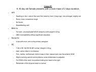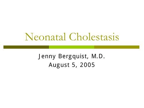
Neonatal Cholestasis
Neonatal Cholestasis
Neonatal Cholestasis

You also want an ePaper? Increase the reach of your titles
YUMPU automatically turns print PDFs into web optimized ePapers that Google loves.
<strong>Neonatal</strong> <strong>Cholestasis</strong><br />
Jenny Bergquist, M.D.<br />
August 5, 2005
Definition: <strong>Neonatal</strong> <strong>Cholestasis</strong><br />
Prolonged conjugated hyperbilirubinemia<br />
in the newborn period<br />
Conjugated hyperbilirubinemia<br />
Conjugated bilirubin >1mg/dL if TB < 5mg/dL<br />
>20% Total Bilirubin if TB is >5 mg/dL<br />
Caused by a group of hepatobiliary<br />
diseases occuring within the first 3 months<br />
of life<br />
Occurs in 1:2500 live births
<strong>Neonatal</strong> <strong>Cholestasis</strong><br />
NASPGHAN Recommendations<br />
Evaluate infants with jaundice at the 2<br />
week visit<br />
Up to 15% of infants are jaundiced at 2 weeks,<br />
the majority due to breast milk jaundice<br />
Timely and accurate diagnosis is crucial for<br />
successful treatment and favorable prognosis<br />
Breast-fed infants may have an evaluation<br />
delayed until 3 weeks if:<br />
Normal exam<br />
No h/o dark urine or light stools<br />
Reliably monitored
Differential Diagnosis:<br />
Obstructive <strong>Cholestasis</strong><br />
Biliary Atresia<br />
* accounts for 30% of all cases of neonatal<br />
cholestasis<br />
Choledochal cysts<br />
Gallstones<br />
Alagille syndrome<br />
<strong>Neonatal</strong> sclerosing cholangitis<br />
Cystic fibrosis<br />
Tumor
Differential Diagnosis:<br />
Hepatocellular <strong>Cholestasis</strong><br />
Idiopathic neonatal hepatitis<br />
Diagnosis based on liver biopsy findings: “giant cell hepatitis”<br />
Was thought to account for ~40% of neonatal cholestasis<br />
(with new diagnostic techniques, % is probably ~10-20%)<br />
60-70% resolve without sequelae<br />
Infectious<br />
Viral: TORCH, CMV, HIV, viral hepatitis<br />
Bacterial: sepsis, syphilis, UTI<br />
Genetic/metabolic<br />
Alpha-1-antitrysin deficiency, galactosemia, tyrosinemia,<br />
hypothyroid, PFIC, CF<br />
Toxic/secondary causes<br />
TPN associated cholestasis
History<br />
Congenital infections<br />
Prenatal ultrasound<br />
ABO incompatibility<br />
<strong>Neonatal</strong> infection<br />
UTI<br />
Dietary history<br />
Weight gain<br />
vomiting<br />
Stool pattern<br />
Delayed: CF,<br />
hypothyroid<br />
Diarrhea: infx,<br />
metabolic disease<br />
Stool and urine color<br />
Excessive bleeding<br />
Irritability or lethargy<br />
+family history
Physical Exam<br />
Weight measurement<br />
General appearance<br />
Ill-appearing: infection, metabolic disease<br />
Well-appearing: biliary atresia<br />
Fundoscopic exam (congenital infection)<br />
Cardiac murmur<br />
Abdominal exam<br />
Ascites, abd wall veins, liver, spleen<br />
Stool/urine for color<br />
Skin exam<br />
Bruising, petechiae<br />
Dysmorphic features<br />
Broad nasal bridge, triangular facies, deep set eyes (Alagille)
Laboratory Studies<br />
Total and direct bilirubin<br />
LFTs + GGTP<br />
Detects liver cell or bile duct injury<br />
PT/PTT, glucose, albumin<br />
Assessment of biosynthetic capacity of liver<br />
CBC, urine and blood culture<br />
Viral serologies<br />
(TORCH infections + HBsAg, CMV, HIV if indicated)<br />
UA for reducing substances<br />
TFTs<br />
Alpha-1-antitrypsin<br />
Sweat chloride or mutation analysis for CF gene
NASPGHAN Recommendations:<br />
Imaging Studies<br />
Ultrasound: initial study recommended<br />
for patients with cholestasis of unknown<br />
etiology<br />
Evaluates for anatomic abnormalities<br />
Liver Biopsy<br />
Recommended for most infants with<br />
cholestasis of unknown etiology<br />
Differentiates b/w extra and intrahepatic processes,<br />
disorders of physiology from anatomy and can<br />
determine need for surgical vs. medical intervention<br />
Scintigraphy (HIDA), ERCP, MRCP
Biliary Atresia<br />
1:8,000-15,000 live births<br />
Accounts for 30% of all cases of<br />
cholestasis in infants<br />
Female>Male<br />
Asians>African Americans>Caucasian<br />
Most frequent cause of chronic end-stage<br />
liver disease in children<br />
Leading indication for liver transplantation<br />
in the pediatric population (40-50% of all<br />
liver transplants)
Biliary Atresia: pathogenesis<br />
Bile duct obstruction due to inflammation<br />
and fibrous obliteration<br />
Perinatal or “classic” type (70-85%):<br />
obstruction begins after birth. Signs/symptoms<br />
develop within ~2-4 weeks of age. No<br />
associated abnormalities<br />
Embryonic type (15-30%): obstructive<br />
process begins in utero. Cholestatic symptoms<br />
present at birth. Associated with congenital<br />
anomalies:<br />
Situs inversus, polysplenia, malrotation, cardiac<br />
anomalies<br />
Unknown etiology: genetic, viral and host<br />
immune factors have been postulated
Variations in Biliary Atresia
Clinical Features<br />
History: Variable degrees of persistent jaundice,<br />
dark urine, light colored stools, +/-poor appetite<br />
* Usually Well-Appearing*<br />
Physical:<br />
Hepatomegaly, +/- splenomegaly<br />
“appear” well-nourised<br />
Usually with decreased fat stores and lean body mass<br />
Enlarged abdomen from HSM may give impression of<br />
“normal weight for age”<br />
Scleral icterus, abdominal wall veins (caput medusa)<br />
Labs:<br />
Total bilirubin rarely is >12mg/dL; CB is usually<br />
Imaging Studies<br />
Ultrasound<br />
Absent gallbladder, “triangular cord” sign<br />
Low sensitivity; operator dependent<br />
Evaluates for other anatomic abnormalities<br />
Hepatobiliary scintigraphy (HIDA)<br />
High sensitivity: normal uptake, but no excretion of<br />
radionuclide tracer into biliary system or bowel in virtually all<br />
patients with BA (exceptions in very early disease)<br />
However, failure of excretion may be seen in both BA and<br />
neonatal hepatitis<br />
Sensitivity and Specificity increase with phenobarbital<br />
administration<br />
ERCP<br />
Invasive, not readily available, technically difficult in infants<br />
Intraoperative use is most common to confirm diagnosis and<br />
document site of obstruction<br />
MRCP<br />
May become an important tool for diagnosis<br />
Further studies are required
“Triangular Cord” Sign
Diagnosis<br />
Percutaneous Liver Biopsy: most reliable test<br />
for diagnosing biliary atresia<br />
Biopsy interpretation is pathologist dependent<br />
Accurate diagnosis made in 90-95% of cases<br />
Liver biopsies made early in the course of disease (
Surgical Management<br />
Kasai Procedure: resection of the<br />
obliterated bile duct w/ creation of a<br />
Roux-n-Y hepatoportoenterostomy<br />
Timing of procedure predicts the<br />
prognosis<br />
90 days- bile flow returned in ~20% cases<br />
Usually require a liver transplant within one year<br />
The experience of the center<br />
performing the Kasai if one of the most<br />
important factors determinig surgical<br />
outcome
Post-operative Management<br />
Prophylactic Abx to prevent cholangitis<br />
Ursodiol: enhance bile flow<br />
No special diet needed unless concern<br />
with poor bile drainageMCT formula<br />
(ie… Alimentum, Pregestimil)<br />
Fat-soluble vitamins: A, D, E, K<br />
+/- short term, high dose steroid therapy
If Kasai Fails?<br />
+/- support for revision of Kasai procedure if fails<br />
Despite clinical improvement after a Kasai, 70-80% pts will<br />
eventually require liver transplantation<br />
Indications for Liver Transplant:<br />
operation not successful in restoring bile flow initially<br />
(~20%)<br />
late referrals (generally >120 days)<br />
develop end-stage liver dz despite bile drainage (ie<br />
portal htn, recurrent cholangitis, ascites, growth failure)<br />
Liver Transplant results:<br />
one-yr survival rates >90% b/c reduced size allografts<br />
and living-related donors
Post-Kasai Complications<br />
Early: Ascending Cholangitis (50%) can lead to<br />
ongoing bile duct injury & re-obstruction<br />
fever, dec. bile secretion, worsening jaundice,<br />
leukocytosis<br />
Late: Portal Hypertension<br />
bleeding esophageal varices, ascites, hypoalbuminemia,<br />
fat-soluble vit def., malabsorbtion of long-chain<br />
triglycerides, encephalopathy<br />
Long term, malignancies screened for<br />
Hepatoblastoma, hepatocarcinoma, cholangiocarcinoma
What happened to our patient?<br />
Kasai Procedure on 8/16/04<br />
Complicated by cholangitis x 2 wks<br />
Portal hypertension<br />
Esophogeal varicessclerotherapy x 2<br />
1/05: TB 32.3/ conjugated bilirubin 18.6<br />
Now on the transplant list awaiting a<br />
liver…
Take-Home Message!<br />
Any Jaundice>2 weeks requires<br />
investigation<br />
ALWAYS ask for fractionated bilirubin<br />
(Total + Direct bilirubin)<br />
Early diagnosis and referral (