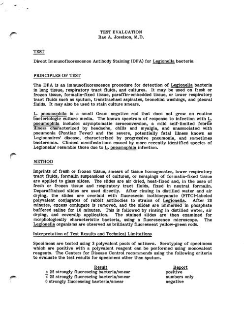
TEST EVALUATION Rae A. Joselson, M.D. TEST Direct ...
TEST EVALUATION Rae A. Joselson, M.D. TEST Direct ...
TEST EVALUATION Rae A. Joselson, M.D. TEST Direct ...

You also want an ePaper? Increase the reach of your titles
YUMPU automatically turns print PDFs into web optimized ePapers that Google loves.
J ^ \<br />
<strong>TEST</strong><br />
<strong>TEST</strong> <strong>EVALUATION</strong><br />
<strong>Rae</strong> A. <strong>Joselson</strong>, M.D.<br />
<strong>Direct</strong> Immunofluorescence Antibody Staining (DFA) for Legionella bacteria<br />
PRINCIPLES OF <strong>TEST</strong><br />
The DFA is an immunofluorescence procedure for detection of Legionella bacteria<br />
in lung tissue, respiratory tract fluids, and cultures. It may be used on fresh or<br />
frozen tissue, formalin-fixed tissue, paraffin-embedded tissue, or lower respiratory<br />
tract fluids such as sputum, transtrachael aspirates, bronchial washings, and pleural<br />
fluids. It may also be used to stain culture smears.<br />
L. pneumophila is a small Gram negative rod that does not grow on routine<br />
bacteriologic culture media. The known spectrum of response to infection with L.<br />
pneumophila includes asymptomatic seroconversion, a mild self-limited febrile<br />
illness characterized by headache, chills and myalgia, and unassociated with<br />
pneumonia (Pontiac Fever) and the severe, potentially fatal illness known as<br />
Legionnaires1 disease, characterized by progressive pneumonia, and sometimes<br />
bacteremia. Clinical manifestations caused by more recently identified species of<br />
Legionella1 resemble those due to L. pneumophila infection.<br />
METHOD<br />
Imprints of fresh or frozen tissue, smears of tissue homogenates, lower respiratory<br />
tract fluids, formalin suspensions of cultures, or scrapings of formalin-fixed tissue<br />
are applied to glass slides. The slides are air dried, heat-fixed and, in the case of<br />
fresh or frozen tissue and respiratory tract fluids, fixed in neutral formalin.<br />
Deparaffinized slides are used directly. After rinsing in distilled water and air<br />
drying, the slides are overlaid with fluorescein isothiocyanate (FITC)-labeled<br />
polyvalent conjugates of rabbit antibodies to strains of Legionella. After 20<br />
minutes, excess conjugate is removed, and the slides are immersed in phosphate<br />
buffered saline for 10 minutes. This is followed by rinsing in distilled water, air<br />
drying, and coverslip application. The stained slides are then examined for<br />
morphologically characteristic bacteria, using a fluorescence microscope. The<br />
Legionella organisms are observed as brilliantly fluorescent yellow-green rods.<br />
Interpretation of Test Results and Technical Limitations<br />
Specimens are tested using 3 polyvalent pools of antisera. Serotyping of specimens<br />
which are positive with a polyvalent reagent can be performed using monovalent<br />
reagents. The Centers for Disease Control recommends using the following criteria<br />
to evaluate the test results for specimens other than sputum.<br />
Result Report<br />
^ 2 5 s t r o n g l y fl u o r e s c i n g b a c t e r i a / s m e a r p o s i t i v e<br />
< 25 strongly fluorescing bacteria/smear numbers only<br />
0 s t r o n g l y fl u o r e s c i n g b a c t e r i a / s m e a r n e g a t i v e
Page 2<br />
With sputum, because organisms are seldom numerous, observation of more than 5<br />
morphologically typical bacteria should be regarded as a positive result. Edelstein<br />
et al. consider the presence of 1 to 4 morphologically characteristic fluorescent<br />
organisms per smear as positive if at least 2 additional smears demonstrate 1<br />
fluorescent organism per smear. They regard the presence of 5 or more<br />
morphologically characteristic fluorescent organisms in any single smear as positive,<br />
regardless of the source of the specimen.<br />
No quantitative data is currently available about the precision and accuracy of the<br />
DFA. The antisera conjugates have been evaluated with over 374 strains of bacteria<br />
and 1 strain of Pseudomonas fluorescens has shown cross reactivity. Other bacteria,<br />
cells, and debris may also fluoresce and be misinterpreted as Legionella organisms,<br />
producing false positive results. Conversely, Legionella bacteria may disintegrate<br />
from the effects of cellular defense mechanisms and exhibit atypical morphology or<br />
appear as fluorescent debris. These forms may go unnoticed, potentially leading to<br />
false negative results. Such discrepancies may be minimized by the use of<br />
counterstains and positive and negative controls, concurrently with the specimen to<br />
be examined. Nevertheless, Edelstein et al. found that considerable experience on<br />
the part of the observer was required to perform the DFA. New serogroups of L.<br />
pneumophila and new species of Legionella organisms are continuing to be identified<br />
and these organisms may not react with currently used conjugates.<br />
The sensitivity and specificity of the DFA have been examined in several papers.<br />
Edelstein et al. evaluated the reliability of the diagnostic tests for Legionnaires'<br />
{ disease in 32 patients with this infection. They reported the following results for<br />
DFA examination.<br />
DFA* vs Culture DFA* vs Serology (Igm & IgG)<br />
Serogroups Serogroup Serogroups Serogroup<br />
i , n , m , i v i i , n , m , i v i<br />
% s e n s i t i v i t y 6 2 6 2 4 3 7 5<br />
% s p e c i fi c i t y 9 4 1 0 0 8 8 8 8<br />
% n e g a t i v e p r e d i c t i v e a c c u r a c y 7 5 7 5 6 6 8 9<br />
% p o s i t i v e p r e d i c t i v e a c c u r a c y 8 9 1 0 0 7 5 9 0<br />
* DFA of respiratory secretions before therapy only<br />
These authors found lung tissue and sputum to be the optimal specimens for the<br />
DFA.<br />
Broome et al. evaluated the specificity of DFA staining in sputum specimens and<br />
found no false positives among 47 controls with pneumonia caused by agents other<br />
than L. pneumophila. However, sensitivity was poor, with only 24% of 21 patients<br />
having a positive DFA and shown to have Legionnaires1 disease by cultural or<br />
serologic criteria. These investigators speculated that the low proportion of<br />
positivity might be due to the absence of Legionnaires' disease bacterium in the
Page 3<br />
sputum at certain times during the course of the disease, the inadequacy of some of<br />
the specimens, or the storage of specimens for months at room temperature which<br />
occurred in their study.<br />
More recently, Zuravleff et al. performed a prospective study of 40 cases of<br />
Legionnaires' disease using newer more selective culture media. When culture<br />
positivity was used as the basis for diagnosis of Legionnaires' disease, the sensitivity<br />
of DFA testing was 33%. When seroconversion was the basis for the diagnosis, the<br />
sensitivity of the DFA was 47%. No attempt was made to evaluate specificity or<br />
predictive value. In discussing the poor sensitivity of the DFA, these authors noted<br />
that the yield from DFA examination is directly related to the number of organisms<br />
recoverable by cultural methods; thus, the DFA may be negative in an early or mild<br />
case of Legionnaires' disease. Treatment with erythromycin can also lead to<br />
negative DFA results.<br />
Evaluation of the sensitivity and specificity of the DFA is hampered somewhat by<br />
the lack of a "gold standard" for the diagnosis of infection due to Legionella<br />
bacteria; the DFA can only be compared with culture and/or serologic methods. In<br />
addition, sensitivity and specificity vary with the source of the DFA specimen, and<br />
studies reporting results for each specimen type are too limited in number for<br />
definitive analysis. Therefore, most investigators recommend that serologic,<br />
cultural, and DFA techniques be used in combination for optimal sensitivity and<br />
specificity in the diagnosis of Legionella infection.<br />
REFERENCES<br />
1. Broome CV, Cherry WB, Winn WC, MacPherson BR. Rapid diagnosis of<br />
Legionnaires' disease by <strong>Direct</strong> Immunofluorescent Staining. Ann Intern Med<br />
90:1-4, 1979.<br />
2. Cherry WB, McKinney RM. Detection of Legionnaires' disease bacteria in<br />
clinical specimens by direct immunofluorescence. Legionnaires; the disease,<br />
the bacterium and methodology. U.S. Dept. of HEW, CDC, 1979.<br />
3. Edelstein PH, Meyer RD, Finegold SM. Laboratory diagnosis of Legionnaires'<br />
disease. Am Rev Resp Dis 121:317-327, 1980.<br />
4. Kirby BD, Snyder KM, Meyer RD, Finegold SM. Legionnaires' Disease: Report<br />
of sixty-five nosocomially acquired cases and review of the literature.<br />
Medicine 59:188-205, 1980.<br />
5. Renner ED, Tseng CH. The species of Legionella: another challenge to<br />
clinical microbiologists. Clin Microbiol News 4:139-142, 1982.<br />
6. Zuravleff JJ, Yu VL, Shonnard JW, Davis BK, Rihs JD. Diagnosis of<br />
Legionnaires' disease. J Am Med Assoc 250:1981-1985, 1983.
■IpPSfy<br />
<strong>TEST</strong><br />
<strong>TEST</strong> <strong>EVALUATION</strong><br />
Stephen Darling, M.D.<br />
<strong>Direct</strong> Anti-globulin Test (<strong>Direct</strong> Coombs) (DAT)<br />
METHOD<br />
Polyspecific antihuman globulin is added to the patients' washed red cells, then<br />
incubated, and centrifuged. Visible agglutination of the red cells indicates the<br />
presence of bound IgG or C3. All positive reactions are further evaluated using<br />
monospecific anti-human IgG and anti-human C3d.<br />
CLINICAL APPLICATIONS<br />
The DAT detects antibodies and/or complement on the surface of patients' red cells<br />
which were bound in vivo. The DAT is clinically useful in the evaluation of possible<br />
immune hemolytic anemia, in the diagnosis of hemolytic disease of the newborn, and<br />
in the investigation of transfusion reactions. A positive reaction indicates red cell<br />
sensitization which could lead to shortened red cell survival. The pattern of<br />
sensitization, i.e., the presence of IgG or complement alone or in combination, gives<br />
additional information regarding the method of sensitization. For example, the<br />
presence of C3 in the absence of IgG is typically seen in immune complex type<br />
(*** sensitization due to drugs (e.g. quinidine), or with cold autoantibodies. Other<br />
patterns are less specific.<br />
CLINICAL AND TECHNICAL LIMITATIONS<br />
The major limitation of the DAT is in the interpretation of results. The sensitivity<br />
of current DATs is such that 100-500 molecules of IgG must be bound to each red<br />
cell in order to get a positive test result. Occasional patients are encountered with<br />
a negative DAT who obviously have shortened red cell survival due to immunologic<br />
destruction. In these cases there must be enough IgG coating of the red cells to<br />
decrease in vivo survival, but not enough to give a positive DAT. If the test were to<br />
be "improved" in order to be 100% sensitive, a significant number of false positives<br />
would result since all normal red cells have a small amount of IgG on their surface.<br />
Unfortunately the strength of the agglutination reaction in positives does not<br />
correlate with the severity of in vivo hemolysis. In fact, 10-15% of all patients<br />
receiving a-methyl dopa for the treatment of hypertension have positive DATs, but<br />
only 1% of these demonstrate shortened red cell survival. This test therefore needs<br />
to be interpreted in light of the patient's clinical status.<br />
A number of technical pitfalls exist in the performance of the DAT which may lead<br />
to false positive or false negative results. False positives may occur if the blood<br />
specimen is not collected in EDTA anticoagulant (lavender top tube). This is due to<br />
nonspecific coating of red cells by complement which is activated during the process<br />
of collection and clot formation. EDTA chelates free Ca# in the serum and thus<br />
{" prevents this activation and binding of complement. False positives may also occur
fpfcv<br />
Page 2<br />
in patients with hyperproteinemic states since rouleaux formation may be<br />
erroneously interpreted as agglutination. Nonspecific red cell adsorption of serum<br />
proteins including immune complexes as well as polyagglutionable red cells, which<br />
are occasionally seen in septic patients and under diverse clinical conditions, are<br />
other possible sources of a false positive DAT.<br />
Technical problems leading to potential false negative results include incomplete<br />
washing of red cells with residual serum neutralizing the added antiserum, the use of<br />
outdated reagents, and the inadvertent elution of the coating antibody during the<br />
washing steps. All of these technical pitfalls are currently more theoretical than<br />
real since appropriate controls and methods are used to avoid or detect the<br />
occasional erroneous result.<br />
REFERENCES<br />
Clinical Practice of Blood Transfusion. Ed. Lawrence Petz and Scott Swisler,<br />
Churchill-Livingstone: New York, New York, 1981.<br />
AABB Technical Manual, 8th Edition. J.B. Lippincott Co, Philadelphia, PA, 1981.<br />
Issitt P, et al: Evaluation of commercial antiglobulin sera over a two year period.<br />
Transfusion 14(2), March-April, 1974.