Epithelial Tumors in the Ovary The Good, The Bad, and the Ugly ...
Epithelial Tumors in the Ovary The Good, The Bad, and the Ugly ...
Epithelial Tumors in the Ovary The Good, The Bad, and the Ugly ...

You also want an ePaper? Increase the reach of your titles
YUMPU automatically turns print PDFs into web optimized ePapers that Google loves.
<strong>Epi<strong>the</strong>lial</strong> <strong>Tumors</strong> <strong>in</strong> <strong>the</strong> <strong>Ovary</strong><br />
<strong>The</strong> <strong>Good</strong>, <strong>The</strong> <strong>Bad</strong>, <strong>and</strong> <strong>the</strong> <strong>Ugly</strong><br />
Charles Zaloudek, MD<br />
Professor, Department of Pathology<br />
University of California, San Francisco<br />
505 Parnassus Ave., M563<br />
San Francisco, CA 94143<br />
charles.zaloudek@ucsf.edu<br />
1
Case 1<br />
High Grade Serous Carc<strong>in</strong>oma<br />
Cl<strong>in</strong>ical History: <strong>The</strong> patient was a 68-year-old woman with bilateral adnexal masses <strong>and</strong> an<br />
elevated CA125 (793). She had had a hysterectomy <strong>in</strong> 2008 for postmenopausal bleed<strong>in</strong>g. In<br />
2011, a CT scan confirmed <strong>the</strong> presence of bilateral adnexal masses <strong>and</strong> revealed multiple small<br />
omental nodules. At operation <strong>in</strong> 2011 she had 6-8 L of ascites. She underwent bilateral<br />
salp<strong>in</strong>go-oophorectomy <strong>and</strong> tumor debulk<strong>in</strong>g <strong>in</strong>clud<strong>in</strong>g a transverse colon resection. Tumor was<br />
noted to extensively <strong>in</strong>volve <strong>the</strong> diaphragm, <strong>the</strong> anterior peritoneum, <strong>and</strong> <strong>the</strong> transverse colon,<br />
<strong>the</strong> mesentery of <strong>the</strong> small bowel, <strong>the</strong> sigmoid colon <strong>and</strong> <strong>the</strong> bladder peritoneum.<br />
Gross Pathology: <strong>The</strong> ovaries were small. <strong>The</strong> right ovary was a pale-tan firm mult<strong>in</strong>odular<br />
solid mass 4 cm <strong>in</strong> maximum dimension with tumor <strong>in</strong>volv<strong>in</strong>g <strong>the</strong> surface. <strong>The</strong> left ovary was 5<br />
cm <strong>in</strong> maximum dimension <strong>and</strong> was cystic <strong>and</strong> solid, with papillary excrescences <strong>in</strong> <strong>the</strong> l<strong>in</strong><strong>in</strong>gs<br />
of some cysts. Solid areas were yellow-white. No abnormalities were noted <strong>in</strong> <strong>the</strong> fallopian<br />
tubes. A 21 cm segment of transverse colon <strong>and</strong> omentum was riddled with small nodules<br />
measur<strong>in</strong>g 0.2 to 1 cm, with a dom<strong>in</strong>ant mass 4.5 cm <strong>in</strong> greatest dimension <strong>in</strong> <strong>the</strong> mesentery. <strong>The</strong><br />
bowel mucosa was unremarkable.<br />
Diagnosis: High-grade serous carc<strong>in</strong>oma of <strong>the</strong> left fallopian tube <strong>and</strong> left <strong>and</strong> right ovaries with<br />
extensive extraovarian tumor spread.<br />
High Grade Serous Carc<strong>in</strong>oma<br />
Serous carc<strong>in</strong>oma is <strong>the</strong> most common type of ovarian cancer, account<strong>in</strong>g for 68% of<br />
ovarian cancers <strong>and</strong> 88% of stage III <strong>and</strong> IV cancers <strong>in</strong> a large population based study. (1)<br />
Mutations of <strong>the</strong> P53 gene are present <strong>in</strong> most cases <strong>and</strong> are thought to be critical events <strong>in</strong> <strong>the</strong><br />
pathogenesis of this type of carc<strong>in</strong>oma. (2) Intraabdom<strong>in</strong>al metastases are usually present at <strong>the</strong><br />
time of diagnosis, as <strong>in</strong> this patient, <strong>in</strong>volv<strong>in</strong>g <strong>the</strong> omentum, <strong>the</strong> peritoneum or <strong>the</strong> abdom<strong>in</strong>al<br />
lymph nodes. (1) Occasional metastases are found <strong>in</strong> unusual distant sites, such as <strong>the</strong> bra<strong>in</strong>,<br />
lung, distant lymph nodes or <strong>the</strong> breast. (3)<br />
General Cl<strong>in</strong>ical Features of <strong>Epi<strong>the</strong>lial</strong> <strong>Tumors</strong>, Especially High Grade Serous Carc<strong>in</strong>oma<br />
Ovarian carc<strong>in</strong>oma occurs ma<strong>in</strong>ly <strong>in</strong> peri- <strong>and</strong> postmenopausal women. About 10% of<br />
patients with ovarian cancer have an <strong>in</strong>herited predisposition to develop <strong>the</strong> cancer (BRCA<br />
mutation or Lynch Syndrome). (4) More than 70 percent of women with ovarian cancer have<br />
extensive extraovarian tumor spread at <strong>the</strong> time of diagnosis. One reason for this is that <strong>the</strong><br />
symptoms caused by epi<strong>the</strong>lial tumors are vague <strong>and</strong> non-specific. Common symptoms <strong>in</strong>clude<br />
pelvic discomfort or pa<strong>in</strong>, a sensation of abdom<strong>in</strong>al fullness or pressure, gastro<strong>in</strong>test<strong>in</strong>al<br />
disturbances, ur<strong>in</strong>ary frequency, <strong>and</strong> occasionally, menstrual abnormalities. Women with<br />
advanced ovarian cancer often have ascites, which <strong>in</strong>terferes with gastro<strong>in</strong>test<strong>in</strong>al function,<br />
lead<strong>in</strong>g to nausea <strong>and</strong> vomit<strong>in</strong>g. Ovarian enlargement <strong>in</strong> a woman over 45 raises <strong>the</strong> question of<br />
ovarian cancer <strong>and</strong> requires fur<strong>the</strong>r evaluation. <strong>The</strong> identification of a solid or complex mass by<br />
sonography or some o<strong>the</strong>r imag<strong>in</strong>g technique is worrisome.<br />
2
<strong>The</strong> CA-125 monoclonal antibody blood test detects an antigenic site on MUC16, a high<br />
molecular weight glycoprote<strong>in</strong> of uncerta<strong>in</strong> function. (5, 6) <strong>The</strong> test is most useful <strong>in</strong> women<br />
with serous carc<strong>in</strong>oma although women with o<strong>the</strong>r types of ovarian cancer sometimes have<br />
elevations of CA-125 as well. <strong>The</strong> CA-125 is usually elevated <strong>in</strong> women with advanced<br />
borderl<strong>in</strong>e <strong>and</strong> malignant epi<strong>the</strong>lial tumors <strong>and</strong> <strong>in</strong> some women with localized disease. (7, 8) <strong>The</strong><br />
response to <strong>the</strong>rapy correlates with <strong>the</strong> serum CA-125 level, with <strong>the</strong> CA-125 dropp<strong>in</strong>g <strong>in</strong>to <strong>the</strong><br />
normal range <strong>in</strong> patients <strong>in</strong> cl<strong>in</strong>ical remission <strong>and</strong> ris<strong>in</strong>g aga<strong>in</strong> if <strong>the</strong> tumor recurs. (9) Increased<br />
CA-125 levels also occur <strong>in</strong> patients with o<strong>the</strong>r types of cancers, <strong>and</strong> <strong>in</strong> some with benign<br />
conditions <strong>in</strong>clud<strong>in</strong>g benign ovarian tumors <strong>and</strong> cysts, pregnancy, endometriosis, pelvic<br />
<strong>in</strong>flammatory disease, leiomyomas, liver disease, <strong>and</strong> some collagen-vascular disorders. (10)<br />
<strong>The</strong> treatment of ovarian tumors is primarily surgical. Invasive carc<strong>in</strong>oma of <strong>the</strong> ovary<br />
directly <strong>in</strong>vades <strong>in</strong>to adjacent organs or spreads via <strong>the</strong> peritoneal fluid to <strong>the</strong> omentum, <strong>the</strong><br />
peritoneum, <strong>the</strong> serosal surfaces of <strong>the</strong> abdom<strong>in</strong>al viscera <strong>and</strong> <strong>the</strong> diaphragm. Lymph node<br />
metastases are common <strong>and</strong> distant metastases are occasionally detected <strong>in</strong> <strong>the</strong> lungs <strong>and</strong> o<strong>the</strong>r<br />
sites. <strong>The</strong> stage, which is based on <strong>the</strong> surgical <strong>and</strong> pathologic f<strong>in</strong>d<strong>in</strong>gs, is <strong>the</strong> most important<br />
prognostic factor.<br />
FIGO Stag<strong>in</strong>g of Ovarian Cancer<br />
IA – One ovary, <strong>in</strong>tact capsule, no tumor on surface, cytology negative<br />
IB ‐ Both ovaries, <strong>in</strong>tact capsule, no tumor on surface, cytology negative<br />
IC – One or both ovaries, with ruptured capsule, tumor on surface or positive cytology<br />
IIA – Spread to uterus <strong>and</strong>/or fallopian tubes, cytology negative<br />
IIB – Spread to o<strong>the</strong>r pelvic tissues, cytology negative<br />
IIC – Pelvic spread, any site, positive cytology<br />
IIIA – Microscopic peritoneal metastases outside <strong>the</strong> pelvis<br />
IIIB ‐ Macroscopic peritoneal metastases outside <strong>the</strong> pelvis ≤ 2 cm<br />
IIIC – Macroscopic peritoneal metastases outside <strong>the</strong> pelvis > 2 cm <strong>and</strong>/or lymph node metastases<br />
IV – Distant metastases (excludes peritoneal metastases)<br />
<strong>The</strong> treatment of ovarian cancer usually <strong>in</strong>cludes surgery <strong>and</strong> chemo<strong>the</strong>rapy. (11-13) <strong>The</strong><br />
st<strong>and</strong>ard surgical treatment is hysterectomy, bilateral salp<strong>in</strong>go-oophorectomy, omentectomy,<br />
pelvic <strong>and</strong> para-aortic lymph node dissection, <strong>and</strong> stag<strong>in</strong>g biopsies <strong>and</strong> appendectomy if<br />
<strong>in</strong>dicated. Gynecologic oncologists generally try to remove as much extraovarian tumor as<br />
possible (“cytoreductive surgery”). <strong>The</strong> prognosis is most favorable <strong>in</strong> early (stage I–II) stage<br />
disease. In this group <strong>the</strong> patient’s age, <strong>the</strong> stage, <strong>the</strong> tumor grade <strong>and</strong> <strong>the</strong> results of peritoneal<br />
cytology studies <strong>in</strong>fluence <strong>the</strong> prognosis. (14) A tumor type of high-grade serous carc<strong>in</strong>oma <strong>and</strong><br />
a positive cytology are <strong>the</strong> most important unfavorable prognostic f<strong>in</strong>d<strong>in</strong>gs <strong>in</strong> early stage disease.<br />
(15) Young women with some types of stage IA or IC adenocarc<strong>in</strong>omas can be treated by<br />
unilateral salp<strong>in</strong>go-oophorectomy, omentectomy, <strong>and</strong> thorough stag<strong>in</strong>g if <strong>the</strong>y are unwill<strong>in</strong>g to<br />
have a hysterectomy. (16-18) Some women with advanced ovarian cancer (stage IIIC-IV)<br />
receive chemo<strong>the</strong>rapy prior to surgery. This is known as neoadjuvant chemo<strong>the</strong>rapy <strong>and</strong> it can<br />
reduce tumor volume <strong>and</strong> make resection easier. Neoadjuvant chemo<strong>the</strong>rapy followed by<br />
cytoreductive surgery resulted <strong>in</strong> approximately <strong>the</strong> same survival rate as primary cytoreductive<br />
surgery followed by chemo<strong>the</strong>rapy <strong>in</strong> a recent European trial, but <strong>the</strong>re was less morbidity. (19)<br />
<strong>The</strong> survival results were less favorable than those currently reported for patients treated with<br />
primary cytoreductive surgery followed by chemo<strong>the</strong>rapy <strong>in</strong> <strong>the</strong> US, <strong>and</strong> American gynecologic<br />
3
oncologists appear to be tak<strong>in</strong>g a cautious approach to adopt<strong>in</strong>g neoadjuvant chemo<strong>the</strong>rapy as<br />
<strong>the</strong> st<strong>and</strong>ard treatment of all patients. (20) Neoadjuvant chemo<strong>the</strong>rapy sometimes causes changes<br />
<strong>in</strong> <strong>the</strong> histologic appearance of a tumor such that it is difficult to classify, (21, 22) although<br />
immunohistochemical sta<strong>in</strong><strong>in</strong>g patterns are less altered <strong>and</strong> may be of some use <strong>in</strong> classify<strong>in</strong>g<br />
treated tumors. (23)<br />
Comb<strong>in</strong>ation chemo<strong>the</strong>rapy with carboplat<strong>in</strong> <strong>and</strong> paclitaxel is <strong>the</strong> st<strong>and</strong>ard first l<strong>in</strong>e<br />
chemo<strong>the</strong>rapy for women with high-grade stage IA carc<strong>in</strong>omas <strong>and</strong> for those with extraovarian<br />
spread or positive peritoneal cytology. (24) Optimally debulked patients with stage III<br />
carc<strong>in</strong>omas may be c<strong>and</strong>idates for treatment with IV paclitaxel followed by <strong>in</strong>traperitoneal<br />
chemo<strong>the</strong>rapy with cisplat<strong>in</strong> <strong>and</strong> paclitaxel. (25, 26) This regimen is reported to result <strong>in</strong><br />
<strong>in</strong>creased survival compared to st<strong>and</strong>ard IV chemo<strong>the</strong>rapy, but it is more toxic. Chemo<strong>the</strong>rapy<br />
results <strong>in</strong> partial or complete cl<strong>in</strong>ical remission <strong>in</strong> about 85 percent of women with advanced<br />
cancer, but most patients relapse with<strong>in</strong> 2–3 years <strong>and</strong> <strong>the</strong> long-term survival rate is less than 20–<br />
30 percent. (27)<br />
Gross Pathology<br />
Serous carc<strong>in</strong>oma tends to be large <strong>and</strong> is often bilateral. <strong>The</strong>re is often a mixture of<br />
cystic, papillary, <strong>and</strong> solid growth. <strong>The</strong> solid areas are tan or white <strong>and</strong> <strong>the</strong>re are foci of<br />
hemorrhage <strong>and</strong> necrosis. Carc<strong>in</strong>oma frequently <strong>in</strong>vades through <strong>the</strong> ovarian capsule <strong>and</strong> grows<br />
on <strong>the</strong> surface of <strong>the</strong> ovary. Serous surface papillary carc<strong>in</strong>oma grows predom<strong>in</strong>antly on <strong>the</strong><br />
surface of <strong>the</strong> ovary, with m<strong>in</strong>imal parenchymal <strong>in</strong>vasion <strong>and</strong> no <strong>in</strong>tracystic growth. When <strong>the</strong>re<br />
is extensive extraovarian serous carc<strong>in</strong>oma with only focal (
admixed with areas of borderl<strong>in</strong>e tumor or low grade carc<strong>in</strong>oma.<br />
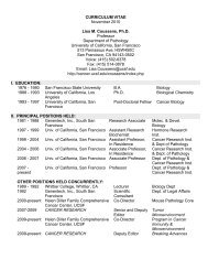
Fig. 1‐1 Papillary growth pattern<br />
<strong>The</strong> histologic classification of high-grade<br />
ovarian carc<strong>in</strong>omas is of controversial. Some<br />
authorities th<strong>in</strong>k that a diagnosis of high-grade<br />
serous carc<strong>in</strong>oma is only appropriate for<br />
predom<strong>in</strong>antly papillary tumors with m<strong>in</strong>or<br />
gl<strong>and</strong>ular or solid areas. (31) Some who favor this<br />
view th<strong>in</strong>k that nei<strong>the</strong>r <strong>the</strong> immunophenotype nor<br />
<strong>the</strong> presence or absence of p53 mutations<br />
separates high grade serous carc<strong>in</strong>oma from high<br />
grade endometrioid carc<strong>in</strong>oma <strong>and</strong> that <strong>the</strong><br />
dist<strong>in</strong>ction must be made based on <strong>the</strong> histology,<br />
such that tumors with papillae <strong>and</strong> slit like gl<strong>and</strong>s<br />
are high grade serous carc<strong>in</strong>omas <strong>and</strong> those where<br />
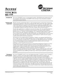
Fig. 1‐2 Solid growth, marked atypia <strong>the</strong>re is sheet like growth of tumor cells or gl<strong>and</strong>s<br />
are high grade endometrioid carc<strong>in</strong>omas. (32)<br />
O<strong>the</strong>rs have adopted a more <strong>in</strong>clusive<br />
classification scheme, <strong>and</strong> view most high grade<br />
carc<strong>in</strong>omas with some features of serous<br />
differentiation as types of high grade serous<br />
carc<strong>in</strong>oma, <strong>in</strong>clud<strong>in</strong>g tumors with extensive solid<br />
areas, tumors with high grade gl<strong>and</strong>ular areas<br />
without squamous differentiation, <strong>and</strong> some<br />
tumors with clear cell or transitional cell areas.<br />
(33-37) High grade serous carc<strong>in</strong>omas with clear<br />
cell areas are variants of high grade serous<br />
carc<strong>in</strong>oma <strong>and</strong> not mixed serous-clear cell carc<strong>in</strong>omas, <strong>and</strong> <strong>the</strong>y should be classified<br />
accord<strong>in</strong>gly.(38) Immunohistochemical <strong>and</strong> molecular studies support <strong>the</strong> concept that <strong>the</strong>se are<br />
variants of high-grade serous carc<strong>in</strong>oma <strong>and</strong> diagnos<strong>in</strong>g <strong>the</strong>m as such results <strong>in</strong> a more<br />
reproducible <strong>and</strong> useful classification system. (33) Tubal <strong>in</strong>traepi<strong>the</strong>lial carc<strong>in</strong>oma is associated<br />
with serous carc<strong>in</strong>oma.<br />
Grad<strong>in</strong>g of Serous Carc<strong>in</strong>oma<br />
Grad<strong>in</strong>g of ovarian carc<strong>in</strong>omas has not been st<strong>and</strong>ardized. Silverberg <strong>and</strong> colleagues<br />
developed a “universal” grad<strong>in</strong>g system that <strong>the</strong>y thought could be used for all types of ovarian<br />
carc<strong>in</strong>omas. (39) In <strong>the</strong>ir system, <strong>the</strong> grade is determ<strong>in</strong>ed by <strong>the</strong> degree of nuclear atypia, <strong>the</strong><br />
mitotic <strong>in</strong>dex, <strong>and</strong> <strong>the</strong> extent to which <strong>the</strong> tumor cells form papillae or gl<strong>and</strong>s. More recently, a<br />
b<strong>in</strong>ary grad<strong>in</strong>g system, <strong>in</strong> which low-grade serous carc<strong>in</strong>oma almost always falls <strong>in</strong>to grade 1 of<br />
<strong>the</strong> “universal” grad<strong>in</strong>g system, has ga<strong>in</strong>ed greater acceptance than <strong>the</strong> “universal” grad<strong>in</strong>g<br />
system because it better reflects <strong>the</strong> current concept that high <strong>and</strong> low grade serous carc<strong>in</strong>omas<br />
represent two different tumor types ra<strong>the</strong>r than two different grades of <strong>the</strong> same tumor. In <strong>the</strong><br />
High Grade vs Low Grade Serous Carc<strong>in</strong>oma<br />
Atypia MF/10 hpf BL<br />
LG Serous Mild‐Mod ≤ 12 +<br />
HG Serous Marked > 12 ‐<br />
BL = associated with borderl<strong>in</strong>e tumor elements<br />
5<br />
b<strong>in</strong>ary system, low-grade serous<br />
carc<strong>in</strong>oma exhibits mild to moderate<br />
nuclear atypia <strong>and</strong> 12 or fewer mitotic<br />
figures per 10 high power fields. (40) In
high-grade serous carc<strong>in</strong>oma <strong>the</strong>re is marked nuclear atypia, considerable pleomorphism <strong>and</strong><br />
often prom<strong>in</strong>ent macronucleoli. Mitotic activity is high; while a floor of 12 mf/10 hpf has been<br />
proposed as a lower limit of mitotic activity <strong>in</strong> high grade serous carc<strong>in</strong>oma, <strong>the</strong>re was a median<br />
of 38 mf/10 hpf <strong>in</strong> one study. (40) Apart from <strong>the</strong>ir differences <strong>in</strong> nuclear atypia <strong>and</strong> mitotic<br />
activity, low <strong>and</strong> high-grade serous carc<strong>in</strong>oma grow <strong>in</strong> different architectural patterns, <strong>and</strong> <strong>the</strong><br />
correct diagnosis can generally be determ<strong>in</strong>ed at low magnification without any need for mitosis<br />
count<strong>in</strong>g. Also, most low-grade serous carc<strong>in</strong>omas are associated with a borderl<strong>in</strong>e serous tumor,<br />
so <strong>in</strong> a primary ovarian tumor <strong>the</strong> f<strong>in</strong>d<strong>in</strong>g of patterns of a borderl<strong>in</strong>e tumor favors low-grade<br />
serous carc<strong>in</strong>oma. <strong>Tumors</strong> with <strong>in</strong>termediate nuclear grades (“grade 2 serous carc<strong>in</strong>oma”) are<br />
similar <strong>in</strong> <strong>the</strong>ir growth patterns <strong>and</strong> molecular features to high-grade serous carc<strong>in</strong>oma <strong>and</strong><br />
exhibit <strong>the</strong> same aggressive behavior, so <strong>the</strong>y are viewed as high-grade tumors. (41)<br />
P53 <strong>and</strong> High Grade Serous Carc<strong>in</strong>oma<br />
Mutations of <strong>the</strong> TP53 gene have long been associated with high-grade serous carc<strong>in</strong>oma.<br />
TP53 mutation has been thought to be an early event <strong>in</strong> serous carc<strong>in</strong>ogenesis, a po<strong>in</strong>t that has<br />
been re-emphasized by recent studies of a p53 positive putative cancer precursor <strong>in</strong> <strong>the</strong> fallopian<br />
tube known as <strong>the</strong> p53 signature lesion. (42) p53 signatures show positive immunosta<strong>in</strong><strong>in</strong>g for<br />
p53 <strong>and</strong> <strong>the</strong>y have TP53 mutations. <strong>The</strong> p53 signature is currently viewed as a non-obligate<br />
premalignant condition, illustrat<strong>in</strong>g just how early <strong>in</strong> <strong>the</strong> carc<strong>in</strong>ogenic process TP53 mutations<br />
may occur. Recent studies of high-grade serous carc<strong>in</strong>oma reveal a high frequency of TP53<br />
mutations <strong>in</strong> such tumors. Yemelyanova et al studied 43 high-grade serous carc<strong>in</strong>omas; only 6<br />
lacked TP53 mutations. (43) Ahmed <strong>and</strong> colleagues found TP53 mutations <strong>in</strong> 96.7% (119/123)<br />
of patients with advanced stage high-grade serous carc<strong>in</strong>omas. (2) <strong>The</strong> Cancer Genome Atlas<br />
Research Network analyzed whole exome DNA sequences <strong>in</strong> 316 high-grade high stage serous<br />
carc<strong>in</strong>omas <strong>and</strong> found that 96% had TP53 mutations. (44) Thus, almost all high-grade serous<br />
carc<strong>in</strong>omas have TP53 mutations. <strong>The</strong> high <strong>and</strong> ra<strong>the</strong>r consistent frequency of TP53 mutations is<br />
somewhat surpris<strong>in</strong>g, s<strong>in</strong>ce I would have expected that variations <strong>in</strong> <strong>the</strong> way high-grade serous<br />
carc<strong>in</strong>oma is diagnosed <strong>and</strong> classified to impact <strong>the</strong> percentage of positive cases.<br />
High-grade serous carc<strong>in</strong>oma frequently shows immunohistochemical evidence of <strong>the</strong><br />
presence of a p53 mutation. This can be manifest as diffuse strong sta<strong>in</strong><strong>in</strong>g of nearly all tumor<br />
cell nuclei. (45) In some cases, <strong>the</strong> p53 mutation results <strong>in</strong> <strong>the</strong> complete absence of p53 prote<strong>in</strong>,<br />
<strong>in</strong> which case <strong>the</strong>re is no sta<strong>in</strong><strong>in</strong>g <strong>in</strong> any tumor cell nuclei. Both of <strong>the</strong>se results are considered<br />
positive test results. <strong>Tumors</strong> that do not have a p53 mutation generally show weak to moderate<br />
Interpretation of p53 Immunosta<strong>in</strong><strong>in</strong>g<br />
Pattern<br />
Diffuse strong positive nuclear sta<strong>in</strong><strong>in</strong>g (Positive<br />
sta<strong>in</strong><strong>in</strong>g <strong>in</strong> > 50% of tumor cell nuclei <strong>and</strong> usually<br />
<strong>in</strong> > 80%)<br />
Patchy weak sta<strong>in</strong><strong>in</strong>g <strong>in</strong> < 50% of tumor cell<br />
nuclei<br />
Absolutely no sta<strong>in</strong><strong>in</strong>g <strong>in</strong> any tumor cell nuclei<br />
Test Result<br />
Positive<br />
Negative<br />
Positive<br />
6
sta<strong>in</strong><strong>in</strong>g <strong>in</strong> some tumor cell nuclei. In one study, <strong>the</strong>re was complete absence of p53 expression<br />
<strong>in</strong> 30.3% of cases, focal expression <strong>in</strong> 12.0% <strong>and</strong> overexpression <strong>in</strong> 57.7%, (46) <strong>and</strong> similar<br />
results were observed <strong>in</strong> ano<strong>the</strong>r study. (43) By recogniz<strong>in</strong>g <strong>the</strong> patterns of positive sta<strong>in</strong><strong>in</strong>g<br />
pathologists can identify which ovarian cancers are likely to have TP53 mutations <strong>and</strong> hence<br />
which are best classified as high-grade serous carc<strong>in</strong>omas. Among patients with probable TP53<br />
mutations, those with complete absence of expression had an <strong>in</strong>creased risk of recurrence<br />
compared with those with overexpression. (46) O<strong>the</strong>r markers that are generally positive <strong>in</strong> highgrade<br />
serous carc<strong>in</strong>oma, <strong>and</strong> that complement p53 as immunohistochemical markers of highgrade<br />
serous carc<strong>in</strong>oma, are p16 <strong>and</strong> HGMA2. A positive sta<strong>in</strong> for p16 <strong>in</strong> this context is diffuse<br />
strong sta<strong>in</strong><strong>in</strong>g of <strong>the</strong> cytoplasm <strong>and</strong> nuclei of all or nearly all tumor cells. (47-49) <strong>The</strong> range of<br />
sta<strong>in</strong><strong>in</strong>g that should be expected for HGMA2 is less well worked out; <strong>in</strong> one study about two<br />
thirds of high grade serous carc<strong>in</strong>omas showed moderate to strong nuclear sta<strong>in</strong><strong>in</strong>g <strong>in</strong> 40-100%<br />
of tumor cells. (50) Low grade serous carc<strong>in</strong>oma, <strong>in</strong> contrast, rarely shows <strong>the</strong> type of strong<br />
sta<strong>in</strong><strong>in</strong>g for p53 or p16 seen <strong>in</strong> high grade serous carc<strong>in</strong>oma, (45, 48, 51, 52) <strong>and</strong> <strong>the</strong>se markers<br />
are generally negative <strong>in</strong> borderl<strong>in</strong>e <strong>and</strong> benign tumors as well.<br />
BRCA Mutations <strong>and</strong> High Grade Serous Carc<strong>in</strong>oma<br />
High-grade serous carc<strong>in</strong>oma is <strong>the</strong> type of ovarian cancer most associated with<br />
mutations <strong>in</strong> <strong>the</strong> BRCA genes. Large, cl<strong>in</strong>ically detected tumors <strong>in</strong> BRCA mutation carriers are<br />
mostly high-grade serous carc<strong>in</strong>omas, although o<strong>the</strong>r types of ovarian cancer occasionally occur<br />
<strong>in</strong> <strong>the</strong>se patients. (53) In <strong>the</strong> range of 10-15% of ovarian carc<strong>in</strong>omas occur <strong>in</strong> women with<br />
germl<strong>in</strong>e mutations of BRCA1 or BRCA2. In a recent analysis of 1342 unselected Canadian<br />
women 176 or 13.3% carried a mutation (107 BRCA1, 67 BRCA2, <strong>and</strong> 2 with mutations <strong>in</strong> both<br />
genes). (54) <strong>The</strong> hereditary breast <strong>and</strong> ovarian cancer syndrome, caused by mutations <strong>in</strong> <strong>the</strong>se<br />
genes, is <strong>in</strong>herited <strong>in</strong> an autosomal dom<strong>in</strong>ant fashion. <strong>The</strong> lifetime risk of a woman with a BRCA<br />
mutation develop<strong>in</strong>g breast cancer is 50-80% <strong>and</strong> <strong>the</strong> lifetime risk of develop<strong>in</strong>g ovarian cancer<br />
is 30-50%. <strong>The</strong> risks reported <strong>in</strong> various studies depend partly on how <strong>the</strong> cases were<br />
ascerta<strong>in</strong>ed, with higher risks reported <strong>in</strong> studies of cancer families <strong>and</strong> lower risks <strong>in</strong> studies<br />
where carriers were identified <strong>in</strong>dependent of <strong>the</strong>ir family histories. Women with BRCA<br />
mutations or who have a family history of ovarian cancer are treated by risk-reduc<strong>in</strong>g bilateral<br />
salp<strong>in</strong>go-oophorectomy (RRSO), which has been shown to markedly lower <strong>the</strong> risk of<br />
develop<strong>in</strong>g carc<strong>in</strong>oma. (55-58) Cancer risk is not elim<strong>in</strong>ated as <strong>the</strong>se patients are still at risk for<br />
peritoneal serous carc<strong>in</strong>oma <strong>and</strong> for breast cancer, although <strong>the</strong> risk for peritoneal cancer is very<br />
low.<br />
<strong>The</strong>re are two BRCA genes, BRCA1 <strong>and</strong> BRCA2. <strong>The</strong>y are classified as tumor<br />
suppressor genes. BRCA1 is located on chromosome 17 at 17q21-22 <strong>and</strong> BRCA2 is located on<br />
chromosome 13 at 13q12-13. <strong>The</strong> BRCA2 gene is larger, hav<strong>in</strong>g about twice as many am<strong>in</strong>o<br />
acids as <strong>the</strong> BRCA1 gene (3148 vs 1863). <strong>The</strong>se genes are both <strong>in</strong>volved <strong>in</strong> many <strong>in</strong>tercellular<br />
processes but <strong>the</strong>ir ma<strong>in</strong> function is to protect <strong>the</strong> genome from double-str<strong>and</strong>ed DNA damage<br />
dur<strong>in</strong>g DNA replication. BRCA1 is <strong>in</strong>volved <strong>in</strong> checkpo<strong>in</strong>t activation <strong>and</strong> DNA repair <strong>and</strong> both<br />
BRCA1 <strong>and</strong> BRCA2 are <strong>in</strong>volved <strong>in</strong> homologous recomb<strong>in</strong>ation as a means to repair double<br />
str<strong>and</strong>ed DNA breaks. (59) When BRCA is deficient, double str<strong>and</strong>ed breaks cannot be repaired<br />
by <strong>the</strong> error free homologous recomb<strong>in</strong>ation process, <strong>and</strong> <strong>the</strong>y are <strong>in</strong>stead repaired by alternate<br />
methods, such as end jo<strong>in</strong><strong>in</strong>g <strong>and</strong> s<strong>in</strong>gle str<strong>and</strong> anneal<strong>in</strong>g, that lead to genomic <strong>in</strong>stability<br />
result<strong>in</strong>g <strong>in</strong> <strong>the</strong> cancer predisposition that is associated with BRCA loss.<br />
7
BRCA associated ovarian cancers <strong>the</strong>oretically might be more responsive to<br />
chemo<strong>the</strong>rapy with drugs like cisplat<strong>in</strong> that damage DNA, s<strong>in</strong>ce <strong>the</strong>ir tumor cells are less able to<br />
repair <strong>the</strong> DNA breaks that such drugs cause. In addition, BRCA associated cancers may be<br />
amenable to <strong>the</strong>rapy with PARP (poly (ADP-ribose) polymerase) <strong>in</strong>hibitors, <strong>the</strong> activity of which<br />
are dependent on loss of BRCA function. In recent analyses, conflict<strong>in</strong>g results have been<br />
published. Analysis of <strong>the</strong> Cancer Genome Atlas Project data <strong>in</strong>dicated that patients with<br />
BRCA1 associated tumors were likely to be slightly younger than those with BRCA2 or wild<br />
type cancers. Patients whose tumors had somatic mutations had <strong>the</strong> same cl<strong>in</strong>ical characteristics<br />
as patients with wild type tumors. Among those with germl<strong>in</strong>e mutations, only patients with<br />
BRCA2 mutations had improved survival <strong>and</strong> better response to plat<strong>in</strong>um based chemo<strong>the</strong>rapy.<br />
(60) On <strong>the</strong> o<strong>the</strong>r h<strong>and</strong>, patients with BRCA1 <strong>in</strong>activation due to germl<strong>in</strong>e mutations or promotor<br />
hypermethylation had <strong>the</strong> same outcome as those with wild type BRCA. <strong>The</strong> authors of ano<strong>the</strong>r<br />
study, which pooled data from 26 studies, concluded that women with BRCA1 <strong>and</strong> BRCA2<br />
germl<strong>in</strong>e mutations both had improved survival relative to those with wild type BRCA, although<br />
those with BRCA2 mutations appeared to have <strong>the</strong> best survival. {Bolton, 2012 #36408}<br />
Loss of BRCA function is often due to a germl<strong>in</strong>e mutation <strong>in</strong> ei<strong>the</strong>r BRCA1 or BRCA2,<br />
but it can also be due to somatic BRCA mutations <strong>and</strong> <strong>in</strong> <strong>the</strong> case of BRCA1, to promotor<br />
hypermethylation. (61) Regardless of <strong>the</strong> cause of BRCA dysfunction, recent pathologic studies<br />
suggest that a comb<strong>in</strong>ation of histologic f<strong>in</strong>d<strong>in</strong>gs may predict that a given ovarian tumor is<br />
BRCA related. One set of histologic criteria was published by Soslow et al, {Soslow, 2012<br />
#36680} <strong>and</strong> ano<strong>the</strong>r was published <strong>in</strong> abstract form only by Fujiwara et al. Histologic features<br />
that may be BRCA related <strong>in</strong>clude serous/undifferentiated histologic type; solid,<br />
pseudoendometrioid or transitional growth patterns; marked atypia <strong>and</strong> giant bizarre nuclei;<br />
numerous tumor <strong>in</strong>filtrat<strong>in</strong>g lymphocytes; <strong>and</strong> a very high mitotic rate. Accord<strong>in</strong>g to <strong>the</strong>se<br />
authors, comb<strong>in</strong>ations of <strong>the</strong>se features, when present, may be <strong>in</strong>dicative of BRCA dysfunction,<br />
but it is not clear that <strong>the</strong> histologic criteria differentiate tumors with BRCA1 mutations from<br />
those with BRCA2 mutations, nor do <strong>the</strong> criteria differentiate tumors with germl<strong>in</strong>e mutations<br />
from those with BRCA dysfunction due to somatic mutations or promotor hypermethylation.<br />
Causes of BRCA Loss of Function<br />
Germl<strong>in</strong>e BRCA mutation 10‐15%<br />
Somatic BRCA mutation 5‐10%<br />
Promotor<br />
30%<br />
Hypermethylation<br />
O<strong>the</strong>r (microRNA, etc) ?<br />
Fallopian Tube Surprise<br />
While small tumors are<br />
occasionally identified <strong>in</strong> <strong>the</strong> ovaries,<br />
most of <strong>the</strong> <strong>in</strong>traepi<strong>the</strong>lial <strong>and</strong> early<br />
<strong>in</strong>vasive serous carc<strong>in</strong>omas detected <strong>in</strong><br />
RRSO specimens have been found <strong>in</strong><br />
<strong>the</strong> fallopian tubes.(62-69) Tubal<br />
<strong>in</strong>traepi<strong>the</strong>lial carc<strong>in</strong>oma is less<br />
common <strong>in</strong> salp<strong>in</strong>gectomy specimens from patients without a BRCA mutation, but it is<br />
occasionally detected; <strong>in</strong> one study tubal <strong>in</strong>traepi<strong>the</strong>lial carc<strong>in</strong>oma was found <strong>in</strong> 8% of patients<br />
with a BRCA mutation <strong>and</strong> <strong>in</strong> 3% of patients with no mutation. (69) Interest<strong>in</strong>gly, complete<br />
section<strong>in</strong>g of <strong>the</strong> fallopian tubes <strong>in</strong> patients with a cl<strong>in</strong>ical diagnosis of a primary ovarian or<br />
primary peritoneal serous carc<strong>in</strong>oma, regardless of whe<strong>the</strong>r it occurs <strong>in</strong> a woman with a germl<strong>in</strong>e<br />
BRCA mutation, frequently reveals foci of serous tubal <strong>in</strong>traepi<strong>the</strong>lial carc<strong>in</strong>oma or small<br />
apparently primary foci of <strong>in</strong>vasive serous carc<strong>in</strong>oma of <strong>the</strong> fallopian tube. (66, 70-72) This has<br />
led to <strong>the</strong> hypo<strong>the</strong>sis that many, if not all, serous carc<strong>in</strong>omas of <strong>the</strong> ovary <strong>and</strong> peritoneum are<br />
8
actually metastases from tubal tumors, not primary neoplasms of <strong>the</strong> ovary or peritoneum. (70,<br />
73, 74) Rare high-grade serous carc<strong>in</strong>omas appear to arise from a low grade serous carc<strong>in</strong>oma or<br />
a borderl<strong>in</strong>e serous tumor. (30) <strong>The</strong> histogenesis of serous carc<strong>in</strong>oma of <strong>the</strong> ovary <strong>and</strong> its<br />
relationship to tumors of <strong>the</strong> fallopian tube is an area of active research.<br />
Immunohistochemistry of Serous <strong>Tumors</strong><br />
All serous tumors have immunohistochemical features <strong>in</strong> common. <strong>The</strong>y usually show<br />
positive sta<strong>in</strong><strong>in</strong>g for cytokerat<strong>in</strong> 7 <strong>and</strong> <strong>the</strong>y do not sta<strong>in</strong> for cytokerat<strong>in</strong> 20 or CDX2. (75) <strong>The</strong>y<br />
show variable sta<strong>in</strong><strong>in</strong>g for estrogen <strong>and</strong> progesterone receptors, with less sta<strong>in</strong><strong>in</strong>g be<strong>in</strong>g present<br />
<strong>in</strong> high-grade serous carc<strong>in</strong>oma than <strong>in</strong> lower grade serous tumors. Borderl<strong>in</strong>e serous tumors <strong>and</strong><br />
serous carc<strong>in</strong>omas generally show membrane sta<strong>in</strong><strong>in</strong>g for OC-125. (76) One of <strong>the</strong> most helpful<br />
features of serous tumors is that <strong>the</strong>y show nuclear sta<strong>in</strong><strong>in</strong>g for WT-1. (77-80) This helps<br />
differentiate <strong>the</strong>m from o<strong>the</strong>r types of ovarian tumors, such as endometrioid <strong>and</strong> clear cell<br />
carc<strong>in</strong>omas. Sta<strong>in</strong><strong>in</strong>g for WT-1 is also helpful <strong>in</strong> differentiat<strong>in</strong>g ovarian serous carc<strong>in</strong>oma from<br />
endometrial serous carc<strong>in</strong>oma, which has a similar microscopic appearance but is less likely to<br />
sta<strong>in</strong> for WT-1. (77, 81, 82) Borderl<strong>in</strong>e serous tumors <strong>and</strong> low grade serous carc<strong>in</strong>omas are more<br />
likely to show nuclear sta<strong>in</strong><strong>in</strong>g for PAX2 than are high grade serous carc<strong>in</strong>omas; variable strong<br />
sta<strong>in</strong><strong>in</strong>g is generally seen <strong>in</strong> <strong>the</strong> former categories of serous tumors while sta<strong>in</strong><strong>in</strong>g is typically<br />
absent <strong>in</strong> high grade carc<strong>in</strong>omas. (83) PAX8 appears to be a reliable marker of female genital<br />
tract tumors, <strong>and</strong> it is almost always positive <strong>in</strong> serous tumors, although sta<strong>in</strong><strong>in</strong>g can vary <strong>in</strong><br />
extent <strong>and</strong> <strong>in</strong>tensity. (84-86) HMGA2 is ano<strong>the</strong>r marker that is positive <strong>in</strong> a majority of highgrade<br />
serous carc<strong>in</strong>omas but uncommon <strong>in</strong> o<strong>the</strong>r types of ovarian cancer. (50) Patients with highgrade<br />
serous carc<strong>in</strong>oma are frequently treated with chemo<strong>the</strong>rapy, ei<strong>the</strong>r after or before surgery.<br />
<strong>The</strong> postchemo<strong>the</strong>rapy immunophenotype is similar to that of untreated serous carc<strong>in</strong>oma. (23)<br />
Immunohistochemical Sta<strong>in</strong><strong>in</strong>g <strong>in</strong> High Grade Serous Carc<strong>in</strong>oma<br />
CK7 +<br />
CK20 ‐<br />
PAX8 +<br />
CA125 +<br />
WT‐1 +<br />
p53<br />
Diffuse strong positive or completely negative<br />
p16<br />
Diffuse strong positive<br />
HMGA2 +<br />
PAX2 ‐<br />
References<br />
1. Köbel M, Kalloger SE, Huntsman DG, Santos JL, Swenerton KD, Seidman JD, et al. Differences <strong>in</strong> Tumor<br />
Type <strong>in</strong> Low-stage Versus High-stage Ovarian Carc<strong>in</strong>omas. Int J Gynecol Pathol. 2010;29(3):203-11.<br />
2. Ahmed AA, Etemadmoghadam D, Temple J, Lynch AG, Riad M, Sharma R, et al. Driver mutations <strong>in</strong><br />
TP53 are ubiquitous <strong>in</strong> high grade serous carc<strong>in</strong>oma of <strong>the</strong> ovary. J Pathol. 2010;221(1):49-56. Epub 2010/03/17.<br />
3. G<strong>in</strong>gell D, Samuel A, Haynik D, McBee W, Kelley J, Zorn K, et al. Metastatic Ovarian Serous Carc<strong>in</strong>oma<br />
Present<strong>in</strong>g as Inflammatory Breast Cancer: A Case Report. Int J Gynecol Pathol. 2010;29(3):243-7.<br />
9
4. F<strong>in</strong>ch A, Be<strong>in</strong>er M, Lub<strong>in</strong>ski J, Lynch HT, Moller P, Rosen B, et al. Salp<strong>in</strong>go-oophorectomy <strong>and</strong> <strong>the</strong> risk of<br />
ovarian, fallopian tube, <strong>and</strong> peritoneal cancers <strong>in</strong> women with a BRCA1 or BRCA2 Mutation. JAMA.<br />
2006;296(2):185-92.<br />
5. Y<strong>in</strong> BWT, Dnistrian A, Lloyd KO. Ovarian cancer antigen CA125 is encoded by <strong>the</strong> MUC16 muc<strong>in</strong> gene.<br />
Int J Cancer. 2002;98(5):737-40.<br />
6. O'Brien TJ, Beard JB, Underwood LJ, Shigemasa K. <strong>The</strong> CA 125 gene: A newly discovered extension of<br />
<strong>the</strong> glycosylated N-term<strong>in</strong>al doma<strong>in</strong> doubles <strong>the</strong> size of this extracellular superstructure. Tumor Biology.<br />
2002;23(3):154-69.<br />
7. Kolwijck E, Thomas CM, Bulten J, Massuger LF. Preoperative CA-125 levels <strong>in</strong> 123 patients with<br />
borderl<strong>in</strong>e ovarian tumors: a retrospective analysis <strong>and</strong> review of <strong>the</strong> literature. Int J Gynecol Cancer.<br />
2009;19(8):1335-8. Epub 2009/12/17.<br />
8. Husse<strong>in</strong>zadeh N. Status of tumor markers <strong>in</strong> epi<strong>the</strong>lial ovarian cancer has <strong>the</strong>re been any progress? A<br />
review. Gynecol Oncol. 2011;120(1):152-7. Epub 2010/10/12.<br />
9. Bast RC, Jr. CA 125 <strong>and</strong> <strong>the</strong> detection of recurrent ovarian cancer: a reasonably accurate biomarker for a<br />
difficult disease. Cancer. 2010;116(12):2850-3. Epub 2010/06/22.<br />
10. Nyante SJ, Black A, Kreimer AR, Duggan MA, Carreon JD, Kessel B, et al. Pathologic f<strong>in</strong>d<strong>in</strong>gs follow<strong>in</strong>g<br />
false-positive screen<strong>in</strong>g tests for ovarian cancer <strong>in</strong> <strong>the</strong> Prostate, Lung, Colorectal <strong>and</strong> Ovarian (PLCO) cancer<br />
screen<strong>in</strong>g trial. Gynecol Oncol. 2011;120(3):474-9. Epub 2010/12/15.<br />
11. Cannistra SA. Cancer of <strong>the</strong> ovary. N Engl J Med. 2004;351(24):2519-29.<br />
12. Fader AN, Rose PG. Role of surgery <strong>in</strong> ovarian carc<strong>in</strong>oma. J Cl<strong>in</strong> Oncol. 2007;25(20):2873-83. Epub<br />
2007/07/10.<br />
13. Morgan RJ, Jr., Alvarez RD, Armstrong DK, Boston B, Burger RA, Chen LM, et al. <strong>Epi<strong>the</strong>lial</strong> ovarian<br />
cancer. J Natl Compr Canc Netw. 2011;9(1):82-113. Epub 2011/01/15.<br />
14. Chan JK, Tian C, Monk BJ, Herzog T, Kapp DS, Bell J, et al. Prognostic factors for high-risk early-stage<br />
epi<strong>the</strong>lial ovarian cancer: a Gynecologic Oncology Group study. Cancer. 2008;112(10):2202-10. Epub 2008/03/19.<br />
15. Seidman JD, Yemelyanova AV, Khedmati F, Bidus MA, Da<strong>in</strong>ty L, Boice CR, et al. Prognostic factors for<br />
stage I ovarian carc<strong>in</strong>oma. Int J Gynecol Pathol. 2010;29(1):1-7. Epub 2009/12/03.<br />
16. Schilder JM, Thompson AM, DePriest PD, Uel<strong>and</strong> FR, Cibull ML, Kryscio RJ, et al. Outcome of<br />
reproductive age women with stage IA or IC <strong>in</strong>vasive epi<strong>the</strong>lial ovarian cancer treated with fertility-spar<strong>in</strong>g <strong>the</strong>rapy.<br />
Gynecol Oncol. 2002;87(1):1-7.<br />
17. Wright JD, Shah M, Ma<strong>the</strong>w L, Burke WM, Culhane J, Goldman N, et al. Fertility preservation <strong>in</strong> young<br />
women with epi<strong>the</strong>lial ovarian cancer. Cancer. 2009;115(18):4118-26. Epub 2009/08/12.<br />
18. Satoh T, Hatae M, Watanabe Y, Yaegashi N, Ishiko O, Kodama S, et al. Outcomes of fertility-spar<strong>in</strong>g<br />
surgery for stage I epi<strong>the</strong>lial ovarian cancer: a proposal for patient selection. J Cl<strong>in</strong> Oncol. 2010;28(10):1727-32.<br />
Epub 2010/03/03.<br />
19. Vergote I, Trope CG, Amant F, Kristensen GB, Ehlen T, Johnson N, et al. Neoadjuvant chemo<strong>the</strong>rapy or<br />
primary surgery <strong>in</strong> stage IIIC or IV ovarian cancer. N Engl J Med. 2010;363(10):943-53. Epub 2010/09/08.<br />
20. Rose PG, Brady MF. EORTC 55971: does it apply to all patients with advanced state ovarian cancer?<br />
Gynecol Oncol. 2011;120(2):300-1. Epub 2010/11/26.<br />
21. Chew I, Soslow RA, Park KJ. Morphologic Changes <strong>in</strong> Ovarian Carc<strong>in</strong>oma After Neoadjuvant<br />
Chemo<strong>the</strong>rapy: Report of a Case Show<strong>in</strong>g Extensive Clear Cell Changes Mimick<strong>in</strong>g Clear Cell Carc<strong>in</strong>oma. Int J<br />
Gynecol Pathol. 2009;28(5):442-6 10.1097/PGP.0b013e3181a071b5.<br />
22. McCluggage WG, Lyness RW, Atk<strong>in</strong>son RJ, Dobbs SP, Harley I, McClell<strong>and</strong> HR, et al. Morphological<br />
effects of chemo<strong>the</strong>rapy on ovarian carc<strong>in</strong>oma. J Cl<strong>in</strong> Pathol. 2002;55(1):27-31.<br />
23. Miller K, Price JH, Dobbs SP, McClell<strong>and</strong> RH, Kennedy K, McCluggage WG. An immunohistochemical<br />
<strong>and</strong> morphological analysis of post-chemo<strong>the</strong>rapy ovarian carc<strong>in</strong>oma. J Cl<strong>in</strong> Pathol. 2008;61(5):652-7. Epub<br />
2007/11/17.<br />
24. Ozols RF. Systemic <strong>the</strong>rapy for ovarian cancer: current status <strong>and</strong> new treatments. Sem<strong>in</strong> Oncol. 2006;33(2<br />
Suppl 6):S3-11.<br />
25. Armstrong DK, Bundy B, Wenzel L, Huang HQ, Baergen R, Lele S, et al. Intraperitoneal cisplat<strong>in</strong> <strong>and</strong><br />
paclitaxel <strong>in</strong> ovarian cancer. N Engl J Med. 2006;354(1):34-43. Epub 2006/01/06.<br />
26. Markman M, Walker JL. Intraperitoneal chemo<strong>the</strong>rapy of ovarian cancer: a review, with a focus on<br />
practical aspects of treatment. J Cl<strong>in</strong> Oncol. 2006;24(6):988-94. Epub 2006/02/08.<br />
27. Lambert HE, Gregory WM, Nelstrop AE, Rust<strong>in</strong> GJ. Long-term survival <strong>in</strong> 463 women treated with<br />
plat<strong>in</strong>um analogs for advanced epi<strong>the</strong>lial carc<strong>in</strong>oma of <strong>the</strong> ovary: life expectancy compared to women of an agematched<br />
normal population. Int J Gynecol Cancer. 2004;14(5):772-8.<br />
10
28. Che M, Tornos C, Deavers MT, Malpica A, Gershenson DM, Silva EG. Ovarian mixed-epi<strong>the</strong>lial<br />
carc<strong>in</strong>omas with a microcystic pattern <strong>and</strong> signet-r<strong>in</strong>g cells. Int J Gynecol Pathol. 2001;20(4):323-8.<br />
29. Boyd C, McCluggage WG. Low-grade ovarian serous neoplasms (low-grade serous carc<strong>in</strong>oma <strong>and</strong> serous<br />
borderl<strong>in</strong>e tumor) associated with high-grade serous carc<strong>in</strong>oma or undifferentiated carc<strong>in</strong>oma: report of a series of<br />
cases of an unusual phenomenon. Am J Surg Pathol. 2012;36(3):368-75. Epub 2011/11/16.<br />
30. Dehari R, Kurman RJ, Logani S, Shih IM. <strong>The</strong> Development of High-grade Serous Carc<strong>in</strong>oma From<br />
Atypical Proliferative (Borderl<strong>in</strong>e) Serous <strong>Tumors</strong> <strong>and</strong> Low-grade Micropapillary Serous Carc<strong>in</strong>oma: A<br />
Morphologic <strong>and</strong> Molecular Genetic Analysis. Am J Surg Pathol. 2007;31(7):1007-12.<br />
31. Westfall D, Roma AA, Silva EG. High-grade serous carc<strong>in</strong>oma of <strong>the</strong> ovary. Ann Diagn Pathol.<br />
2009;13(4):285-90. Epub 2009/07/18.<br />
32. Roh MH, Yass<strong>in</strong> Y, Miron A, Mehra KK, Mehrad M, Monte NM, et al. High-grade fimbrial-ovarian<br />
carc<strong>in</strong>omas are unified by altered p53, PTEN <strong>and</strong> PAX2 expression. Mod Pathol. 2010;23(10):1316-24. Epub<br />
2010/06/22.<br />
33. Kobel M, Kalloger SE, Baker PM, Ewanowich CA, Arseneau J, Zherebitskiy V, et al. Diagnosis of ovarian<br />
carc<strong>in</strong>oma cell type is highly reproducible: a transcanadian study. Am J Surg Pathol. 2010;34(7):984-93. Epub<br />
2010/05/28.<br />
34. Kobel M, Kalloger SE, Santos JL, Huntsman DG, Gilks CB, Swenerton KD. Tumor type <strong>and</strong> substage<br />
predict survival <strong>in</strong> stage I <strong>and</strong> II ovarian carc<strong>in</strong>oma: <strong>in</strong>sights <strong>and</strong> implications. Gynecol Oncol. 2010;116(1):50-6.<br />
Epub 2009/10/14.<br />
35. Gilks CB, Ionescu DN, Kalloger SE, Kobel M, Irv<strong>in</strong>g J, Clarke B, et al. Tumor cell type can be<br />
reproducibly diagnosed <strong>and</strong> is of <strong>in</strong>dependent prognostic significance <strong>in</strong> patients with maximally debulked ovarian<br />
carc<strong>in</strong>oma. Hum Pathol. 2008;39(8):1239-51. Epub 2008/07/08.<br />
36. Soslow RA. Histologic subtypes of ovarian carc<strong>in</strong>oma: an overview. Int J Gynecol Pathol. 2008;27(2):161-<br />
74.<br />
37. McCluggage WG. My approach to <strong>and</strong> thoughts on <strong>the</strong> typ<strong>in</strong>g of ovarian carc<strong>in</strong>omas. J Cl<strong>in</strong> Pathol.<br />
2008;61(2):152-63. Epub 2007/08/21.<br />
38. Han G, Gilks CB, Leung S, Ewanowich CA, Irv<strong>in</strong>g JA, Longacre TA, et al. Mixed ovarian epi<strong>the</strong>lial<br />
carc<strong>in</strong>omas with clear cell <strong>and</strong> serous components are variants of high-grade serous carc<strong>in</strong>oma: an <strong>in</strong>terobserver<br />
correlative <strong>and</strong> immunohistochemical study of 32 cases. Am J Surg Pathol. 2008;32(7):955-64.<br />
39. Silverberg SG. Histopathologic grad<strong>in</strong>g of ovarian carc<strong>in</strong>oma: a review <strong>and</strong> proposal. Int J Gynecol Pathol.<br />
2000;19(1):7-15.<br />
40. Malpica A, Deavers MT, Lu K, Bodurka DC, Atk<strong>in</strong>son EN, Gershenson DM, et al. Grad<strong>in</strong>g ovarian serous<br />
carc<strong>in</strong>oma us<strong>in</strong>g a two-tier system. Am J Surg Pathol. 2004;28(4):496-504.<br />
41. Ayhan A, Kurman RJ, Yemelyanova A, Vang R, Logani S, Seidman JD, et al. Def<strong>in</strong><strong>in</strong>g <strong>the</strong> Cut Po<strong>in</strong>t<br />
Between Low-grade <strong>and</strong> High-grade Ovarian Serous Carc<strong>in</strong>omas: A Cl<strong>in</strong>icopathologic <strong>and</strong> Molecular Genetic<br />
Analysis. Am J Surg Pathol. 2009;33(8):1220-4.<br />
42. Jarboe EA, Pizer ES, Miron A, Monte N, Mutter GL, Crum CP. Evidence for a latent precursor (p53<br />
signature) that may precede serous endometrial <strong>in</strong>traepi<strong>the</strong>lial carc<strong>in</strong>oma. Mod Pathol. 2009;22(3):345-50. Epub<br />
2009/01/20.<br />
43. Yemelyanova A, Vang R, Kshirsagar M, Lu D, Marks MA, Shih IM, et al. Immunohistochemical sta<strong>in</strong><strong>in</strong>g<br />
patterns of p53 can serve as a surrogate marker for TP53 mutations <strong>in</strong> ovarian carc<strong>in</strong>oma: an immunohistochemical<br />
<strong>and</strong> nucleotide sequenc<strong>in</strong>g analysis. Mod Pathol. 2011;24(9):1248-53. Epub 2011/05/10.<br />
44. Cancer Genome Atlas Research N. Integrated genomic analyses of ovarian carc<strong>in</strong>oma. Nature.<br />
2011;474(7353):609-15. Epub 2011/07/02.<br />
45. O'Neill CJ, Deavers MT, Malpica A, Foster H, McCluggage WG. An Immunohistochemical Comparison<br />
Between Low-Grade <strong>and</strong> High-Grade Ovarian Serous Carc<strong>in</strong>omas: Significantly Higher Expression of p53, MIB1,<br />
BCL2, HER-2/neu, <strong>and</strong> C-KIT <strong>in</strong> High-Grade Neoplasms. Am J Surg Pathol. 2005;29(8):1034-41.<br />
46. Kobel M, Reuss A, Bois A, Kommoss S, Kommoss F, Gao D, et al. <strong>The</strong> biological <strong>and</strong> cl<strong>in</strong>ical value of<br />
p53 expression <strong>in</strong> pelvic high-grade serous carc<strong>in</strong>omas. J Pathol. 2010;222(2):191-8. Epub 2010/07/16.<br />
47. Phillips V, Kelly P, McCluggage WG. Increased p16 Expression <strong>in</strong> High-grade Serous <strong>and</strong><br />
Undifferentiated Carc<strong>in</strong>oma Compared With O<strong>the</strong>r Morphologic Types of Ovarian Carc<strong>in</strong>oma. Int J Gynecol Pathol.<br />
2009;28(2):179-86.<br />
48. O'Neill CJ, McBride HA, Connolly LE, Deavers MT, Malpica A, McCluggage WG. High-grade ovarian<br />
serous carc<strong>in</strong>oma exhibits significantly higher p16 expression than low-grade serous carc<strong>in</strong>oma <strong>and</strong> serous<br />
borderl<strong>in</strong>e tumour. Histopathology. 2007;50(6):773-9.<br />
11
49. Armes JE, Lourie R, De SM, Stamaratis G, Boyd A, Kumar B, et al. Abnormalities of <strong>the</strong> RB1 pathway <strong>in</strong><br />
ovarian serous papillary carc<strong>in</strong>oma as determ<strong>in</strong>ed by overexpression of <strong>the</strong> p16(INK4A) prote<strong>in</strong>. Int J Gynecol<br />
Pathol. 2005;24(4):363-8.<br />
50. Mahajan A, Liu Z, Gellert L, Zou X, Yang G, Lee P, et al. HMGA2: a biomarker significantly<br />
overexpressed <strong>in</strong> high-grade ovarian serous carc<strong>in</strong>oma. Mod Pathol. 2010;23(5):673-81. Epub 2010/03/17.<br />
51. Wong KK, Lu KH, Malpica A, Bodurka DC, Shvartsman HS, Schm<strong>and</strong>t RE, et al. Significantly Greater<br />
Expression of ER, PR, <strong>and</strong> ECAD <strong>in</strong> Advanced-Stage Low-Grade Ovarian Serous Carc<strong>in</strong>oma as Revealed by<br />
Immunohistochemical Analysis. Int J Gynecol Pathol. 2007;26(4):404-9.<br />
52. S<strong>in</strong>ger G, Stohr R, Cope L, Dehari R, Hartmann A, Cao DF, et al. Patterns of p53 mutations separate<br />
ovarian serous borderl<strong>in</strong>e tumors <strong>and</strong> low- <strong>and</strong> high-grade carc<strong>in</strong>omas <strong>and</strong> provide support for a new model of<br />
ovarian carc<strong>in</strong>ogenesis: a mutational analysis with immunohistochemical correlation. Am J Surg Pathol.<br />
2005;29(2):218-24.<br />
53. Piek JM, Torrenga B, Hermsen B, Verheijen RH, Zweemer RP, Gille JJ, et al. Histopathological<br />
characteristics of BRCA1- <strong>and</strong> BRCA2-associated <strong>in</strong>traperitoneal cancer: a cl<strong>in</strong>ic-based study. FamCancer.<br />
2003;2(2):73-8.<br />
54. Zhang S, Royer R, Li S, McLaughl<strong>in</strong> JR, Rosen B, Risch HA, et al. Frequencies of BRCA1 <strong>and</strong> BRCA2<br />
mutations among 1,342 unselected patients with <strong>in</strong>vasive ovarian cancer. Gynecol Oncol. 2011;121(2):353-7. Epub<br />
2011/02/18.<br />
55. Kauff ND, Satagopan JM, Robson ME, Scheuer L, Hensley M, Hudis CA, et al. Risk-reduc<strong>in</strong>g salp<strong>in</strong>gooophorectomy<br />
<strong>in</strong> women with a BRCA1 or BRCA2 mutation. N Engl J Med. 2002;346(21):1609-15.<br />
56. Rebbeck TR, Lynch HT, Neuhausen SL, Narod SA, Van't Veer L, Garber JE, et al. Prophylactic<br />
oophorectomy <strong>in</strong> carriers of BRCA1 or BRCA2 mutations. N Engl J Med. 2002;346(21):1616-22.<br />
57. Rutter JL, Wacholder S, Chetrit A, Lub<strong>in</strong> F, Menczer J, Ebbers S, et al. Gynecologic surgeries <strong>and</strong> risk of<br />
ovarian cancer <strong>in</strong> women with BRCA1 <strong>and</strong> BRCA2 Ashkenazi founder mutations: an Israeli population-based casecontrol<br />
study. JNatlCancer Inst. 2003;95(14):1072-8.<br />
58. Kauff ND, Domchek SM, Friebel TM, Robson ME, Lee J, Garber JE, et al. Risk-reduc<strong>in</strong>g salp<strong>in</strong>gooophorectomy<br />
for <strong>the</strong> prevention of BRCA1- <strong>and</strong> BRCA2-associated breast <strong>and</strong> gynecologic cancer: a multicenter,<br />
prospective study. J Cl<strong>in</strong> Oncol. 2008;26(8):1331-7.<br />
59. Roy RC, J.;Powell,N. BRCA1 <strong>and</strong> BRCA2: different roles <strong>in</strong> a common pathway of genome protection.<br />
Nature Reviews Cancer. 2012;12(1):68-78.<br />
60. Yang D, Khan S, Sun Y, Hess K, Shmulevich I, Sood AK, et al. Association of BRCA1 <strong>and</strong> BRCA2<br />
mutations with survival, chemo<strong>the</strong>rapy sensitivity, <strong>and</strong> gene mutator phenotype <strong>in</strong> patients with ovarian cancer.<br />
JAMA. 2011;306(14):1557-65. Epub 2011/10/13.<br />
61. Press JZ, De Luca A, Boyd N, Young S, Troussard A, Ridge Y, et al. Ovarian carc<strong>in</strong>omas with genetic <strong>and</strong><br />
epigenetic BRCA1 loss have dist<strong>in</strong>ct molecular abnormalities. BMC Cancer. 2008;8:17. Epub 2008/01/23.<br />
62. Callahan MJ, Crum CP, Medeiros F, K<strong>in</strong>delberger DW, Elv<strong>in</strong> JA, Garber JE, et al. Primary fallopian tube<br />
malignancies <strong>in</strong> BRCA-positive women undergo<strong>in</strong>g surgery for ovarian cancer risk reduction. J Cl<strong>in</strong> Oncol.<br />
2007;25(25):3985-90.<br />
63. Carcangiu ML, Radice P, Manoukian S, Spatti G, Gobbo M, Pensotti V, et al. Atypical epi<strong>the</strong>lial<br />
proliferation <strong>in</strong> fallopian tubes <strong>in</strong> prophylactic salp<strong>in</strong>go-oophorectomy specimens from BRCA1 <strong>and</strong> BRCA2<br />
germl<strong>in</strong>e mutation carriers. Int J Gynecol Pathol. 2004;23(1):35-40.<br />
64. Colgan TJ, Murphy J, Cole DEC, Narod S, Rosen B. Occult carc<strong>in</strong>oma <strong>in</strong> prophylactic oophorectomy<br />
specimens - Prevalence <strong>and</strong> association with BRCA germl<strong>in</strong>e mutation status. Am J Surg Pathol. 2001;25(10):1283-<br />
9.<br />
65. F<strong>in</strong>ch A, Shaw P, Rosen B, Murphy J, Narod SA, Colgan TJ. Cl<strong>in</strong>ical <strong>and</strong> pathologic f<strong>in</strong>d<strong>in</strong>gs of<br />
prophylactic salp<strong>in</strong>go-oophorectomies <strong>in</strong> 159 BRCA1 <strong>and</strong> BRCA2 carriers. Gynecol Oncol. 2006;100(1):58-64.<br />
66. Medeiros F, Muto MG, Lee Y, Elv<strong>in</strong> JA, Callahan MJ, Feltmate C, et al. <strong>The</strong> Tubal Fimbria Is a Preferred<br />
Site for Early Adenocarc<strong>in</strong>oma <strong>in</strong> Women With Familial Ovarian Cancer Syndrome. Am J Surg Pathol.<br />
2006;30(2):230-6.<br />
67. Leeper K, Garcia R, Swisher E, Goff B, Greer B, Paley P. Pathologic f<strong>in</strong>d<strong>in</strong>gs <strong>in</strong> prophylactic<br />
oophorectomy specimens <strong>in</strong> high-risk women. Gynecol Oncol. 2002;87(1):52-6.<br />
68. Paley PJ, Swisher EM, Garcia RL, Agoff SN, Greer BE, Peters KL, et al. Occult cancer of <strong>the</strong> fallopian<br />
tube <strong>in</strong> BRCA-1 germl<strong>in</strong>e mutation carriers at prophylactic oophorectomy: a case for recommend<strong>in</strong>g hysterectomy<br />
at surgical prophylaxis. Gynecol Oncol. 2001;80(2):176-80.<br />
69. Shaw PA, Rouzbahman M, Pizer ES, P<strong>in</strong>tilie M, Begley H. C<strong>and</strong>idate serous cancer precursors <strong>in</strong> fallopian<br />
tube epi<strong>the</strong>lium of BRCA1/2 mutation carriers. Mod Pathol. 2009;22(9):1133-8. Epub 2009/06/23.<br />
12
70. K<strong>in</strong>delberger DW, Lee Y, Miron A, Hirsch MS, Feltmate C, Medeiros F, et al. Intraepi<strong>the</strong>lial Carc<strong>in</strong>oma of<br />
<strong>the</strong> Fimbria <strong>and</strong> Pelvic Serous Carc<strong>in</strong>oma: Evidence for a Causal Relationship. Am J Surg Pathol. 2007;31(2):161-9.<br />
71. Maeda D, Ota S, Takazawa Y, Ohashi K, Mori M, Imamura T, et al. Mucosal carc<strong>in</strong>oma of <strong>the</strong> fallopian<br />
tube coexists with ovarian cancer of serous subtype only: a study of Japanese cases. Virchows Arch.<br />
2010;457(5):597-608. Epub 2010/09/28.<br />
72. Przybyc<strong>in</strong> CG, Kurman RJ, Ronnett BM, Shih Ie M, Vang R. Are all pelvic (nonuter<strong>in</strong>e) serous carc<strong>in</strong>omas<br />
of tubal orig<strong>in</strong>? Am J Surg Pathol. 2010;34(10):1407-16. Epub 2010/09/24.<br />
73. Kurman RJ, Shih Ie M. <strong>The</strong> orig<strong>in</strong> <strong>and</strong> pathogenesis of epi<strong>the</strong>lial ovarian cancer: a proposed unify<strong>in</strong>g<br />
<strong>the</strong>ory. Am J Surg Pathol. 2010;34(3):433-43. Epub 2010/02/16.<br />
74. Lee Y, Miron A, Drapk<strong>in</strong> R, Nucci MR, Medeiros F, Saleemudd<strong>in</strong> A, et al. A c<strong>and</strong>idate precursor to serous<br />
carc<strong>in</strong>oma that orig<strong>in</strong>ates <strong>in</strong> <strong>the</strong> distal fallopian tube. J Pathol. 2007;211(1):26-35.<br />
75. Cathro HP, Stoler MH. Expression of cytokerat<strong>in</strong>s 7 <strong>and</strong> 20 <strong>in</strong> ovarian neoplasia. Am J Cl<strong>in</strong> Pathol.<br />
2002;117(6):944-51.<br />
76. Tornos C, Soslow R, Chen S, Akram M, Hummer AJ, bu-Rustum N, et al. Expression of WT1, CA 125,<br />
<strong>and</strong> GCDFP-15 as Useful Markers <strong>in</strong> <strong>the</strong> Differential Diagnosis of Primary Ovarian Carc<strong>in</strong>omas Versus Metastatic<br />
Breast Cancer to <strong>the</strong> <strong>Ovary</strong>. Am J Surg Pathol. 2005;29(11):1482-9.<br />
77. Nofech-Mozes S, Khalifa MA, Ismiil N, Saad RS, Hanna WM, Covens A, et al. Immunophenotyp<strong>in</strong>g of<br />
serous carc<strong>in</strong>oma of <strong>the</strong> female genital tract. Mod Pathol. 2008;21(9):1147-55. Epub 2008/06/24.<br />
78. Waldstrom M, Grove A. Immunohistochemical expression of wilms tumor gene prote<strong>in</strong> <strong>in</strong> different<br />
histologic subtypes of ovarian carc<strong>in</strong>omas. Arch Pathol Lab Med. 2005;129(1):85-8.<br />
79. Hwang H, Quenneville L, Yaziji H, Gown AM. Wilms tumor gene product: sensitive <strong>and</strong> contextually<br />
specific marker of serous carc<strong>in</strong>omas of ovarian surface epi<strong>the</strong>lial orig<strong>in</strong>. Appl Immunohistochem Mol Morphol.<br />
2004;12(2):122-6.<br />
80. Hashi A, Yum<strong>in</strong>amochi T, Murata S, Iwamoto H, Honda T, Hoshi K. Wilms tumor gene immunoreactivity<br />
<strong>in</strong> primary serous carc<strong>in</strong>omas of <strong>the</strong> fallopian tube, ovary, endometrium, <strong>and</strong> peritoneum. Int J Gynecol Pathol.<br />
2003;22(4):374-7.<br />
81. Acs G, Pasha T, Zhang PJ. WT1 is differentially expressed <strong>in</strong> serous, endometrioid, clear cell, <strong>and</strong><br />
muc<strong>in</strong>ous carc<strong>in</strong>omas of <strong>the</strong> peritoneum, fallopian tube, ovary, <strong>and</strong> endometrium. Int J Gynecol Pathol.<br />
2004;23(2):110-8.<br />
82. Al Hussa<strong>in</strong>i M, Stockman A, Foster H, McCluggage WG. WT-1 assists <strong>in</strong> dist<strong>in</strong>guish<strong>in</strong>g ovarian from<br />
uter<strong>in</strong>e serous carc<strong>in</strong>oma <strong>and</strong> <strong>in</strong> dist<strong>in</strong>guish<strong>in</strong>g between serous <strong>and</strong> endometrioid ovarian carc<strong>in</strong>oma.<br />
Histopathology. 2004;44(2):109-15.<br />
83. Tung CS, Mok SC, Tsang YT, Zu Z, Song H, Liu J, et al. PAX2 expression <strong>in</strong> low malignant potential<br />
ovarian tumors <strong>and</strong> low-grade ovarian serous carc<strong>in</strong>omas. Mod Pathol. 2009;22(9):1243-50. Epub 2009/06/16.<br />
84. Laury AR, Hornick JL, Perets R, Krane JF, Corson J, Drapk<strong>in</strong> R, et al. PAX8 reliably dist<strong>in</strong>guishes ovarian<br />
serous tumors from malignant meso<strong>the</strong>lioma. Am J Surg Pathol. 2010;34(5):627-35. Epub 2010/04/24.<br />
85. Nonaka D, Chiriboga L, Soslow RA. Expression of pax8 as a useful marker <strong>in</strong> dist<strong>in</strong>guish<strong>in</strong>g ovarian<br />
carc<strong>in</strong>omas from mammary carc<strong>in</strong>omas. Am J Surg Pathol. 2008;32(10):1566-71.<br />
86. Bowen NJ, Logani S, Dickerson EB, Kapa LB, Akhtar M, Benigno BB, et al. Emerg<strong>in</strong>g roles for PAX8 <strong>in</strong><br />
ovarian cancer <strong>and</strong> endosalp<strong>in</strong>geal development. Gynecol Oncol. 2007;104(2):331-7. Epub 2006/10/27.<br />
13
Case 2<br />
Borderl<strong>in</strong>e Serous Tumor, Micropapillary Type<br />
Cl<strong>in</strong>ical History: None provided.<br />
Gross Pathology: <strong>The</strong> left ovary was 29 cm <strong>in</strong> maximum diameter. <strong>The</strong> surface was smooth to<br />
f<strong>in</strong>ely granular but <strong>the</strong>re were areas of surface papillary growth measur<strong>in</strong>g up to 5 cm <strong>in</strong><br />
maximum dimension. Most of <strong>the</strong> cyst l<strong>in</strong><strong>in</strong>g was smooth, but areas of soft tan papillary growth<br />
up to 2 cm were present <strong>in</strong> <strong>the</strong> l<strong>in</strong><strong>in</strong>g. <strong>The</strong> right ovary was 10 cm <strong>in</strong> diameter. An area of<br />
papillary growth on <strong>the</strong> surface measured 8 cm <strong>in</strong> maximum dimension. <strong>The</strong> cut surface showed<br />
tan cysts up to 0.3 cm.<br />
By report, <strong>the</strong> tumor spread beyond <strong>the</strong> ovaries to <strong>in</strong>volve <strong>the</strong> omentum, <strong>the</strong> uter<strong>in</strong>e serosa <strong>and</strong><br />
<strong>the</strong> cul de sac.<br />
Diagnosis: Borderl<strong>in</strong>e Serous Tumor, Micropapillary Variant.<br />
Borderl<strong>in</strong>e Serous Tumor<br />
Tumor is conf<strong>in</strong>ed to <strong>the</strong> ovaries <strong>in</strong> most patients (60-85%). (1, 2) A m<strong>in</strong>ority of patients<br />
has pelvic or peritoneal implants <strong>and</strong> pelvic or abdom<strong>in</strong>al lymph nodes conta<strong>in</strong> tumor <strong>in</strong> up to<br />
40% of patients. (3). Spread to parenchymal organs or outside <strong>the</strong> abdom<strong>in</strong>al cavity is<br />
uncommon, although it does rarely occur. (4) Long-term survival is more than 90% for patients<br />
with all stages of disease, (1, 2, 5, 6) but <strong>the</strong> longer patients with extraovarian disease are<br />
followed <strong>the</strong> greater <strong>the</strong> percentage who will have recurrences, s<strong>in</strong>ce many recurrences are<br />
detected more than 5 years after <strong>in</strong>itial diagnosis. (7) Survival approaches 100 percent for<br />
patients with tumor conf<strong>in</strong>ed to <strong>the</strong> ovaries (stage I). Tumor-related deaths fall <strong>in</strong>to three ma<strong>in</strong><br />
categories: 1) <strong>the</strong> patient develops low grade serous carc<strong>in</strong>oma <strong>and</strong> dies of carc<strong>in</strong>oma; 2) <strong>the</strong><br />
patient develops a fatal complication of a borderl<strong>in</strong>e tumor, such as fibrous adhesions lead<strong>in</strong>g to<br />
bowel obstruction; or 3) <strong>the</strong> patient dies of a complication of treatment.<br />
Conservative treatment is generally <strong>in</strong>dicated for patients with borderl<strong>in</strong>e tumors, except<br />
for <strong>the</strong> small proportion whose tumors exhibit features consistently associated with aggressive<br />
behavior, such as <strong>in</strong>vasive peritoneal implants or recurrence as low-grade serous carc<strong>in</strong>oma.<br />
Oncologists treat patients with <strong>in</strong>vasive implants or low-grade serous carc<strong>in</strong>oma with<br />
chemo<strong>the</strong>rapy although such patients tend to have long survivals regardless of <strong>the</strong>rapy <strong>and</strong> tumor<br />
response to chemo<strong>the</strong>rapy is less than satisfactory (i.e., available chemo<strong>the</strong>rapy is less than<br />
optimal for such patients).<br />
<strong>The</strong> st<strong>and</strong>ard surgical treatment for a borderl<strong>in</strong>e tumor is total abdom<strong>in</strong>al hysterectomy,<br />
bilateral salp<strong>in</strong>go-oophorectomy, omentectomy, <strong>and</strong> complete stag<strong>in</strong>g with resection of<br />
extraovarian tumor implants if any are present. Many women with borderl<strong>in</strong>e tumors are young<br />
<strong>and</strong> want to reta<strong>in</strong> <strong>the</strong>ir childbear<strong>in</strong>g capability. Unilateral salp<strong>in</strong>go-oophorectomy, or even<br />
cystectomy, can be considered as a treatment option <strong>in</strong> some circumstances, although patients<br />
treated <strong>in</strong> this way have about a 25 percent risk of recurrence <strong>in</strong> <strong>the</strong> contralateral ovary. (1, 8)<br />
Laparoscopic management has been utilized, even <strong>in</strong> women with small peritoneal implants. (9,<br />
10) Conservative surgery to preserve fertility appears possible <strong>in</strong> some circumstances, even <strong>in</strong><br />
14
patients with extraovarian tumor spread. Patients with micropapillary borderl<strong>in</strong>e serous tumors<br />
are more likely than those with typical borderl<strong>in</strong>e serous tumors to have tumor spread beyond <strong>the</strong><br />
ovary. When corrected for stage <strong>and</strong> implant type, however, no difference <strong>in</strong> survival has been<br />
demonstrated.(11) Whe<strong>the</strong>r women who have been <strong>in</strong>adequately staged dur<strong>in</strong>g <strong>the</strong>ir <strong>in</strong>itial<br />
operation should be restaged is controversial. <strong>The</strong> tumor stage is changed <strong>in</strong> about 15% of<br />
women who have restag<strong>in</strong>g surgery, but <strong>the</strong> risk of recurrence appears to <strong>the</strong> same <strong>in</strong> women<br />
who are restaged <strong>and</strong> those who are not. Restag<strong>in</strong>g is most valuable <strong>in</strong> women with<br />
micropapillary borderl<strong>in</strong>e serous tumors, s<strong>in</strong>ce <strong>the</strong>y have a greater risk of hav<strong>in</strong>g <strong>in</strong>vasive<br />
peritoneal implants. Recurrences are generally detected many years after primary <strong>the</strong>rapy, <strong>and</strong><br />
recurrent disease can be slowly progressive. Recurrent tumor can be as borderl<strong>in</strong>e serous tumor,<br />
low grade serous carc<strong>in</strong>oma, or, rarely, high grade serous carc<strong>in</strong>oma. (12, 13) Some authors have<br />
reported an <strong>in</strong>creased rate of disease progression when recurrence is as low-grade serous<br />
carc<strong>in</strong>oma. (1) Surgical resection of contralateral or extraovarian tumor deposits is <strong>the</strong> most<br />
effective treatment for women with progressive or recurrent tumors. Oncologists are not <strong>in</strong><br />
complete agreement, but most give chemo<strong>the</strong>rapy or radio<strong>the</strong>rapy only when <strong>the</strong>re is progressive<br />
disease that cannot be resected. <strong>The</strong> survival rate is high, even for women with advanced stage<br />
tumors. (14)<br />
Gross Pathology<br />
Borderl<strong>in</strong>e serous tumors are large, usually multilocular cystic neoplasms. <strong>The</strong>y are<br />
bilateral <strong>in</strong> 35–40% of cases. Coarse papillary excrescences arise from <strong>the</strong> cyst l<strong>in</strong><strong>in</strong>gs <strong>and</strong>, <strong>in</strong><br />
40–50% of cases, from <strong>the</strong> surface of <strong>the</strong> ovary. Papillary growth is a focal f<strong>in</strong>d<strong>in</strong>g <strong>in</strong> some<br />
tumors <strong>and</strong> an extensive confluent one <strong>in</strong> o<strong>the</strong>rs. Areas of solid growth are unusual except <strong>in</strong> <strong>the</strong><br />
form of adenofibromatous <strong>and</strong> hemorrhage or necrosis are seldom present.<br />
Microscopic Pathology<br />
Borderl<strong>in</strong>e serous tumors have a characteristic pattern of papillary growth with a complex<br />
“hierarchical” branch<strong>in</strong>g pattern (Fig. 2-1). <strong>The</strong> papillae grow from <strong>the</strong> cyst l<strong>in</strong><strong>in</strong>gs <strong>in</strong>to <strong>the</strong><br />
lum<strong>in</strong>a, or from <strong>the</strong> surface of <strong>the</strong> ovary. Complex papillary <strong>and</strong> gl<strong>and</strong>ular patterns <strong>and</strong><br />
Fig. 2‐1 Branch<strong>in</strong>g papillary growth<br />
secondary cyst formation are typical. <strong>The</strong><br />
papillae have fibrovascular cores that are<br />
conspicuous even <strong>in</strong> smaller branches. <strong>The</strong><br />
papillae are l<strong>in</strong>ed by proliferat<strong>in</strong>g columnar cells<br />
that pile up <strong>in</strong>to several layers. Ciliated cells are<br />
not uncommon. <strong>The</strong> cells form tufts from which<br />
clusters of cells <strong>and</strong> s<strong>in</strong>gle cells are detached<br />
<strong>in</strong>to <strong>the</strong> cyst lumens. <strong>The</strong>re is low grade nuclear<br />
atypia <strong>and</strong> scattered mitotic figures are present.<br />
Cells with abundant eos<strong>in</strong>ophilic cytoplasm, <strong>the</strong><br />
“<strong>in</strong>different” or “metaplastic” cells, are scattered<br />
s<strong>in</strong>gly or <strong>in</strong> small clusters among <strong>the</strong> columnar<br />
tumor cells; <strong>the</strong>y tend to be most conspicuous at<br />
<strong>the</strong> tips of <strong>the</strong> papillae. Plaques or nodules of<br />
loose fibrous tissue conta<strong>in</strong><strong>in</strong>g gl<strong>and</strong>s <strong>and</strong> papillae are occasionally noted <strong>in</strong> borderl<strong>in</strong>e serous<br />
tumors, ei<strong>the</strong>r <strong>in</strong>side <strong>the</strong> tumor or more commonly on <strong>the</strong> surface of <strong>the</strong> ovary. <strong>The</strong>y resemble<br />
desmoplastic peritoneal implants (see below) <strong>and</strong> have been termed “autoimplants”.<br />
15
Autoimplants tend to be detected <strong>in</strong> high stage tumors, but do not appear to have prognostic<br />
significance. (1, 15) <strong>The</strong> wall of a borderl<strong>in</strong>e serous tumor is generally thicker than that of a<br />
cystadenoma. Some tumors have a fibromatous stromal component that is prom<strong>in</strong>ent enough that<br />
<strong>the</strong>y can be classified as borderl<strong>in</strong>e serous adenofibromas or cystadenofibromas.<br />
<strong>The</strong> ma<strong>in</strong> microscopic f<strong>in</strong>d<strong>in</strong>g that differentiates a serous borderl<strong>in</strong>e tumor from a serous<br />
carc<strong>in</strong>oma is <strong>the</strong> absence of diffuse stromal <strong>in</strong>vasion <strong>in</strong> <strong>the</strong> borderl<strong>in</strong>e tumor. In a borderl<strong>in</strong>e<br />
tumor, papillae <strong>and</strong> gl<strong>and</strong>s that appear to be with<strong>in</strong> <strong>the</strong> stroma are usually an artifact result<strong>in</strong>g<br />
from tangential cutt<strong>in</strong>g of complicated <strong>in</strong>fold<strong>in</strong>gs of <strong>the</strong> cyst l<strong>in</strong><strong>in</strong>g. Such gl<strong>and</strong>s are not<br />
<strong>in</strong>filtrative <strong>and</strong> <strong>the</strong>re is no stromal fibroblastic or <strong>in</strong>flammatory reaction around <strong>the</strong>m.<br />
Stromal Micro<strong>in</strong>vasion<br />
Limited foci of stromal <strong>in</strong>vasion are occasionally identified <strong>in</strong> a borderl<strong>in</strong>e serous tumor,<br />
called micro<strong>in</strong>vasion by analogy with foci of m<strong>in</strong>imal <strong>in</strong>vasion seen <strong>in</strong> o<strong>the</strong>r sites, such as <strong>the</strong><br />
cervix. Various arbitrary size limits have been proposed for micro<strong>in</strong>vasion, rang<strong>in</strong>g from 3 mm<br />
to 5 mm, but <strong>in</strong> practice foci of micro<strong>in</strong>vasion are almost always smaller than 3 mm. (16) A<br />
largest dimension of <strong>in</strong>vasive growth of 3 mm is accord<strong>in</strong>gly a reasonable maximum size limit<br />
for micro<strong>in</strong>vasion. (17) Multiple foci of micro<strong>in</strong>vasion are typically present, usually 2 or 3 but<br />
rang<strong>in</strong>g up to 10 or even more. A number of patterns of micro<strong>in</strong>vasive growth have been<br />
described. <strong>The</strong> most common is one <strong>in</strong> which small clusters <strong>and</strong> cords of cells with eos<strong>in</strong>ophilic<br />
cytoplasm, round vesicular nuclei, <strong>and</strong> prom<strong>in</strong>ent nucleoli are haphazardly distributed <strong>in</strong> <strong>the</strong><br />
fibrous stroma of <strong>the</strong> cyst wall or a papilla. A stromal reaction usually is not seen around <strong>the</strong><br />
micro<strong>in</strong>vasive cells. <strong>The</strong>y are occasionally found with<strong>in</strong> lymphatic spaces, but <strong>the</strong> cl<strong>in</strong>ical<br />
significance of this f<strong>in</strong>d<strong>in</strong>g is unclear. (18) A systematic study of borderl<strong>in</strong>e serous tumors us<strong>in</strong>g<br />
sections sta<strong>in</strong>ed with <strong>the</strong> lymphovascular endo<strong>the</strong>lial marker D2-40 revealed <strong>in</strong>tratumoral<br />
lymphovascular space <strong>in</strong>vasion <strong>in</strong> 60% of borderl<strong>in</strong>e tumors with micro<strong>in</strong>vasion but <strong>in</strong> none of<br />
<strong>the</strong> tumors without it. (19) In a second pattern <strong>the</strong> stroma is <strong>in</strong>vaded by papillae, small gl<strong>and</strong>s,<br />
cribriform gl<strong>and</strong>s, cords of cells, or even confluent nests of tumor cells. (18) Less common<br />
patterns <strong>in</strong>clude complex branch<strong>in</strong>g micropapillae <strong>in</strong> <strong>the</strong> stroma, usually with<strong>in</strong> clear clefts, <strong>and</strong> a<br />
macropapillary pattern of <strong>in</strong>vasion <strong>in</strong> large papillae with thick fibrous cores, also generally<br />
surrounded by clear spaces, are l<strong>in</strong>ed by one or two layers of tumor cells. Intrastromal tumor<br />
cells can lie <strong>in</strong> unremarkable stroma or <strong>the</strong>y can be surrounded by <strong>in</strong>flamed or myxoid fibrous<br />
stroma. When micro<strong>in</strong>vasion is detected <strong>the</strong> pathologist should conduct a thorough evaluation to<br />
exclude larger areas of <strong>in</strong>vasion that would be <strong>in</strong>dicative of low-grade serous carc<strong>in</strong>oma. It is<br />
difficult to identify stromal micro<strong>in</strong>vasion at low magnification; immunosta<strong>in</strong>s for cytokerat<strong>in</strong> or<br />
epi<strong>the</strong>lial membrane antigen can be used to accentuate <strong>the</strong> micro<strong>in</strong>vasive epi<strong>the</strong>lial cells <strong>in</strong> <strong>the</strong><br />
stroma <strong>in</strong> questionable cases.<br />
<strong>The</strong> cl<strong>in</strong>ical significance of micro<strong>in</strong>vasion is unclear. Most patients have been reported to<br />
have an uneventful course <strong>and</strong> an unfavorable outcome observed <strong>in</strong> a few patients has been<br />
attributed to o<strong>the</strong>r factors, such as <strong>in</strong>vasive implants or <strong>in</strong>complete stag<strong>in</strong>g. (18, 20-23) <strong>The</strong><br />
authors of recent studies of borderl<strong>in</strong>e serous tumors have noted progressive disease <strong>in</strong> a few<br />
patients with micro<strong>in</strong>vasion, (24, 25) <strong>and</strong> one group found micro<strong>in</strong>vasion to be a significant<br />
adverse f<strong>in</strong>d<strong>in</strong>g. (1)<br />
<strong>The</strong> epi<strong>the</strong>lium <strong>in</strong> borderl<strong>in</strong>e serous tumors <strong>in</strong>cludes eos<strong>in</strong>ophilic “metaplastic” or<br />
“<strong>in</strong>different” cells. <strong>The</strong>se cells tend to be more numerous <strong>in</strong> tumors <strong>in</strong> which micro<strong>in</strong>vasion is<br />
identified, <strong>and</strong> <strong>the</strong>y are often prom<strong>in</strong>ent <strong>in</strong> tumors removed from pregnant patients. For unknown<br />
reasons, micro<strong>in</strong>vasion seems to be detected more frequently <strong>in</strong> tumors from pregnant women.<br />
16
(26) Muc<strong>in</strong> secretion <strong>and</strong> stromal decidual reactions have also been noted <strong>in</strong> tumors removed<br />
from pregnant women.<br />
Micropapillary Variant of Borderl<strong>in</strong>e Serous Tumor<br />
<strong>The</strong> term “micropapillary serous carc<strong>in</strong>oma” was proposed for a group of proliferative<br />
serous tumors with excessive epi<strong>the</strong>lial proliferation, <strong>in</strong>clud<strong>in</strong>g some non-<strong>in</strong>vasive tumors <strong>in</strong> <strong>the</strong><br />
borderl<strong>in</strong>e category as well as some low grade <strong>in</strong>vasive serous carc<strong>in</strong>omas. (27, 28) While no<br />
one doubts that <strong>in</strong>vasive micropapillary tumors are forms of low grade serous carc<strong>in</strong>oma, <strong>the</strong><br />
nature of <strong>the</strong> non-<strong>in</strong>vasive tumors is still controversial, with most gynecologic pathologists<br />
consider<strong>in</strong>g <strong>the</strong>m to be variants of borderl<strong>in</strong>e serous tumors. Gene expression analysis <strong>in</strong>dicates<br />
that micropapillary borderl<strong>in</strong>e serous tumors are more closely related to low grade serous<br />
carc<strong>in</strong>oma than to typical borderl<strong>in</strong>e serous tumors. (29) Cl<strong>in</strong>ical follow-up of patients with<br />
micropapillary serous tumors <strong>in</strong>dicates that most patients with stage I tumors are cured by<br />
surgery. (30, 31) However, patients with micropapillary borderl<strong>in</strong>e serous tumors are more likely<br />
to have bilateral tumors, surface papillary growth, extraovarian disease <strong>and</strong> <strong>in</strong>vasive implants.<br />
Survival rates appear to be similar to those for conventional borderl<strong>in</strong>e serous tumors, once<br />
adjusted for stage <strong>and</strong> implant type. (11, 24, 31-34) Although it rema<strong>in</strong>s a somewhat<br />
controversial entity, I view non-<strong>in</strong>vasive micropapillary serous tumors as “high grade” or<br />
“excessively proliferative” variants of borderl<strong>in</strong>e serous tumors.<br />
Grossly, micropapillary borderl<strong>in</strong>e serous tumors are cystic <strong>and</strong> solid tumors that average<br />
8–9 cm <strong>in</strong> diameter. <strong>The</strong>y tend to be bilateral, <strong>and</strong> to exhibit both <strong>in</strong>tracystic <strong>and</strong> surface<br />
papillary tumor growth.<br />
Microscopically, micropapillary borderl<strong>in</strong>e serous tumors are characterized by foci of<br />
micropapillary growth <strong>in</strong> <strong>the</strong> background of a typical borderl<strong>in</strong>e serous tumor. <strong>The</strong><br />
Fig. 2‐2. Micropapillary growth.<br />
micropapillary growth must be > 0.5 cm (an<br />
arbitrary size) for a borderl<strong>in</strong>e serous tumor to<br />
be classified as <strong>the</strong> micropapillary type. (30) If<br />
<strong>the</strong>re is micropapillary growth < 0.5 cm <strong>the</strong><br />
tumor is simply classified as a borderl<strong>in</strong>e<br />
serous tumor.<br />
This pattern is characterized by <strong>the</strong><br />
presence of long micropapillary tufts of<br />
epi<strong>the</strong>lial cells with little or no fibrovascular<br />
stroma sprout<strong>in</strong>g from bulbous fibrovascular<br />
stromal papillae, from <strong>the</strong> cyst wall. When<br />
proliferative micropapillae are tangentially<br />
sectioned, <strong>the</strong>y are packed toge<strong>the</strong>r with larger<br />
papillae result<strong>in</strong>g <strong>in</strong> a very busy histologic<br />
appearance. However, <strong>the</strong> pattern is one of<br />
proliferation <strong>in</strong>to cystic spaces ra<strong>the</strong>r than growth <strong>in</strong>to <strong>the</strong> stroma, so <strong>the</strong> proliferation is not an<br />
<strong>in</strong>vasive one. In ano<strong>the</strong>r arbitrary def<strong>in</strong>ition, <strong>the</strong> papillae supposedly should be five or more<br />
times longer than <strong>the</strong>y are wide. <strong>Tumors</strong> with foci of cribriform growth along <strong>the</strong> surfaces of<br />
papillae are also classified as micropapillary borderl<strong>in</strong>e serous tumors. <strong>The</strong> tumor cells are often<br />
somewhat different from those <strong>in</strong> <strong>the</strong> background borderl<strong>in</strong>e serous tumor. <strong>The</strong>y tend to be<br />
cuboidal, hobnail-shaped, or low columnar, ra<strong>the</strong>r than <strong>the</strong> taller columnar cells seen <strong>in</strong> typical<br />
borderl<strong>in</strong>e serous tumors. <strong>The</strong> tumor cell nuclei are uniform <strong>and</strong> <strong>the</strong>re is only mild or moderate<br />
17
atypia. Mitotic figures are <strong>in</strong>frequent. Ciliated cells are seen less often <strong>in</strong> micropapillary<br />
borderl<strong>in</strong>e serous tumors than <strong>in</strong> typical borderl<strong>in</strong>e serous tumors.<br />
Peritoneal <strong>and</strong> Omental Implants<br />
Microscopic or macroscopic peritoneal or omental tumor implants are found <strong>in</strong> 15–30<br />
percent of women with serous borderl<strong>in</strong>e tumors. <strong>The</strong>se are most often small superficial<br />
excrescences measur<strong>in</strong>g only a few millimeters, although larger solid or cystic implants are<br />
occasionally present. <strong>The</strong>re is controversy as to whe<strong>the</strong>r <strong>the</strong>se represent metastases from <strong>the</strong><br />
ovarian tumor or sites of synchronous peritoneal neoplasia, although an <strong>in</strong>creas<strong>in</strong>g number of<br />
studies show similar genetic profiles <strong>in</strong> <strong>the</strong> ovarian <strong>and</strong> extraovarian tumors, favor<strong>in</strong>g <strong>the</strong> view<br />
that <strong>the</strong>y represent spread from <strong>the</strong> ovarian tumor. (35-39) Several types of implants occur.<br />
In non-<strong>in</strong>vasive epi<strong>the</strong>lial implants, papillary serous borderl<strong>in</strong>e tumor grows on <strong>the</strong><br />
Classification of Peritoneal/Omental Implants of Borderl<strong>in</strong>e Serous <strong>Tumors</strong><br />
Non‐<strong>in</strong>vasive epi<strong>the</strong>lial implant<br />
Non‐<strong>in</strong>vasive desmoplastic implant<br />
Indeterm<strong>in</strong>ate implant<br />
Invasive implant (generally viewed as a form of low grade serous carc<strong>in</strong>oma)<br />
surface of <strong>the</strong> peritoneum or <strong>in</strong> cystic spaces just beneath it. <strong>The</strong> implants are circumscribed <strong>and</strong><br />
do not <strong>in</strong>vade <strong>the</strong> underly<strong>in</strong>g stroma. Often, immunosta<strong>in</strong>s for calret<strong>in</strong><strong>in</strong> del<strong>in</strong>eate meso<strong>the</strong>lial<br />
cells <strong>in</strong> <strong>the</strong> l<strong>in</strong><strong>in</strong>g of <strong>the</strong> subsurface cysts, although <strong>the</strong> tumor cells are negative.<br />
Non-<strong>in</strong>vasive desmoplastic implants are plaques of vascular fibrous stroma that conta<strong>in</strong> a<br />
few epi<strong>the</strong>lial cells, small clusters of cells, or scattered small gl<strong>and</strong>s l<strong>in</strong>ed by bl<strong>and</strong> epi<strong>the</strong>lial<br />
cells. <strong>The</strong> implants appear plastered on to <strong>the</strong> peritoneal surface <strong>and</strong> <strong>the</strong>re is no <strong>in</strong>vasion <strong>in</strong>to <strong>the</strong><br />
underly<strong>in</strong>g stroma.<br />
Mixtures of <strong>the</strong>se two types of non-<strong>in</strong>vasive implants are common; sometimes non<strong>in</strong>vasive<br />
papillary growth overlies a desmoplastic implant or <strong>the</strong> patterns may lie side by side.<br />
Patients with non-<strong>in</strong>vasive implants tend to have a favorable prognosis, although a small<br />
percentage develop progressive disease <strong>and</strong> die of tumor. (1, 7, 33) Non-<strong>in</strong>vasive implants can<br />
also cause adhesions lead<strong>in</strong>g to bowel obstruction.<br />
Invasive implants are rare (5–10 percent) but, toge<strong>the</strong>r with advanced tumor stage, are <strong>the</strong><br />
most significant adverse prognostic f<strong>in</strong>d<strong>in</strong>gs <strong>in</strong> patients with borderl<strong>in</strong>e serous tumors. (6, 11, 24,<br />
34, 40) Some studies are difficult to evaluate because <strong>the</strong> authors did not use st<strong>and</strong>ard def<strong>in</strong>itions<br />
of <strong>in</strong>vasive implants. No <strong>in</strong>vasive implants were found <strong>in</strong> one hospital-based study of 57 patients<br />
that did not <strong>in</strong>clude any consultation cases, <strong>in</strong>dicat<strong>in</strong>g that practic<strong>in</strong>g pathologists will not see<br />
<strong>in</strong>vasive implants very often. (41) <strong>The</strong> dist<strong>in</strong>guish<strong>in</strong>g features of <strong>in</strong>vasive implants are 1) an<br />
<strong>in</strong>filtrative pattern of growth <strong>in</strong>to <strong>the</strong> surround<strong>in</strong>g tissues <strong>and</strong> 2) abundant epi<strong>the</strong>lium, often <strong>in</strong> <strong>the</strong><br />
form of micropapillae surrounded by clear clefts. Invasive implants essentially have <strong>the</strong><br />
microscopic appearance of low grade serous carc<strong>in</strong>oma. In practice, I like to use <strong>the</strong> term<br />
“<strong>in</strong>vasive implant” for microscopic foci of <strong>in</strong>vasive growth most typically seen <strong>in</strong> <strong>the</strong> omentum<br />
of patients with borderl<strong>in</strong>e serous tumors <strong>in</strong> <strong>the</strong> ovary, <strong>and</strong> “low grade serous carc<strong>in</strong>oma” for<br />
macroscopic amounts of <strong>in</strong>vasive growth as seen <strong>in</strong> <strong>the</strong> ovaries or <strong>the</strong> peritoneum/omentum.<br />
Exp<strong>and</strong>ed criteria for <strong>in</strong>vasive implants have been proposed, but rema<strong>in</strong> controversial. (42)<br />
<strong>The</strong>se <strong>in</strong>clude a micropapillary pattern of growth, clear spaces or clefts around clusters of tumor<br />
cells <strong>and</strong> <strong>in</strong>filtrative gl<strong>and</strong>s that do not extend <strong>in</strong>to underly<strong>in</strong>g tissues. It can be very difficult to<br />
18
classify implants. When clear cut <strong>in</strong>vasion is not present <strong>the</strong> term “<strong>in</strong>determ<strong>in</strong>ate” implant is<br />
often used. Pathologists need to realize that a diagnosis of “<strong>in</strong>vasive implant” generally leads to<br />
chemo<strong>the</strong>rapy. S<strong>in</strong>ce chemo<strong>the</strong>rapy <strong>in</strong> this sett<strong>in</strong>g is not particularly effective, I prefer to be<br />
conservative with <strong>the</strong> diagnosis of <strong>in</strong>vasive implants; I like to see tissue <strong>in</strong>vasion as well as<br />
<strong>in</strong>creased cellularity before mak<strong>in</strong>g such a diagnosis.<br />
Pathologic F<strong>in</strong>d<strong>in</strong>gs <strong>in</strong> Lymph Nodes<br />
Tumor is present <strong>in</strong> pelvic or para-aortic lymph nodes <strong>in</strong> up to a third of patients with<br />
advanced borderl<strong>in</strong>e tumors. (3, 43) <strong>The</strong>re are two ma<strong>in</strong> patterns of lymph node <strong>in</strong>volvement. In<br />
one, <strong>the</strong> tumor grows <strong>in</strong> a branch<strong>in</strong>g papillary pattern similar to that observed <strong>in</strong> a primary<br />
ovarian borderl<strong>in</strong>e serous tumor. In <strong>the</strong> o<strong>the</strong>r, tumor cells are present s<strong>in</strong>gly or <strong>in</strong> small nests <strong>in</strong><br />
<strong>the</strong> subcapsular s<strong>in</strong>uses. Lymph node <strong>in</strong>volvement by hyperplastic meso<strong>the</strong>lial cells is a rare<br />
mimic of <strong>the</strong> latter pattern. (44) This is a very rare f<strong>in</strong>d<strong>in</strong>g that I have never encountered. <strong>The</strong><br />
presence of peritoneal meso<strong>the</strong>lial hyperplasia, <strong>the</strong> appearance of <strong>the</strong> cells, <strong>and</strong> positive<br />
immunosta<strong>in</strong><strong>in</strong>g for meso<strong>the</strong>lial markers such as calret<strong>in</strong><strong>in</strong> differentiate hyperplastic meso<strong>the</strong>lial<br />
cells from tumor cells. A rare but prognostically unfavorable pattern of lymph node <strong>in</strong>volvement<br />
by borderl<strong>in</strong>e serous tumor is one <strong>in</strong> which <strong>the</strong> lymph node parenchyma is replaced by a nodular<br />
proliferation of tumor cells grow<strong>in</strong>g <strong>in</strong> reactive fibroblastic stroma. This pattern represents low<br />
grade serous carc<strong>in</strong>oma <strong>in</strong> <strong>the</strong> lymph node <strong>and</strong> is similar <strong>in</strong> appearance to an <strong>in</strong>vasive peritoneal<br />
implant. Borderl<strong>in</strong>e serous tumors ma<strong>in</strong>ly <strong>in</strong>volve <strong>in</strong>tra-abdom<strong>in</strong>al lymph nodes, but tumor is<br />
rarely found <strong>in</strong> extra-abdom<strong>in</strong>al lymph nodes, ei<strong>the</strong>r at <strong>the</strong> time <strong>the</strong> ovarian tumors are removed<br />
or at some later time. (4, 45)<br />
<strong>Epi<strong>the</strong>lial</strong> <strong>in</strong>clusions l<strong>in</strong>ed by low columnar cells, many of which are ciliated, are found<br />
<strong>in</strong> <strong>the</strong> pelvic or para-aortic lymph nodes <strong>in</strong> 5–25% of patients who are operated on for uter<strong>in</strong>e<br />
cancers. <strong>The</strong>se <strong>in</strong>clusions, which are called benign epi<strong>the</strong>lial <strong>in</strong>clusions or endosalp<strong>in</strong>giosis, are<br />
much more common <strong>in</strong> patients with borderl<strong>in</strong>e serous tumors than <strong>in</strong> <strong>the</strong> general population. It<br />
has been proposed that <strong>the</strong>y represent a precursor lesion or a type of lymph node <strong>in</strong>volvement by<br />
a borderl<strong>in</strong>e serous tumor.(3, 46, 47) Some pathologists have proposed that lymph node<br />
<strong>in</strong>volvement by borderl<strong>in</strong>e serous tumors represents synchronous neoplasia aris<strong>in</strong>g <strong>in</strong> epi<strong>the</strong>lial<br />
<strong>in</strong>clusions ra<strong>the</strong>r than metastases from <strong>the</strong> ovarian tumor. For stag<strong>in</strong>g purposes <strong>the</strong> <strong>in</strong>clusions are<br />
viewed as benign <strong>and</strong> not as lymph node <strong>in</strong>volvement. Regardless of its orig<strong>in</strong>, lymph node<br />
<strong>in</strong>volvement by a borderl<strong>in</strong>e serous tumor is most often found <strong>in</strong> women who have peritoneal or<br />
omental implants <strong>and</strong> it does not appear to be an <strong>in</strong>dependent adverse prognostic f<strong>in</strong>d<strong>in</strong>g. (3, 48,<br />
49) Foci of low grade serous carc<strong>in</strong>oma <strong>in</strong> a lymph node on <strong>the</strong> o<strong>the</strong>r h<strong>and</strong> are an unfavorable<br />
f<strong>in</strong>d<strong>in</strong>g.(3)<br />
Low Grade Serous Carc<strong>in</strong>oma<br />
Low-grade serous carc<strong>in</strong>oma is relatively uncommon, account<strong>in</strong>g for no more than 10%<br />
of serous carc<strong>in</strong>omas. (50) In a large population based study only 3.4% of ovarian cancers were<br />
low grade serous carc<strong>in</strong>omas. (51) Although clearly of serous type, this tumor is now recognized<br />
as a dist<strong>in</strong>ct type of ovarian carc<strong>in</strong>oma <strong>and</strong> it is no longer viewed simply as <strong>the</strong> low grade end of<br />
a cont<strong>in</strong>uum with high grade serous carc<strong>in</strong>oma at <strong>the</strong> most malignant end of <strong>the</strong> spectrum. Lowgrade<br />
serous carc<strong>in</strong>oma has a different molecular basis than high-grade serous carc<strong>in</strong>oma. It is<br />
characterized by BRAF or KRAS mutations, with <strong>the</strong> latter <strong>the</strong> most common, ra<strong>the</strong>r than TP53<br />
mutations. (52) <strong>The</strong> DNA ploidy pattern of low grade serous carc<strong>in</strong>oma is more similar to that of<br />
borderl<strong>in</strong>e serous tumor than high grade serous carc<strong>in</strong>oma. (53) Low grade serous carc<strong>in</strong>oma has<br />
19
een proposed to arise via stepwise progression of a serous tumor from benign to borderl<strong>in</strong>e to<br />
low-grade carc<strong>in</strong>oma. (54) Low-grade serous carc<strong>in</strong>oma is typically <strong>in</strong>termixed with areas of<br />
borderl<strong>in</strong>e serous tumor, support<strong>in</strong>g its orig<strong>in</strong> from <strong>the</strong> borderl<strong>in</strong>e tumor. Low-grade serous<br />
carc<strong>in</strong>oma does not appear to be l<strong>in</strong>ked to BRCA mutations. (55) Low-grade serous carc<strong>in</strong>oma<br />
occurs <strong>in</strong> younger patients than high-grade serous carc<strong>in</strong>oma (average age <strong>in</strong> <strong>the</strong> mid-40’s) <strong>and</strong><br />
<strong>the</strong> cl<strong>in</strong>ical course is usually more <strong>in</strong>dolent. <strong>The</strong> median survival is considerably greater than for<br />
patients with high-grade serous carc<strong>in</strong>oma; (56) <strong>in</strong> <strong>the</strong> largest series published to date <strong>the</strong> median<br />
overall survival of patients with stage II-IV low-grade serous carc<strong>in</strong>oma was more than 80<br />
months. (57) Although women with low-grade serous carc<strong>in</strong>oma tend to have slowly progressive<br />
disease, it is difficult to treat. Primary <strong>and</strong> recurrent low grade serous carc<strong>in</strong>oma tends to be<br />
resistant to st<strong>and</strong>ard ovarian cancer chemo<strong>the</strong>rapy regimens.(58, 59) <strong>The</strong> cl<strong>in</strong>ical behavior of<br />
low-grade serous carc<strong>in</strong>oma that develops as a recurrence of a borderl<strong>in</strong>e serous tumor appears to<br />
be similar to that of de novo low-grade serous carc<strong>in</strong>oma. (60)<br />
Low-grade serous carc<strong>in</strong>oma cells are cuboidal to low columnar <strong>and</strong> <strong>the</strong>y have relatively<br />
uniform round to oval nuclei with evenly distributed chromat<strong>in</strong> <strong>and</strong> small nucleoli. <strong>The</strong>re is mild<br />
to moderate nuclear atypia, <strong>and</strong> <strong>the</strong> mitotic rate is low (
References<br />
1. Longacre TA, McKenney JK, Tazelaar HD, Kempson RL, Hendrickson MR. Ovarian Serous <strong>Tumors</strong> of<br />
Low Malignant Potential (Borderl<strong>in</strong>e <strong>Tumors</strong>): Outcome-Based Study of 276 Patients With Long-Term (>/=5-Year)<br />
Follow-Up. Am J Surg Pathol. 2005;29(6):707-23.<br />
2. Kaern J, Trope CG, Abeler VM. A retrospective study of 370 borderl<strong>in</strong>e tumors of <strong>the</strong> ovary treated at <strong>the</strong><br />
Norwegian Radium Hospital from 1970 to 1982: A review of cl<strong>in</strong>icopathologic features <strong>and</strong> treatment modalities.<br />
Cancer. 1993;71:1810-20.<br />
3. McKenney JK, Balzer BL, Longacre TA. Lymph node <strong>in</strong>volvement <strong>in</strong> ovarian serous tumors of low<br />
malignant potential (borderl<strong>in</strong>e tumors): pathology, prognosis, <strong>and</strong> proposed classification. Am J Surg Pathol.<br />
2006;30(5):614-24.<br />
4. Malpica A, Deavers MT, Gershenson D, Tortolero-Luna G, Silva EG. Serous tumors <strong>in</strong>volv<strong>in</strong>g extraabdom<strong>in</strong>al/extra-pelvic<br />
sites after <strong>the</strong> diagnosis of an ovarian serous neoplasm of low malignant potential. Am J<br />
Surg Pathol. 2001;25(8):988-96.<br />
5. Kennedy AW, Hart WR. Ovarian papillary serous tumors of low malignant potential (serous borderl<strong>in</strong>e<br />
tumors) - A long term follow-up study, <strong>in</strong>clud<strong>in</strong>g patients with micro<strong>in</strong>vasion, lymph node metastasis, <strong>and</strong><br />
transformation to <strong>in</strong>vasive serous carc<strong>in</strong>oma. Cancer. 1996;78(2):278-86.<br />
6. Seidman JD, Kurman RJ. Ovarian serous borderl<strong>in</strong>e tumors: A critical review of <strong>the</strong> literature with<br />
emphasis on prognostic <strong>in</strong>dicators. Hum Pathol. 2000;31(5):539-57.<br />
7. Silva EG, Gershenson DM, Malpica A, Deavers M. <strong>The</strong> Recurrence <strong>and</strong> <strong>the</strong> Overall Survival Rates of<br />
Ovarian Serous Borderl<strong>in</strong>e Neoplasms With Non<strong>in</strong>vasive Implants is Time Dependent. Am J Surg Pathol.<br />
2006;30(11):1367-71.<br />
8. Zanetta G, Rota S, Chiari S, Bonazzi C, Brat<strong>in</strong>a G, Mangioni C. Behavior of borderl<strong>in</strong>e tumors with<br />
particular <strong>in</strong>terest to persistence, recurrence, <strong>and</strong> progression to <strong>in</strong>vasive carc<strong>in</strong>oma: A prospective study. J Cl<strong>in</strong><br />
Oncol. 2001;19(10):2658-64.<br />
9. Deffieux X, Morice P, Camatte S, Fourchotte V, Duvillard P, Castaigne D. Results after laparoscopic<br />
management of serous borderl<strong>in</strong>e tumor of <strong>the</strong> ovary with peritoneal implants. Gynecol Oncol. 2005;97(1):84-9.<br />
10. Maneo A, Vignali M, Chiari S, Colombo A, Mangioni C, L<strong>and</strong>oni F. Are borderl<strong>in</strong>e tumors of <strong>the</strong> ovary<br />
safely treated by laparoscopy? Gynecol Oncol. 2004;94(2):387-92.<br />
11. Gilks CB, Alkushi A, Yue JJ, Lanv<strong>in</strong> D, Ehlen TG, Miller DM. Advanced-stage serous borderl<strong>in</strong>e tumors<br />
of <strong>the</strong> ovary: a cl<strong>in</strong>icopathological study of 49 cases. Int J Gynecol Pathol. 2003;22(1):29-36.<br />
12. Uzan C, Kane A, Rey A, Gouy S, Pautier P, Lhomme C, et al. How to follow up advanced-stage borderl<strong>in</strong>e<br />
tumours? Mode of diagnosis of recurrence <strong>in</strong> a large series stage II-III serous borderl<strong>in</strong>e tumours of <strong>the</strong> ovary. Ann<br />
Oncol. 2011;22(3):631-5. Epub 2010/08/18.<br />
13. Parker RL, Clement PB, Chercover DJ, Sornarajah T, Gilks CB. Early recurrence of ovarian serous<br />
borderl<strong>in</strong>e tumor as high-grade carc<strong>in</strong>oma: a report of two cases. Int J Gynecol Pathol. 2004;23(3):265-72.<br />
14. Sherman ME, M<strong>in</strong>k PJ, Curtis R, Cote TR, Brooks S, Hartge P, et al. Survival among women with<br />
borderl<strong>in</strong>e ovarian tumors <strong>and</strong> ovarian carc<strong>in</strong>oma: a population-based analysis. Cancer. 2004;100(5):1045-52.<br />
15. Roll<strong>in</strong>s SE, Young RH, Bell DA. Autoimplants <strong>in</strong> serous borderl<strong>in</strong>e tumors of <strong>the</strong> ovary: a<br />
cl<strong>in</strong>icopathologic study of 30 cases of a process to be dist<strong>in</strong>guished from serous adenocarc<strong>in</strong>oma. Am J Surg Pathol.<br />
2006;30(4):457-62.<br />
16. McKenney JK, Balzer BL, Longacre TA. Patterns of stromal <strong>in</strong>vasion <strong>in</strong> ovarian serous tumors of low<br />
malignant potential (borderl<strong>in</strong>e tumors): a reevaluation of <strong>the</strong> concept of stromal micro<strong>in</strong>vasion. Am J Surg Pathol.<br />
2006;30(10):1209-21.<br />
17. Seidman JD, Soslow RA, Vang R, Berman JJ, Stoler MH, Sherman ME, et al. Borderl<strong>in</strong>e ovarian tumors:<br />
diverse contemporary viewpo<strong>in</strong>ts on term<strong>in</strong>ology <strong>and</strong> diagnostic criteria with illustrative images. Hum Pathol.<br />
2004;35(8):918-33.<br />
18. Bell DA, Scully RE. Ovarian serous borderl<strong>in</strong>e tumors with stromal micro<strong>in</strong>vasion: a report of 21 cases.<br />
Hum Pathol. 1990;21:397-403.<br />
19. Sangoi AR, McKenney JK, Dadras SS, Longacre TA. Lymphatic vascular <strong>in</strong>vasion <strong>in</strong> ovarian serous<br />
tumors of low malignant potential with stromal micro<strong>in</strong>vasion: a case control study. Am J Surg Pathol.<br />
2008;32(2):261-8.<br />
20. Laurent I, Uzan C, Gouy S, Pautier P, Duvillard P, Morice P. Results after conservative treatment of serous<br />
borderl<strong>in</strong>e tumours of <strong>the</strong> ovary with stromal micro<strong>in</strong>vasion but without micropapillary pattern. BJOG : an<br />
<strong>in</strong>ternational journal of obstetrics <strong>and</strong> gynaecology. 2009;116(6):860-2. Epub 2009/05/13.<br />
21
21. Hogg R, Scurry J, Kim SN, Friedl<strong>and</strong>er M, Hacker N. Micro<strong>in</strong>vasion l<strong>in</strong>ks ovarian serous borderl<strong>in</strong>e tumor<br />
<strong>and</strong> grade 1 <strong>in</strong>vasive carc<strong>in</strong>oma. Gynecol Oncol. 2007;106(1):44-51.<br />
22. Tavassoli FA. Serous tumor of low malignant potential with early stromal <strong>in</strong>vasion (serous LMP with<br />
micro<strong>in</strong>vasion). Mod Pathol. 1988;1:407-14.<br />
23. Nayar R, Siriaunkgul S, Robb<strong>in</strong>s KM, McGowan L, G<strong>in</strong>zan S, Silverberg SG. Micro<strong>in</strong>vasion <strong>in</strong> low<br />
malignant potential tumors of <strong>the</strong> ovary. Hum Pathol. 1996;27(6):521-7.<br />
24. Prat J, De Nictolis M. Serous borderl<strong>in</strong>e tumors of <strong>the</strong> ovary: a long-term follow-up study of 137 cases,<br />
<strong>in</strong>clud<strong>in</strong>g 18 with a micropapillary pattern <strong>and</strong> 20 with micro<strong>in</strong>vasion. Am J Surg Pathol. 2002;26(9):1111-28.<br />
25. Butt<strong>in</strong> BM, Herzog TJ, Powell MA, Rader JS, Mutch DG. <strong>Epi<strong>the</strong>lial</strong> ovarian tumors of low malignant<br />
potential: <strong>The</strong> role of micro<strong>in</strong>vasion. Obstet Gynecol. 2002;99(1):11-7.<br />
26. Mooney J, Silva E, Tornos C, Gershenson D. Unusual features of serous neoplasms of low malignant<br />
potential dur<strong>in</strong>g pregnancy. Gynecol Oncol. 1997;65:30-5.<br />
27. Burks RT, Sherman ME, Kurman RJ. Micropapillary serous carc<strong>in</strong>oma of <strong>the</strong> ovary - A dist<strong>in</strong>ctive lowgrade<br />
carc<strong>in</strong>oma related to serous borderl<strong>in</strong>e tumors. Am J Surg Pathol. 1996;20(11):1319-30.<br />
28. Seidman JD, Kurman RJ. Subclassification of serous borderl<strong>in</strong>e tumors of <strong>the</strong> ovary <strong>in</strong>to benign <strong>and</strong><br />
malignant types - A cl<strong>in</strong>icopathologic study of 65 advanced stage cases. Am J Surg Pathol. 1996;20(11):1331-45.<br />
29. May T, Virtanen C, Sharma M, Milea A, Begley H, Rosen B, et al. Low malignant potential tumors with<br />
micropapillary features are molecularly similar to low-grade serous carc<strong>in</strong>oma of <strong>the</strong> ovary. Gynecol Oncol.<br />
2010;117(1):9-17. Epub 2010/02/02.<br />
30. Smith Sehdev AE, Sehdev PS, Kurman RJ. Non<strong>in</strong>vasive <strong>and</strong> <strong>in</strong>vasive micropapillary (low-grade) serous<br />
carc<strong>in</strong>oma of <strong>the</strong> ovary: a cl<strong>in</strong>icopathologic analysis of 135 cases. Am J Surg Pathol. 2003;27(6):725-36.<br />
31. Eichhorn JH, Bell DA, Young RH, Scully RE. Ovarian serous borderl<strong>in</strong>e tumors with micropapillary <strong>and</strong><br />
cribriform patterns - A study of 40 cases <strong>and</strong> comparison with 44 cases without <strong>the</strong>se patterns. Am J Surg Pathol.<br />
1999;23(4):397-409.<br />
32. Uzan C, Kane A, Rey A, Gouy S, Camatte S, Pautier P, et al. Prognosis <strong>and</strong> prognostic factors of <strong>the</strong><br />
micropapillary pattern <strong>in</strong> patients treated for stage II <strong>and</strong> III serous borderl<strong>in</strong>e tumors of <strong>the</strong> ovary. Oncologist.<br />
2011;16(2):189-96. Epub 2011/01/29.<br />
33. Deavers MT, Gershenson DM, Tortolero-Luna G, Malpica A, Lu KH, Silva EG. Micropapillary <strong>and</strong><br />
cribriform patterns <strong>in</strong> ovarian serous tumors of low malignant potential: a study of 99 advanced stage cases. Am J<br />
Surg Pathol. 2002;26(9):1129-41.<br />
34. Longacre TA, Kempson RL, Hendrickson MR. Serous tumours of low malignant potential (serous<br />
borderl<strong>in</strong>e tumours): mov<strong>in</strong>g toward detente. Histopathology. 2005;47(3):315-8.<br />
35. Emerson RE, Wang M, Liu F, Lawrence WD, bdul-Karim FW, Cheng L. Molecular Genetic Evidence of<br />
an Independent Orig<strong>in</strong> of Serous Low Malignant Potential Implants <strong>and</strong> Lymph Node Inclusions. Int J Gynecol<br />
Pathol. 2007;26(4):387-94.<br />
36. Sieben NL, Roemen GM, Oost<strong>in</strong>g J, Fleuren GJ, van EM, Prat J. Clonal analysis favours a monoclonal<br />
orig<strong>in</strong> for serous borderl<strong>in</strong>e tumours with peritoneal implants. J Pathol. 2006;210(4):405-11.<br />
37. Diebold J, Seemueller F, Loehrs U. K-RAS mutations <strong>in</strong> ovarian <strong>and</strong> extraovarian lesions of serous tumors<br />
of borderl<strong>in</strong>e malignancy. Lab Invest. 2003;83(2):251-8.<br />
38. Krishnamurti U, Sasatomi E, Swalsky PA, Jones MW, F<strong>in</strong>kelste<strong>in</strong> SD. Microdissection-based mutational<br />
genotyp<strong>in</strong>g of serous borderl<strong>in</strong>e tumors of <strong>the</strong> ovary. Int J Gynecol Pathol. 2005;24(1):56-61.<br />
39. Gu J, Roth LM, Younger C, Michael H, Abdul-Karim FW, Zhang SB, et al. Molecular evidence for <strong>the</strong><br />
<strong>in</strong>dependent orig<strong>in</strong> of extra-ovarian papillary serous tumors of low malignant potential. JNatlCancer Inst.<br />
2001;93(15):1147-52.<br />
40. Bell DA, We<strong>in</strong>stock MA, Scully RE. Peritoneal implants of ovarian serous borderl<strong>in</strong>e tumors. Histologic<br />
features <strong>and</strong> prognosis. Cancer. 1988;62:2212-22.<br />
41. Slomovitz BM, Caputo TA, Gretz HF, III, Economos K, Tortoriello DV, Schlosshauer PW, et al. A<br />
comparative analysis of 57 serous borderl<strong>in</strong>e tumors with <strong>and</strong> without a non<strong>in</strong>vasive micropapillary component. Am<br />
J Surg Pathol. 2002;26(5):592-600.<br />
42. Bell KA, Sehdev AES, Kurman RJ. Ref<strong>in</strong>ed diagnostic criteria for implants associated with ovarian<br />
atypical proliferative serous tumors (borderl<strong>in</strong>e) <strong>and</strong> micropapillary serous carc<strong>in</strong>omas. Am J Surg Pathol.<br />
2001;25(4):419-32.<br />
43. Fadare O. Recent developments on <strong>the</strong> significance <strong>and</strong> pathogenesis of lymph node <strong>in</strong>volvement <strong>in</strong><br />
ovarian serous tumors of low malignant potential (borderl<strong>in</strong>e tumors). Int J Gynecol Cancer. 2009;19(1):103-8.<br />
Epub 2009/03/05.<br />
22
44. Clement PB, Young RH, Oliva E, Sumner HW, Scully RE. Hyperplastic meso<strong>the</strong>lial cells with<strong>in</strong><br />
abdom<strong>in</strong>al lymph nodes: mimic of metastatic ovarian carc<strong>in</strong>oma <strong>and</strong> serous borderl<strong>in</strong>e tumor- -a report of two cases<br />
associated with ovarian neoplasms. Mod Pathol. 1996;9:879-86.<br />
45. Verbruggen MB, Verheijen RH, van de Goot FR, van BM, Dorsman JC, Van Diest PJ. Serous borderl<strong>in</strong>e<br />
tumor of <strong>the</strong> ovary present<strong>in</strong>g with cervical lymph node <strong>in</strong>volvement: a report of 3 cases. Am J Surg Pathol.<br />
2006;30(6):739-43.<br />
46. Djordjevic B, Clement-Kruzel S, Atk<strong>in</strong>son NE, Malpica A. Nodal endosalp<strong>in</strong>giosis <strong>in</strong> ovarian serous<br />
tumors of low malignant potential with lymph node <strong>in</strong>volvement: a case for a precursor lesion. Am J Surg Pathol.<br />
2010;34(10):1442-8. Epub 2010/09/28.<br />
47. Moore WF, Bentley RC, Berchuck A, Robboy SJ. Some mullerian <strong>in</strong>clusion cysts <strong>in</strong> lymph nodes may<br />
sometimes be metastases from serous borderl<strong>in</strong>e tumors of <strong>the</strong> ovary. Am J Surg Pathol. 2000;24(5):710-8.<br />
48. Djordjevic B, Malpica A. Lymph node <strong>in</strong>volvement <strong>in</strong> ovarian serous tumors of low malignant potential: a<br />
cl<strong>in</strong>icopathologic study of thirty-six cases. Am J Surg Pathol. 2010;34(1):1-9. Epub 2009/11/10.<br />
49. Lesieur B, Kane A, Duvillard P, Gouy S, Pautier P, Lhomme C, et al. Prognostic value of lymph node<br />
<strong>in</strong>volvement <strong>in</strong> ovarian serous borderl<strong>in</strong>e tumors. Am J Obstet Gynecol. 2011. Epub 2011/02/26.<br />
50. Schmeler KM, Gershenson DM. Low-grade serous ovarian cancer: a unique disease. Current oncology<br />
reports. 2008;10(6):519-23. Epub 2008/10/22.<br />
51. Köbel M, Kalloger SE, Huntsman DG, Santos JL, Swenerton KD, Seidman JD, et al. Differences <strong>in</strong> Tumor<br />
Type <strong>in</strong> Low-stage Versus High-stage Ovarian Carc<strong>in</strong>omas. Int J Gynecol Pathol. 2010;29(3):203-11.<br />
52. Wong KK, Tsang YT, Deavers MT, Mok SC, Zu Z, Sun C, et al. BRAF mutation is rare <strong>in</strong> advanced-stage<br />
low-grade ovarian serous carc<strong>in</strong>omas. Am J Pathol. 2010;177(4):1611-7. Epub 2010/08/31.<br />
53. Pradhan M, Davidson B, Trope CG, Danielsen HE, Abeler VM, Risberg B. Gross genomic alterations<br />
differ between serous borderl<strong>in</strong>e tumors <strong>and</strong> serous adenocarc<strong>in</strong>omas--an image cytometric DNA ploidy analysis of<br />
307 cases with histogenetic implications. Virchows Arch. 2009;454(6):677-83. Epub 2009/05/08.<br />
54. Vang R, Shih Ie M, Kurman RJ. Ovarian low-grade <strong>and</strong> high-grade serous carc<strong>in</strong>oma: pathogenesis,<br />
cl<strong>in</strong>icopathologic <strong>and</strong> molecular biologic features, <strong>and</strong> diagnostic problems. Adv Anat Pathol. 2009;16(5):267-82.<br />
Epub 2009/08/25.<br />
55. V<strong>in</strong>eyard MA, Daniels MS, Urbauer DL, Deavers MT, Sun CC, Boerw<strong>in</strong>kle E, et al. Is low-grade serous<br />
ovarian cancer part of <strong>the</strong> tumor spectrum of hereditary breast <strong>and</strong> ovarian cancer? Gynecol Oncol.<br />
2011;120(2):229-32. Epub 2010/12/04.<br />
56. Malpica A, Deavers MT, Lu K, Bodurka DC, Atk<strong>in</strong>son EN, Gershenson DM, et al. Grad<strong>in</strong>g ovarian serous<br />
carc<strong>in</strong>oma us<strong>in</strong>g a two-tier system. Am J Surg Pathol. 2004;28(4):496-504.<br />
57. Gershenson DM, Sun CC, Lu KH, Coleman RL, Sood AK, Malpica A, et al. Cl<strong>in</strong>ical behavior of stage II-<br />
IV low-grade serous carc<strong>in</strong>oma of <strong>the</strong> ovary. Obstet Gynecol. 2006;108(2):361-8.<br />
58. Gershenson DM, Sun CC, Bodurka D, Coleman RL, Lu KH, Sood AK, et al. Recurrent low-grade serous<br />
ovarian carc<strong>in</strong>oma is relatively chemoresistant. Gynecol Oncol. 2009;114(1):48-52. Epub 2009/04/14.<br />
59. Schmeler KM, Sun CC, Bodurka DC, Deavers MT, Malpica A, Coleman RL, et al. Neoadjuvant<br />
chemo<strong>the</strong>rapy for low-grade serous carc<strong>in</strong>oma of <strong>the</strong> ovary or peritoneum. Gynecol Oncol. 2008;108(3):510-4.<br />
60. Shvartsman HS, Sun CC, Bodurka DC, Mahajan V, Crispens M, Lu KH, et al. Comparison of <strong>the</strong> cl<strong>in</strong>ical<br />
behavior of newly diagnosed stages II-IV low-grade serous carc<strong>in</strong>oma of <strong>the</strong> ovary with that of serous ovarian<br />
tumors of low malignant potential that recur as low-grade serous carc<strong>in</strong>oma. Gynecol Oncol. 2007;105(3):625-9.<br />
61. Yemelyanova A, Mao TL, Nakayama N, Shih Ie M, Kurman RJ. Low-grade serous carc<strong>in</strong>oma of <strong>the</strong> ovary<br />
display<strong>in</strong>g a macropapillary pattern of <strong>in</strong>vasion. Am J Surg Pathol. 2008;32(12):1800-6. Epub 2008/09/10.<br />
62. Silva EG, Deavers MT, Malpica A. Patterns of low-grade serous carc<strong>in</strong>oma with emphasis on <strong>the</strong><br />
nonepi<strong>the</strong>lial-l<strong>in</strong>ed spaces pattern of <strong>in</strong>vasion <strong>and</strong> <strong>the</strong> disorganized orphan papillae. Int J Gynecol Pathol.<br />
2010;29(6):507-12. Epub 2010/10/01.<br />
63. Moran CA, Suster S, Silva EG. Low-Grade Serous Carc<strong>in</strong>oma of <strong>the</strong> <strong>Ovary</strong> Metastatic to <strong>the</strong> Anterior<br />
Mediast<strong>in</strong>um Simulat<strong>in</strong>g Multilocular Thymic Cysts: A Cl<strong>in</strong>icopathologic <strong>and</strong> Immunohistochemical Study of 3<br />
Cases. Am J Surg Pathol. 2005;29(4):496-9.<br />
64. Rec<strong>in</strong>e MA, Deavers MT, Middleton LP, Silva EG, Malpica A. Serous carc<strong>in</strong>oma of <strong>the</strong> ovary <strong>and</strong><br />
peritoneum with metastases to <strong>the</strong> breast <strong>and</strong> axillary lymph nodes: a potential pitfall. Am J Surg Pathol.<br />
2004;28(12):1646-51.<br />
65. Gilks CB, Bell DA, Scully RE. Serous psammocarc<strong>in</strong>oma of <strong>the</strong> ovary <strong>and</strong> peritoneum. Int J Gynecol<br />
Pathol. 1990;9:110-21.<br />
23
Case 3<br />
Clear Cell Carc<strong>in</strong>oma<br />
Cl<strong>in</strong>ical History: <strong>The</strong> patient was a 72 year old woman admitted to <strong>the</strong> hospital for evaluation of<br />
a large pelvic mass. A pelvic exam<strong>in</strong>ation performed 8 months prior to admission had not<br />
revealed a mass. She was treated by TAHBSO followed by <strong>in</strong>stillation of radioactive chromic<br />
phosphate <strong>in</strong>to <strong>the</strong> abdom<strong>in</strong>al cavity.<br />
Gross Pathology: <strong>The</strong> left ovary was enlarged <strong>and</strong> cystic, measur<strong>in</strong>g 12 cm <strong>in</strong> diameter. <strong>The</strong><br />
capsule was <strong>in</strong>tact, but <strong>the</strong>re were adhesions to <strong>the</strong> uterus. <strong>The</strong> cut surfaces revealed many cysts<br />
as well as solid areas. <strong>The</strong> cysts were up to 10 cm <strong>and</strong> solid areas were up to 3 cm. Small dark<br />
p<strong>in</strong>k velvety tumor masses l<strong>in</strong>ed <strong>the</strong> <strong>in</strong>ner surfaces of <strong>the</strong> cysts.<br />
Diagnosis: Clear Cell Carc<strong>in</strong>oma<br />
Clear Cell <strong>Tumors</strong><br />
Clear cell tumors were for many years thought to be of mesonephric orig<strong>in</strong>, but were<br />
shown by Scully <strong>and</strong> Barlow to arise from endometriosis or from <strong>the</strong> surface epi<strong>the</strong>lium or<br />
epi<strong>the</strong>lial <strong>in</strong>clusions. (1) Most clear cell tumors are carc<strong>in</strong>omas, but benign <strong>and</strong> borderl<strong>in</strong>e clear<br />
cell tumors also occur. (2-6) Women with borderl<strong>in</strong>e clear cell tumors generally have a favorable<br />
prognosis. Rare patients with borderl<strong>in</strong>e clear cell tumors have developed metastases or died of<br />
<strong>the</strong>ir tumors. (6, 7) While borderl<strong>in</strong>e clear cell tumors, even those with <strong>in</strong>traepi<strong>the</strong>lial carc<strong>in</strong>oma,<br />
are thought to have a favorable prognosis, it is important to keep <strong>in</strong> m<strong>in</strong>d that few cases have<br />
been studied, <strong>and</strong> even fewer have had long follow-up, so <strong>the</strong> behavior of such tumors should be<br />
viewed as <strong>in</strong>completely determ<strong>in</strong>ed.<br />
Clear Cell <strong>Tumors</strong> of <strong>the</strong> <strong>Ovary</strong><br />
Clear Cell Adenofibroma<br />
Borderl<strong>in</strong>e Clear Cell Adenofibroma<br />
Borderl<strong>in</strong>e Clear Cell Adenofibroma with Intraepi<strong>the</strong>lial Carc<strong>in</strong>oma<br />
Clear Cell Carc<strong>in</strong>oma<br />
More than 90 percent of clear cell tumors are carc<strong>in</strong>omas. Clear cell carc<strong>in</strong>oma comprises<br />
from 5 to 12 percent of ovarian cancers <strong>in</strong> North America <strong>and</strong> o<strong>the</strong>r western countries, with <strong>the</strong><br />
highest percentages hav<strong>in</strong>g been reported most recently. (8) Clear cell carc<strong>in</strong>oma is often<br />
detected at an early stage, so studies that deal only with advanced stage tumors conta<strong>in</strong> a lower<br />
percentage of clear cell carc<strong>in</strong>omas. Clear cell carc<strong>in</strong>oma constitutes a higher percentage of<br />
epi<strong>the</strong>lial ovarian cancers (20-25 percent) <strong>in</strong> Japan than <strong>in</strong> western countries. (9, 10) Survival<br />
rates that are favorable, or at least equivalent to those observed with o<strong>the</strong>r types of ovarian<br />
carc<strong>in</strong>oma have been found <strong>in</strong> many studies of clear cell carc<strong>in</strong>oma, particularly for patients with<br />
early stage tumors. (11-14) <strong>The</strong> trend recently, however, has been to view clear cell carc<strong>in</strong>oma as<br />
an unfavorable histologic type because of a worse prognosis <strong>in</strong> advanced stages compared to<br />
o<strong>the</strong>r common epi<strong>the</strong>lial tumors, <strong>and</strong> a poor response to plat<strong>in</strong>um-based chemo<strong>the</strong>rapy. (9, 15-<br />
18) Never<strong>the</strong>less, current NCCN guidel<strong>in</strong>es call for treatment of patients with clear cell<br />
carc<strong>in</strong>oma, even <strong>in</strong> stage IA, with 3-6 cycles of IV taxane/carboplat<strong>in</strong> <strong>the</strong>rapy. Recurrent clear<br />
24
cell carc<strong>in</strong>oma appears to be particularly resistant to chemo<strong>the</strong>rapy <strong>and</strong> it is accord<strong>in</strong>gly difficult<br />
to treat. (19)<br />
Several paraneoplastic syndromes occur <strong>in</strong> women with clear cell carc<strong>in</strong>oma. (20) Clear<br />
cell carc<strong>in</strong>oma is more likely than o<strong>the</strong>r types of epi<strong>the</strong>lial carc<strong>in</strong>oma to be associated with<br />
hypercalcemia, (21) <strong>and</strong> women with clear cell carc<strong>in</strong>oma also are more likely to have<br />
thromboembolic events such as deep venous thrombosis <strong>and</strong> pulmonary emboli. (22)<br />
<strong>The</strong>re is a strong association between clear cell carc<strong>in</strong>oma <strong>and</strong> endometriosis, with clear<br />
cell carc<strong>in</strong>oma patients often hav<strong>in</strong>g endometriosis <strong>in</strong> <strong>the</strong> ovaries or elsewhere <strong>in</strong> <strong>the</strong> pelvis. (23-<br />
25) Atypical endometriosis has been proposed as a possible precursor of clear cell carc<strong>in</strong>oma,<br />
(24, 26). Recently, mutations <strong>in</strong> <strong>the</strong> ARID1A gene have been detected <strong>in</strong> ovarian cancers<br />
associated with endometriosis, <strong>and</strong> <strong>in</strong> endometriosis itself, <strong>in</strong>clud<strong>in</strong>g 46-57 per cent of patients<br />
with clear cell carc<strong>in</strong>oma. (27, 28). An orig<strong>in</strong> <strong>in</strong> or an association with endometriosis has<br />
correlated with a more favorable outcome <strong>in</strong> some studies. (24, 29) Clear cell carc<strong>in</strong>oma appears<br />
overrepresented among women with <strong>the</strong> Lynch syndrome. (30)<br />
Benign Clear Cell <strong>Tumors</strong><br />
Benign clear cell tumors are unilateral solid tumors. <strong>The</strong>y range from small tumors 3 or 4<br />
cm <strong>in</strong> diameter to large ones measur<strong>in</strong>g up to15cm <strong>in</strong> diameter. Small cysts are usually visible on<br />
close <strong>in</strong>spection of <strong>the</strong> white or tan cut surfaces. Microscopically, all benign clear cell tumors are<br />
adenofibromas. (4, 6). <strong>The</strong> fibromatous stroma conta<strong>in</strong>s sp<strong>in</strong>dle-shaped fibroblasts <strong>and</strong> collagen<br />
bundles. Scattered with<strong>in</strong> <strong>the</strong> stroma are small tubules or cysts l<strong>in</strong>ed by cuboidal or hobnail cells<br />
with clear or granular eos<strong>in</strong>ophilic cytoplasm. <strong>The</strong>re is no cytologic atypia or mitotic activity.<br />
Borderl<strong>in</strong>e Clear Cell <strong>Tumors</strong><br />
Borderl<strong>in</strong>e clear cell tumors are unilateral <strong>and</strong> predom<strong>in</strong>antly solid. <strong>The</strong>y typically<br />
measure 10–15cm <strong>in</strong> diameter. <strong>The</strong> cut surface is white, gray, or tan <strong>and</strong> conta<strong>in</strong>s small to<br />
medium sized cysts. Borderl<strong>in</strong>e clear cell tumors, like <strong>the</strong> benign ones, are all adenofibromas. (4,<br />
6) Two tumors reported as purely cystic borderl<strong>in</strong>e clear cell tumors might also be <strong>in</strong>terpreted as<br />
cysts l<strong>in</strong>ed by <strong>in</strong>traepi<strong>the</strong>lial clear cell carc<strong>in</strong>oma. (31) Tubules <strong>and</strong> cysts are irregularly<br />
distributed <strong>in</strong> a fibrous stroma. <strong>The</strong> tubules <strong>and</strong> cysts are l<strong>in</strong>ed by cuboidal or hobnail cells that<br />
have clear or eos<strong>in</strong>ophilic cytoplasm. Occasionally, <strong>the</strong> epi<strong>the</strong>lium is stratified or tufted, or grows<br />
as small solid circumscribed nests. Some tumor cells exhibit mild to moderate nuclear atypia <strong>and</strong><br />
<strong>the</strong>re may be scattered mitotic figures (usually
Clear Cell Carc<strong>in</strong>oma<br />
Gross Pathology<br />
Clear cell carc<strong>in</strong>omas are usually large tumors, rang<strong>in</strong>g from 10 to 30cm <strong>in</strong> diameter.<br />
<strong>The</strong>y can be solid or cystic with solid gray-tan nodular areas <strong>in</strong> <strong>the</strong>ir walls. <strong>The</strong> cut surfaces are<br />
soft <strong>and</strong> tan or gray-white. Some clear cell carc<strong>in</strong>omas may arise from clear cell adenofibromas<br />
like Case 3, (32) while o<strong>the</strong>rs appear to arise from endometriosis. In one study, cystic tumors<br />
were more likely to be diagnosed at an early stage, to be associated with endometriosis, to<br />
exhibit a papillary growth pattern <strong>and</strong> to have a more favorable outcome than solid<br />
adenofibromatous tumors, (24) while <strong>in</strong> ano<strong>the</strong>r study adenofibromatous tumors were more<br />
likely to be of low stage at diagnosis <strong>and</strong> to have a more favorable prognosis. (33) <strong>The</strong><br />
significance, if any, of <strong>the</strong> various growth patterns is thus unclear <strong>and</strong> needs fur<strong>the</strong>r study.<br />
<strong>Tumors</strong> that are conf<strong>in</strong>ed to <strong>the</strong> ovaries (stage I) are usually unilateral, but when tumors of all<br />
stages are considered, about 30 percent are bilateral.<br />
Microscopic Pathology<br />
Clear cells carc<strong>in</strong>oma grows <strong>in</strong> a variety of patterns <strong>and</strong> conta<strong>in</strong>s various cell types,<br />
<strong>in</strong>clud<strong>in</strong>g, usually, cells with clear cytoplasm. <strong>The</strong> diagnosis is not based simply on <strong>the</strong> presence<br />
of cells with clear cytoplasm, as many types of ovarian tumors conta<strong>in</strong> clear cells, but on <strong>the</strong><br />
comb<strong>in</strong>ation of <strong>the</strong> presence of characteristic cell types grow<strong>in</strong>g <strong>in</strong> <strong>the</strong> typical patterns of clear<br />
cell carc<strong>in</strong>oma.<br />
Clear cell carc<strong>in</strong>oma conta<strong>in</strong>s clear cells, cells with granular eos<strong>in</strong>ophilic cytoplasm, <strong>and</strong><br />
hobnail cells; usually, a mixture of <strong>the</strong> various cell types is present. Clear cells are flat, cuboidal,<br />
low columnar, or polygonal <strong>and</strong> have abundant clear cytoplasm, central often fairly uniform<br />
vesicular nuclei <strong>and</strong>, usually, conspicuous nucleoli. PAS sta<strong>in</strong>s <strong>and</strong> electron microscopic studies<br />
reveal that <strong>the</strong>y conta<strong>in</strong> abundant cytoplasmic glycogen, which accounts for <strong>the</strong> cytoplasmic<br />
clear<strong>in</strong>g. Cells with eos<strong>in</strong>ophilic cytoplasm are similar <strong>in</strong> size, shape, <strong>and</strong> nuclear features to <strong>the</strong><br />
clear cells, but <strong>the</strong>y have granular eos<strong>in</strong>ophilic cytoplasm. Hobnail cells are columnar <strong>and</strong> have<br />
ei<strong>the</strong>r granular eos<strong>in</strong>ophilic or clear cytoplasm. <strong>The</strong>ir most dramatic feature is <strong>the</strong>ir<br />
hyperchromatic apical nuclei that bulge <strong>in</strong>to <strong>the</strong> lum<strong>in</strong>a of cysts or tubules. Mitotic activity tends<br />
to be lower <strong>in</strong> clear cell carc<strong>in</strong>oma than <strong>in</strong> o<strong>the</strong>r types of ovarian carc<strong>in</strong>oma (average, about 5<br />
mf/10 hpf); (34) <strong>the</strong> low proliferation rate has been proposed as a possible reason for <strong>the</strong> poor<br />
response to chemo<strong>the</strong>rapy. (35)<br />
Cytologic Features of Clear Cell Carc<strong>in</strong>oma<br />
Cell Types<br />
Flat<br />
Cuboidal<br />
Low Columnar<br />
Polygonal<br />
Hobnail<br />
Cytoplasm Types<br />
Clear<br />
Eos<strong>in</strong>ophilic<br />
Oxyphilic<br />
26
Fig. 3‐1 Papillary growth pattern<br />
<strong>The</strong> rare oxyphilic clear cell carc<strong>in</strong>oma is<br />
composed predom<strong>in</strong>antly of large polygonal cells<br />
with abundant eos<strong>in</strong>ophilic cytoplasm.(36)<br />
Thorough sampl<strong>in</strong>g is necessary to reveal areas<br />
that are more typical of clear cell carc<strong>in</strong>oma,<br />
<strong>the</strong>reby establish<strong>in</strong>g <strong>the</strong> diagnosis.<br />
<strong>The</strong>re is typically a mixture of growth<br />
patterns <strong>in</strong> clear cell carc<strong>in</strong>oma.(34) <strong>The</strong> tumor<br />
cells l<strong>in</strong>e short stubby papillae with fibrous or<br />
sometimes hyal<strong>in</strong>e cores <strong>in</strong> <strong>the</strong> papillary pattern<br />
(Fig. 3-1) <strong>and</strong> l<strong>in</strong>e tubules or cysts <strong>in</strong> <strong>the</strong><br />
tubulocystic pattern (Fig. 3-2). Occasionally, long<br />
Fig. 3‐2 Tubulocystic growth pattern branch<strong>in</strong>g papillae are present, but marked<br />
stratification or tuft<strong>in</strong>g of tumor cells on <strong>the</strong><br />
surfaces of papillae is not a feature of clear cell<br />
carc<strong>in</strong>oma. R<strong>in</strong>g-like tubules l<strong>in</strong>ed by cuboidal<br />
cells with clear cytoplasm <strong>and</strong> filled with<br />
eos<strong>in</strong>ophilic secretions are particularly<br />
characteristic. <strong>The</strong>re can be areas of solid growth<br />
composed of polygonal cells with clear or<br />
eos<strong>in</strong>ophilic cytoplasm. Adenofibromatous clear<br />
cell carc<strong>in</strong>omas conta<strong>in</strong> crowded angulated <strong>and</strong><br />
<strong>in</strong>filtrative tubules grow<strong>in</strong>g through fibrous<br />
stroma. Compared to what is seen <strong>in</strong> a borderl<strong>in</strong>e clear cell adenofibroma, <strong>the</strong> tubules are more<br />
crowded <strong>and</strong> <strong>in</strong>filtrative <strong>in</strong> carc<strong>in</strong>oma, <strong>and</strong> <strong>the</strong>re are usually typical areas of confluent<br />
tubulocystic or papillary growth, or sheets of tumor cells.<br />
Histologic Patterns of Clear Cell Carc<strong>in</strong>oma<br />
Tubulocystic<br />
Not gl<strong>and</strong>s l<strong>in</strong>ed by columnar cells<br />
Papillary<br />
Not papillae with surface stratification or tufted growth<br />
Solid sheets of polygonal cells<br />
Adenofibromatous<br />
A few ancillary f<strong>in</strong>d<strong>in</strong>gs can help suggest <strong>the</strong> possibility of clear cell carc<strong>in</strong>oma.<br />
Eos<strong>in</strong>ophilic hyal<strong>in</strong>e globules are often scattered among <strong>the</strong> tumor cells.(37) Amorphous<br />
eos<strong>in</strong>ophilic hyal<strong>in</strong>e material is a frequent f<strong>in</strong>d<strong>in</strong>g <strong>in</strong> <strong>the</strong> stroma <strong>and</strong> <strong>the</strong> cores of <strong>the</strong> papillae. <strong>The</strong><br />
hyal<strong>in</strong>e material is PAS positive <strong>and</strong> appears to be basement membrane-like material, based on<br />
its ultrastructural appearance <strong>and</strong> immunohistochemical sta<strong>in</strong><strong>in</strong>g for type IV collagen <strong>and</strong><br />
lam<strong>in</strong><strong>in</strong>.<br />
27
Clear cell carc<strong>in</strong>oma can be graded us<strong>in</strong>g <strong>the</strong> universal grad<strong>in</strong>g system proposed by<br />
Silverberg <strong>and</strong> his colleagues, but histologic grad<strong>in</strong>g has not proven to be of prognostic value <strong>in</strong><br />
most studies. (38, 39) Clear cell carc<strong>in</strong>oma is accord<strong>in</strong>gly not graded.<br />
Clear cell carc<strong>in</strong>oma is strongly associated with endometriosis, ei<strong>the</strong>r with<strong>in</strong> <strong>the</strong> <strong>in</strong>volved<br />
ovary or elsewhere <strong>in</strong> <strong>the</strong> pelvis, <strong>and</strong> occasional examples of clear cell carc<strong>in</strong>oma arise directly<br />
from endometriosis. <strong>The</strong> percentage of cases associated with endometriosis, <strong>in</strong>clud<strong>in</strong>g atypical<br />
endometriosis, approaches or exceeds 50 percent <strong>in</strong> some series. (24, 29, 40, 41)<br />
Immunohistochemistry of Clear Cell Carc<strong>in</strong>oma<br />
Sta<strong>in</strong> Result Pattern<br />
CK7 Positive Cytoplasm/membranes<br />
EMA Positive Cytoplasm/membranes<br />
HNF‐1β Positive Nucleus<br />
ER Usually negative Nucleus<br />
PR Usually negative Nucleus<br />
P53<br />
Negative; weak moderate sta<strong>in</strong><strong>in</strong>g Nucleus<br />
<strong>in</strong> some tumor cells<br />
P16 Negative; patchy positive sta<strong>in</strong><strong>in</strong>g Cytoplasm <strong>and</strong> nucleus<br />
WT1 Negative Nucleus<br />
PAX8 Positive Nucleus<br />
SALL4 Negative or focal <strong>and</strong> weak Nucleus<br />
Glypican‐3 Negative or focal <strong>and</strong> weak Cytoplasm<br />
Immunohistochemistry <strong>and</strong> Molecular Pathology of Clear Cell <strong>Tumors</strong><br />
Clear cell tumors share many immunohistochemical features with o<strong>the</strong>r primary epi<strong>the</strong>lial<br />
tumors of <strong>the</strong> ovary. Clear cell carc<strong>in</strong>oma is typically positive for cytokerat<strong>in</strong>, cytokerat<strong>in</strong> 7 <strong>and</strong><br />
epi<strong>the</strong>lial membrane antigen. (42, 43) It sta<strong>in</strong>s for CD15 <strong>and</strong> is usually cytokerat<strong>in</strong> 20 negative. (42)<br />
Sta<strong>in</strong><strong>in</strong>g for estrogen <strong>and</strong> progesterone receptors is m<strong>in</strong>imal or totally absent.(43, 44) A new<br />
antibody, hepatocyte nuclear factor-1beta (HNF-1β) has emerged as a sensitive marker for clear cell<br />
tumors, particularly clear cell carc<strong>in</strong>oma. (44-46) HNF-1β is a nuclear antigen, <strong>and</strong> diffuse strong<br />
nuclear sta<strong>in</strong><strong>in</strong>g is observed <strong>in</strong> more than 80% of clear cell carc<strong>in</strong>omas. A panel that <strong>in</strong>cludes HNF-<br />
1β, WT-1, <strong>and</strong> ER may help differentiate between clear cell carc<strong>in</strong>oma <strong>and</strong> serous carc<strong>in</strong>oma. (44)<br />
HNF-1β is likely to be positive <strong>in</strong> clear cell carc<strong>in</strong>oma, while WT-1 <strong>and</strong> ER are likely to be positive<br />
<strong>in</strong> serous carc<strong>in</strong>oma. HNF-1β has been reported as be<strong>in</strong>g highly specific for clear cell tumors <strong>and</strong><br />
unlikely to sta<strong>in</strong> serous carc<strong>in</strong>oma (95% specific). (44) However, <strong>the</strong> most commonly used antibody<br />
is a polyclonal antibody <strong>and</strong> we have recently begun to observe positive sta<strong>in</strong><strong>in</strong>g <strong>in</strong> cases of serous<br />
<strong>and</strong> endometrioid carc<strong>in</strong>oma. <strong>The</strong>refore, diffuse strong nuclear sta<strong>in</strong><strong>in</strong>g for HNF-1β supports a<br />
diagnosis of a clear cell tumor, but I do completely rely on positive sta<strong>in</strong><strong>in</strong>g to exclude a serous or<br />
endometrioid tumor. Diffuse positive nuclear sta<strong>in</strong><strong>in</strong>g for p53 has been reported by some, (42) but<br />
not by o<strong>the</strong>rs. (47, 48) We f<strong>in</strong>d that sta<strong>in</strong><strong>in</strong>g for p53, when present, is usually irregular <strong>in</strong><br />
distribution <strong>and</strong> less <strong>in</strong>tense than is observed <strong>in</strong> serous carc<strong>in</strong>oma, <strong>in</strong> which nuclear sta<strong>in</strong><strong>in</strong>g tends to<br />
be strong <strong>and</strong> diffuse when present. Clear cell carc<strong>in</strong>oma frequently conta<strong>in</strong>s waxy eos<strong>in</strong>ophilic<br />
hyal<strong>in</strong>e material <strong>in</strong> its stroma. This material appears to be basement membrane material based on its<br />
electron microscopic appearance <strong>and</strong> positive sta<strong>in</strong><strong>in</strong>g for lam<strong>in</strong><strong>in</strong> <strong>and</strong> type IV collagen. (49, 50) In<br />
summary, <strong>the</strong> typical clear cell tumor immunophenotype is cytokerat<strong>in</strong> 7, EMA, <strong>and</strong> hepatocyte<br />
28
nuclear factor-1beta positive <strong>and</strong> estrogen receptor, progesterone receptor, WT-1 <strong>and</strong> p53 negative.<br />
(34)<br />
Recent developments <strong>in</strong> molecular pathology have provided <strong>in</strong>terest<strong>in</strong>g <strong>in</strong>formation about<br />
clear cells carc<strong>in</strong>oma, some of use to surgical pathologists. <strong>The</strong> observation that HNF-1β is one of<br />
<strong>the</strong> genes that is upregulated <strong>in</strong> clear cell carc<strong>in</strong>oma led to <strong>the</strong> <strong>in</strong>troduction of HNF-1β<br />
immunosta<strong>in</strong><strong>in</strong>g as a diagnostic aid. (51) More recently, it has been observed that mutations of <strong>the</strong><br />
tumor suppressor gene ARID1A occur <strong>in</strong> clear cell carc<strong>in</strong>oma <strong>and</strong> result <strong>in</strong> loss of ARID1A<br />
immunosta<strong>in</strong><strong>in</strong>g. (27, 28) ARID1A mutations also occur <strong>in</strong> endometrioid carc<strong>in</strong>oma, but it is<br />
possible that immunosta<strong>in</strong><strong>in</strong>g for ARID1A may prove helpful <strong>in</strong> <strong>the</strong> differential diagnosis with<br />
serous carc<strong>in</strong>oma <strong>and</strong> o<strong>the</strong>r types of ovarian tumors with clear cells. In tumors that show loss of<br />
ARID1A, <strong>the</strong> prote<strong>in</strong> is also generally lost <strong>in</strong> adjacent precursors, such as atypical endometriosis,<br />
endometriosis, <strong>and</strong> clear cell adenofibromas, suggest<strong>in</strong>g that ARID1A mutation, along with<br />
mutations of PIK3CA are early events <strong>in</strong> tumorigenesis. (52) Mutations are known to be present <strong>in</strong><br />
<strong>the</strong> follow<strong>in</strong>g genes <strong>in</strong> clear cell carc<strong>in</strong>oma: PIK3CA, KRAS, ARID1A <strong>and</strong> PPP2R1A. (28)<br />
Differential Diagnosis<br />
A wide variety of tumors that can <strong>in</strong>volve <strong>the</strong> ovary conta<strong>in</strong> clear cells <strong>and</strong> can be<br />
considered <strong>in</strong> <strong>the</strong> differential diagnosis of clear cell carc<strong>in</strong>oma.<br />
Clear Cell Carc<strong>in</strong>oma Differential Diagnosis<br />
Serous Carc<strong>in</strong>oma<br />
Borderl<strong>in</strong>e Serous Tumor<br />
Endometrioid Carc<strong>in</strong>oma<br />
Yolk Sac Tumor<br />
Dysgerm<strong>in</strong>oma<br />
Steroid Cell Tumor<br />
Metastatic Clear Cell Carc<strong>in</strong>oma<br />
Melanoma<br />
O<strong>the</strong>rs<br />
Some clear cell carc<strong>in</strong>omas conta<strong>in</strong> m<strong>in</strong>or admixtures of endometrioid carc<strong>in</strong>oma or o<strong>the</strong>r<br />
types of surface epi<strong>the</strong>lial carc<strong>in</strong>oma, but this is a diagnosis that should be made with caution.<br />
O<strong>the</strong>r types of epi<strong>the</strong>lial ovarian tumors, especially serous carc<strong>in</strong>oma <strong>and</strong> endometrioid<br />
carc<strong>in</strong>oma, occasionally conta<strong>in</strong> clear cells <strong>and</strong> pathologists are frequently tempted to diagnose<br />
<strong>the</strong>m as hav<strong>in</strong>g a component of clear cell carc<strong>in</strong>oma. (53) <strong>The</strong> diagnosis of clear cell carc<strong>in</strong>oma<br />
should only be made if <strong>the</strong>re is a comb<strong>in</strong>ation of <strong>the</strong> characteristic cytologic features,<br />
architectural growth patterns, <strong>and</strong> immunophenotype, ra<strong>the</strong>r than simply <strong>the</strong> presence of tumor<br />
cells with cytoplasmic clear<strong>in</strong>g. Carc<strong>in</strong>omas with overlapp<strong>in</strong>g features of clear cell carc<strong>in</strong>oma<br />
<strong>and</strong> serous carc<strong>in</strong>oma are occasionally encountered; <strong>the</strong>se are best regarded as variants of serous<br />
carc<strong>in</strong>oma. (54) Papillary clear cell carc<strong>in</strong>omas are occasionally mistaken for borderl<strong>in</strong>e serous<br />
tumors, a significant diagnostic error. (55) Features that suggest <strong>the</strong> correct diagnosis <strong>in</strong>clude a<br />
unilateral tumor, non-branch<strong>in</strong>g papillae, a monotonous tumor cell population, <strong>the</strong> presence of<br />
more characteristic patterns of clear cell carc<strong>in</strong>oma, <strong>and</strong> an association with endometriosis.<br />
Yolk sac tumor <strong>and</strong> clear cell carc<strong>in</strong>oma were thought for many years to be variants of <strong>the</strong><br />
same tumor type, <strong>the</strong> mesonephroma, so it is not surpris<strong>in</strong>g that yolk sac tumor is sometimes<br />
29
considered <strong>in</strong> <strong>the</strong> differential diagnosis of clear cell carc<strong>in</strong>oma. Knowledge of <strong>the</strong> cl<strong>in</strong>ical details of<br />
<strong>the</strong> case, especially <strong>the</strong> patient’s age <strong>and</strong> <strong>the</strong> serum alpha-fetoprote<strong>in</strong> level is helpful <strong>in</strong> arriv<strong>in</strong>g at<br />
<strong>the</strong> correct diagnosis. Clear cell carc<strong>in</strong>oma is generally alpha-fetoprote<strong>in</strong> negative while yolk sac<br />
tumor is positive although sta<strong>in</strong><strong>in</strong>g for alpha-fetoprote<strong>in</strong> can be weak or focal. (56) O<strong>the</strong>r recently<br />
<strong>in</strong>troduced sta<strong>in</strong>s for yolk sac tumor such as glypican-3 <strong>and</strong> SALL4 tend to be negative <strong>in</strong> clear cell<br />
carc<strong>in</strong>oma or to sta<strong>in</strong> only focally <strong>and</strong> weakly. (57, 58) Keep <strong>in</strong> m<strong>in</strong>d that both clear cell carc<strong>in</strong>oma<br />
<strong>and</strong> yolk sac tumor show positive nuclear sta<strong>in</strong><strong>in</strong>g for HNF-1β. Clear cell carc<strong>in</strong>oma sta<strong>in</strong>s strongly<br />
for epi<strong>the</strong>lial markers such as CD15, (59) EMA <strong>and</strong> cytokerat<strong>in</strong> 7, (56), which are generally<br />
negative <strong>in</strong> yolk sac tumor, although exceptions occasionally occur. (60)<br />
Metastatic clear cell renal cell carc<strong>in</strong>oma is generally not a diagnostic issue as renal cell<br />
carc<strong>in</strong>oma rarely spreads to <strong>the</strong> ovaries. (61, 62) Ovarian clear cell carc<strong>in</strong>oma shows positive<br />
sta<strong>in</strong><strong>in</strong>g for CK7 <strong>and</strong> CA125 while metastatic clear cell renal cell carc<strong>in</strong>oma is rarely CK7 positive,<br />
(43, 63, 64) although it shows positive sta<strong>in</strong><strong>in</strong>g for CD10. (65) Renal cell carc<strong>in</strong>oma antigen (RCC),<br />
<strong>and</strong> PAX2 are generally positive <strong>in</strong> metastatic renal cell carc<strong>in</strong>oma, (43, 66) but ovarian clear cell<br />
tumors also occasionally sta<strong>in</strong> for <strong>the</strong>se markers. (67) Both renal <strong>and</strong> ovarian clear cell carc<strong>in</strong>omas<br />
show positive nuclear sta<strong>in</strong><strong>in</strong>g for PAX8 <strong>and</strong> hepatocyte nuclear factor-1beta. (68)<br />
References<br />
1. Scully RE, Barlow JF. "Mesonephroma" of ovary: tumor of Mullerian nature related to endometrioid<br />
carc<strong>in</strong>oma. Cancer. 1967;20(9):1405-17.<br />
2. Kao GF, Norris HJ. Cystadenofibromas of <strong>the</strong> ovary with epi<strong>the</strong>lial atypia. Am J Surg Pathol. 1978;2:357-<br />
63.<br />
3. Kao GF, Norris HJ. Unusual cystadenofibromas: endometrioid, muc<strong>in</strong>ous, <strong>and</strong> clear cell type. Obstet<br />
Gynecol. 1979;54:729-36.<br />
4. Roth LM, Langley FA, Fox H, Wheeler JE, Czernobilsky B. Ovarian clear cell adenofibromatous tumors:<br />
benign, low malignant potential, <strong>and</strong> associated with <strong>in</strong>vasive clear cell carc<strong>in</strong>oma. Cancer. 1984;53:1156-63.<br />
5. Russell P, Merkur H. Proliferat<strong>in</strong>g ovarian "epi<strong>the</strong>lial" tumours: a cl<strong>in</strong>icopathological analysis of 144 cases.<br />
Aust N Z J Obstet Gynaecol. 1979;19:45-51.<br />
6. Bell DA, Scully RE. Benign <strong>and</strong> borderl<strong>in</strong>e clear cell adenofibromas of <strong>the</strong> ovary. Cancer. 1985;56:2922-<br />
31.<br />
7. Ji H, Yliskoski M, Anttila M, Syrjanen K, Saarikoski S. Management of stage-I borderl<strong>in</strong>e ovarian tumors.<br />
Int JGynaecol Obstet. 1996;54(1):37-44.<br />
8. Köbel M, Kalloger SE, Huntsman DG, Santos JL, Swenerton KD, Seidman JD, et al. Differences <strong>in</strong> Tumor<br />
Type <strong>in</strong> Low-stage Versus High-stage Ovarian Carc<strong>in</strong>omas. Int J Gynecol Pathol. 2010;29(3):203-11.<br />
9. Sugiyama T, Kamura T, Kigawa J, Terakawa N, Kikuchi Y, Kita T, et al. Cl<strong>in</strong>ical characteristics of clear<br />
cell carc<strong>in</strong>oma of <strong>the</strong> ovary - A dist<strong>in</strong>ct histologic type with poor prognosis <strong>and</strong> resistance to plat<strong>in</strong>um-based<br />
chemo<strong>the</strong>rapy. Cancer. 2000;88(11):2584-9.<br />
10. Anglesio MS, Carey MS, Kobel M, Mackay H, Huntsman DG. Clear cell carc<strong>in</strong>oma of <strong>the</strong> ovary: a report<br />
from <strong>the</strong> first Ovarian Clear Cell Symposium, June 24th, 2010. Gynecol Oncol. 2011;121(2):407-15. Epub<br />
2011/02/01.<br />
11. Chan JK, Teoh D, Hu JM, Sh<strong>in</strong> JY, Osann K, Kapp DS. Do clear cell ovarian carc<strong>in</strong>omas have poorer<br />
prognosis compared to o<strong>the</strong>r epi<strong>the</strong>lial cell types? A study of 1411 clear cell ovarian cancers. Gynecol Oncol.<br />
2008;109(3):370-6.<br />
12. Kobel M, Kalloger SE, Santos JL, Huntsman DG, Gilks CB, Swenerton KD. Tumor type <strong>and</strong> substage<br />
predict survival <strong>in</strong> stage I <strong>and</strong> II ovarian carc<strong>in</strong>oma: <strong>in</strong>sights <strong>and</strong> implications. Gynecol Oncol. 2010;116(1):50-6.<br />
Epub 2009/10/14.<br />
13. Timmers PJ, Zw<strong>in</strong>derman AH, Teodorovic I, Vergote I, Trimbos JB. Clear cell carc<strong>in</strong>oma compared to<br />
serous carc<strong>in</strong>oma <strong>in</strong> early ovarian cancer: same prognosis <strong>in</strong> a large r<strong>and</strong>omized trial. Int J Gynecol Cancer.<br />
2009;19(1):88-93. Epub 2009/03/05.<br />
30
14. Bamias A, Psaltopoulou T, Sotiropoulou M, Haidopoulos D, Lianos E, Bournakis E, et al. Muc<strong>in</strong>ous but<br />
not clear cell histology is associated with <strong>in</strong>ferior survival <strong>in</strong> patients with advanced stage ovarian carc<strong>in</strong>oma treated<br />
with plat<strong>in</strong>um-paclitaxel chemo<strong>the</strong>rapy. Cancer. 2010;116(6):1462-8. Epub 2010/01/29.<br />
15. Mizuno M, Kikkawa F, Shibata K, Kajiyama H, Ino K, Kawai M, et al. Long-term follow-up <strong>and</strong><br />
prognostic factor analysis <strong>in</strong> clear cell adenocarc<strong>in</strong>oma of <strong>the</strong> ovary. J Surg Oncol. 2006;94(2):138-43. Epub<br />
2006/07/19.<br />
16. Takano M, Kikuchi Y, Yaegashi N, Kuzuya K, Ueki M, Tsuda H, et al. Clear cell carc<strong>in</strong>oma of <strong>the</strong> ovary: a<br />
retrospective multicentre experience of 254 patients with complete surgical stag<strong>in</strong>g. Br J Cancer. 2006;94(10):1369-<br />
74. Epub 2006/04/28.<br />
17. Utsunomiya H, Akahira J, Tanno S, Moriya T, Toyoshima M, Niikura H, et al. Paclitaxel-plat<strong>in</strong>um<br />
comb<strong>in</strong>ation chemo<strong>the</strong>rapy for advanced or recurrent ovarian clear cell adenocarc<strong>in</strong>oma: a multicenter trial. Int J<br />
Gynecol Cancer. 2006;16(1):52-6.<br />
18. Lee YY, Kim TJ, Kim MJ, Kim HJ, Song T, Kim MK, et al. Prognosis of ovarian clear cell carc<strong>in</strong>oma<br />
compared to o<strong>the</strong>r histological subtypes: A meta-analysis. Gynecol Oncol. 2011;122(3):541-7. Epub 2011/06/07.<br />
19. Crotzer DR, Sun CC, Coleman RL, Wolf JK, Levenback CF, Gershenson DM. Lack of effective systemic<br />
<strong>the</strong>rapy for recurrent clear cell carc<strong>in</strong>oma of <strong>the</strong> ovary. Gynecol Oncol. 2007;105(2):404-8.<br />
20. Clement PB, Young RH, Scully RE. Cl<strong>in</strong>ical syndromes associated with tumors of <strong>the</strong> female genital tract.<br />
Sem<strong>in</strong> Diagn Pathol. 1991;8:204-33.<br />
21. Kennedy AW, Biscotti CV, Hart WR, Tuason LJ. Histologic correlates of progression-free <strong>in</strong>terval <strong>and</strong><br />
survival <strong>in</strong> ovarian clear cell adenocarc<strong>in</strong>oma. Gynecol Oncol. 1993;50:334-8.<br />
22. Duska LR, Garrett L, Henretta M, Ferriss JS, Lee L, Horowitz N. When 'never-events' occur despite<br />
adherence to cl<strong>in</strong>ical guidel<strong>in</strong>es: <strong>the</strong> case of venous thromboembolism <strong>in</strong> clear cell cancer of <strong>the</strong> ovary compared<br />
with o<strong>the</strong>r epi<strong>the</strong>lial histologic subtypes. Gynecol Oncol. 2010;116(3):374-7. Epub 2009/11/20.<br />
23. Kennedy AW, Biscotti CV, Hart WR, Webster KD. Ovarian clear cell adenocarc<strong>in</strong>oma. Gynecol Oncol.<br />
1989;32:342-9.<br />
24. Veras E, Mao TL, Ayhan A, Ueda S, Lai H, Hayran M, et al. Cystic <strong>and</strong> adenofibromatous clear cell<br />
carc<strong>in</strong>omas of <strong>the</strong> ovary: dist<strong>in</strong>ctive tumors that differ <strong>in</strong> <strong>the</strong>ir pathogenesis <strong>and</strong> behavior: a cl<strong>in</strong>icopathologic<br />
analysis of 122 cases. Am J Surg Pathol. 2009;33(6):844-53. Epub 2009/04/04.<br />
25. Kumar S, Munkarah A, Arabi H, B<strong>and</strong>yopadhyay S, Semaan A, Hayek K, et al. Prognostic analysis of<br />
ovarian cancer associated with endometriosis. Am J Obstet Gynecol. 2011;204(1):63 e1-7. Epub 2010/11/16.<br />
26. LaGrenade A, Silverberg SG. Ovarian tumors associated with atypical endometriosis. Hum Pathol.<br />
1988;19:1080-4.<br />
27. Wieg<strong>and</strong> KC, Shah SP, Al-Agha OM, Zhao Y, Tse K, Zeng T, et al. ARID1A mutations <strong>in</strong> endometriosisassociated<br />
ovarian carc<strong>in</strong>omas. N Engl J Med. 2010;363(16):1532-43. Epub 2010/10/15.<br />
28. Jones S, Wang TL, Shih Ie M, Mao TL, Nakayama K, Roden R, et al. Frequent mutations of chromat<strong>in</strong><br />
remodel<strong>in</strong>g gene ARID1A <strong>in</strong> ovarian clear cell carc<strong>in</strong>oma. Science. 2010;330(6001):228-31. Epub 2010/09/10.<br />
29. Orezzoli JP, Russell AH, Oliva E, del Carmen MG, Eichhorn J, Fuller AF. Prognostic implication of<br />
endometriosis <strong>in</strong> clear cell carc<strong>in</strong>oma of <strong>the</strong> ovary. Gynecol Oncol. 2008;110(3):336-44.<br />
30. Ketabi Z, Bartuma K, Bernste<strong>in</strong> I, Mal<strong>and</strong>er S, Gronberg H, Bjorck E, et al. Ovarian cancer l<strong>in</strong>ked to<br />
Lynch syndrome typically presents as early-onset, non-serous epi<strong>the</strong>lial tumors. Gynecol Oncol. 2011;121(3):462-5.<br />
Epub 2011/03/11.<br />
31. Suzuki A, Shiozawa T, Mori A, Kimura K, Konishi I. Cystic clear cell tumor of borderl<strong>in</strong>e malignancy of<br />
<strong>the</strong> ovary lack<strong>in</strong>g fibromatous components: report of two cases <strong>and</strong> a possible new histological subtype. Gynecol<br />
Oncol. 2006;101(3):540-4.<br />
32. Yamamoto S, Tsuda H, Takano M, Hase K, Tamai S, Matsubara O. Clear-cell adenofibroma can be a<br />
clonal precursor for clear-cell adenocarc<strong>in</strong>oma of <strong>the</strong> ovary: a possible alternative ovarian clear-cell carc<strong>in</strong>ogenic<br />
pathway. J Pathol. 2008;216(1):103-10. Epub 2008/07/08.<br />
33. Yamamoto S, Tsuda H, Yoshikawa T, Kudoh K, Kita T, Furuya K, et al. Clear Cell Adenocarc<strong>in</strong>oma<br />
Associated With Clear Cell Adenofibromatous Components: A Subgroup of Ovarian Clear Cell Adenocarc<strong>in</strong>oma<br />
With Dist<strong>in</strong>ct Cl<strong>in</strong>icopathologic Characteristics. Am J Surg Pathol. 2007;31(7):999-1006.<br />
34. Delair D, Oliva E, Kobel M, Macias A, Gilks CB, Soslow RA. Morphologic spectrum of<br />
immunohistochemically characterized clear cell carc<strong>in</strong>oma of <strong>the</strong> ovary: a study of 155 cases. Am J Surg Pathol.<br />
2011;35(1):36-44. Epub 2010/12/18.<br />
35. Itamochi H, Kigawa J, Sugiyama T, Kikuchi Y, Suzuki M, Terakawa N. Low proliferation activity may be<br />
associated with chemoresistance <strong>in</strong> clear cell carc<strong>in</strong>oma of <strong>the</strong> ovary. Obstet Gynecol. 2002;100(2):281-7.<br />
31
36. Young RH, Scully RE. Oxyphilic clear cell carc<strong>in</strong>oma of <strong>the</strong> ovary. A report of n<strong>in</strong>e cases. Am J Surg<br />
Pathol. 1987;11:661-7.<br />
37. Al-Nafussi AI, Hughes DE, Williams AR. Hyal<strong>in</strong>e globules <strong>in</strong> ovarian tumours. Histopathology.<br />
1993;23:563-6.<br />
38. Sato Y, Shimamoto T, Amada S, Asada Y, Hayashi T. Prognostic value of histologic grad<strong>in</strong>g of ovarian<br />
carc<strong>in</strong>omas. Int J Gynecol Pathol. 2003;22(1):52-6.<br />
39. Yamamoto S, Kasajima A, Takano M, Yaegashi N, Fujiwara H, Kuzuya K, et al. Validation of <strong>the</strong><br />
histologic grad<strong>in</strong>g for ovarian clear cell adenocarc<strong>in</strong>oma: a retrospective multi-<strong>in</strong>stitutional study by <strong>the</strong> Japan Clear<br />
Cell Carc<strong>in</strong>oma Study Group. Int J Gynecol Pathol. 2011;30(2):129-38. Epub 2011/02/05.<br />
40. Jenison EL, Montag AG, Griffiths CT, Welch WR, Lav<strong>in</strong> PT, Greer J, et al. Clear cell adenocarc<strong>in</strong>oma of<br />
<strong>the</strong> ovary: a cl<strong>in</strong>ical analysis <strong>and</strong> comparison with serous carc<strong>in</strong>oma. Gynecol Oncol. 1989;32:65-71.<br />
41. Fukunaga M, Nomura K, Ishikawa E, Ushigome S. Ovarian atypical endometriosis: Its close association<br />
with malignant epi<strong>the</strong>lial tumours. Histopathology. 1997;30(3):249-55.<br />
42. Vang R, Whitaker BP, Farhood AI, Silva EG, Ro JY, Deavers MT. Immunohistochemical analysis of clear<br />
cell carc<strong>in</strong>oma of <strong>the</strong> gynecologic tract. Int J Gynecol Pathol. 2001;20(3):252-9.<br />
43. Cameron RI, Ashe P, O'Rourke DM, Foster H, McCluggage WG. A panel of immunohistochemical sta<strong>in</strong>s<br />
assists <strong>in</strong> <strong>the</strong> dist<strong>in</strong>ction between ovarian <strong>and</strong> renal clear cell carc<strong>in</strong>oma. Int J Gynecol Pathol. 2003;22(3):272-6.<br />
44. Kobel M, Kalloger SE, Carrick J, Huntsman D, Asad H, Oliva E, et al. A limited panel of immunomarkers<br />
can reliably dist<strong>in</strong>guish between clear cell <strong>and</strong> high-grade serous carc<strong>in</strong>oma of <strong>the</strong> ovary. Am J Surg Pathol.<br />
2009;33(1):14-21. Epub 2008/10/03.<br />
45. Yamamoto S, Tsuda H, Aida S, Shimazaki H, Tamai S, Matsubara O. Immunohistochemical detection of<br />
hepatocyte nuclear factor 1beta <strong>in</strong> ovarian <strong>and</strong> endometrial clear-cell adenocarc<strong>in</strong>omas <strong>and</strong> nonneoplastic<br />
endometrium. Hum Pathol. 2007;38(7):1074-80.<br />
46. Kato N, Sasou S, Motoyama T. Expression of hepatocyte nuclear factor-1beta (HNF-1beta) <strong>in</strong> clear cell<br />
tumors <strong>and</strong> endometriosis of <strong>the</strong> ovary. Mod Pathol. 2006;19(1):83-9.<br />
47. Ho ES, Lai CR, Hsieh YT, Chen JT, L<strong>in</strong> AJ, Hung MH, et al. p53 mutation is <strong>in</strong>frequent <strong>in</strong> clear cell<br />
carc<strong>in</strong>oma of <strong>the</strong> ovary. Gynecol Oncol. 2001;80(2):189-93.<br />
48. Shimizu M, Nikaido T, Toki T, Shiozawa T, Fujii S. Clear cell carc<strong>in</strong>oma has an expression pattern of cell<br />
cycle regulatory molecules that is unique among ovarian adenocarc<strong>in</strong>omas. Cancer. 1999;85(3):669-77.<br />
49. Kwon TJ, Ro JY, Tornos C, Ordonez NG. Reduplicated basal lam<strong>in</strong>a <strong>in</strong> clear-cell carc<strong>in</strong>oma of <strong>the</strong> ovary:<br />
An immunohistochemical <strong>and</strong> electron microscopic study. Ultrastruct Pathol. 1996;20(6):529-36.<br />
50. Mikami Y, Hata S, Melamed J, Moriya T, Manabe T. Basement membrane material <strong>in</strong> ovarian clear cell<br />
carc<strong>in</strong>oma: correlation with growth pattern <strong>and</strong> nuclear grade. Int J Gynecol Pathol. 1999;18(1):52-7.<br />
51. Tsuchiya A, Sakamoto M, Yasuda J, Chuma M, Ohta T, Ohki M, et al. Expression profil<strong>in</strong>g <strong>in</strong> ovarian<br />
clear cell carc<strong>in</strong>oma: identification of hepatocyte nuclear factor-1 beta as a molecular marker <strong>and</strong> a possible<br />
molecular target for <strong>the</strong>rapy of ovarian clear cell carc<strong>in</strong>oma. Am J Pathol. 2003;163(6):2503-12.<br />
52. Yamamoto S, Tsuda H, Takano M, Tamai S, Matsubara O. Loss of ARID1A prote<strong>in</strong> expression occurs as<br />
an early event <strong>in</strong> ovarian clear-cell carc<strong>in</strong>oma development <strong>and</strong> frequently coexists with PIK3CA mutations. Mod<br />
Pathol. 2012;25(4):615-24. Epub 2011/12/14.<br />
53. Silva EG, Young RH. Endometrioid Neoplasms With Clear Cells: A Report of 21 Cases <strong>in</strong> Which <strong>the</strong><br />
Alteration is not of Typical Secretory Type. Am J Surg Pathol. 2007;31(8):1203-8.<br />
54. Han G, Gilks CB, Leung S, Ewanowich CA, Irv<strong>in</strong>g JA, Longacre TA, et al. Mixed ovarian epi<strong>the</strong>lial<br />
carc<strong>in</strong>omas with clear cell <strong>and</strong> serous components are variants of high-grade serous carc<strong>in</strong>oma: an <strong>in</strong>terobserver<br />
correlative <strong>and</strong> immunohistochemical study of 32 cases. Am J Surg Pathol. 2008;32(7):955-64.<br />
55. Sangoi AR, Soslow RA, Teng NN, Longacre TA. Ovarian clear cell carc<strong>in</strong>oma with papillary features: a<br />
potential mimic of serous tumor of low malignant potential. Am J Surg Pathol. 2008;32(2):269-74.<br />
56. Ramal<strong>in</strong>gam P, Malpica A, Silva EG, Gershenson DM, Liu JL, Deavers MT. <strong>The</strong> Use of Cytokerat<strong>in</strong> 7 <strong>and</strong><br />
EMA <strong>in</strong> Differentiat<strong>in</strong>g Ovarian Yolk Sac <strong>Tumors</strong> From Endometrioid <strong>and</strong> Clear Cell Carc<strong>in</strong>omas. Am J Surg<br />
Pathol. 2004;28(11):1499-505.<br />
57. Esheba GE, Pate LL, Longacre TA. Oncofetal prote<strong>in</strong> glypican-3 dist<strong>in</strong>guishes yolk sac tumor from clear<br />
cell carc<strong>in</strong>oma of <strong>the</strong> ovary. Am J Surg Pathol. 2008;32(4):600-7.<br />
58. Maeda D, Ota S, Takazawa Y, Aburatani H, Nakagawa S, Yano T, et al. Glypican-3 expression <strong>in</strong> clear cell<br />
adenocarc<strong>in</strong>oma of <strong>the</strong> ovary. Mod Pathol. 2009;22(6):824-32. Epub 2009/03/31.<br />
59. Zirker TA, Silva EG, Morris M, Ord¢§ez NG. Immunohistochemical differentiation of clear-cell carc<strong>in</strong>oma<br />
of <strong>the</strong> female genital tract <strong>and</strong> endodermal s<strong>in</strong>us tumor with <strong>the</strong> use of alpha-fetoprote<strong>in</strong> <strong>and</strong> Leu-M1. Am J Cl<strong>in</strong><br />
Pathol. 1989;91:511-4.<br />
32
60. Devouassoux-Shisheboran M, Schammel DP, Tavassoli FA. Ovarian hepatoid yolk sac tumours:<br />
morphological, immunohistochemical <strong>and</strong> ultrastructural features. Histopathology. 1999;34(5):462-9.<br />
61. Young RH, Hart WR. Renal cell carc<strong>in</strong>oma metastatic to <strong>the</strong> ovary: a report of three cases emphasiz<strong>in</strong>g<br />
possible confusion with ovarian clear cell adenocarc<strong>in</strong>oma. Int J Gynecol Pathol. 1992;11:96-104.<br />
62. Young RH. From Krukenberg to today: <strong>the</strong> ever present problems posed by metastatic tumors <strong>in</strong> <strong>the</strong> ovary.<br />
Part II. Adv Anat Pathol. 2007;14(3):149-77.<br />
63. Nolan LP, Heatley MK. <strong>The</strong> value of immunocytochemistry <strong>in</strong> dist<strong>in</strong>guish<strong>in</strong>g between clear cell carc<strong>in</strong>oma<br />
of <strong>the</strong> kidney <strong>and</strong> ovary. Int J Gynecol Pathol. 2001;20(2):155-9.<br />
64. Hammock L, Ghorab Z, Gomez-Fern<strong>and</strong>ez CR. Metastatic renal cell carc<strong>in</strong>oma to <strong>the</strong> ovary: a case report<br />
<strong>and</strong> discussion of differential diagnoses. Arch Pathol Lab Med. 2003;127(3):e123-e6.<br />
65. Ordi J, Romagosa C, Tavassoli FA, Nogales F, Palac<strong>in</strong> A, Condom E, et al. CD10 Expression <strong>in</strong> <strong>Epi<strong>the</strong>lial</strong><br />
Tissues <strong>and</strong> <strong>Tumors</strong> of <strong>the</strong> Gynecologic Tract: A Useful Marker <strong>in</strong> <strong>the</strong> Diagnosis of Mesonephric, Trophoblastic,<br />
<strong>and</strong> Clear Cell <strong>Tumors</strong>. Am J Surg Pathol. 2003;27(2):178-86.<br />
66. Sangoi AR, Karamch<strong>and</strong>ani J, Kim J, Pai RK, McKenney JK. <strong>The</strong> use of immunohistochemistry <strong>in</strong> <strong>the</strong><br />
diagnosis of metastatic clear cell renal cell carc<strong>in</strong>oma: a review of PAX-8, PAX-2, hKIM-1, RCCma, <strong>and</strong> CD10.<br />
Adv Anat Pathol. 2010;17(6):377-93. Epub 2010/10/23.<br />
67. Gokden N, Gokden M, Phan DC, McKenney JK. <strong>The</strong> utility of PAX-2 <strong>in</strong> dist<strong>in</strong>guish<strong>in</strong>g metastatic clear<br />
cell renal cell carc<strong>in</strong>oma from its morphologic mimics: an immunohistochemical study with comparison to renal cell<br />
carc<strong>in</strong>oma marker. Am J Surg Pathol. 2008;32(10):1462-7.<br />
68. Laury AR, Perets R, Piao H, Krane JF, Barletta JA, French C, et al. A comprehensive analysis of PAX8<br />
expression <strong>in</strong> human epi<strong>the</strong>lial tumors. Am J Surg Pathol. 2011;35(6):816-26. Epub 2011/05/10.<br />
33
Case 4<br />
Metastatic Adenocarc<strong>in</strong>oma<br />
Cl<strong>in</strong>ical History: <strong>The</strong> patient was a 43-year-old Mexican woman. She had bilateral ovarian<br />
tumors <strong>and</strong> peritoneal metastases. Review of <strong>the</strong> history revealed that <strong>the</strong> patient had had a<br />
gastric resection 7-8 years previously <strong>in</strong> Mexico, but <strong>the</strong> pathologic f<strong>in</strong>d<strong>in</strong>gs were unknown.<br />
Gross Pathology: <strong>The</strong> ovaries were solid <strong>and</strong> moderately enlarged, <strong>the</strong> left measur<strong>in</strong>g 7 cm <strong>and</strong><br />
<strong>the</strong> right 8 cm <strong>in</strong> maximum diameter. <strong>The</strong> cut surfaces were rubbery <strong>and</strong> lobulated, with graywhite<br />
glisten<strong>in</strong>g trabeculated cut surfaces.<br />
Diagnosis: Metastatic adenocarc<strong>in</strong>oma, signet r<strong>in</strong>g cell type, compatible with a gastric primary<br />
(Krukenberg Tumor).<br />
Metastatic <strong>Tumors</strong> <strong>in</strong> <strong>the</strong> <strong>Ovary</strong><br />
Metastatic tumors <strong>in</strong>volve <strong>the</strong> ovaries more often than any o<strong>the</strong>r part of <strong>the</strong> female genital<br />
tract. It is often said that 5–10% of all malignant ovarian tumors are metastatic, so <strong>the</strong> possibility<br />
that an ovarian tumor might be metastatic always has to be considered, particularly if <strong>the</strong> gross or<br />
microscopic pattern is unusual for a primary tumor. Possible metastatic pathways to <strong>the</strong> ovary<br />
<strong>in</strong>clude retrograde lymphatic spread, transperitoneal spread, <strong>and</strong> hematogenous metastasis. (2)<br />
Based on <strong>the</strong> appearance of <strong>the</strong> <strong>in</strong>volved ovaries <strong>and</strong> on cl<strong>in</strong>ical <strong>and</strong> operative f<strong>in</strong>d<strong>in</strong>gs,<br />
lymphatic <strong>and</strong> transperitoneal spread account for most ovarian metastases, particularly from<br />
primary sites with<strong>in</strong> <strong>the</strong> abdomen. (3) Adenocarc<strong>in</strong>omas of <strong>the</strong> breast, large <strong>in</strong>test<strong>in</strong>e,<br />
endometrium, <strong>and</strong> stomach are <strong>the</strong> most common primary sites, but a wide variety of malignant<br />
tumors can metastasize to <strong>the</strong> ovaries. (1) <strong>The</strong>se <strong>in</strong>clude cancers of <strong>the</strong> cervix, (4-6) appendix, (7,<br />
8) pancreas, (9, 10) bile duct <strong>and</strong> gallbladder, (11, 12) liver, (13, 14) kidney, (15-18) ur<strong>in</strong>ary<br />
tract, (19) <strong>and</strong> lung, (20) as well as melanoma, (21, 22) malignant lymphoma <strong>and</strong> various types<br />
of soft tissue <strong>and</strong> gastro<strong>in</strong>test<strong>in</strong>al sarcomas. (21, 23-26) It is often difficult to differentiate<br />
metastatic endometrial adenocarc<strong>in</strong>oma <strong>in</strong> <strong>the</strong> ovary from synchronous primary endometrioid<br />
adenocarc<strong>in</strong>omas of <strong>the</strong> endometrium <strong>and</strong> ovary.<br />
Cl<strong>in</strong>ically significant ovarian metastases are most often detected <strong>in</strong> women who are<br />
already known to have an extraovarian cancer, most often one of <strong>the</strong> colon or rectum. (27) <strong>The</strong><br />
average time between diagnosis of <strong>the</strong> primary tumor <strong>and</strong> detection of <strong>the</strong> ovarian metastases is<br />
about 2 years, but <strong>the</strong> length of <strong>the</strong> <strong>in</strong>terval varies <strong>and</strong> ranges from a few months to many years.<br />
Nongenital Primary Sites of Ovarian Metastases (1)<br />
Primary Site %<br />
Colorectal 32.3<br />
Appendix 20.3<br />
Breast 8.5<br />
Stomach 6.8<br />
Small <strong>in</strong>test<strong>in</strong>e 6.8<br />
Pancreas 5.1<br />
O<strong>the</strong>r or unknown 20.3<br />
(28) Some patients have no history of a primary extraovarian tumor. <strong>The</strong>y present with<br />
34
symptoms of a pelvic tumor, such as pelvic or abdom<strong>in</strong>al pa<strong>in</strong>, gastro<strong>in</strong>test<strong>in</strong>al or ur<strong>in</strong>ary<br />
disturbances, abdom<strong>in</strong>al distention, or abnormal uter<strong>in</strong>e bleed<strong>in</strong>g, <strong>and</strong> are at least <strong>in</strong>itially<br />
thought to have primary ovarian cancer. In most such patients a locally advanced gastro<strong>in</strong>test<strong>in</strong>al<br />
primary cancer is discovered at <strong>the</strong> same time as <strong>the</strong> ovarian metastases but occasionally <strong>the</strong><br />
primary site is only discovered months or years after <strong>the</strong> ovarian tumors are removed. Not all<br />
ovarian tumors that are detected <strong>in</strong> women with a history of cancer are metastases. For example,<br />
when a woman with a history of colorectal cancer develops a new pelvic mass <strong>the</strong> most likely<br />
diagnosis is metastatic adenocarc<strong>in</strong>oma, but primary benign or malignant ovarian tumors are<br />
discovered <strong>in</strong> a significant percentage of cases (26 percent benign <strong>and</strong> 17 percent primary<br />
ovarian cancers <strong>in</strong> one study). (28) <strong>The</strong> possibility of treat<strong>in</strong>g women with cancers that have a<br />
significant risk of metastasiz<strong>in</strong>g to <strong>the</strong> ovaries with prophylactic oophorectomy is sometimes<br />
considered, but it has not been shown to improve survival <strong>and</strong> its efficacy is at present unclear.<br />
(29, 30) <strong>The</strong> prognosis is poor for women with ovarian metastases, (27) but resection appears to<br />
leng<strong>the</strong>n survival <strong>and</strong> relieve symptoms. Some have found that ovarian metastases are more<br />
likely to develop <strong>in</strong> premenopausal women, but this has not been confirmed by o<strong>the</strong>rs.<br />
Gross Pathology<br />
Metastatic tumors <strong>in</strong> <strong>the</strong> ovary vary <strong>in</strong> appearance depend<strong>in</strong>g on <strong>the</strong> primary site.<br />
Metastatic colorectal cancer is bilateral <strong>in</strong> 50–70 percent of cases. <strong>The</strong> ovarian metastases<br />
average 10–11 cm <strong>in</strong> diameter, have a smooth surface, <strong>and</strong> tend to be cystic or solid <strong>and</strong> cystic.<br />
(31-33) <strong>The</strong> cysts are unilocular or multilocular <strong>and</strong> are filled with muc<strong>in</strong>. In one study, 90% of<br />
muc<strong>in</strong>ous carc<strong>in</strong>omas were correctly classified as primary or metastatic by assum<strong>in</strong>g that all<br />
bilateral muc<strong>in</strong>ous tumors or muc<strong>in</strong>ous tumors smaller than 10 cm were metastatic, <strong>and</strong> all<br />
unilateral muc<strong>in</strong>ous tumors larger than 10 cm were primary. (34) <strong>The</strong> criteria were subsequently<br />
modified by <strong>in</strong>creas<strong>in</strong>g <strong>the</strong> size cutoff to 13 cm, result<strong>in</strong>g <strong>in</strong> improved performance of <strong>the</strong><br />
algorithm. (35) Metastatic colorectal <strong>and</strong> cervical adenocarc<strong>in</strong>omas proved to be <strong>the</strong> most<br />
difficult to correctly identify.<br />
Metastatic stomach cancer usually has <strong>the</strong> appearance of a Krukenberg tumor. <strong>The</strong><br />
ovaries tend to reta<strong>in</strong> <strong>the</strong>ir shape but are symmetrically or asymmetrically enlarged. <strong>The</strong>y are<br />
firm <strong>and</strong> have areas of nodularity on <strong>the</strong> surface. <strong>The</strong> cut surface is gray, tan, or white <strong>and</strong><br />
edematous <strong>and</strong> honeycombed with small muc<strong>in</strong>ous cysts. Most o<strong>the</strong>r types of metastatic tumors<br />
tend to be solid <strong>and</strong> grow as multiple nodules <strong>in</strong> <strong>the</strong> cortex <strong>and</strong> medulla, often with implants on<br />
<strong>the</strong> serosa.<br />
Microscopic Pathology<br />
General microscopic features that raise <strong>the</strong> possibility that an ovarian tumor might be<br />
metastatic <strong>in</strong>clude bilaterality, a mult<strong>in</strong>odular growth pattern, implants on <strong>the</strong> surface of <strong>the</strong><br />
ovary or just beneath it, numerous emboli of metastatic carc<strong>in</strong>oma <strong>in</strong> lymphatic spaces,<br />
especially <strong>in</strong> <strong>the</strong> hilum <strong>and</strong> mesovarium, a desmoplastic or unusually fibrous or myxoid stromal<br />
reaction, an unusual microscopic pattern for a primary ovarian tumor, such as a signet r<strong>in</strong>g cell<br />
appearance, a colloid carc<strong>in</strong>oma, a l<strong>in</strong>ear so-called “Indian file” pattern of <strong>in</strong>vasion, <strong>and</strong> a higher<br />
nuclear grade than would be expected for <strong>the</strong> architectural pattern (for example, high grade<br />
nuclei <strong>and</strong> frequent mitotic figures <strong>in</strong> a purely gl<strong>and</strong>ular tumor with low grade architecture). (36)<br />
Lute<strong>in</strong>ization of <strong>the</strong> stroma occurs around some metastatic tumors, but it also occurs around<br />
primary ovarian tumors <strong>and</strong> does not necessarily imply that a tumor is metastatic. <strong>The</strong> lute<strong>in</strong>ized<br />
35
General Features Suggest<strong>in</strong>g that an Ovarian Tumor Could Be Metastatic<br />
History of an extraovarian primary carc<strong>in</strong>oma<br />
Cl<strong>in</strong>ical evidence of an extraovarian primary carc<strong>in</strong>oma<br />
Bilateral tumors<br />
Small size (for muc<strong>in</strong>ous tumors < 13 cm)<br />
Tumor or muc<strong>in</strong> grossly present on <strong>the</strong> surface of <strong>the</strong> ovary<br />
Microscopic implants on or just beneath <strong>the</strong> surface of <strong>the</strong> ovary<br />
Mult<strong>in</strong>odular pattern of <strong>in</strong>vasive growth<br />
Desmoplastic or myxoid stromal reaction<br />
High nuclear grade or frequent mitotic figures but low architectural grade<br />
Unusual histologic pattern – signet r<strong>in</strong>g cells, colloid carc<strong>in</strong>oma pattern, etc.<br />
Lymphovascular space <strong>in</strong>vasion, especially <strong>in</strong> <strong>the</strong> hilum of <strong>the</strong> ovary<br />
cells occasionally secrete sufficient amounts of estrogen or <strong>and</strong>rogen to cause cl<strong>in</strong>ical symptoms.<br />
(37)<br />
Krukenberg Tumor <strong>and</strong> Metastatic Stomach Cancer<br />
A Krukenberg tumor is a form of metastatic adenocarc<strong>in</strong>oma, sometimes found <strong>in</strong> a<br />
young woman, <strong>in</strong> which malignant signet r<strong>in</strong>g cells comprise more than 10 percent of <strong>the</strong> tumor<br />
<strong>and</strong> often <strong>in</strong>filtrate a hypercellular stroma. (38, 39) <strong>The</strong> last case of Krukenberg tumor that we<br />
saw was an unexpected f<strong>in</strong>d<strong>in</strong>g <strong>in</strong> a 25 year old woman <strong>in</strong> who a gastric primary was<br />
subsequently detected at endoscopy. Gastric cancer is not common <strong>in</strong> young patients, but when it<br />
occurs, it occurs <strong>in</strong> women more often than me <strong>and</strong> more than 50% of young women with signet<br />
r<strong>in</strong>g cell carc<strong>in</strong>oma of <strong>the</strong> stomach develop Krukenberg tumors. While <strong>the</strong> primary carc<strong>in</strong>oma is<br />
usually <strong>in</strong> <strong>the</strong> stomach, signet-r<strong>in</strong>g cell carc<strong>in</strong>omas of <strong>the</strong> breast, colon, gallbladder, <strong>and</strong> o<strong>the</strong>r<br />
sites can also give rise to metastases of this type. Krukenberg tumors are relatively rare <strong>in</strong> <strong>the</strong><br />
USA <strong>and</strong> Europe, but are common <strong>in</strong> populations <strong>in</strong> which <strong>the</strong>re is a high <strong>in</strong>cidence of gastric<br />
carc<strong>in</strong>oma, such as <strong>in</strong> Japan <strong>and</strong> <strong>in</strong> women of Japanese extraction.<br />
Microscopically, <strong>the</strong> signet-r<strong>in</strong>g cells grow as s<strong>in</strong>gle cells, <strong>in</strong> variably sized nests<br />
or cords, or <strong>in</strong> widely scattered tubules. <strong>The</strong> malignant cells conta<strong>in</strong> cytoplasmic muc<strong>in</strong> globules<br />
that compress <strong>and</strong> flatten <strong>the</strong> hyperchromatic nucleus aga<strong>in</strong>st <strong>the</strong> cell wall (Fig. 4-1). Small<br />
polygonal or cuboidal cells with eos<strong>in</strong>ophilic cytoplasm <strong>and</strong> eccentric nuclei are present <strong>in</strong> some<br />
tumors. Some Krukenberg tumors conta<strong>in</strong> prom<strong>in</strong>ent tubular gl<strong>and</strong>s <strong>in</strong> addition to signet r<strong>in</strong>g<br />
cells. <strong>The</strong>se have been designated tubular Krukenberg tumors. (40) <strong>The</strong> hilar lymphatics often<br />
conta<strong>in</strong> tumor cells, which may be cohesive or form gl<strong>and</strong>s. <strong>The</strong> stroma of a Krukenberg tumor<br />
is abundant, hypercellular, <strong>and</strong> focally edematous,<br />
Fig. 4‐1 Metastatic signet r<strong>in</strong>g cell carc<strong>in</strong>oma <strong>and</strong> it may conta<strong>in</strong> pools of muc<strong>in</strong>. <strong>The</strong> malignant<br />
cells can be obscured by <strong>the</strong> stroma, but <strong>the</strong>y are<br />
easily be identified <strong>in</strong> sections sta<strong>in</strong>ed for neutral<br />
muc<strong>in</strong>s with PAS or mucicarm<strong>in</strong>e, or with<br />
immunohistochemical sta<strong>in</strong>s for cytokerat<strong>in</strong> or<br />
epi<strong>the</strong>lial membrane antigen. <strong>The</strong> tumor cells tend<br />
to be cytokerat<strong>in</strong> 7 positive, but sta<strong>in</strong><strong>in</strong>g for<br />
cytokerat<strong>in</strong> 20 <strong>and</strong> CDX2 is variable. Diffuse types<br />
of gastric carc<strong>in</strong>oma can lack membrane sta<strong>in</strong><strong>in</strong>g<br />
36
Krukenberg Tumor Primary Sites<br />
%<br />
Stomach 76<br />
Colon <strong>and</strong> rectum 11<br />
Breast 4<br />
Gallbladder <strong>and</strong> biliary 3<br />
O<strong>the</strong>rs 6<br />
“O<strong>the</strong>r” primary sites <strong>in</strong>cludes small <strong>in</strong>test<strong>in</strong>e,<br />
appendix, pancreas, uterus, bladder, renal pelvis<br />
for e-cadher<strong>in</strong>, similar to what is seen <strong>in</strong><br />
lobular carc<strong>in</strong>oma of <strong>the</strong> breast. It is<br />
unclear whe<strong>the</strong>r sta<strong>in</strong><strong>in</strong>g for e-cadher<strong>in</strong>,<br />
along with ER <strong>and</strong> o<strong>the</strong>r breast markers,<br />
might help to p<strong>in</strong>po<strong>in</strong>t <strong>the</strong> primary site if<br />
it is unknown. <strong>The</strong> e-cadher<strong>in</strong> sta<strong>in</strong> was<br />
negative <strong>in</strong> this case. <strong>The</strong> reactive<br />
stromal cells may show strong sta<strong>in</strong><strong>in</strong>g<br />
for <strong>in</strong>hib<strong>in</strong>. Rare Krukenberg tumors<br />
have been designated as “primary<br />
Krukenberg tumors” because an<br />
extraovarian primary could not be<br />
identified. (41) Rare ovarian muc<strong>in</strong>ous tumors have a component of signet r<strong>in</strong>g cells <strong>and</strong> have<br />
overlapp<strong>in</strong>g features with a Krukenberg tumor, although <strong>the</strong>y are generally not classified as such.<br />
(42) Gastro<strong>in</strong>test<strong>in</strong>al primary cancers, particularly those of <strong>the</strong> stomach, can rema<strong>in</strong> undetected<br />
even after careful <strong>in</strong>vestigation, so it is best to consider all Krukenberg tumors to be metastatic<br />
until proven o<strong>the</strong>rwise by cl<strong>in</strong>ical follow-up or autopsy. Never<strong>the</strong>less, a rare patient survives<br />
long-term after resection of a Krukenberg tumor or no primary site is identified at autopsy,<br />
rais<strong>in</strong>g <strong>the</strong> possibility of <strong>the</strong> existence of a very rare primary type of signet r<strong>in</strong>g Krukenberg<br />
tumor. (43)<br />
Metastatic Colorectal Adenocarc<strong>in</strong>oma<br />
Colorectal carc<strong>in</strong>oma is <strong>the</strong> cancer that most often metastasizes to <strong>the</strong> ovaries. Metastatic<br />
colorectal adenocarc<strong>in</strong>oma often simulates a primary adenocarc<strong>in</strong>oma of <strong>the</strong> ovary. (31-33, 44,<br />
45) <strong>The</strong> typical colorectal adenocarc<strong>in</strong>oma, <strong>in</strong> which absorptive cells predom<strong>in</strong>ate <strong>and</strong> goblet<br />
cells are <strong>in</strong>conspicuous, mimics endometrioid adenocarc<strong>in</strong>oma. Metastatic colorectal<br />
Fig. 4‐2 Dirty necrosis <strong>in</strong> metastatic colon cancer<br />
adenocarc<strong>in</strong>oma forms large complex gl<strong>and</strong>s <strong>and</strong><br />
cysts that conta<strong>in</strong> necrotic debris. <strong>The</strong> debris is<br />
coarsely granular <strong>and</strong> conta<strong>in</strong>s nuclear fragments<br />
<strong>and</strong> <strong>in</strong>flammatory cells, result<strong>in</strong>g <strong>in</strong> a dist<strong>in</strong>ctive<br />
appearance, termed “dirty” necrosis (Fig. 4-2).<br />
While extensive necrosis is characteristic of<br />
metastatic colorectal adenocarc<strong>in</strong>oma, it can also<br />
be seen <strong>in</strong> a primary ovarian adenocarc<strong>in</strong>oma,<br />
especially an endometrioid adenocarc<strong>in</strong>oma. <strong>The</strong><br />
malignant cells l<strong>in</strong><strong>in</strong>g <strong>the</strong> gl<strong>and</strong>s <strong>and</strong> cysts stratify<br />
or grow <strong>in</strong> cartwheel, garl<strong>and</strong>, or cribriform<br />
patterns with foci of segmental epi<strong>the</strong>lial necrosis. <strong>The</strong> degree of nuclear atypia <strong>and</strong> mitotic<br />
activity tends to be greater than is seen <strong>in</strong> an endometrioid adenocarc<strong>in</strong>oma of similar<br />
architectural grade. F<strong>in</strong>d<strong>in</strong>gs that are typical of primary endometrioid carc<strong>in</strong>oma, such as<br />
squamous metaplasia, a focal adenofibromatous pattern of growth <strong>and</strong> adjacent endometriosis are<br />
generally absent.<br />
Metastatic colorectal adenocarc<strong>in</strong>oma mimics primary muc<strong>in</strong>ous carc<strong>in</strong>oma of <strong>the</strong> ovary<br />
when goblet cells are numerous <strong>and</strong> <strong>the</strong>re is abundant muc<strong>in</strong> production. (34) Metastatic<br />
adenocarc<strong>in</strong>oma from <strong>the</strong> pancreas or biliary tract typically has a muc<strong>in</strong>ous phenotype <strong>and</strong> thus<br />
also enters <strong>the</strong> differential diagnosis, (9-12, 45) as can metastases from urachal adenocarc<strong>in</strong>omas<br />
37
of <strong>the</strong> bladder. (46, 47) Metastatic muc<strong>in</strong>ous adenocarc<strong>in</strong>omas exhibit <strong>the</strong> general gross <strong>and</strong><br />
microscopic features of metastases, <strong>in</strong>clud<strong>in</strong>g surface implants <strong>and</strong> at least focally <strong>in</strong>filtrative<br />
growth, (36) but it may be necessary to compare <strong>the</strong> microscopic appearance of <strong>the</strong> metastasis<br />
with that of <strong>the</strong> primary or to perform immunohistochemical studies <strong>in</strong> order to arrive at <strong>the</strong><br />
correct diagnosis.<br />
Immunohistochemical sta<strong>in</strong>s are often essential <strong>in</strong> <strong>the</strong> differential diagnosis between a<br />
primary adenocarc<strong>in</strong>oma of <strong>the</strong> ovary <strong>and</strong> metastatic colorectal adenocarc<strong>in</strong>oma.(2, 48, 49)<br />
Immunosta<strong>in</strong>s for cytokerat<strong>in</strong> subtypes <strong>and</strong> CDX2 are <strong>the</strong> most useful ones. Primary<br />
adenocarc<strong>in</strong>oma of <strong>the</strong> ovary is almost always strongly positive for cytokerat<strong>in</strong> 7, (50) while<br />
colorectal adenocarc<strong>in</strong>oma is usually cytokerat<strong>in</strong> 7-negative. (33, 51) Immunosta<strong>in</strong>s for<br />
cytokerat<strong>in</strong> 20 <strong>and</strong> CDX2 are negative <strong>in</strong> endometrioid carc<strong>in</strong>oma. Sta<strong>in</strong>s for cytokerat<strong>in</strong> 20 <strong>and</strong><br />
CDX2 are generally positive <strong>in</strong> ovarian muc<strong>in</strong>ous adenocarc<strong>in</strong>oma but sta<strong>in</strong><strong>in</strong>g is often only<br />
weak to moderate <strong>and</strong> focal (50, 52) <strong>in</strong> contrast to <strong>the</strong> diffuse strong positive sta<strong>in</strong><strong>in</strong>g for<br />
cytokerat<strong>in</strong> 20 <strong>and</strong> CDX2 <strong>in</strong> most cases of metastatic colorectal adenocarc<strong>in</strong>oma. (33, 50, 53)<br />
Thus, a cytokerat<strong>in</strong> 7 positive/cytokerat<strong>in</strong> 20 negative or weak/CDX2 negative or weak<br />
immunophenotype strongly suggests a primary ovarian carc<strong>in</strong>oma, while a cytokerat<strong>in</strong> 7<br />
Immunohistochemistry of Metastatic Colorectal Adenocarc<strong>in</strong>oma<br />
Sta<strong>in</strong> Metastatic Colon Endometrioid Muc<strong>in</strong>ous<br />
CK7 ‐ + +<br />
CK20 + ‐ +/‐<br />
CDX2 + ‐ +/‐<br />
PAX8 ‐ + +/‐<br />
CA125 ‐ + ‐<br />
ER ‐ + ‐<br />
negative/cytokerat<strong>in</strong> 20 positive/CDX2 positive immunophenotype strongly favors metastatic<br />
adenocarc<strong>in</strong>oma.(50, 51, 53, 54) While colonic adenocarc<strong>in</strong>oma is almost <strong>in</strong>variably cytokerat<strong>in</strong><br />
7 negative, some rectal adenocarc<strong>in</strong>omas <strong>and</strong> adenocarc<strong>in</strong>omas from more proximal portions of<br />
<strong>the</strong> gastro<strong>in</strong>test<strong>in</strong>al tract, <strong>in</strong>clud<strong>in</strong>g <strong>the</strong> appendix, small <strong>in</strong>test<strong>in</strong>e, <strong>and</strong> stomach, can be cytokerat<strong>in</strong><br />
7 positive. (54-56) O<strong>the</strong>r immunohistochemical sta<strong>in</strong>s that may help differentiate between<br />
primary <strong>and</strong> metastatic neoplasms <strong>in</strong>clude beta-caten<strong>in</strong>, (57, 58) which shows positive nuclear<br />
sta<strong>in</strong><strong>in</strong>g <strong>in</strong> some colorectal <strong>and</strong> endometrioid carc<strong>in</strong>omas, but which is usually negative <strong>in</strong><br />
primary muc<strong>in</strong>ous carc<strong>in</strong>oma; OC 125 (CA 125), which shows positive membrane sta<strong>in</strong><strong>in</strong>g <strong>in</strong><br />
endometrioid carc<strong>in</strong>oma, but is usually negative <strong>in</strong> primary muc<strong>in</strong>ous carc<strong>in</strong>oma <strong>and</strong> metastatic<br />
colorectal carc<strong>in</strong>oma; (59, 60) PAX8, which shows positive nuclear sta<strong>in</strong><strong>in</strong>g <strong>in</strong> endometrioid<br />
adenocarc<strong>in</strong>oma <strong>and</strong> often to at least a limited degree <strong>in</strong> muc<strong>in</strong>ous carc<strong>in</strong>oma, but which is<br />
negative <strong>in</strong> metastatic colorectal adenocarc<strong>in</strong>oma; (61) alpha-methylacyl-CoA racemase (also<br />
known as P504S), (58) which shows granular cytoplasmic sta<strong>in</strong><strong>in</strong>g <strong>in</strong> some metastatic colorectal<br />
adenocarc<strong>in</strong>omas <strong>and</strong> primary muc<strong>in</strong>ous adenocarc<strong>in</strong>omas, but which tends to be negative <strong>in</strong><br />
endometrioid adenocarc<strong>in</strong>omas; <strong>and</strong> carc<strong>in</strong>oembryonic antigen (CEA), where negative or weak<br />
sta<strong>in</strong><strong>in</strong>g favors an ovarian primary over metastatic colorectal adenocarc<strong>in</strong>oma.(32)<br />
Metastatic <strong>Tumors</strong> from <strong>the</strong> Appendix <strong>and</strong> Pseudomyxoma Peritonei<br />
Metastatic tumors from <strong>the</strong> appendix are rare <strong>and</strong> account for only 1 percent of ovarian<br />
metastases. (62) Most appendiceal tumors that metastasize to <strong>the</strong> ovary are signet r<strong>in</strong>g cell<br />
38
adenocarc<strong>in</strong>omas that arise from <strong>the</strong> base of <strong>the</strong> mucosa <strong>and</strong> <strong>in</strong>filtrate <strong>the</strong> wall of <strong>the</strong> appendix.<br />
<strong>The</strong>y conta<strong>in</strong> signet r<strong>in</strong>g cells, tubules, <strong>and</strong> goblet cells <strong>in</strong> vary<strong>in</strong>g proportions. (7, 8) Carc<strong>in</strong>omas<br />
with a prom<strong>in</strong>ent tubular component l<strong>in</strong>ed by goblet cells have been classified as goblet cell or<br />
muc<strong>in</strong>ous carc<strong>in</strong>oids, or as mixed carc<strong>in</strong>oid-adenocarc<strong>in</strong>omas <strong>in</strong> <strong>the</strong> past. (63) Such tumors are<br />
biologically aggressive <strong>and</strong> do not always exhibit <strong>the</strong> typical sta<strong>in</strong><strong>in</strong>g pattern of a carc<strong>in</strong>oid<br />
tumor (argentaff<strong>in</strong> positive <strong>and</strong> immunoreactive for chromogran<strong>in</strong> <strong>and</strong>/or synaptophys<strong>in</strong>). Such<br />
metastases often fulfill criteria for classification as a classical or tubular Krukenberg tumor (see<br />
above). O<strong>the</strong>r metastatic appendiceal adenocarc<strong>in</strong>omas are gl<strong>and</strong>ular adenocarc<strong>in</strong>omas of<br />
colorectal or muc<strong>in</strong>ous <strong>in</strong>test<strong>in</strong>al types. (7) <strong>The</strong>se metastases are usually immunoreactive for<br />
cytokerat<strong>in</strong> 20, but are also cytokerat<strong>in</strong> 7 positive <strong>in</strong> about 50 percent of <strong>the</strong> cases, so <strong>the</strong>y can<br />
potentially be mistaken for primary ovarian carc<strong>in</strong>omas. (7)<br />
Pseudomyxoma peritonei is an uncommon condition <strong>in</strong> which muc<strong>in</strong>ous ascites causes<br />
progressive abdom<strong>in</strong>al distention <strong>and</strong> gastro<strong>in</strong>test<strong>in</strong>al dysfunction. <strong>The</strong> mucus is viscous,<br />
loculated, yellow-brown or red, <strong>and</strong> conta<strong>in</strong>s foci of fibrous organization that form adhesions to<br />
adjacent structures. Pseudomyxoma peritonei <strong>in</strong> women is often associated with ovarian tumors<br />
that resemble borderl<strong>in</strong>e muc<strong>in</strong>ous tumors or, less often, a muc<strong>in</strong>ous cystadenoma, <strong>and</strong> <strong>in</strong> <strong>the</strong><br />
past it was thought to be <strong>the</strong> cause of death of nearly all women who die of borderl<strong>in</strong>e muc<strong>in</strong>ous<br />
tumors. It also occurs <strong>in</strong> patients with tumors of <strong>the</strong> gastro<strong>in</strong>test<strong>in</strong>al tract, particularly those of<br />
<strong>the</strong> appendix, <strong>and</strong> rarely with tumors of o<strong>the</strong>r sites.<br />
Three different microscopic patterns have been described <strong>in</strong> pseudomyxoma peritonei.<br />
(64-67) In <strong>the</strong> first, <strong>the</strong> muc<strong>in</strong> is superficial. It lies on <strong>the</strong> surface of <strong>the</strong> ovaries, on <strong>the</strong> peritoneal<br />
surfaces or on <strong>the</strong> omentum <strong>and</strong> may conta<strong>in</strong> <strong>in</strong>flammatory cells, macrophages, fibroblasts <strong>and</strong><br />
an <strong>in</strong>growth of capillaries. In some cases no epi<strong>the</strong>lium is present, while <strong>in</strong> o<strong>the</strong>rs <strong>the</strong>re is a small<br />
amount of low-grade muc<strong>in</strong>ous epi<strong>the</strong>lium. This has been termed <strong>the</strong> “acellular” or “superficial<br />
organiz<strong>in</strong>g” pattern of pseudomyxoma peritonei, depend<strong>in</strong>g on whe<strong>the</strong>r or not epi<strong>the</strong>lium is<br />
present. Second, <strong>and</strong> most common, is a pattern <strong>in</strong> which <strong>the</strong> mucus dissects through <strong>the</strong><br />
<strong>in</strong>volved tissue. B<strong>and</strong>s of fibrous tissue surround <strong>the</strong> muc<strong>in</strong> <strong>and</strong> <strong>the</strong>re is organization, with<br />
<strong>in</strong>growth of fibroblasts <strong>and</strong> capillaries. Occasional strips of low-grade muc<strong>in</strong>ous epi<strong>the</strong>lium are<br />
present <strong>in</strong> <strong>the</strong> mucus or <strong>in</strong> or adjacent to <strong>the</strong> fibrous b<strong>and</strong>s. (68) This is <strong>the</strong> pattern that has been<br />
designated as dissem<strong>in</strong>ated peritoneal adenomuc<strong>in</strong>osis (DPAM) by some authors. (65) It is found<br />
<strong>in</strong> women with benign or borderl<strong>in</strong>e-appear<strong>in</strong>g muc<strong>in</strong>ous tumors of <strong>the</strong> ovary. F<strong>in</strong>ally, <strong>in</strong> a third<br />
pattern, <strong>the</strong> mucus conta<strong>in</strong>s more epi<strong>the</strong>lial cells <strong>and</strong> <strong>the</strong> cells exhibit high-grade nuclear atypia<br />
or <strong>the</strong>re are clear-cut features of muc<strong>in</strong>ous adenocarc<strong>in</strong>oma, such as proliferative malignant<br />
gl<strong>and</strong>s, solid growth, or signet r<strong>in</strong>g cells. This pattern can be designated as metastatic muc<strong>in</strong>ous<br />
carc<strong>in</strong>oma or peritoneal muc<strong>in</strong>ous carc<strong>in</strong>omatosis. <strong>The</strong> prognosis is related to <strong>the</strong> type of<br />
peritoneal muc<strong>in</strong>ous lesion present. Patients with acellular or superficial organiz<strong>in</strong>g muc<strong>in</strong> have<br />
<strong>the</strong> best prognosis. A diagnosis of “acellular muc<strong>in</strong>” requires careful study, s<strong>in</strong>ce epi<strong>the</strong>lial cells<br />
are usually found if sufficient slides are studied. Patients with so-called DPAM have an<br />
<strong>in</strong>termediate prognosis with a high rate of recurrence, a protracted cl<strong>in</strong>ical course <strong>and</strong>, <strong>in</strong> many<br />
cases, eventual death due to complications of pseudomyxoma peritonei. Patients with markedly<br />
atypical or malignant cells <strong>in</strong> <strong>the</strong> mucus have an unfavorable prognosis, <strong>and</strong> usually die of<br />
carc<strong>in</strong>omatosis with<strong>in</strong> a few years of diagnosis. Radical forms of <strong>the</strong>rapy for pseudomyxoma<br />
peritonei <strong>in</strong>volv<strong>in</strong>g peritonectomy <strong>and</strong> <strong>in</strong>tra-abdom<strong>in</strong>al chemo<strong>the</strong>rapy have been developed <strong>in</strong><br />
recent years <strong>and</strong> appear to have some <strong>the</strong>rapeutic benefit. (69-71)<br />
<strong>The</strong> nature of <strong>the</strong> ovarian tumors that occur <strong>in</strong> women with pseudomyxoma peritonei has<br />
been a source of controversy. Recent cl<strong>in</strong>icopathologic studies have shown that nearly all women<br />
39
with pseudomyxoma peritonei have a tumor <strong>in</strong> <strong>the</strong> appendix or, <strong>in</strong> a few cases, elsewhere <strong>in</strong> <strong>the</strong><br />
gastro<strong>in</strong>test<strong>in</strong>al tract. <strong>The</strong>se studies have led to <strong>the</strong> conclusion that <strong>the</strong> ovarian tumors are<br />
secondary, with <strong>the</strong> primary site usually be<strong>in</strong>g <strong>in</strong> <strong>the</strong> appendix. (64, 72, 73) F<strong>in</strong>d<strong>in</strong>gs that favor<br />
this hypo<strong>the</strong>sis <strong>in</strong>clude: <strong>the</strong> tumors <strong>in</strong> <strong>the</strong> ovary <strong>and</strong> appendix are usually synchronous; <strong>the</strong><br />
tumors <strong>in</strong> <strong>the</strong> ovary <strong>and</strong> appendix are histologically similar; <strong>the</strong> ovarian tumors are frequently<br />
bilateral, a f<strong>in</strong>d<strong>in</strong>g that is more <strong>in</strong> keep<strong>in</strong>g with a secondary neoplasm than with a primary<br />
borderl<strong>in</strong>e muc<strong>in</strong>ous tumor; when unilateral, <strong>the</strong> ovarian tumors are most often right sided, near<br />
<strong>the</strong> appendix; implants are present on <strong>the</strong> surfaces of <strong>the</strong> ovaries, a f<strong>in</strong>d<strong>in</strong>g characteristic of<br />
metastases; <strong>the</strong> gross <strong>and</strong> microscopic appearance is not exactly typical of a primary muc<strong>in</strong>ous<br />
tumor; <strong>the</strong> ovarian <strong>in</strong>volvement may be superficial; pseudomyxoma ovarii is almost always<br />
present, <strong>and</strong> frequently conta<strong>in</strong>s epi<strong>the</strong>lial cells with an appearance identical to those <strong>in</strong> <strong>the</strong><br />
abdom<strong>in</strong>al muc<strong>in</strong>; immunosta<strong>in</strong>s for cytokerat<strong>in</strong> 7 are negative <strong>in</strong> some ovarian tumors; (74) <strong>the</strong><br />
ovarian <strong>and</strong> appendiceal tumors show immunohistochemical sta<strong>in</strong><strong>in</strong>g <strong>and</strong> molecular mark<strong>in</strong>g for<br />
MUC2, a marker of <strong>in</strong>test<strong>in</strong>al neoplasms, while primary ovarian muc<strong>in</strong>ous tumors express<br />
MUC5AC but not MUC2; (75, 76) <strong>and</strong> <strong>the</strong>re are molecular similarities between <strong>the</strong> ovarian <strong>and</strong><br />
appendiceal tumors <strong>in</strong> some cases. (77-79) <strong>The</strong> current consensus is that <strong>the</strong> ovarian tumors <strong>in</strong><br />
women with pseudomyxoma peritonei are secondary to a gastro<strong>in</strong>test<strong>in</strong>al neoplasm, usually one<br />
located <strong>in</strong> <strong>the</strong> appendix, <strong>in</strong> almost all cases. (68, 80, 81) <strong>The</strong> pathologist should keep <strong>in</strong> m<strong>in</strong>d that<br />
<strong>the</strong> appendix tends to be abnormal <strong>in</strong> patients with pseudomyxoma peritonei, even when it is<br />
grossly unremarkable <strong>and</strong> ovarian tumors are present. If possible, <strong>the</strong> appendix should be<br />
removed <strong>and</strong> processed <strong>in</strong> its entirety for histologic study. <strong>The</strong> type of tumor present <strong>in</strong> <strong>the</strong><br />
appendix, if any, <strong>and</strong> <strong>the</strong> histologic appearance of <strong>the</strong> tumor cells <strong>in</strong> <strong>the</strong> peritoneal muc<strong>in</strong> are<br />
important prognostic factors <strong>in</strong> pseudomyxoma peritonei, <strong>and</strong> should be described <strong>in</strong> <strong>the</strong><br />
pathology report. In most patients, <strong>the</strong> appendiceal tumor is a low-grade appendiceal muc<strong>in</strong>ous<br />
neoplasm, sometimes associated with a diverticulum, (82) although <strong>in</strong> a m<strong>in</strong>ority of patients it is<br />
an adenocarc<strong>in</strong>oma. (83-85) In a small m<strong>in</strong>ority of patients, no extraovarian primary tumor can<br />
be identified. In some of <strong>the</strong>se patients, <strong>the</strong> explanation for <strong>the</strong> pseudomyxoma peritonei appears<br />
to be that an <strong>in</strong>test<strong>in</strong>al muc<strong>in</strong>ous type tumor aris<strong>in</strong>g <strong>in</strong> a benign cystic teratoma has spread to <strong>the</strong><br />
peritoneum <strong>and</strong> given rise to pseudomyxoma peritonei. (86-88) Such tumors can have <strong>the</strong><br />
immunophenotype of <strong>in</strong>test<strong>in</strong>al (cytokerat<strong>in</strong> 7 negative, cytokerat<strong>in</strong> 20 positive, CDX-2<br />
positive), not ovarian (cytokerat<strong>in</strong> 7 positive, cytokerat<strong>in</strong> 20 <strong>and</strong> CDX-2 variable) muc<strong>in</strong>ous<br />
tumors. In a few patients, <strong>the</strong> derivation of <strong>the</strong> pseudomyxoma peritonei is difficult to reconcile<br />
with current concepts of <strong>the</strong> disease <strong>and</strong>, <strong>in</strong> such cases, <strong>the</strong> pseudomyxoma peritonei may arise<br />
from an ovarian muc<strong>in</strong>ous tumor or from an occult primary muc<strong>in</strong>ous tumor <strong>in</strong> some o<strong>the</strong>r organ.<br />
Fig. 4‐3. Metastatic breast carc<strong>in</strong>oma of ductal type<br />
Metastatic Breast Cancer<br />
Breast cancer is a common<br />
metastatic tumor of <strong>the</strong> ovary, but it is<br />
rarely cl<strong>in</strong>ically significant. Historically,<br />
most examples of metastatic breast cancer<br />
have been small, sometimes even<br />
microscopic, foci of tumor cells found <strong>in</strong><br />
ovaries removed for hormonal <strong>the</strong>rapy of<br />
breast cancer. (89, 90) Adnexal masses <strong>in</strong><br />
women with a history of breast cancer are<br />
more likely to be primary ovarian<br />
40
neoplasms than metastases. (91) It is unusual to f<strong>in</strong>d metastatic breast cancer <strong>in</strong> risk reduc<strong>in</strong>g<br />
salp<strong>in</strong>go-oophorectomy specimens, even though many patients who undergo this procedure have<br />
been diagnosed with breast cancer. (92) Metastatic breast cancers that present cl<strong>in</strong>ically as<br />
primary ovarian tumors, prior to discovery of <strong>the</strong> breast primary, often pose diagnostic problems.<br />
(27, 93) Metastases from <strong>the</strong> breast are sometimes an <strong>in</strong>cidental microscopic f<strong>in</strong>d<strong>in</strong>g, but <strong>in</strong> about<br />
one-third of cases <strong>the</strong> ovaries are enlarged by solid diffuse or nodular metastases. Most<br />
metastases resemble <strong>in</strong>filtrat<strong>in</strong>g ductal (Fig. 4-3) or lobular carc<strong>in</strong>oma, <strong>and</strong> are easy to recognize<br />
as metastatic from <strong>the</strong> breast. Uncommon microscopic patterns that can be difficult to<br />
differentiate from primary tumors or metastases from o<strong>the</strong>r organs <strong>in</strong>clude a s<strong>in</strong>gle cell pattern, a<br />
Metastatic Carc<strong>in</strong>oma ‐ Breast vs Ovarian<br />
Breast <strong>Ovary</strong><br />
CK7 + +<br />
ER + +<br />
GCDFP‐15 + ‐<br />
Mammaglob<strong>in</strong> + ‐<br />
PAX8 ‐ +<br />
WT‐1 ‐ +<br />
CA125 ‐ +<br />
diffuse, solid growth pattern, <strong>and</strong> a<br />
cribriform growth pattern. (89)<br />
Metastatic breast cancer needs to be<br />
differentiated from ovarian cancer not only<br />
<strong>in</strong> <strong>the</strong> ovary but also <strong>in</strong> various <strong>in</strong>traabdom<strong>in</strong>al<br />
metastatic sites. Considerable<br />
study has been devoted to <strong>the</strong> use of<br />
immunohistochemistry to resolve this<br />
differential diagnosis, <strong>and</strong> antibodies are<br />
now available that make it possible to<br />
correctly diagnose most cases. Breast cancer<br />
is typically cytokerat<strong>in</strong> 7 positive,<br />
cytokerat<strong>in</strong> 20 negative, <strong>and</strong> it may show nuclear sta<strong>in</strong><strong>in</strong>g for estrogen <strong>and</strong> progesterone<br />
receptors, an immunophenotype that overlaps with that of many primary ovarian carc<strong>in</strong>omas.<br />
Positive immunohistochemical sta<strong>in</strong><strong>in</strong>g for gross cystic disease fluid prote<strong>in</strong> (GCDFP)-15 <strong>and</strong><br />
mammaglob<strong>in</strong> provide support for a diagnosis of metastatic breast cancer, (94) although it should<br />
be noted that mammaglob<strong>in</strong> is occasionally positive <strong>in</strong> primary tumors of <strong>the</strong> female genital<br />
tract. (95) Metastatic breast cancer is generally negative for WT-1, OC125 <strong>and</strong> PAX8, (96, 97)<br />
so sta<strong>in</strong><strong>in</strong>g for GCDFP-15, mammaglob<strong>in</strong>, WT-1, OC125 <strong>and</strong> PAX8 can generally clarify<br />
whe<strong>the</strong>r a tumor is a primary ovarian neoplasm or metastatic breast cancer. Metastases from a<br />
signet r<strong>in</strong>g cell type of <strong>in</strong>filtrat<strong>in</strong>g lobular carc<strong>in</strong>oma are a rare cause of Krukenberg tumors <strong>in</strong><br />
<strong>the</strong> ovary; <strong>the</strong> primary site can generally be correctly identified due to positive sta<strong>in</strong><strong>in</strong>g for ER,<br />
mammaglob<strong>in</strong>, <strong>and</strong>, sometimes GCDFP-15. (98)<br />
Female Genital Tract<br />
Carc<strong>in</strong>omas of <strong>the</strong> endocervix <strong>and</strong> endometrium not <strong>in</strong>frequently metastasize to <strong>the</strong><br />
ovaries where <strong>the</strong>y can be difficult to differentiate from primary neoplasms.<br />
Synchronous endometrioid carc<strong>in</strong>omas <strong>in</strong> <strong>the</strong> endometrium <strong>and</strong> ovary are not uncommon;<br />
<strong>the</strong>re is endometrioid carc<strong>in</strong>oma <strong>in</strong> <strong>the</strong> endometrium <strong>in</strong> 10–20% of women who have ovarian<br />
endometrioid carc<strong>in</strong>omas. <strong>The</strong>se are thought most often to be separate synchronous primary<br />
tumors. <strong>The</strong> endometrial cancer is usually superficial <strong>and</strong> well-differentiated, <strong>and</strong> would not be<br />
expected to spread to <strong>the</strong> ovary. <strong>The</strong> survival rate is high when <strong>the</strong> carc<strong>in</strong>omas are conf<strong>in</strong>ed to<br />
<strong>the</strong> uterus <strong>and</strong> ovary. (99, 100) This favors <strong>the</strong> <strong>in</strong>terpretation that <strong>the</strong> ovarian <strong>and</strong> endometrial<br />
carc<strong>in</strong>omas are separate, synchronous primary tumors s<strong>in</strong>ce advanced stage endometrial or<br />
ovarian carc<strong>in</strong>oma would have such a favorable prognosis. In some patients with simultaneous<br />
tumors, however, <strong>the</strong> ovarian tumor is a metastasis from <strong>the</strong> endometrial carc<strong>in</strong>oma. Features<br />
that suggest that <strong>the</strong> ovarian carc<strong>in</strong>oma might be metastatic <strong>in</strong>clude: <strong>the</strong> endometrial carc<strong>in</strong>oma<br />
41
is high grade, <strong>the</strong> endometrial cancer is deeply <strong>in</strong>vasive <strong>in</strong>to <strong>the</strong> myometrium, <strong>the</strong>re is<br />
myometrial or ovarian hilar lymphovascular <strong>in</strong>vasion; aggregates of malignant cells are present<br />
<strong>in</strong> <strong>the</strong> fallopian tube lumen, <strong>the</strong> ovarian tumor is small; <strong>the</strong> ovarian tumor is mult<strong>in</strong>odular <strong>and</strong><br />
solid, <strong>the</strong>re is a desmoplastic stromal reaction, <strong>the</strong> ovarian tumor is bilateral, surface implants are<br />
present on <strong>the</strong> ovary, <strong>and</strong> extraovarian metastases are present <strong>in</strong> a distribution characteristic of<br />
endometrial adenocarc<strong>in</strong>oma (i.e., lymph node metastases more likely than peritoneal<br />
metastases). Immunosta<strong>in</strong>s do not help determ<strong>in</strong>e whe<strong>the</strong>r <strong>the</strong> ovarian tumor is primary or<br />
metastatic. In <strong>the</strong> future molecular diagnostic techniques may provide more def<strong>in</strong>itive diagnostic<br />
<strong>in</strong>formation but at this time <strong>the</strong>y are not <strong>in</strong> rout<strong>in</strong>e cl<strong>in</strong>ical use.<br />
Endocervical adenocarc<strong>in</strong>oma metastasizes to <strong>the</strong> ovaries, but less often than endometrial<br />
carc<strong>in</strong>oma. When it <strong>in</strong>volves <strong>the</strong> ovaries cervical adenocarc<strong>in</strong>oma can mimic ei<strong>the</strong>r endometrioid<br />
adenocarc<strong>in</strong>oma or muc<strong>in</strong>ous adenocarc<strong>in</strong>oma. <strong>The</strong> ovarian tumors average about 13 cm <strong>in</strong><br />
diameter, are usually multicystic, <strong>and</strong> are unilateral <strong>in</strong> two thirds of cases. <strong>The</strong>se features would<br />
certa<strong>in</strong>ly prompt consideration of a primary ovarian tumor <strong>in</strong> many <strong>in</strong>stances, as would <strong>the</strong><br />
microscopic resemblance to a primary borderl<strong>in</strong>e tumor or low-grade adenocarc<strong>in</strong>oma. Clues to<br />
<strong>the</strong> correct diagnosis <strong>in</strong>clude knowledge that <strong>the</strong> patient has or had a cervical adenocarc<strong>in</strong>oma<br />
<strong>and</strong> recognition <strong>in</strong> <strong>the</strong> ovarian tumor of certa<strong>in</strong> morphologic features of endocervical<br />
adenocarc<strong>in</strong>oma, <strong>in</strong>clud<strong>in</strong>g an endometrioid appearance at low magnification but apical<br />
muc<strong>in</strong>ous cytoplasm at high magnification, numerous mitotic figures <strong>and</strong> elongated<br />
hyperchromatic nuclei that are more abnormal than expected for a tumor with well formed<br />
gl<strong>and</strong>s, “float<strong>in</strong>g” mitoses (mitotic figures <strong>in</strong> <strong>the</strong> apical cytoplasm above <strong>the</strong> nuclei) <strong>and</strong><br />
apoptotic bodies along <strong>the</strong> base of <strong>the</strong> epi<strong>the</strong>lium. Somewhat surpris<strong>in</strong>gly, <strong>the</strong> cervical<br />
adenocarc<strong>in</strong>oma is not always deeply <strong>in</strong>vasive, (4) <strong>and</strong> even cases of apparently <strong>in</strong> situ cervical<br />
adenocarc<strong>in</strong>oma have been associated with ovarian metastases. (101) Immunohistochemical<br />
sta<strong>in</strong><strong>in</strong>g for p16 <strong>and</strong> <strong>in</strong> situ hybridization for HPV can help confirm that an ovarian<br />
adenocarc<strong>in</strong>oma is metastatic from <strong>the</strong> cervix.<br />
References<br />
1. Moore RG, Chung M, Granai CO, Gajewski W, Ste<strong>in</strong>hoff MM. Incidence of metastasis to <strong>the</strong> ovaries from<br />
nongenital tract primary tumors. Gynecol Oncol. 2004;93(1):87-91.<br />
2. McCluggage WG, Wilk<strong>in</strong>son N. Metastatic neoplasms <strong>in</strong>volv<strong>in</strong>g <strong>the</strong> ovary: a review with an emphasis on<br />
morphological <strong>and</strong> immunohistochemical features. Histopathology. 2005;47(3):231-47.<br />
3. Chang TC, Changchien CC, Tseng CW, Lai CH, Tseng CJ, L<strong>in</strong> SE, et al. Retrograde lymphatic spread: a<br />
likely route for metastatic ovarian cancers of gastro<strong>in</strong>test<strong>in</strong>al orig<strong>in</strong>. Gynecol Oncol. 1997;66:372-7.<br />
4. Ronnett BM, Yemelyanova AV, Vang R, Gilks CB, Miller D, Gravitt PE, et al. Endocervical<br />
adenocarc<strong>in</strong>omas with ovarian metastases: analysis of 29 cases with emphasis on m<strong>in</strong>imally <strong>in</strong>vasive cervical<br />
tumors <strong>and</strong> <strong>the</strong> ability of <strong>the</strong> metastases to simulate primary ovarian neoplasms. Am J Surg Pathol.<br />
2008;32(12):1835-53. Epub 2008/09/25.<br />
5. L<strong>and</strong>oni F, Zanagnolo V, Lovato-Diaz L, Maneo A, Rossi R, Gadducci A, et al. Ovarian metastases <strong>in</strong><br />
early-stage cervical cancer (IA2-IIA): a multicenter retrospective study of 1965 patients (a Cooperative Task Force<br />
study). Int J Gynecol Cancer. 2007;17(3):623-8.<br />
6. Shimada M, Kigawa J, Nishimura R, Yamaguchi S, Kuzuya K, Nakanishi T, et al. Ovarian metastasis <strong>in</strong><br />
carc<strong>in</strong>oma of <strong>the</strong> uter<strong>in</strong>e cervix. Gynecol Oncol. 2006;101(2):234-7.<br />
7. Ronnett BM, Kurman RJ, Shmookler BM, Sugarbaker PH, Young RH. <strong>The</strong> morphologic spectrum of<br />
ovarian metastases of appendiceal adenocarc<strong>in</strong>omas - A cl<strong>in</strong>icopathologic <strong>and</strong> immunohistochemical analysis of<br />
tumors often mis<strong>in</strong>terpreted as primary ovarian tumors or metastatic tumors from o<strong>the</strong>r gastro<strong>in</strong>test<strong>in</strong>al sites. Am J<br />
Surg Pathol. 1997;21(10):1144-55.<br />
42
8. Hristov AC, Young RH, Vang R, Yemelyanova AV, Seidman JD, Ronnett BM. Ovarian Metastases of<br />
Appendiceal <strong>Tumors</strong> With Goblet Cell Carc<strong>in</strong>oidlike <strong>and</strong> Signet R<strong>in</strong>g Cell Patterns: A Report of 30 Cases. Am J<br />
Surg Pathol. 2007;31(10):1502-11.<br />
9. Young RH, Hart WR. Metastases from carc<strong>in</strong>omas of <strong>the</strong> pancreas simulat<strong>in</strong>g primary muc<strong>in</strong>ous tumors of<br />
<strong>the</strong> ovary. A report of seven cases. Am J Surg Pathol. 1989;13:748-56.<br />
10. Meriden Z, Yemelyanova AV, Vang R, Ronnett BM. Ovarian metastases of pancreaticobiliary tract<br />
adenocarc<strong>in</strong>omas: analysis of 35 cases, with emphasis on <strong>the</strong> ability of metastases to simulate primary ovarian<br />
muc<strong>in</strong>ous tumors. Am J Surg Pathol. 2011;35(2):276-88. Epub 2011/01/26.<br />
11. Khunamornpong S, Lerwill MF, Siriaunkgul S, Suprasert P, Pojchamarnwiputh S, Chiangmai WN, et al.<br />
Carc<strong>in</strong>oma of extrahepatic bile ducts <strong>and</strong> gallbladder metastatic to <strong>the</strong> ovary: a report of 16 cases. Int J Gynecol<br />
Pathol. 2008;27(3):366-79.<br />
12. Khunamornpong S, Siriaunkgul S, Suprasert P, Pojchamarnwiputh S, Na CW, Young RH. Intrahepatic<br />
Cholangiocarc<strong>in</strong>oma Metastatic to <strong>the</strong> <strong>Ovary</strong>: A Report of 16 Cases of an Underemphasized Form of Secondary<br />
Tumor <strong>in</strong> <strong>the</strong> <strong>Ovary</strong> That May Mimic Primary Neoplasia. Am J Surg Pathol. 2007;31(12):1788-99.<br />
13. Pitman MB, Triratanachat S, Young RH, Oliva E. Hepatocyte paraff<strong>in</strong> 1 antibody does not dist<strong>in</strong>guish<br />
primary ovarian tumors with hepatoid differentiation from metastatic hepatocellular carc<strong>in</strong>oma. Int J Gynecol<br />
Pathol. 2004;23(1):58-64.<br />
14. Young RH, Gersell DJ, Clement PB, Scully RE. Hepatocellular carc<strong>in</strong>oma metastatic to <strong>the</strong> ovary: A report<br />
of three cases discovered dur<strong>in</strong>g life with discussion of <strong>the</strong> differential diagnosis of hepatoid tumors of <strong>the</strong> ovary.<br />
Hum Pathol. 1992;23:574-80.<br />
15. Young RH, Hart WR. Renal cell carc<strong>in</strong>oma metastatic to <strong>the</strong> ovary: a report of three cases emphasiz<strong>in</strong>g<br />
possible confusion with ovarian clear cell adenocarc<strong>in</strong>oma. Int J Gynecol Pathol. 1992;11:96-104.<br />
16. Oliva E, Musulen E, Prat J, Young RH. Transitional cell carc<strong>in</strong>oma of <strong>the</strong> renal pelvis with symptomatic<br />
ovarian metastases. Int J Surg Pathol. 1995;2:231-6.<br />
17. Cameron RI, Ashe P, O'Rourke DM, Foster H, McCluggage WG. A panel of immunohistochemical sta<strong>in</strong>s<br />
assists <strong>in</strong> <strong>the</strong> dist<strong>in</strong>ction between ovarian <strong>and</strong> renal clear cell carc<strong>in</strong>oma. Int J Gynecol Pathol. 2003;22(3):272-6.<br />
18. Insabato L, De Rosa G, Franco R, D'Onofrio V, Di Vizio D. Ovarian metastasis from renal cell carc<strong>in</strong>oma:<br />
a report of three cases. Int J Surg Pathol. 2003;11(4):309-12.<br />
19. Lee M, Jung YW, Kim SW, Kim SH, Kim YT. Metastasis to <strong>the</strong> ovaries from transitional cell carc<strong>in</strong>oma of<br />
<strong>the</strong> bladder <strong>and</strong> renal pelvis: a report of two cases. Journal of gynecologic oncology. 2010;21(1):59-61. Epub<br />
2010/04/10.<br />
20. Irv<strong>in</strong>g JA, Young RH. Lung Carc<strong>in</strong>oma Metastatic to <strong>the</strong> <strong>Ovary</strong>: A Cl<strong>in</strong>icopathologic Study of 32 Cases<br />
Emphasiz<strong>in</strong>g <strong>The</strong>ir Morphologic Spectrum <strong>and</strong> Problems <strong>in</strong> Differential Diagnosis. Am J Surg Pathol.<br />
2005;29(8):997-1006.<br />
21. Young RH. From Krukenberg to today: <strong>the</strong> ever present problems posed by metastatic tumors <strong>in</strong> <strong>the</strong> ovary.<br />
Part II. Adv Anat Pathol. 2007;14(3):149-77.<br />
22. Gupta D, Deavers MT, Silva EG, Malpica A. Malignant melanoma <strong>in</strong>volv<strong>in</strong>g <strong>the</strong> ovary: a cl<strong>in</strong>icopathologic<br />
<strong>and</strong> immunohistochemical study of 23 cases. Am J Surg Pathol. 2004;28(6):771-80.<br />
23. Irv<strong>in</strong>g JA, Lerwill MF, Young RH. Gastro<strong>in</strong>test<strong>in</strong>al stromal tumors metastatic to <strong>the</strong> ovary: a report of five<br />
cases. Am J Surg Pathol. 2005;29(7):920-6.<br />
24. Young RH, Scully RE. Sarcomas metastatic to <strong>the</strong> ovary: a report of 21 cases. Int J Gynecol Pathol.<br />
1990;9:231-52.<br />
25. Young RH, Scully RE. Alveolar rhabdomyosarcoma metastatic to <strong>the</strong> ovary. A report of two cases <strong>and</strong> a<br />
discussion of <strong>the</strong> differential diagnosis of small cell malignant tumors of <strong>the</strong> ovary. Cancer. 1989;64:899-904.<br />
26. Eltabbakh GH, Bel<strong>in</strong>son JL, Biscotti CV. Osteosarcoma metastatic to <strong>the</strong> ovary: a case report <strong>and</strong> review of<br />
<strong>the</strong> literature. Int J Gynecol Pathol. 1997;16:76-8.<br />
27. Demopoulos RI, Touger L, Dub<strong>in</strong> N. Secondary ovarian carc<strong>in</strong>oma: a cl<strong>in</strong>ical <strong>and</strong> pathological evaluation.<br />
Int J Gynecol Pathol. 1987;6:166-75.<br />
28. Abu-Rustum N, Barakat RR, Curt<strong>in</strong> JP. Ovarian <strong>and</strong> uter<strong>in</strong>e disease <strong>in</strong> women with colorectal cancer.<br />
Obstet Gynecol. 1997;89:85-7.<br />
29. Banerjee S, Kapur S, Moran BJ. <strong>The</strong> role of prophylactic oophorectomy <strong>in</strong> women undergo<strong>in</strong>g surgery for<br />
colorectal cancer. Colorectal Dis. 2005;7(3):214-7.<br />
30. Tentes A, Markakidis S, Mirelis C, Leventis C, Mitrousi K, Gosev A, et al. Oophorectomy dur<strong>in</strong>g surgery<br />
for colorectal carc<strong>in</strong>oma. Tech Coloproctol. 2004;8 Suppl 1:s214-s6.<br />
31. Lash RH, Hart WR. Intest<strong>in</strong>al adenocarc<strong>in</strong>omas metastatic to <strong>the</strong> ovaries. A cl<strong>in</strong>icopathologic evaluation of<br />
22 cases. Am J Surg Pathol. 1987;11:114-21.<br />
43
32. Daya D, Nazerali L, Frank GL. Metastatic ovarian carc<strong>in</strong>oma of large <strong>in</strong>test<strong>in</strong>al orig<strong>in</strong> simulat<strong>in</strong>g primary<br />
ovarian carc<strong>in</strong>oma: A cl<strong>in</strong>icopathologic study of 25 cases. Am J Cl<strong>in</strong> Pathol. 1992;97:751-8.<br />
33. Lewis MR, Deavers MT, Silva EG, Malpica A. Ovarian <strong>in</strong>volvement by metastatic colorectal<br />
adenocarc<strong>in</strong>oma: still a diagnostic challenge. Am J Surg Pathol. 2006;30(2):177-84.<br />
34. Seidman JD, Kurman RJ, Ronnett BM. Primary <strong>and</strong> metastatic muc<strong>in</strong>ous adenocarc<strong>in</strong>omas <strong>in</strong> <strong>the</strong> ovaries:<br />
<strong>in</strong>cidence <strong>in</strong> rout<strong>in</strong>e practice with a new approach to improve <strong>in</strong>traoperative diagnosis. Am J Surg Pathol.<br />
2003;27(7):985-93.<br />
35. Yemelyanova AV, Vang R, Judson K, Wu LS, Ronnett BM. Dist<strong>in</strong>ction of Primary <strong>and</strong> Metastatic<br />
Muc<strong>in</strong>ous <strong>Tumors</strong> Involv<strong>in</strong>g <strong>the</strong> <strong>Ovary</strong>: Analysis of Size <strong>and</strong> Laterality Data by Primary Site With Reevaluation of<br />
an Algorithm for Tumor Classification. Am J Surg Pathol. 2008;32(1):128-38.<br />
36. Lee KR, Young RH. <strong>The</strong> dist<strong>in</strong>ction between primary <strong>and</strong> metastatic muc<strong>in</strong>ous carc<strong>in</strong>omas of <strong>the</strong> ovary:<br />
gross <strong>and</strong> histologic f<strong>in</strong>d<strong>in</strong>gs <strong>in</strong> 50 cases. Am J Surg Pathol. 2003;27(3):281-92.<br />
37. Scully RE, Richardson GS. Lute<strong>in</strong>ization of <strong>the</strong> stroma of metastatic cancer <strong>in</strong>volv<strong>in</strong>g <strong>the</strong> ovary <strong>and</strong> its<br />
endocr<strong>in</strong>e significance. Cancer. 1961;14:827-40.<br />
38. Kiyokawa T, Young RH, Scully RE. Krukenberg tumors of <strong>the</strong> ovary: a cl<strong>in</strong>icopathologic analysis of 120<br />
cases with emphasis on <strong>the</strong>ir variable pathologic manifestations. Am J Surg Pathol. 2006;30(3):277-99.<br />
39. Young RH. From Krukenberg to today: <strong>the</strong> ever present problems posed by metastatic tumors <strong>in</strong> <strong>the</strong> ovary:<br />
part I. Historical perspective, general pr<strong>in</strong>ciples, muc<strong>in</strong>ous tumors <strong>in</strong>clud<strong>in</strong>g <strong>the</strong> krukenberg tumor. Adv Anat<br />
Pathol. 2006;13(5):205-27.<br />
40. Bullon A, Arseneau J, Prat J, Young RH, Scully RE. Tubular Krukenberg tumor: a problem <strong>in</strong><br />
histopathologic diagnosis. Am J Surg Pathol. 1981;5:225-32.<br />
41. Joshi VV. Primary Krukenberg tumor of ovary. Review of literature <strong>and</strong> case report. Cancer.<br />
1968;22:1199-207.<br />
42. McCluggage WG, Young RH. Primary ovarian muc<strong>in</strong>ous tumors with signet r<strong>in</strong>g cells: report of 3 cases<br />
with discussion of so-called primary Krukenberg tumor. Am J Surg Pathol. 2008;32(9):1373-9.<br />
43. Holtz F, Hart WR. Krukenberg tumors of <strong>the</strong> ovary. A cl<strong>in</strong>icopathologic analysis of 27 cases. Cancer.<br />
1982;50:2438-47.<br />
44. Dionigi A, Facco C, Tibiletti MG, Bernasconi B, Riva C, Capella C. Ovarian metastases from colorectal<br />
carc<strong>in</strong>oma - Cl<strong>in</strong>icopathologic profile, immunophenotype, <strong>and</strong> karyotype analysis. Am J Cl<strong>in</strong> Pathol.<br />
2000;114(1):111-22.<br />
45. Hart WR. Diagnostic challenge of secondary (metastatic) ovarian tumors simulat<strong>in</strong>g primary endometrioid<br />
<strong>and</strong> muc<strong>in</strong>ous neoplasms. Pathol Int. 2005;55(5):231-43.<br />
46. Lee W. Urachal adenocarc<strong>in</strong>oma metastatic to <strong>the</strong> ovaries resembl<strong>in</strong>g primary ovarian muc<strong>in</strong>ous<br />
carc<strong>in</strong>oma: a case report with <strong>the</strong> immunohistochemical study. Int J Cl<strong>in</strong> Exp Pathol. 2010;4(1):118-23. Epub<br />
2011/01/14.<br />
47. Ohira S, Shiohara S, Itoh K, Ashida T, Fukushima M, Konishi I. Urachal adenocarc<strong>in</strong>oma metastatic to <strong>the</strong><br />
ovaries: case report <strong>and</strong> literature review. Int J Gynecol Pathol. 2003;22(2):189-93.<br />
48. Baker PM, Oliva E. Immunohistochemistry as a tool <strong>in</strong> <strong>the</strong> differential diagnosis of ovarian tumors: an<br />
update. Int J Gynecol Pathol. 2005;24(1):39-55.<br />
49. McCluggage WG. Immunohistochemistry <strong>in</strong> <strong>the</strong> dist<strong>in</strong>ction between primary <strong>and</strong> metastatic ovarian<br />
muc<strong>in</strong>ous neoplasms. J Cl<strong>in</strong> Pathol. 2011. Epub 2011/07/20.<br />
50. Vang R, Gown AM, Barry TS, Wheeler DT, Yemelyanova A, Seidman JD, et al. Cytokerat<strong>in</strong>s 7 <strong>and</strong> 20 <strong>in</strong><br />
primary <strong>and</strong> secondary muc<strong>in</strong>ous tumors of <strong>the</strong> ovary: analysis of coord<strong>in</strong>ate immunohistochemical expression<br />
profiles <strong>and</strong> sta<strong>in</strong><strong>in</strong>g distribution <strong>in</strong> 179 cases. Am J Surg Pathol. 2006;30(9):1130-9.<br />
51. Loy TS, Calaluce RD, Keeney GL. Cytokerat<strong>in</strong> immunosta<strong>in</strong><strong>in</strong>g <strong>in</strong> differentiat<strong>in</strong>g primary ovarian<br />
carc<strong>in</strong>oma from metastatic colonic adenocarc<strong>in</strong>oma. Mod Pathol. 1996;9:1040-4.<br />
52. Ji H, Isacson C, Seidman JD, Kurman RJ, Ronnett BM. Cytokerat<strong>in</strong>s 7 <strong>and</strong> 20, Dpc4, <strong>and</strong> MUC5AC <strong>in</strong> <strong>the</strong><br />
dist<strong>in</strong>ction of metastatic muc<strong>in</strong>ous carc<strong>in</strong>omas <strong>in</strong> <strong>the</strong> ovary from primary ovarian muc<strong>in</strong>ous tumors: Dpc4 assists <strong>in</strong><br />
identify<strong>in</strong>g metastatic pancreatic carc<strong>in</strong>omas. Int J Gynecol Pathol. 2002;21(4):391-400.<br />
53. Vang R, Gown AM, Wu LS, Barry TS, Wheeler DT, Yemelyanova A, et al. Immunohistochemical<br />
expression of CDX2 <strong>in</strong> primary ovarian muc<strong>in</strong>ous tumors <strong>and</strong> metastatic muc<strong>in</strong>ous carc<strong>in</strong>omas <strong>in</strong>volv<strong>in</strong>g <strong>the</strong> ovary:<br />
comparison with CK20 <strong>and</strong> correlation with coord<strong>in</strong>ate expression of CK7. Mod Pathol. 2006;19(11):1421-8.<br />
54. Park SY, Kim HS, Hong EK, Kim WH. Expression of cytokerat<strong>in</strong>s 7 <strong>and</strong> 20 <strong>in</strong> primary carc<strong>in</strong>omas of <strong>the</strong><br />
stomach <strong>and</strong> colorectum <strong>and</strong> <strong>the</strong>ir value <strong>in</strong> <strong>the</strong> differential diagnosis of metastatic carc<strong>in</strong>omas to <strong>the</strong> ovary. Hum<br />
Pathol. 2002;33(11):1078-85.<br />
44
55. Chen ZM, Wang HL. Alteration of cytokerat<strong>in</strong> 7 <strong>and</strong> cytokerat<strong>in</strong> 20 expression profile is uniquely<br />
associated with tumorigenesis of primary adenocarc<strong>in</strong>oma of <strong>the</strong> small <strong>in</strong>test<strong>in</strong>e. Am J Surg Pathol.<br />
2004;28(10):1352-9.<br />
56. Wang NP, Zee S, Zarbo RJ, Bacchi CE, Gown AM. Coord<strong>in</strong>ate expression of cytokerat<strong>in</strong>s 7 <strong>and</strong> 20 def<strong>in</strong>es<br />
unique subsets of carc<strong>in</strong>omas. Appl Immunohistochem Mol Morphol. 1995;3:99-107.<br />
57. Chou YY, Jeng YM, Kao HL, Chen T, Mao TL, L<strong>in</strong> MC. Differentiation of ovarian muc<strong>in</strong>ous carc<strong>in</strong>oma<br />
<strong>and</strong> metastatic colorectal adenocarc<strong>in</strong>oma by immunosta<strong>in</strong><strong>in</strong>g with beta-caten<strong>in</strong>. Histopathology. 2003;43(2):151-6.<br />
58. Logani S, Oliva E, Arnell PM, Am<strong>in</strong> MB, Young RH. Use of novel immunohistochemical markers<br />
expressed <strong>in</strong> colonic adenocarc<strong>in</strong>oma to dist<strong>in</strong>guish primary ovarian tumors from metastatic colorectal carc<strong>in</strong>oma.<br />
Mod Pathol. 2005;18(1):19-25.<br />
59. Loy TS, Quesenberry JT, Sharp SC. Distribution of CA 125 <strong>in</strong> adenocarc<strong>in</strong>omas: An immunohistochemical<br />
study of 481 cases. Am J Cl<strong>in</strong> Pathol. 1992;98:175-9.<br />
60. Leake J, Woolas RP, Daniel J, Oram DH, Brown CL. Immunocytochemical <strong>and</strong> serological expression of<br />
CA 125: A cl<strong>in</strong>icopathological study of 40 malignant ovarian epi<strong>the</strong>lial tumours. Histopathology. 1994;24:57-64.<br />
61. Laury AR, Perets R, Piao H, Krane JF, Barletta JA, French C, et al. A comprehensive analysis of PAX8<br />
expression <strong>in</strong> human epi<strong>the</strong>lial tumors. Am J Surg Pathol. 2011;35(6):816-26. Epub 2011/05/10.<br />
62. Mer<strong>in</strong>o MJ, Edmonds P, LiVolsi V. Appendiceal carc<strong>in</strong>oma metastatic to <strong>the</strong> ovaries <strong>and</strong> mimick<strong>in</strong>g<br />
primary ovarian tumors. Int J Gynecol Pathol. 1985;4:110-20.<br />
63. Burke AP, Sob<strong>in</strong> LH, Federspiel BH, Shekitka KM, Helwig EB. Goblet cell carc<strong>in</strong>oids <strong>and</strong> related tumors<br />
of <strong>the</strong> vermiform appendix. Am J Cl<strong>in</strong> Pathol. 1990;94:27-35.<br />
64. Prayson RA, Hart WR, Petras RE. Pseudomyxoma peritonei: A cl<strong>in</strong>icopathologic study of 19 cases with<br />
emphasis on site of orig<strong>in</strong> <strong>and</strong> nature of associated ovarian tumors. Am J Surg Pathol. 1994;18:591-603.<br />
65. Ronnett BM, Zahn CM, Kurman RJ, Kass ME, Sugarbaker PH, Shmookler BM. Dissem<strong>in</strong>ated peritoneal<br />
adenomuc<strong>in</strong>osis <strong>and</strong> peritoneal muc<strong>in</strong>ous carc<strong>in</strong>omatosis - A cl<strong>in</strong>icopathologic analysis of 109 cases with emphasis<br />
on dist<strong>in</strong>guish<strong>in</strong>g pathologic features, site of orig<strong>in</strong>, prognosis, <strong>and</strong> relationship to "Pseudomyxoma peritonei". Am J<br />
Surg Pathol. 1995;19:1390-408.<br />
66. Lee KR, Scully RE. Muc<strong>in</strong>ous tumors of <strong>the</strong> ovary: a cl<strong>in</strong>icopathologic study of 196 borderl<strong>in</strong>e tumors (of<br />
<strong>in</strong>test<strong>in</strong>al type) <strong>and</strong> carc<strong>in</strong>omas, <strong>in</strong>clud<strong>in</strong>g an evaluation of 11 cases with 'pseudomyxoma peritonei'. Am J Surg<br />
Pathol. 2000;24(11):1447-64.<br />
67. Bradley RF, Stewart JH, Russell GB, Lev<strong>in</strong>e EA, Geis<strong>in</strong>ger KR. Pseudomyxoma peritonei of appendiceal<br />
orig<strong>in</strong>: a cl<strong>in</strong>icopathologic analysis of 101 patients uniformly treated at a s<strong>in</strong>gle <strong>in</strong>stitution, with literature review.<br />
Am J Surg Pathol. 2006;30(5):551-9.<br />
68. Young RH. Pseudomyxoma peritonei <strong>and</strong> selected o<strong>the</strong>r aspects of <strong>the</strong> spread of appendiceal neoplasms.<br />
Sem<strong>in</strong> Diagn Pathol. 2004;21(2):134-50.<br />
69. Youssef H, Newman C, Ch<strong>and</strong>rakumaran K, Mohamed F, Cecil TD, Moran BJ. Operative f<strong>in</strong>d<strong>in</strong>gs, early<br />
complications, <strong>and</strong> long-term survival <strong>in</strong> 456 patients with pseudomyxoma peritonei syndrome of appendiceal<br />
orig<strong>in</strong>. Dis Colon Rectum. 2011;54(3):293-9. Epub 2011/02/10.<br />
70. Sugarbaker PH. Twenty-three years of progress <strong>in</strong> <strong>the</strong> management of a rare disease. Dis Colon Rectum.<br />
2011;54(3):265-6. Epub 2011/02/10.<br />
71. Elias D, Gilly F, Quenet F, Bereder JM, Sideris L, Mansvelt B, et al. Pseudomyxoma peritonei: a French<br />
multicentric study of 301 patients treated with cytoreductive surgery <strong>and</strong> <strong>in</strong>traperitoneal chemo<strong>the</strong>rapy. Eur J Surg<br />
Oncol. 2010;36(5):456-62. Epub 2010/03/17.<br />
72. Young RH, Gilks CB, Scully RE. Muc<strong>in</strong>ous tumors of <strong>the</strong> appendix associated with muc<strong>in</strong>ous tumors of<br />
<strong>the</strong> ovary <strong>and</strong> pseudomyxoma peritonei. A cl<strong>in</strong>icopathological analysis of 22 cases support<strong>in</strong>g an orig<strong>in</strong> <strong>in</strong> <strong>the</strong><br />
appendix. Am J Surg Pathol. 1991;15:415-29.<br />
73. Ronnett BM, Kurman RJ, Zahn CM, Shmookler BM, Jablonski KA, Kass ME, et al. Pseudomyxoma<br />
peritonei <strong>in</strong> women: A cl<strong>in</strong>icopathologic analysis of 30 cases with emphasis on site of orig<strong>in</strong>, prognosis, <strong>and</strong><br />
relationship to ovarian muc<strong>in</strong>ous tumors of low malignant potential. Hum Pathol. 1995;26:509-24.<br />
74. Ronnett BM, Shmookler BM, Diener-West M, Sugarbaker PH, Kurman RJ. Immunohistochemical<br />
evidence support<strong>in</strong>g <strong>the</strong> appendiceal orig<strong>in</strong> of pseudomyxoma peritonei <strong>in</strong> women. Int J Gynecol Pathol. 1997;16:1-<br />
9.<br />
75. O'Connell JT, Hacker CM, Barsky SH. MUC2 Is a Molecular Marker for Pseudomyxoma Peritonei. Mod<br />
Pathol. 2002;15(9):958-72.<br />
76. O'Connell JT, Toml<strong>in</strong>son JS, Roberts AA, McGonigle KF, Barsky SH. Pseudomyxoma Peritonei is a<br />
disease of MUC2-express<strong>in</strong>g goblet cells. Am J Pathol. 2002;161(2):551-64.<br />
45
77. Chuaqui RF, Zhuang ZP, Emmert-buck MR, Bryant BR, Nogales F, Tavassoli FA, et al. Genetic analysis<br />
of synchronous muc<strong>in</strong>ous tumors of <strong>the</strong> ovary <strong>and</strong> appendix. Hum Pathol. 1996;27:165-71.<br />
78. Cuatrecasas M, Matias-Guiu X, Prat J. Synchronous muc<strong>in</strong>ous tumors of <strong>the</strong> appendix <strong>and</strong> <strong>the</strong> ovary<br />
associated with pseudomyxoma peritonei - A cl<strong>in</strong>icopathologic study of six cases with comparative analysis of c-Kiras<br />
mutations. Am J Surg Pathol. 1996;20(6):739-46.<br />
79. Szych C, Staebler A, Connolly DC, Wu R, Cho KR, Ronnett BM. Molecular genetic evidence support<strong>in</strong>g<br />
<strong>the</strong> clonality <strong>and</strong> appendiceal orig<strong>in</strong> of Pseudomyxoma peritonei <strong>in</strong> women. Am J Pathol. 1999;154(6):1849-55.<br />
80. Misdraji J, Young RH. Primary epi<strong>the</strong>lial neoplasms <strong>and</strong> o<strong>the</strong>r epi<strong>the</strong>lial lesions of <strong>the</strong> appendix (exclud<strong>in</strong>g<br />
carc<strong>in</strong>oid tumors). Sem<strong>in</strong> Diagn Pathol. 2004;21(2):120-33.<br />
81. Mukherjee A, Parvaiz A, Cecil TD, Moran BJ. Pseudomyxoma peritonei usually orig<strong>in</strong>ates from <strong>the</strong><br />
appendix: a review of <strong>the</strong> evidence. Eur J Gynaecol Oncol. 2004;25(4):411-4.<br />
82. Lamps LW, Gray GF, Jr., Dilday BR, Wash<strong>in</strong>gton MK. <strong>The</strong> coexistence of low-grade muc<strong>in</strong>ous neoplasms<br />
of <strong>the</strong> appendix <strong>and</strong> appendiceal diverticula: a possible role <strong>in</strong> <strong>the</strong> pathogenesis of pseudomyxoma peritonei. Mod<br />
Pathol. 2000;13(5):495-501.<br />
83. Misdraji J. Appendiceal muc<strong>in</strong>ous neoplasms: controversial issues. Arch Pathol Lab Med.<br />
2010;134(6):864-70. Epub 2010/06/09.<br />
84. Pai RK, Beck AH, Norton JA, Longacre TA. Appendiceal Muc<strong>in</strong>ous Neoplasms: Cl<strong>in</strong>icopathologic Study<br />
of 116 Cases With Analysis of Factors Predict<strong>in</strong>g Recurrence. Am J Surg Pathol. 2009;33(10):1425-39.<br />
85. Panarelli NC, Yantiss RK. Muc<strong>in</strong>ous neoplasms of <strong>the</strong> appendix <strong>and</strong> peritoneum. Arch Pathol Lab Med.<br />
2011;135(10):1261-8. Epub 2011/10/06.<br />
86. McKenney JK, Soslow RA, Longacre TA. Ovarian mature teratomas with muc<strong>in</strong>ous epi<strong>the</strong>lial neoplasms:<br />
morphologic heterogeneity <strong>and</strong> association with pseudomyxoma peritonei. Am J Surg Pathol. 2008;32(5):645-55.<br />
87. Stewart CJ, Tsukamoto T, Cooke B, Leung YC, Hammond IG. Ovarian muc<strong>in</strong>ous tumour aris<strong>in</strong>g <strong>in</strong> mature<br />
cystic teratoma <strong>and</strong> associated with pseudomyxoma peritonei: report of two cases <strong>and</strong> comparison with ovarian<br />
<strong>in</strong>volvement by low-grade appendiceal muc<strong>in</strong>ous tumour. Pathology. 2006;38(6):534-8. Epub 2007/03/31.<br />
88. Ronnett BM, Seidman JD. Muc<strong>in</strong>ous tumors aris<strong>in</strong>g <strong>in</strong> ovarian mature cystic teratomas: relationship to <strong>the</strong><br />
cl<strong>in</strong>ical syndrome of pseudomyxoma peritonei. Am J Surg Pathol. 2003;27(5):650-7.<br />
89. Gagnon Y, Tâtu B. Ovarian metastases of breast carc<strong>in</strong>oma. A cl<strong>in</strong>icopathologic study of 59 cases. Cancer.<br />
1989;64:892-8.<br />
90. Mazur MT, Hsueh S, Gersell DJ. Metastases to <strong>the</strong> female genital tract. Analysis of 325 cases. Cancer.<br />
1984;53:1978-84.<br />
91. Simpk<strong>in</strong>s F, Zahurak M, Armstrong D, Grumb<strong>in</strong>e F, Bristow R. Ovarian malignancy <strong>in</strong> breast cancer<br />
patients with an adnexal mass. Obstet Gynecol. 2005;105(3):507-13.<br />
92. Rabban JT, Barnes M, Chen L-M, Powell CB, Crawford B, Zaloudek CJ. Ovarian Pathology <strong>in</strong> Riskreduc<strong>in</strong>g<br />
Salp<strong>in</strong>go-oophorectomies From Women With BRCA Mutations, Emphasiz<strong>in</strong>g <strong>the</strong> Differential Diagnosis<br />
of Occult Primary <strong>and</strong> Metastatic Carc<strong>in</strong>oma. Am J Surg Pathol. 2009;33(8):1125-36.<br />
93. Young RH, Carey RW, Robboy SJ. Breast carc<strong>in</strong>oma masquerad<strong>in</strong>g as primary ovarian neoplasm. Cancer.<br />
1981;48:210-2.<br />
94. Kanner WA, Galgano MT, Stoler MH, Mills SE, Atk<strong>in</strong>s KA. Dist<strong>in</strong>guish<strong>in</strong>g breast carc<strong>in</strong>oma from<br />
Mullerian serous carc<strong>in</strong>oma with mammaglob<strong>in</strong> <strong>and</strong> meso<strong>the</strong>l<strong>in</strong>. Int J Gynecol Pathol. 2008;27(4):491-5.<br />
95. Onuma K, Dabbs DJ, Bhargava R. Mammaglob<strong>in</strong> expression <strong>in</strong> <strong>the</strong> female genital tract:<br />
immunohistochemical analysis <strong>in</strong> benign <strong>and</strong> neoplastic endocervix <strong>and</strong> endometrium. Int J Gynecol Pathol.<br />
2008;27(3):418-25.<br />
96. Nonaka D, Chiriboga L, Soslow RA. Expression of pax8 as a useful marker <strong>in</strong> dist<strong>in</strong>guish<strong>in</strong>g ovarian<br />
carc<strong>in</strong>omas from mammary carc<strong>in</strong>omas. Am J Surg Pathol. 2008;32(10):1566-71.<br />
97. Tornos C, Soslow R, Chen S, Akram M, Hummer AJ, bu-Rustum N, et al. Expression of WT1, CA 125,<br />
<strong>and</strong> GCDFP-15 as Useful Markers <strong>in</strong> <strong>the</strong> Differential Diagnosis of Primary Ovarian Carc<strong>in</strong>omas Versus Metastatic<br />
Breast Cancer to <strong>the</strong> <strong>Ovary</strong>. Am J Surg Pathol. 2005;29(11):1482-9.<br />
98. Le Bouâdec G, De Latour M, Levrel O, Dauplat J. Ovarian carc<strong>in</strong>oma mucocellular (Krukenberg's tumor):<br />
Ten cases of breast cancer metastasis. Presse Med. 1997;26(10):454-7.<br />
99. Za<strong>in</strong>o R, Whitney C, Brady MF, DeGeest K, Burger RA, Buller RE. Simultaneously detected endometrial<br />
<strong>and</strong> ovarian carc<strong>in</strong>omas- a prospective cl<strong>in</strong>icopathologic study of 74 cases: a gynecologic oncology group study.<br />
Gynecol Oncol. 2001;83(2):355-62.<br />
100. Soliman PT, Slomovitz BM, Broaddus RR, Sun CC, Oh JC, Eifel PJ, et al. Synchronous primary cancers of<br />
<strong>the</strong> endometrium <strong>and</strong> ovary: a s<strong>in</strong>gle <strong>in</strong>stitution review of 84 cases. Gynecol Oncol. 2004;94(2):456-62.<br />
46
101. Chang MC, Nevadunsky NS, Viswanathan AN, Crum CP, Feltmate CM. Endocervical adenocarc<strong>in</strong>oma <strong>in</strong><br />
situ with ovarian metastases: a unique variant with potential for long-term survival. Int J Gynecol Pathol.<br />
2010;29(1):88-92. Epub 2009/12/03.<br />
47