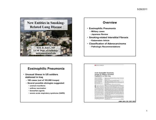
New Entities in Smoking Related Lung Disease
New Entities in Smoking Related Lung Disease
New Entities in Smoking Related Lung Disease

Create successful ePaper yourself
Turn your PDF publications into a flip-book with our unique Google optimized e-Paper software.
<strong>New</strong> <strong>Entities</strong> <strong>in</strong> Smok<strong>in</strong>g-<br />
<strong>Related</strong> <strong>Lung</strong> <strong>Disease</strong><br />
Kirk D. Jones, MD<br />
UCSF Dept. of Pathology<br />
kirk.jones@ucsf.edu<br />
Eos<strong>in</strong>ophilic Pneumonia<br />
• Unusual illness <strong>in</strong> US soldiers<br />
stationed <strong>in</strong> Iraq<br />
– 100 cases (out of 183,000 troops)<br />
– Several possible etiologies suggested:<br />
• uranium munitions<br />
• anthrax vacc<strong>in</strong>ation<br />
• biowarfare agents<br />
• severe acute respiratory syndrome (SARS)<br />
Overview<br />
• Eos<strong>in</strong>ophilic Pneumonia<br />
– Military cases<br />
– Japanese Review<br />
• Smok<strong>in</strong>g-related Interstitial Fibrosis<br />
– Katzenste<strong>in</strong> Article<br />
• Classification of Adenocarc<strong>in</strong>oma<br />
– Pathologic Recommendations<br />
JAMA 2004; 292: 2997-3005<br />
5/28/2011<br />
1
• 18 cases <strong>in</strong> series<br />
• All smokers<br />
– 14 recently started<br />
Iraq Cases<br />
• All but one exposed to dusts<br />
• 6 had BAL<br />
– 8-42% eos<strong>in</strong>ophils<br />
• 12 mechanically ventilated<br />
• 2 died<br />
5/28/2011<br />
2
The Military Experience<br />
• Analysis of 15 different brands of<br />
tobacco available <strong>in</strong> the theater of<br />
operation revealed no unusual<br />
components, tox<strong>in</strong>s, pesticide residues<br />
or significant microbial colonization.<br />
CHEST 2008; 133: 1174-1180<br />
Diagnosis of Eos<strong>in</strong>ophilic<br />
Pneumonia<br />
• Triad of f<strong>in</strong>d<strong>in</strong>gs with<strong>in</strong> airspaces<br />
– Macrophages (often mult<strong>in</strong>ucleate)<br />
– Fibr<strong>in</strong><br />
– Eos<strong>in</strong>ophils<br />
• Often, the clue will be the macrophages<br />
rather than the eos<strong>in</strong>ophils<br />
• Be aware of steroid adm<strong>in</strong>istration<br />
– Hypereos<strong>in</strong>ophilic, granular fibr<strong>in</strong><br />
5/28/2011<br />
3
Overlap with other diseases<br />
• Smok<strong>in</strong>g related diseases<br />
– Desquamative <strong>in</strong>terstitial pneumonia<br />
• Smoker’s macrophages with<strong>in</strong> airspaces<br />
• Clues on the CT scan<br />
– Pulmonary Langerhans cell histiocytosis<br />
• Bronchiolocentric disease<br />
• Cysts and nodules on CT<br />
• Langerhans cell <strong>in</strong>filtrate<br />
• Fibr<strong>in</strong>ous organiz<strong>in</strong>g pneumonia<br />
• Plugs of fibr<strong>in</strong>, but lacks eos<strong>in</strong>ophils<br />
5/28/2011<br />
4
5/28/2011<br />
5
Conclusions on EP<br />
• Smok<strong>in</strong>g can be added to the list of<br />
causes of eos<strong>in</strong>ophilic pneumonia<br />
– Start<strong>in</strong>g smok<strong>in</strong>g<br />
– Increas<strong>in</strong>g frequency of cigarettes<br />
– Restart<strong>in</strong>g smok<strong>in</strong>g<br />
– Chang<strong>in</strong>g brands<br />
• Infection (e.g. Coccidioides), Drug<br />
reaction, Churg-Strauss, idiopathic<br />
Smok<strong>in</strong>g-related <strong>in</strong>terstitial<br />
fibrosis (SRIF)<br />
• Emphysema the textbook<br />
– “abnormal permanent enlargement of the<br />
airspaces distal to the term<strong>in</strong>al bronchiole,<br />
accompanied by destruction of their walls,<br />
and without obvious fibrosis”<br />
• Emphysema as experienced <strong>in</strong> reality<br />
– Often with associated fibrosis<br />
• Bronchiolocentric (as <strong>in</strong> respiratory bronchiolitis)<br />
• Patchy<br />
• Peripheral<br />
5/28/2011<br />
6
Human Pathology 2010; 41: 316-325<br />
SRIF – Patterns of Fibrosis<br />
• Variable widen<strong>in</strong>g of the alveolar septa<br />
by dense, relatively acellular collagen<br />
– Patchy <strong>in</strong> distribution<br />
– Accentuated subpleurally and around<br />
bronchioles<br />
– Associated respiratory bronchiolitis<br />
– May resemble “burnt out LCH”<br />
• Stellate bronchiolocentric scars<br />
Smok<strong>in</strong>g <strong>Related</strong> Interstitial<br />
Fibrosis (SRIF)<br />
• Exam<strong>in</strong>ed 23 lobectomy specimens<br />
– Excised for primary pulmonary neoplasms<br />
• 10 AD, 6 SQ, 1 ADSQ, 2 SCC, 1 LCNEC, 2 TC, 1 AC<br />
• 3 nonsmokers<br />
– 27 sections per lobe<br />
– 12 cases with significant fibrosis (all smokers)<br />
• 1 case: Usual <strong>in</strong>terstitial pneumonia pattern<br />
• 1 case: Asbestosis<br />
• 1 case: Langerhans cell histiocytosis<br />
• 9 cases: Termed SRIF<br />
5/28/2011<br />
7
Histopathology 2008, 53: 707-714<br />
5/28/2011<br />
8
SRIF - Conclusions<br />
• Smok<strong>in</strong>g can result <strong>in</strong> a surpris<strong>in</strong>g<br />
variety of fibrotic patterns.<br />
• Important to correlate fibrotic cases<br />
with cl<strong>in</strong>ical and radiologic data.<br />
– Particularly if fibroblast foci and<br />
honeycomb<strong>in</strong>g are present.<br />
• A new name for a common f<strong>in</strong>d<strong>in</strong>g<br />
– “Irregular emphysema”<br />
– “Airspace enlargement with fibrosis”<br />
Airspace Enlargement with Fibrosis<br />
• AEF def<strong>in</strong>ed as:<br />
– Fibrous (frequently hyal<strong>in</strong>ized) <strong>in</strong>terstitium with<br />
structural remodel<strong>in</strong>g<br />
– Emphysematous changes<br />
– Frequent bronchiolocentric location<br />
– Absence of fibroblast foci<br />
• AEF is associated with smok<strong>in</strong>g<br />
– May be observed <strong>in</strong> some occupational<br />
exposures/pneumoconioses<br />
• Can be associated with a UIP pattern<br />
Classification of <strong>Lung</strong><br />
Adenocarc<strong>in</strong>oma<br />
• International multidiscipl<strong>in</strong>ary effort by:<br />
– IASLC: International Association for the<br />
Study of <strong>Lung</strong> Cancer<br />
– ATS: American Thoracic Society<br />
– ERS: European Respiratory Society<br />
• Representatives from thoracic<br />
oncology, pulmonology, radiology,<br />
molecular biology, thoracic surgery,<br />
and pathology<br />
5/28/2011<br />
9
Journal of Thoracic Oncology 2011; 6: 244-285<br />
Pathology Recommendation 1<br />
• “We recommend discont<strong>in</strong>u<strong>in</strong>g the use<br />
of the term “BAC”<br />
– Five situations where it is used:<br />
• Current WHO def<strong>in</strong>ition (lacks <strong>in</strong>vasion)<br />
• Lesions with small regions of <strong>in</strong>vasion<br />
• Lesions with predom<strong>in</strong>ant surface growth but<br />
central <strong>in</strong>vasive component<br />
• Lesions with prom<strong>in</strong>ent <strong>in</strong>vasive component<br />
and peripheral alveolar surface growth<br />
• In muc<strong>in</strong>ous tumors (with <strong>in</strong>vasion)<br />
Classification: Summary<br />
• Elim<strong>in</strong>ate bronchioloalveolar carc<strong>in</strong>oma<br />
• Def<strong>in</strong>e adenocarc<strong>in</strong>oma <strong>in</strong> situ<br />
• Def<strong>in</strong>e m<strong>in</strong>imally <strong>in</strong>vasive adenocarc<strong>in</strong>oma<br />
• Resurrect the term “lepidic”<br />
• Promote comprehensive histologic subtyp<strong>in</strong>g<br />
• Emphasize micropapillary carc<strong>in</strong>oma<br />
• Detach muc<strong>in</strong>ous adenocarc<strong>in</strong>omas<br />
• Discourage term NSCLC – subclassify if possible<br />
Journal of Thoracic Oncology. 6(2):244-285, February 2011.<br />
5/28/2011<br />
10
Pathology Recommendations 2/3<br />
• Small (≤3 cm) solitary<br />
adenocarc<strong>in</strong>omas with pure lepidic<br />
growth termed adenocarc<strong>in</strong>oma <strong>in</strong> situ.<br />
• Small (≤3 cm) solitary<br />
adenocarc<strong>in</strong>omas with predom<strong>in</strong>ant<br />
lepidic growth and foci of <strong>in</strong>vasion<br />
measur<strong>in</strong>g ≤ 0.5 cm termed m<strong>in</strong>imally<br />
<strong>in</strong>vasive adenocarc<strong>in</strong>oma.<br />
Lepidic Growth<br />
• Ma<strong>in</strong>ta<strong>in</strong>s alveolar architecture<br />
– No destruction or effacement<br />
• No central or broad scar<br />
• Often has thickened alveolar septa<br />
• Cuboidal epithelium<br />
• Little to no stratification or tuft<strong>in</strong>g<br />
• No papillary structures<br />
5/28/2011<br />
11
From Whence “Lepidic”<br />
• J. George Adami, Pr<strong>in</strong>ciples<br />
of Pathology, 1908<br />
– Novel classification of<br />
cancers:<br />
• Lepidic: Tumors derived from<br />
“l<strong>in</strong><strong>in</strong>g membranes”<br />
– From “λεπιδοσ” mean<strong>in</strong>g scale.<br />
• Hylic: Tumors derived from<br />
“pulps”<br />
– From “ύλη” mean<strong>in</strong>g crude<br />
undifferentiated material<br />
5/28/2011<br />
12
From Whence “Lepidic”<br />
• 1962: H. Spencer, Pathology of the <strong>Lung</strong><br />
– “Malignant pulmonary adenomatosis show<strong>in</strong>g the<br />
lepidic nature of the tumour cells.”<br />
– “In the more rapidly grow<strong>in</strong>g and anaplastic<br />
tumours the cells may grow <strong>in</strong> a hylic fashion.”<br />
– “Other cells grow out <strong>in</strong>to the surround<strong>in</strong>g alveoli<br />
either fill<strong>in</strong>g them with a solid mass of malignant<br />
cells (a hilic growth) or l<strong>in</strong><strong>in</strong>g their walls (a lepidic<br />
growth)”.<br />
While it is tempt<strong>in</strong>g to<br />
use me as a visual<br />
metaphor, I have noth<strong>in</strong>g<br />
to do with the term<br />
lepidic other than our<br />
common Lat<strong>in</strong> root.<br />
Lepidic – the temptation<br />
• Lepidic = scale-like<br />
• Lepidoptera = scale w<strong>in</strong>g<br />
• BAC: Aerogenous spread, rests on<br />
alveolar septa<br />
• Butterfly: Aerogenous travel, rests on<br />
grasses and flowers<br />
Judg<strong>in</strong>g Invasion<br />
• Several features may be used to diagnose<br />
regions of <strong>in</strong>vasion; however, this can<br />
occasionally be difficult<br />
• Broad regions of scarr<strong>in</strong>g/central scar<br />
– Not simply alveolar wall thicken<strong>in</strong>g<br />
• Abnormal gland architecture<br />
– Odd alveolar shapes, lack of airspace<br />
macrophages<br />
• Blood or lymphatic vascular, pleural <strong>in</strong>vasion<br />
• Architectural patterns which denote <strong>in</strong>vasion<br />
5/28/2011<br />
13
Patterns which denote <strong>in</strong>vasion<br />
• Solid with muc<strong>in</strong><br />
– Includ<strong>in</strong>g clear cell, signet r<strong>in</strong>g<br />
• Papillary<br />
• Ac<strong>in</strong>ar<br />
• Micropapillary (Pathology recommendation 7)<br />
– Prognostic data<br />
5/28/2011<br />
14
Pathology Recommendation 4/5<br />
• Comprehensive histologic subtyp<strong>in</strong>g used<br />
to assess patterns semiquantitatively <strong>in</strong><br />
5% <strong>in</strong>crements, choos<strong>in</strong>g a s<strong>in</strong>gle<br />
predom<strong>in</strong>ant pattern.<br />
• In multiple lung adenocarc<strong>in</strong>omas,<br />
comprehensive histologic subtyp<strong>in</strong>g may<br />
aid <strong>in</strong> determ<strong>in</strong><strong>in</strong>g whether tumors are<br />
metastases or separate primaries.<br />
Lesions larger than 3cm?<br />
• The data <strong>in</strong> the IASLC analysis were<br />
for tumors less than 2 or 3 cm.<br />
• If greater than 3 cm<br />
– “Lepidic predom<strong>in</strong>ant adenocarc<strong>in</strong>oma,<br />
suspect AIS or MIA.”<br />
– “Lepidic predom<strong>in</strong>ant adenocarc<strong>in</strong>oma.”<br />
Am J Surg Pathol. 2009 Dec;33(12):1752-64.<br />
5/28/2011<br />
15
Pathology Recommendation 6<br />
• Tumors with a predom<strong>in</strong>ant surface<br />
alveolar growth pattern and more than<br />
5 mm <strong>in</strong>vasion (but still m<strong>in</strong>or<br />
component) should be termed:<br />
– Lepidic predom<strong>in</strong>ant adenocarc<strong>in</strong>oma<br />
– “Mixed subtype” should be discont<strong>in</strong>ued<br />
Pathology Recommendation 8<br />
• Former muc<strong>in</strong>ous BAC will be termed<br />
muc<strong>in</strong>ous AIS, muc<strong>in</strong>ous MIA, or<br />
<strong>in</strong>vasive muc<strong>in</strong>ous adenocarc<strong>in</strong>oma<br />
5/28/2011<br />
16
Pathology Recommendation 9/10<br />
• NSCLC be classified <strong>in</strong>to more specific<br />
histologic type whenever possible<br />
• NSCLC-NOS should be used as little as<br />
possible<br />
Why Does This Matter?<br />
• AIS and MIA, as def<strong>in</strong>ed, have a much<br />
better prognosis when compared to<br />
<strong>in</strong>vasive tumors of the same size.<br />
• Some histologic subtypes have<br />
prognostic significance.<br />
– Lepidic > Ac<strong>in</strong>ar / Papillary > Solid / Micropapillary<br />
5/28/2011<br />
17
How to <strong>in</strong>corporate <strong>in</strong>to practice<br />
• Tumor size for stag<strong>in</strong>g<br />
– If not lepidic pattern – use gross<br />
measurement or measure off slide.<br />
– If lepidic pattern:<br />
– I currently report both <strong>in</strong>vasive and total<br />
diameter. I stage accord<strong>in</strong>g to <strong>in</strong>vasive<br />
regions when tumor less than 3 cm (1a vs.<br />
1b)<br />
How to <strong>in</strong>corporate <strong>in</strong>to practice<br />
• Histologic typ<strong>in</strong>g<br />
– I am a partial fan of comprensive<br />
histologic typ<strong>in</strong>g. I f<strong>in</strong>d it useful for<br />
determ<strong>in</strong><strong>in</strong>g met vs. multiple primary, but I<br />
don’t like us<strong>in</strong>g 5% <strong>in</strong>cremental diagnosis.<br />
– Diagnose adenocarc<strong>in</strong>oma, then list the<br />
subtypes <strong>in</strong> the comment.<br />
How to <strong>in</strong>corporate <strong>in</strong>to practice<br />
• Tumor size for stag<strong>in</strong>g<br />
– If not lepidic pattern – use gross<br />
measurement or measure off slide.<br />
– If lepidic pattern:<br />
• Less than 3 cm: I currently report both <strong>in</strong>vasive<br />
and total diameter. I stage accord<strong>in</strong>g to<br />
<strong>in</strong>vasive regions (1a vs. 1b).<br />
• Greater than 3 cm report as usual (for now).<br />
<strong>New</strong>: Lepidic predom<strong>in</strong>ant adenocarc<strong>in</strong>oma.<br />
Old: Adenocarc<strong>in</strong>oma, mixed bronchioloalveolar, papillary and ac<strong>in</strong>ar type.<br />
Possible: Adenocarc<strong>in</strong>oma, see comment.<br />
5/28/2011<br />
18
5/28/2011<br />
19