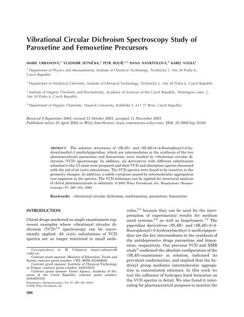
Vibrational Circular Dichroism Spectroscopy Study of ... - Quick.cz
Vibrational Circular Dichroism Spectroscopy Study of ... - Quick.cz
Vibrational Circular Dichroism Spectroscopy Study of ... - Quick.cz

Create successful ePaper yourself
Turn your PDF publications into a flip-book with our unique Google optimized e-Paper software.
<strong>Vibrational</strong> <strong>Circular</strong> <strong>Dichroism</strong> <strong>Spectroscopy</strong> <strong>Study</strong> <strong>of</strong><br />
Paroxetine and Femoxetine Precursors<br />
MARIE URBANOVÁ, 1 VLADIMÍR SETNIČKA, 2 PETR BOURˇ , 2,3 HANA NAVRÁTILOVÁ, 4 KAREL VOLKA 2<br />
1<br />
Department <strong>of</strong> Physics and Measurements, Institute <strong>of</strong> Chemical Technology, Technická 5, 166 28 Praha 6,<br />
Czech Republic<br />
2 Department <strong>of</strong> Analytical Chemistry, Institute <strong>of</strong> Chemical Technology, Technická 5, 166 28 Praha 6, Czech Republic<br />
3<br />
Institute <strong>of</strong> Organic Chemistry and Biochemistry, Academy <strong>of</strong> Sciences <strong>of</strong> the Czech Republic, Flemingovo nám. 2,<br />
166 10 Praha 6, Czech Republic<br />
4 Department <strong>of</strong> Organic Chemistry, Masaryk University, Kotlárˇská 2, 611 37 Brno, Czech Republic<br />
Received 8 September 2001; revised 31 October 2001; accepted 11 November 2001<br />
Published online 25 April 2002 in Wiley InterScience (www.interscience.wiley.com). DOI: 10.1002/bip.10104<br />
INTRODUCTION<br />
ABSTRACT: The solution structures <strong>of</strong> (3R,4S)- and (3S,4R)-4-(4-fluorophenyl)-3-hydroxylmethyl-1-methylpiperidine,<br />
which are intermediates in the synthesis <strong>of</strong> the two<br />
pharmaceuticals paroxetine and femoxetine, were studied by vibrational circular dichroism<br />
(VCD) spectroscopy. In addition, six derivatives with different substituents<br />
attached to the C3 atom were prepared and their VCD and absorption spectra discussed<br />
with the aid <strong>of</strong> ab initio simulations. The VCD spectra were found to be sensitive to the<br />
geometry changes. In addition, a subtle variation caused by intermolecular aggregation<br />
was apparent in the spectra. The VCD technique can be applied for structural analysis<br />
<strong>of</strong> chiral pharmaceuticals in solutions. © 2002 Wiley Periodicals, Inc. Biopolymers (Biospectroscopy)<br />
67: 298–301, 2002<br />
Keywords: vibrational circular dichroism; conformation; paroxetine; femoxetine<br />
Chiral drugs marketed as single enantiomers represent<br />
examples where vibrational circular dichroism<br />
(VCD) 1,2 spectroscopy can be conveniently<br />
applied. Ab initio calculations <strong>of</strong> VCD<br />
spectra are no longer restricted to small mole-<br />
Correspondence to: M. Urbanová (marie.urbanova@<br />
vscht.<strong>cz</strong>).<br />
Contract grant sponsor: Ministry <strong>of</strong> Education, Youth and<br />
Sports; contract grant number: CEZ: MSM 223400008.<br />
Contract grant sponsor: Institute <strong>of</strong> Chemical Technology<br />
at Prague; contract grant number: 444010015.<br />
Contract grant sponsor: Grant Agency, Academy <strong>of</strong> Sciences<br />
<strong>of</strong> the Czech Republic; contract grant number:<br />
IAA4055104.<br />
Biopolymers (Biospectroscopy), Vol. 67, 298–301 (2002)<br />
© 2002 Wiley Periodicals, Inc.<br />
298<br />
cules, 3,4 because they can be used for the interpretation<br />
<strong>of</strong> experimental results for medium<br />
sized systems, 5,6 as well as biopolymers. 7,8 The<br />
piperidine derivatives (3S,4R)- and (3R,4S)-4-(4fluorophenyl)-3-hydroxylmethyl-1-methylpiperidine<br />
are the key intermediates in the synthesis <strong>of</strong><br />
the antidepressive drugs paroxetine and femoxetine,<br />
respectively. Our previous VCD and NMR<br />
study 5 confirmed the absolute configuration <strong>of</strong> the<br />
(3R,4S)-enantiomer in solution, indicated its<br />
prevalent conformation, and implied that the hydroxyl<br />
group mediates intermolecular aggregation<br />
in concentrated solutions. In this work we<br />
test the influence <strong>of</strong> hydrogen bond formation on<br />
the VCD spectra in detail. We also found it interesting<br />
for pharmaceutical purposes to monitor the
Figure 1. The structures <strong>of</strong> the (3S,4R)-4-(4-fluorophenyl)-3-hydroxylmethyl-1-methylpiperidinederivatives.<br />
substitutions <strong>of</strong> the hydroxyl group by other functional<br />
groups and to follow possible conformation<br />
changes using VCD.<br />
MATERIALS AND METHODS<br />
Samples (Fig. 1) <strong>of</strong> (3S,4R)-3 (100% ee) 9 and<br />
(3R,4S)-3 (96% ee) were kind gifts from Synthon<br />
BV, The Netherlands. Compounds 4–8 were prepared<br />
as described elsewhere. 10,11 The solutions<br />
in CCl 4 and CDCl 3 within 0.64–0.064 mol L 1<br />
were used for the VCD measurements. The VCD<br />
and absorption spectra were scanned with a resolution<br />
<strong>of</strong> 4 cm 1 using a Bruker FTIR IFS 66/S<br />
spectrometer equipped with the VCD/IRRAS<br />
module PMA 37 as previously described in detail.<br />
12 A demountable cell separated by a 50- or<br />
200-m Teflon spacer was used.<br />
A simulation <strong>of</strong> the VCD intensities based on<br />
the Gaussian program package 13 was performed<br />
6,7 for geometries optimized with the aid <strong>of</strong><br />
the conformer searching routine implemented in<br />
the Spartan program. 14 The vibrational frequencies<br />
and intensities were calculated at the<br />
BPW91/6-31G** density functional theory 15<br />
level at the harmonic approximation using the<br />
MFP/GIAO theory 16 for VCD. The spectra were<br />
simulated using Lorentzian pr<strong>of</strong>ileswitha5cm 1<br />
bandwidth. Normal mode assignment is based on<br />
a visual inspection <strong>of</strong> the dynamic displacements.<br />
VCD STUDY OF PAROXETINE AND FEMOXETINE 299<br />
RESULTS AND DISCUSSION<br />
Figure 2 shows the experimental VCD spectra <strong>of</strong><br />
derivatives 3, 4, and 8 defined in Figure 1. All<br />
spectra were recorded with a high S/N ratio. Recording<br />
<strong>of</strong> the VCD spectra <strong>of</strong> (3R,4S)-3 and<br />
(3S,4R)-3 reduces the risk <strong>of</strong> artifacts and confirms<br />
the quality <strong>of</strong> the experimental data: the<br />
opposite enantiomers exhibit mirror-image VCD<br />
spectra as shown for (3S,4R)-3 and (3R,4S)-3 (Fig.<br />
2). For the sake <strong>of</strong> brevity, we deal only with the<br />
(3R,4S) enantiomers in the following text.<br />
As an example, the agreement between the<br />
experiment and calculation is shown for derivative<br />
7 in Figure 3. The numbers indicate the corresponding<br />
vibrational modes. Such a comparison<br />
<strong>of</strong> the absorption and VCD spectra (Fig. 2) for all<br />
the derivatives reveals the common features summarized<br />
in Table I. The mode numbers <strong>of</strong> compound<br />
3 are used also for the other compounds,<br />
because the ordering <strong>of</strong> the normal modes differs<br />
only slightly. Comparing the VCD spectra in Figure<br />
2, we find that the individual derivatives differ<br />
in certain regions where the vibration modes<br />
involve the chiral centers C3 and C4 and the<br />
atoms in their near vicinity (modes 33–39, 47–49,<br />
and 58–59).<br />
The VCD measurements require rather high<br />
concentrations <strong>of</strong> the compounds and enable only<br />
a narrow interval for concentration variations. In<br />
our case we recorded the VCD <strong>of</strong> 3 in CDCl 3 and<br />
CCl 4 solutions with a reasonable S/N ratio in the<br />
range <strong>of</strong> 0.64–0.064 mol L 1 [Fig. 4(B)]. The observed<br />
decrease <strong>of</strong> the absorption <strong>of</strong> the free OH<br />
Figure 2. The experimental VCD spectra <strong>of</strong> derivatives<br />
3, 4, and 8 (0.64 mol L 1 in CDCl 3) and the typical<br />
noise spectrum (N).
300 URBANOVÁ ET AL.<br />
Figure 3. The (A) VCD and (B) absorption spectra <strong>of</strong> compound 7. The experimental<br />
spectra <strong>of</strong> the CDCl 3 solution (c 0.64 mol L 1 ; spectra a) and the simulated spectra<br />
(spectra b).<br />
group at 3627 cm 1 and its increase at 3350–3150<br />
cm 1 for aggregated OH groups with increasing<br />
concentration [Fig. 4(A)] confirmed the hydrogen<br />
bond formation. Although the intensity <strong>of</strong> the free<br />
(OH) increases about 2 times as the concentration<br />
decreases from 0.64 to 0.064 mol L 1 , only<br />
slight changes were observed in the VCD in the<br />
mid-IR region (cf. Fig. 4). This is in accord with<br />
our detailed analysis, 5 which revealed that only<br />
limited parts <strong>of</strong> the spectral region used are affected<br />
by the OH vibrations, for example, the<br />
VCD features at 1250–1200 and 1000 cm 1 (the<br />
gray areas in Fig. 4), which originate in skeletal<br />
deformation coupled to or including the COO<br />
bond. Also, based on the concentration dependence<br />
<strong>of</strong> the VCD spectra, we can conclude that<br />
the aggregation does not have a significant influence<br />
on the conformation.<br />
Table I. VCD and Absorption Bands Common to All Derivatives<br />
Mode<br />
VCD and Absorption Frequencies (cm 1 )<br />
Experimental Calculated<br />
CONCLUSIONS<br />
The simulated and experimental spectra are in a<br />
very good agreement over the entire region <strong>of</strong><br />
recorded frequencies. The observed variations in<br />
the regions specific to the particular substituents<br />
could be identified and reasonably explained using<br />
the normal mode assignment. In all <strong>of</strong> the<br />
derivatives studied, the substituents do not significantly<br />
influence the spectral response <strong>of</strong> the<br />
piperidine skeleton. The theoretical and experimental<br />
results suggest that the phenyl and<br />
methyl groups are in equatorial positions and the<br />
torsion angle C(substituent)-C3-C4-C(piperidine)<br />
is close to 180° (except 8). The concentration dependence<br />
<strong>of</strong> the VCD and absorption reveals<br />
strong intermolecular interactions through hy-<br />
Assignment<br />
19–21 1604, 1510 1604, 1496–1498 Phenyl deformation<br />
27 1439–1440 1437–1447 CH bend (in neighborhood <strong>of</strong> C3)<br />
30 1379–1384 1369–1372 CH bend on phenyl<br />
41 1281–1289 1263–1271 CH 2 twist and CH 3 deformation<br />
43 1221–1225 1211–1214 CF and phenyl deformation<br />
52 1096 1092–1097 No specific skeletal vibration<br />
55 1060–1070 1052–1060 Skeletal vibration including CO
Figure 4. The concentration dependence <strong>of</strong> (A) the<br />
normalized absorption spectra in the (OH) region and<br />
(B) the normalized VCD spectra <strong>of</strong> compound 3 in<br />
CDCl 3 at concentrations <strong>of</strong> 0.64 (spectrum a), 0.128<br />
(spectrum b), and 0.064 mol L 1 (spectrum c) and the<br />
noise (N) spectrum for a concentration <strong>of</strong> 0.128 mol<br />
L 1 .<br />
drogen bridges with a minor effect on the molecular<br />
conformation.<br />
REFERENCES<br />
1. Keiderling, T. A. In Practical Fourier Transform<br />
Infrared <strong>Spectroscopy</strong>; Ferraro, J. R., Krishnan, K.,<br />
Eds.; Academic: San Diego, CA, 1990; pp 203–284.<br />
2. Nafie, L. A.; Freedman, T. B. In <strong>Circular</strong> <strong>Dichroism</strong>:<br />
Principles and Applications; Berova, N., Nakanishi,<br />
K., Eds.; Wiley: New York, 2000; pp 97–132.<br />
VCD STUDY OF PAROXETINE AND FEMOXETINE 301<br />
3. Bourˇ, P.; McCann, J.; Wieser, H. J Phys Chem A<br />
1998, 102, 102–110.<br />
4. Tam, C. N.; Bourˇ, P.; Keiderling, T. A. J Am Chem<br />
Soc 1996, 118, 10285–10293.<br />
5. Bourˇ, P.; Navrátilová, H.; Setnička, V.; Urbanová,<br />
M.; Volka, K., J Org Chem 2002, 67, 161–168.<br />
6. Setnička, V.; Urbanová, M.; Bourˇ, P.; Král, V.;<br />
Volka, K. J Phys Chem A 2001, 105.<br />
7. Bourˇ, P.; Záruba, K.; Urbanová, M.; Setnička, V.;<br />
Matéjka, P.; Fiedler, Z.; Volka, K. Chirality 2000,<br />
12, 191–198.<br />
8. Silva, R. A. G. D.; Kubelka, J.; Bourˇ, P.; Decatur,<br />
S. M.; Keiderling, T. A. Proc Natl Acad Sci USA<br />
2000, 97, 8318–8323.<br />
9. Christensen, J. A.; Squires, R. F. U.S. Pat.<br />
4,007,196, 1977.<br />
10. Navrátilová, H. Ph.D. Thesis, Masaryk University,<br />
2001.<br />
11. Navrátilová, H. Chirality 2001, 13.<br />
12. Urbanová, M.; Setnička, V.; Volka, K. Chirality<br />
2000, 12, 199–203.<br />
13. Frisch, M. J.; Trucks, G. W.; Schlegel, H. B.; Scuseria,<br />
G. E.; Robb, M. A.; Cheeseman, J. R.; Zakrzewski,<br />
V. G.; Montgomery, J. A.; Stratmann, R. E.;<br />
Burant, J. C.; Dapprich, S.; Millam, J. M.; Daniels,<br />
A. D.; Kudin, K. N.; Strain, M. C.; Farkas, O.;<br />
Tomasi, J.; Barone, V.; Cossi, M.; Cammi, R.; Mennucci,<br />
B.; Pomelli, C.; Adamo, C.; Clifford, S.;<br />
Ochterski, J.; Petersson, G. A.; Ayala, P. Y.; Cui,<br />
Q.; Morokuma, K.; Malick, D. K.; Rabuck, A. D.;<br />
Raghavachari, K.; Foresman, J. B.; Cioslowski, J.;<br />
Ortiz, J. V.; Stefanov, B. B.; Liu, G.; Liashenko, A.;<br />
Piskorz, P.; Komaromi, I.; Gomperts, R.; Martin,<br />
R. L.; Fox, D. J.; Keith, T.; Al-Laham, M. A.; Peng,<br />
C. Y.; Nanayakkara, A.; Gonzalez, C.; Challacombe,<br />
M.; Gill, P. M. W.; Johnson, B.; Chen, W.;<br />
Wong, M. W.; Andres, J. L.; Gonzalez, C.; Head-<br />
Gordon, M.; Replogle, E. S.; Pople, J. A. Gaussian<br />
98. Revision A.3 and A.7; Gaussian Inc.: Pittsburgh,<br />
PA, 1998.<br />
14. Spartan, P. C. PC Spartan Pro 2000, Pro 1.0.5;<br />
Wavefunction Inc.: Irvine, CA, 2000.<br />
15. Becke, A. D. J Chem Phys 1993, 98, 5648–5652.<br />
16. Devlin, F. J.; Stephens, P. J.; Cheeseman, J. R.;<br />
Frisch, M. J. J Phys Chem A 1997, 101, 9912–<br />
9924.