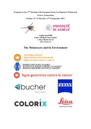
espcrbulletin - European Society for Pigment Cell Research
espcrbulletin - European Society for Pigment Cell Research
espcrbulletin - European Society for Pigment Cell Research

Create successful ePaper yourself
Turn your PDF publications into a flip-book with our unique Google optimized e-Paper software.
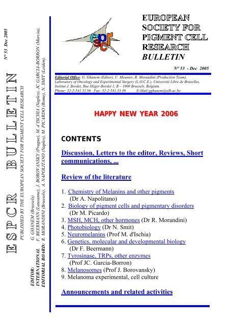
N° 53 Dec 2005<br />
E S P C R B U L L E T I N<br />
PUBLISHED BY THE EUROPEAN SOCIETY FOR PIGMENT CELL RESEARCH<br />
EDITOR: G. GHANEM (Brussels)<br />
INTERNATIONAL F. BEERMANN (Lausanne), J. BOROVANSKY (Prague), M. d’ISCHIA (Naples), JC GARCIA-BORRON (Murcia),<br />
EDITORIAL BOARD: R. MORANDINI (Brussels), A. NAPOLITANO (Naples), M. PICARDO (Rome), N. SMIT (Leiden).<br />
E<br />
S<br />
P<br />
R<br />
BULLETIN<br />
N° 53 - Dec 2005<br />
Editorial Office: G. Ghanem (Editor), C. Meunier, R. Morandini (Production Team),<br />
Laboratory of Oncology and Experimental Surgery (L.O.C.E.), Université Libre de Bruxelles,<br />
Institut J. Bordet, Rue Héger-Bordet 1, B – 1000 Brussels, Belgium.<br />
Phone: 32-2-541.32.96 Fax: 32-2-541.33.49 E-Mail:gghanem@ulb.ac.be<br />
CONTENTS<br />
HAPPY NEW YEAR 2006<br />
Discussion, Letters to the editor, Reviews, Short<br />
communications, ...<br />
Review of the literature<br />
1. Chemistry of Melanins and other pigments<br />
(Dr A. Napolitano)<br />
2. Biology of pigment cells and pigmentary disorders<br />
(Dr M. Picardo)<br />
3. MSH, MCH, other hormones (Dr R. Morandini)<br />
4. Photobiology (Dr N. Smit)<br />
5. Neuromelanins (Prof M. d'Ischia)<br />
6. Genetics, molecular and developmental biology<br />
(Dr F. Beermann)<br />
7. Tyrosinase, TRPs, other enzymes<br />
(Prof JC. Garcia-Borron)<br />
8. Melanosomes (Prof J. Borovansky)<br />
9. Melanoma experimental, cell culture<br />
Announcements and related activities
LETTER TO THE EDITOR<br />
DISCUSSION, REVIEW,<br />
SHORT COMMUNICATION, ...<br />
A Jubilee Year <strong>for</strong> Two Famous Scientists and<br />
Distinguished ESPCR Members<br />
By Jan Borovanský<br />
This year two legends of the ESPCR have reached important anniversaries. They both<br />
originated from families with medical connections and graduated as M.D. They both were born in a<br />
different country from that where they permanently live nowadays. One of them started his career as a<br />
dermatologist to become top scientist in the field of pigment cell research and cell pathology, the other<br />
at first was a biochemist interested in<br />
melanogenesis and melanogens to<br />
become first class dermatologist and<br />
photobiologist. They share both a<br />
common interest in the toxicity of<br />
melanin precursors and a great hobby<br />
in per<strong>for</strong>ming music. As students they<br />
both played in the collegiate jazz<br />
bands as a clarinet and alto saxophone<br />
and clarinet players, respectively.<br />
They share a common weakness <strong>for</strong><br />
the piano and both of them are my<br />
best friends. I introduced them to each<br />
other on December 5, 1984.<br />
Professor Patrick Anthony Riley, M.B., B.S., Ph.D., D.Sc., F.I. Biol., F.R.C. Pathol. Was born on<br />
22nd March 1935 in Neuilly-sur-Seine, France. His father Bertrand Hurrell Riley belonged to founders<br />
of the World Health Organization and spent many years in Geneva. Patrick Riley visited him regularly<br />
during holidays because he studied at a boarding school in the U.K. From childhood he has had a deep<br />
fascination <strong>for</strong> the way things work which can be illustrated e.g. by a set of magnets and one-penny<br />
coins he developed much later in his adulthood <strong>for</strong> a commercial purpose to teach children the<br />
phenomenon of magnetism in a playful way. He has always been interested in and fascinated by<br />
mechanisms of biological phenomena and his preoccupation in living cells was deepened and<br />
„saturated“ by many prominent teachers, scientists and friends, especially at the University College<br />
London, which was a powerhouse of biological learning. During medical studies he met his charming<br />
wife and life-long supporter, Christine. After graduation from University College Hospital Medical<br />
School (UCHMS) in 1960 he spent a half year as a house-physician in the Department of<br />
Dermatology and later, having obtained a Rockefeller scholarship, he joined Dr. Arthur Jarrett in the<br />
Department of Dermatological Histopathology at UCHMS where he started his research activities by<br />
studying various enzyme activities in dendritic epidermal cells. He was awarded a Medical <strong>Research</strong><br />
Council Junior Clinical <strong>Research</strong> Fellowship and pursued studies of epidermal depigmentation and the<br />
relationship between melanocytes and Langerhans cells. In 1965 he obtained his Ph.D. having<br />
defended a thesis entitled Studies of Melanocyte Function and co-authored a textbook of dermatology.<br />
In 1966 he was awarded a Beit Memorial Fellowship and moved to the Department of Pathological<br />
Chemistry where he shared an office with Trevor Slater, a brilliant and enthusiastic biochemist and<br />
advocate of free radical reactions. I suppose that this famous pioneer of free radical research ignited in<br />
Patrick Riley his permanent fascination <strong>for</strong> free radical reactions <strong>for</strong> in the same year the seminal<br />
1403
paper on photosensitization and lysosomal damage, prepared in cooperation with Professor Trevor<br />
Slater, appeared in Nature. In the succeeding period Patrick Riley published a series of depigmentation<br />
studies with 4-hydroxyanisole with the aim of utilizing its selective melanocytotoxicity to treat<br />
advanced melanoma. After many years research ef<strong>for</strong>t this goal was reached: In 1991 a paper<br />
describing results of a preliminary clinical trial with hydroxyanisole per<strong>for</strong>med by a surgeon and<br />
friend, Brian Morgan, partly confirmed the expectations, although subsequent experience has been<br />
disappointing. When Riley joined the Department of Pathological Chemistry (re-named later the Dept.<br />
of Chemical Pathology and subsequently the Dept. of Molecular Pathology) it was chaired by<br />
Professor Claude Rimington. Here I must repeat a well-known proverb“; The World is small.“ As a<br />
student of medicine I used to work voluntarily (1963-1966) in the laboratory of Professor A.F. Richter<br />
who, like Claude Rimington, was interested in porhyrins and in nineteen fifties they had been close<br />
pen-friends. In 1970s many of Riley’s papers were devoted to the growth of cell populations, to<br />
mechanisms of growth inhibition and functional properties of cells in relation to the cell cycle. He was<br />
also interested in carcinogenesis and mechanisms of DNA transcription. When the British Council<br />
arranged my first meeting with Patrick Riley on March 2, 1978, huge model of double helix was a<br />
dominant object in his office. Although our meeting had been planned <strong>for</strong> half an hour, it lasted a<br />
terribly short four hours because our mutual discussion not only revealed plenty of common scientific<br />
interests but also very similar „blood group“. After 27 years since that meeting I can confirm that it<br />
was one of decisive moments of my life which influenced me <strong>for</strong> ever. Thanks to the British Council<br />
Bursary in 1980 and British Council support <strong>for</strong> Cooperation in Higher Education in 1984 (catalysed<br />
by Patrick) I could spend several months in his lab at the UCL in University Street and in Wellington<br />
Arms (a discussion pub) just opposite the Dept. of Biochemical Pathology building, studying the effect<br />
of zinc and other cations on cultured melanoma and other cells which led to the discovery of the<br />
remarkably high zinc cytotoxicity in vitro. Since that time I have been able to watch closely the<br />
progress of scientific activities in his lab. In 1980s Patrick (with M.Hola) used time-lapse<br />
cinematography to study mitoses in cell cultures and cell genealogy. Later the National Foundation <strong>for</strong><br />
Cancer <strong>Research</strong> provided his lab with image analysis equipment and Patrick (with D. Spargo) set<br />
about trying to devise a quantitative index of cell shape that could be used to generate parameters that<br />
could be exploited as diagnostic criteria. In 1985 Patrick Riley became professor at the University<br />
College and delivered an inaugural lecture “Pathological Migration: >From Melanin to Malignancy“.<br />
In 1991 we published together a paper which emphasized the role of leakage of toxic melanin<br />
precursors from aberrant melanosomes in cytotoxic phenomena in melanoma cells. Patrick´s studies,<br />
together with S. Naish and C.J. Cooksey, revealed participation of quinones, semiquinones and other<br />
radicals in the toxic action of 4-hydroxyanisole. In cooperation with C.J. Cooksey, Stan Pavel, Nico<br />
Smit and others, Patrick continued with studies of the cytotoxicity of a series of substituted phenols<br />
towards cultured melanoma cells and <strong>for</strong>mulated melanogenesis-targeted antimelanoma therapy and,<br />
together with A.M. Jordan and others, the melanocyte-directed prodrug strategy. In 1993 an<br />
international research organization devoted to the quinone chemistry - The Quintox Group - was set up<br />
(with Patrick Riley as its first chairman and with E.J.Land , C.A.Ramsden and C.J.Cooksey as the<br />
most active members) which conducted a range of important studies of melanin biogenesis leading e.g.<br />
to rearrangement of first reactions in Raper-Mason scheme. After his retirement in 2000 Patrick did<br />
not finish his scientific career; on the contrary he founded Totteridge Institute <strong>for</strong> Advanced Studies<br />
and as spiritus movens he has continued to stimulate the research activities of the Quintox Group.<br />
Prof Patrick Riley deserves special thanks <strong>for</strong> what he had done <strong>for</strong> the <strong>European</strong> <strong>Society</strong> <strong>for</strong><br />
<strong>Pigment</strong> <strong>Cell</strong> <strong>Research</strong>. Together with G. Prota and N. Cascinelli he founded the <strong>Society</strong> and acted as<br />
its secretary <strong>for</strong> many years. Since 1995 he has been an honorary member of the ESPCR.<br />
When I was a Ph.D. student and young teacher, there was no internet and I used to visit the<br />
Central Medical Library in Prague to search <strong>for</strong> what´s new in the melanin/melanosome field in the<br />
Current Contents regularly on Friday afternoon. At that occasion I came across Patrick Riley´s name.<br />
His University College London affiliation induced an imagination of an honorable traditional old<br />
1404
monumental building with very serious, perhaps stiff, scientists in my mind. My very first meeting<br />
with Patrick immediately convinced me how wrong I had been. Patrick is typical gentleman constantly<br />
radiating English humour around him. He is a charming companion, a „fountain“ of encyclopedic<br />
knowledge, an excellent speaker and kind friend. Nothing is too much trouble <strong>for</strong> him in supporting<br />
others. It is no surprise that, at scientific meetings, he is always surrounded by many people longing to<br />
speak to him. He influenced me not only scientifically but also in other respects. Thanks to him I could<br />
repeatedly listen to discourses at the Royal Institution; he introduced me to London cultural life and, in<br />
a period when a harsh regime existed in my country, he kept me saturated with <strong>for</strong>bidden Solzenicyn<br />
novels.<br />
Assoc. Prof. Stanislav Pavel, M.D., Ph.D., Ph.D. was born on July 4, 1945 in Prague,<br />
Czechoslovakia. In his younger days he was excellent student and passionate skier (this hobby has<br />
survived till present time and his young colleagues have problems to cope with his speed even now).<br />
He was vacillating whether to study chemistry, medicine or at the academy of music. In 1963 he<br />
decided on medicine (probably under the influence of the family milieu). From the second year of his<br />
medical studies at the Faculty of General Medicine (1st Faculty of Medicine), Charles University,<br />
Prague he joined students´research activities in the field of melanogens and biochemical markers of<br />
melanoma at the 2nd Institute of Medical Chemistry and Biochemistry (renamed later Dept. of<br />
Biochemistry and Experimental Oncology). Having graduated in 1969 he became postgraduate student<br />
supervised by Professor J. Duchon M.D., PhD. and continued in his research. At the same time he was<br />
involved in teaching medical biochemistry. Together with the neurologist, Prof. J. Tichy, M.D., Ph.D.,<br />
he founded the Medical Dixieland band which is well-known to the pigment cell community from its<br />
concerts on the occasions of the the ESPCR Meeting in Amsterdam (1991) and the IPCC in Egmont<br />
aan See (2003).<br />
In 1979 he defended his Ph.D. Thesis „Melanogenesis in hamsters with transplanted<br />
melanomas“ and in 1980 he submitted his Habilitation Thesis that he could not defend: In the summer<br />
of 1981 he was <strong>for</strong>ced to defect to the Netherlands in order to have a clean slate because the secret<br />
police had repeatedly pressed him to become an in<strong>for</strong>mer. He settled in Groningen, where he had been<br />
on a scientific visit couple years be<strong>for</strong>e, and continued his studies concerning metabolic manifestations<br />
of melanogenesis. He was the first to prove the presence of 5,6-dihydroxyindole in biological material<br />
.He demonstrated that melanoma cells excreted O-methylated derivatives of 5,6-dihydroxyindole and<br />
determined the chemical structure of conjugates of 5,6-dihydroxyindole and its O-methylated<br />
derivatives isolated from urine. In 1986 he moved to Amsterdam and was enrolled into specialization<br />
program at the Department of Dermatovenerology to become a clinical dermatologist. In 1988 at the<br />
University of Amsterdam he defended his second PhD thesis „Eumelanin-related compounds, their<br />
metabolism and clinical relevance“. In January 1992 he joined Department of Dermatology at the<br />
University of Leiden as a head of the phototherapy section and director of the Ward section and<br />
responsible <strong>for</strong> allergology. A year later his coworker, Dr Nico Smit, followed him to Leiden and<br />
intensive pigment cell research was launched supported in all the respects by the head of<br />
Dermatological Department of that time Prof. Bert-Jan Vermeer (distant descendant of famous Dutch<br />
painter Vermeer). I was lucky to meet this charming man during my 1993 winter stay in Leiden (and<br />
many times after that), when we and friends from Dept. of Electron Microscopy studied melanosome<br />
architecture in tyrosinase-transfected fibroblasts where melanogenesis takes place in the absence of<br />
melanosomal structural proteins in lysosomes. In Leiden, Dr Pavel has been widely engaged in<br />
dermatological practice, in teaching both medical and postgraduate students and has been in charge of<br />
many national and international grant projects. He has been involved in much of the research leading<br />
to a better understanding of melanogenesis as a potentially toxic process. Nowadays Dr Pavel belongs<br />
amongst prominent Dutch dermatologists with deep chemical and biochemical backgrounds. His<br />
chemical interest has been further deepened by active research and debates in the Quintox group.<br />
1405
The <strong>Pigment</strong> <strong>Cell</strong> Community knows Dr Pavel from his activities in the <strong>European</strong> <strong>Society</strong> <strong>for</strong><br />
<strong>Pigment</strong> <strong>Cell</strong> <strong>Research</strong> Council, in which he was secretary 1994-1997, President 1997-2000 and also<br />
Vice-President of the Federation of <strong>Pigment</strong> <strong>Cell</strong> Societies.<br />
I shared an office in Prague with Stan <strong>for</strong> many years. As we had children of similar age, we<br />
used to take them regularly during the summer holidays to spend some time in a small village in the<br />
hilly southernmost part of Bohemia (leaving our wives in Prague) where, with a glass of red wine in<br />
front of a log fire, we had long discussions about the beauties of life. I greatly miss his presence in our<br />
department, because he has a gregarious nature with a thoughtful attitude to all his colleagues and<br />
students.<br />
I wish Patrick and Stan many more years of productive work and continued good health and<br />
happiness both in their professional and personal life.<br />
ASPCR MEETING REPORT<br />
www.aspcr.org/first.htm<br />
1406
Message from the IFPCS President to ESPCR Members<br />
Dear friends, members of the ESPCR,<br />
I am greatly honored to serve as the President of the IFPCS, and I look <strong>for</strong>ward <strong>for</strong> the next<br />
three years of leading our federation towards bigger successes and further recognition in the world of<br />
scientific research. I begin by expressing my gratitude to the leadership and tremendous ef<strong>for</strong>ts of the<br />
Past-President, Professor Dot Bennett, who invested so much of her time and energy towards<br />
strengthening the ties of the various societies, and facilitating many of the activities of the IFPCS, and<br />
to Professor Yasu Tomita <strong>for</strong> his great service as IFPCS Secretary-Treasurer.<br />
I wish to welcome to the IFPCS the new sister-society, the ASPCR, who undoubtedly will add<br />
a new and important dimension to our federation. Vitiligo is a disease that is as challenging to ASPCR<br />
scientists and clinicians as melanoma is to those of us in Western countries. The more we learn about<br />
the melanocyte, the more knowledge we gain about these two extreme diseases that inflict millions<br />
around the world. I hope all of us will continue to appreciate the importance of collaborations, and<br />
extend our collaborations to ASPCR members. I am a firm believer that collaborations are synergistic,<br />
and the whole is always greater than the sum of its parts. In a world when research funding is<br />
challenging, yet means of communication are so easy, it only makes sense to interact scientifically and<br />
share resources.<br />
Attending the IPCC, in September 2005, was quite gratifying, not only by the excellent<br />
research presented, but also by witnessing how the IFPCS has made us a community, a large<br />
international family with close-knit ties. Witnessing the ease of communication and the delight of<br />
meeting each other made the IPCC more like a family reunion, without jeopardizing its high<br />
professional and scientific standards. We are all indebted to Dr. Vince Hearing <strong>for</strong> his outstanding<br />
organization of such a world-class meeting. But, the success of the meeting would not have been<br />
achieved without your attendance and participation. Here, I want to emphasize a very important point,<br />
an attitude that prevails in all of the <strong>Pigment</strong> <strong>Cell</strong> <strong>Research</strong> Societies, and transcends to the IFPCS,<br />
namely the participation of young scientists, students, postdoctoral fellows and junior faculty<br />
members. We always strive to give them a podium, and a chance to present their work at our meetings<br />
by providing travel awards, and perhaps equally, or more importantly, a friendly and supportive<br />
audience. Many of us, including myself, still fondly remember attending our first IPCC, personally<br />
meeting leaders of our field of research, who served as our role models. I sincerely hope that you take<br />
it as a special responsibility to introduce young scientists in your laboratories to the communities of<br />
ESPCR and IFPCS, by encouraging them to join and become active members. The young scientists of<br />
today are the leaders of our specialty in the <strong>for</strong>eseen future. Let’s invest in the future of our young<br />
scientists to insure the continuity of pigment cell research.<br />
We should all be proud of <strong>Pigment</strong> <strong>Cell</strong> <strong>Research</strong>, the official journal of the IFPCS. Thanks to<br />
the dedication of the Past Editor-in-Chief, Vince Hearing, and the ef<strong>for</strong>ts of the current Editor-in-<br />
Chief, Colin Goding, we have a very highly respected journal with an impact factor of 3.00. I urge<br />
you to make a strong commitment to publish in PCR. The continued success of the journal depends on<br />
each one of us. Special thanks go to Colin Goding <strong>for</strong> giving PCR a new refreshing look, and insuring<br />
the expedited review of manuscripts.<br />
Lastly, I want to remind you of acknowledging the importance of research specialty and our<br />
model, the pigment cell. It is gratifying to see prominent laboratories, not traditionally known or<br />
affiliated with the IFPCS, conducting first class research on the melanocyte. We need to reach out <strong>for</strong><br />
1407
such scientists and recruit them to our pigment cell community. By strengthening the individual<br />
<strong>Pigment</strong> <strong>Cell</strong> Societies, in your case ESPCR, you will ultimately be contributing to the strength of the<br />
IFPCS.<br />
I wish you a very happy holiday season and a successful and healthy 2006.<br />
Respectfully yours,<br />
Zalfa Abdel-Malek<br />
IFPCS President<br />
1408
CURRENT LITERATURE<br />
___________________________________<br />
1.. Chemiisttry off Mellaniins and otther Piigmentts<br />
(Dr. A. Napolitano)<br />
The free radical chemistry of melanin of human retinal pigment epithelium (RPE) and its reactivity<br />
under aerobic and anaerobic conditions was investigated in detail by time resolved ESR (Seagle et al. J<br />
Am. Chem. Soc.). Only one of the different free radical species reacted efficiently with reactive oxygen<br />
species, and this was observed with either intrinsic semiquinone-like radicals or extrinsic<br />
photogenerated semiquinone radicals. This study confirms the proposed protective role of RPE by<br />
melanin from toxic species in both the light and dark.<br />
ESR spectroscopy was also employed: a) to characterize the splenic eumelanin of black C57BL/6 mice<br />
and evidencing difference of this pigment from the hair and skin melanin (Plonka et al, Acta Biochim.<br />
Pol.) and b) to study the effect of pH on paramagnetic centers in the pigmented soil fungi<br />
Cladosporium cladosporioides (Pilawa, B. et al., Acta Phys. Pol).<br />
The reactivity of another fungal melanin from Aspergillus nidulans against hydrogen peroxide and<br />
hypochlorous acid was examined in comparison with the standard antioxidant 5-thio-2-nitrobenzoic<br />
acid (De Cassia R. Goncalves and Pombeiro-Sponchado, Biol. Pharm. Bull). Because its marked<br />
ability as hypochlorous acid scavenger this pigment was taken as a promising material <strong>for</strong> cosmetic<br />
industry.<br />
A large number of studies have focused on the inhibition/stimulation of tyrosinase activity and melanin<br />
<strong>for</strong>mation by several compounds of the shikimate pathway from various plant sources. Thus extracts<br />
from Umbelliferae showed a potent stimulatory effect on melanogenesis with significant enhancement<br />
of cell proliferation (Matsuda et al. Biol.Pharm Bull). Linear and angular furocoumarins proved the<br />
most active. Structure-activity relationship indicated that H or methoxy substituents at the 5 or 8<br />
positions was a requisite <strong>for</strong> the melanogenesis stimulation activity. Among inhibitors of tyrosinase<br />
activity procyanidins trimers and pentamers from apple (Toshihiko et al. J. Agr. Food. Chem.) and<br />
flavones from Glycyrrhiza uralensis (Kim et al, Planta Medica) were reported. The compounds<br />
responsible <strong>for</strong> the strong melanin synthesis inhibitory activity were identified and are of potential<br />
interest as skin whitening agents. A dihydroderivative of chlorophorin from Chlorophora excelsa was<br />
found to be more stable to light than the parent compound and exhibited higher inhibitory effects and<br />
lower toxicity toward B16 melanoma cells (Arung et al., Holz<strong>for</strong>schung).<br />
A systematic investigation of the structural requirements <strong>for</strong> the inhibitory effects was carried out on a<br />
series of chalcones showing the importance of the 2,4 pattern of hydroxylation on the B ring of these<br />
compounds <strong>for</strong> the highest inhibitory potency (Khatib et al. Bioorg. Med. Chem.).<br />
REACTIVITY AND PROPERTIES<br />
- Buszman E, Pilawa B, Zdybel M, Wrzesniok D, Grzegorczyk A, Wilczok T.<br />
Paramagnetic centers in DOPA-melanin-dihydrostreptomycin complexes. Acta Physica<br />
Polonica, A 108(2), 353-356, 2005.<br />
- De Cassia R. Goncalves R, Pombeiro-Sponchiado SR.<br />
Antioxidant activity of the melanin pigment extracted from Aspergillus nidulans. Biol. Pharm.<br />
Bull. 28(6), 1129-1131, 2005.<br />
- Perna G, Gallone A, Capozzi V, Biagi PF, Fratello A, Guida G, Zanna P, Argenzio E, Cicero R.<br />
1409
Optical spectra of melanin films extracted from Rana esculenta L. Physica Scripta, (2005),<br />
T118 (1st International Meeting on Applied Physics, 2003), 89-92.<br />
- Pilawa B, Buszman E, Gondzik A, Wilczynski S, Zdybel M, Witoszynska T, Wilczok T.<br />
Effect of pH on paramagnetic centers in Cladosporium cladosporioides melanin. Acta Physica<br />
Polonica, A 108(1), 147-150, 2005.<br />
- Plonka PM, Michalczyk D, Popik M, Handjiski B, Slominski A, Paus R.<br />
Splenic eumelanin differs from hair eumelanin in C57BL/6 mice. Acta Biochimica Polonica<br />
52(2), 433-441, 2005.<br />
- Seagle BL, Rezai KA, Gasyna EM, Kobori Y, Rezaei KA, Norris JR Jr.<br />
Time-Resolved Detection of Melanin Free Radicals Quenching Reactive Oxygen Species. J.<br />
Am. Chem. Soc. 127(32), 11220-11221, 2005.<br />
- Subianto S, Will G, Meredith P.<br />
Electrochemical synthesis of melanin free-standing films. Polymer 46(25), 11505-11509,<br />
2005.<br />
BIOSYNTHESIS/CONTROL<br />
- Arung ET, Yoshikawa K, Shimizu K, Kondo R.<br />
The effect of chlorophorin and its derivative on melanin biosynthesis. Holz<strong>for</strong>schung 59(5),<br />
514-518, 2005.<br />
- Ha SK, Koketsu M, Lee K, Choi SY, Park J-H, Ishihara H, Kim SY.<br />
Inhibition of tyrosinase activity by N,N-unsubstituted selenourea derivatives. Biol. Pharm.<br />
Bull. 28(5), 838-840, 2005.<br />
- Khatib S, Nerya O, Musa R, Shmuel M, Tamir S, Vaya J.<br />
Chalcones as potent tyrosinase inhibitors: the importance of a 2,4-substituted resorcinol.<br />
Bioorg. Med. Chem. 13(2), 433-41, 2005.<br />
- Kim HJ, Seo SH, Lee BG, Lee YS.<br />
Identification of tyrosinase inhibitors from Glycyrrhiza uralensis. Planta Medica 71(8), 785-<br />
787, 2005.<br />
- Matsuda H, Hirata N, Kawaguchi Y, Yamazaki M, Naruto S, Shibano M; Taniguchi M, Baba K,<br />
Kubo M.<br />
Melanogenesis stimulation in murine b16 melanoma cells by umberiferae plant extracts and<br />
their coumarin constituents. Biol. Pharm. Bull. 28(7), 1229-33, 2005.<br />
- Shoji T, Masumoto S, Moriichi N, Kobori M, Kanda T, Shinmoto H, Tsushida T.<br />
Procyanidin Trimers to Pentamers Fractionated from Apple Inhibit Melanogenesis in B16<br />
Mouse Melanoma <strong>Cell</strong>s. J. Agric. Food Chem. 53(15), 6105-6111, 2005.<br />
MELANIN ANALYSIS<br />
- Butler MJ, Gardiner RB, Day AW.<br />
Fungal melanin detection by the use of copper sulfide-silver. Mycologia 97(2), 312-319, 2005.<br />
OTHER PIGMENTS<br />
1410
- Abu El-Souod SM, Mahmoud YAG, Bedaiwy YM, Abd El-Zaher EHF.<br />
Melanin contents and applications of some aphyllophorales. African J. Mycol. Biotechnol.<br />
13(2), 1-21, 2005.<br />
- Morris-Jones R, Gomez BL, Diez S, Uran M, Morris-Jones SD, Casadevall A, Nosanchuk JD,<br />
Hamilton AJ.<br />
Synthesis of melanin pigment by Candida albicans in vitro and during infection. Infection and<br />
Immunity 73(9), 6147-50, 2005.<br />
1411
2.. Biiollogy off piigmentt celllls and piigmenttary diisorders<br />
(Dr. M. Picardo)<br />
The actual update aims to focus on basic melanocyte biology (and in vitro manipulation) and on the<br />
vitiligo/melanoma dichotomy.<br />
As regards the basic biology of the melanocytes some relevant papers, proposing monolayer or<br />
tridimensional models, should be considered. Hachiya provides a tridimensional model of the skin, the<br />
human skin substitute (HSS) with melanocytes spontaneously sorted to the basal layer. In this HSS<br />
model a physiological, melanocyte phototype-dependent pigmentation takes place. Moreover, as<br />
indicated by RT-PCR, SCF/MITF/MAPK/ET1 signal pathway occurs. Consistent with these findings<br />
the authors propose this HSS as a useful approach to study hyper- and hypo-pigmentation disorders.<br />
Finally, the HSS could help to evaluate the melanocyte stem-like cells. In these last months a lot of<br />
papers provide further in<strong>for</strong>mation about MITF. Miller, using molecular biology approaches, focusing<br />
on the significance of the post-translational SUMO modifications, indicating that MITF have two<br />
conserved sumoylation consensus sites and interacts with two components of the sumoylation pathway<br />
(UBC9 and PIAS3). The results of it’s study suggest that sumoylation affects MITF activity only when<br />
multiple E-boxes are present on MITF targets. Bordogna describes development and execution of a<br />
functional screen on melanocytes aiming to identify genes involved in the biology of the pigment cells.<br />
He reports that Emx1/2 factors down-regulates MITF transcription and induce a de-differentiation<br />
process. A downstream target of Emx2 is Wnt1 which could be the mediator of MITF inhibition.<br />
MITF, as indicated by Murakami, also regulates the expression of the plasminogen activator involved<br />
in the remodelling of the extracellular matric and thus promoting the cell invasion. Finally, it is<br />
probably irrelevant to mention: the intriguing in vitro study on MITF of Bismuth.<br />
A new finding on basic biology of the melanocyte from the Gilchrest team is provided. Indeed, Kim<br />
by means of WB demonstrates that melanocytes are able to express the receptors <strong>for</strong> VEGF, depending<br />
also on culture conditions. The biological significance remains to be clarified as there isno<br />
proliferation, morphological changes, or melanin production. Krengel investigated the relatioship<br />
between UVB exposure and expression of melanocyte-adhesion molecules, particularly 6-integrin.<br />
An interesting overview of the biological significance of the cadherin-mediated adhesion is proposed<br />
by Lilien & Balsamo and by Junghans.<br />
The studies on vitiligo pathogenesis appear to assign a relevant role to the genetic backround both in<br />
immunologic and redox status alterations.<br />
Miyamura generated a database based on 200 alleles of the tyrosinase gene from normally pigmented<br />
Indian and Japanese subjects. The database was created by sequencing and analysis of 5 exons and a<br />
polymorphism study on promoter and exons. The authors describe 12 polymorphisms (11 are racialdependent)<br />
and suggest that the database could help to confirm new mutations easily by checking this<br />
database.<br />
Kemp reports that 1858T allele in PTPN22 gene is over-represented in vitiligo patients and PTPN22<br />
encodes the lymphoid protein tyrosine phosphatase already involved in some autoimmune disorders.<br />
The same authors (Akhtar S) also evaluate the polymorphism in ACE gene but they did not find any<br />
significant difference in the frequency of insertion/deletion of a 287-base pair repetitive sequence in<br />
intron 16 in contrast with that reported <strong>for</strong> vitiligo associated with autoimmune disease. This one and<br />
another previous discrepancy between the results obtained by different genetic studies, underline a<br />
polyfactorial pathogenesis as an univocal positivity <strong>for</strong> a defined set of gene polymorphisms cannot be<br />
indicated <strong>for</strong> the world vitiligo population. The observation appears to be in agreement with the idea of<br />
concomitant genetic, metabolic and external factors which may lead or not to the loss of melanocyte<br />
depending on the reciprocal balance. The review of Zhang and coworkers on the possible genetic<br />
background <strong>for</strong> vitiligo represents a helpful guide even <strong>for</strong> non geneticists as it provides an overview<br />
on the actual approaches.<br />
Jadali, starting from the idea of a prevalent immunological pathogenesis, per<strong>for</strong>med a random peptide<br />
library <strong>for</strong> the determination of antibody specificity found in the serum of vitiligo patients. However,<br />
the results obtained appear to be too distant from the defintion of a pathogenetic role. Schallreuter<br />
provides a new piece in the puzzle of vitiligo. She shows that in vitiligo patients there is an altered<br />
1412
calcium-dependent uptake of phenylalanine, both in vivo and in vitro, thus affecting the initial steps of<br />
the melanogenesis.<br />
Now, from the pathogenesis to the treatment. Tacrolimus has been succesfully used <strong>for</strong> the treatment<br />
of the vitiligo but how it works and why? Lan observes like in vitro, tacrolimus is able to act directly<br />
on keratinocytes by inducing CSF and MMP2 release. Through this mechanism it promotes<br />
melanocyte growth and proliferation and it could represent the cellular basis <strong>for</strong> the effectiveness in<br />
the treatment of the vitiligo. Falabella proposes an overview of the actual surgical methods and<br />
remarks that the appropriate selection of the patients is necessary to achieve the best results. Mulekar<br />
reports a 6-year follow-up of 142 patients treated with autologous non-cultured<br />
melanocyte/keratinocyte transplantation. He shows that the transplantation of a fresh non-cultured<br />
suspension of melanocyte/keratinocyte can give good repigmentation but the result obtained is<br />
independent of previous repigmentation in another area or test graft. It is thus possible that the disease<br />
can be active even if the lesions are apparently stable. The suggestion of Mulekar is in agreement with<br />
the idea of an intrinsic melanocyte defect which is more or less clinically evident. However, until now<br />
the panel of methods used <strong>for</strong> melanocyte or melanocyte/keratinocyte isolation is wide and the culture<br />
<strong>for</strong> in vitro or in vivo study is not univocal and obvious. Lin suggests the utilization of chitosan-coated<br />
membranes to improve the growth of the melanocytes. The chitosan-induced spheroid morphology<br />
does not affect the phenotype of the melanocytes that are able to return to the normal dendrititc<br />
morphology after the reinoculation on collagen-coated surface. The method could be useful both <strong>for</strong><br />
preliminary culture <strong>for</strong> cell transplantation in vitiligo patients and in vitro culture of melanocytes from<br />
vitiligo subjects. Hartmann per<strong>for</strong>med a trial vs placebo (four-quarters comparison) <strong>for</strong> the evaluation<br />
of the effectiveness of a combinatory therapy narrow-band UVB plus topical calcipotriol. In agreement<br />
with current literature, the authors report a higher effectiveness of the narrow-band UVB compared<br />
with broad-band UVB but they cannot indicate an improvement of UVB therapy by the topical<br />
application of calcipotriol.<br />
A lot of in vivo and in vitro evidence suggest that melanoma and vitiligo can be considered the<br />
opposite face of the same phenomenon. The understanding of vitiligo pathogenesis could represent a<br />
lesson <strong>for</strong> the approaches to the melanoma. Palermo hypothesizes that if in both diseases there is an<br />
abnormal (different degree of avidity) immune response against the same antigen, it is possible to<br />
utilize the peripheral cytotoxic melanocyte-specific TCD8 cells from vitiligo subjects <strong>for</strong> the treatment<br />
of melanoma. Obviously, a HLA compatiblity between vitiligo and melanoma patients is the crucial<br />
requisite,. The results of the in vivo trial could open a new scenario <strong>for</strong> melanoma treatment in the near<br />
future. Regarding melanoma: Tonks demonstrates a different role in vivo and in vitro <strong>for</strong> pocket<br />
proteins during proliferation of the melanocytes and progression of melanoma. A further update on the<br />
role of a-MSH and ET-1 was provided by Kadekaro. She indicates a possible intracellular pathway<br />
involving a-MSH, MC1R, Akt, CREB and Mitf and counteracting the apoptotic effect of UV. In<br />
normal melanocytes, thus, the UV-induced ET-1 production protects the cells from the apoptosis and<br />
from mutagenesis (associated with hydrogen peroxide production and DNA damage) whereas loss of<br />
function mutations of MC1R increases a risk of melanoma. An in vitro study (from a molecular<br />
approach) of the role of p53 in melanoma, paying partucular attention to its changes in stability,<br />
localization, and activity based on different DNA-damaging agents, represents the aim of the paper of<br />
Razorenova. Using two different melanoma cell lines, Mangahas shows that ET1, through ETB<br />
receptor, activates CXCL1 and CXCL8 in melanoma but not in normal melanocyte cell lines. The<br />
biological relevance is evident because ET1 downregulates e-cadherin, which inhibits the tumor<br />
invasion, and activates metalloproteinases, which promote the migration and invasion processes.<br />
Two papers sorted from PubMed are clinical studies. Guerra-Tapia describes, in a young subject, a<br />
case of vitiligo in congenital divided nevus evolving in halo nevus. The clinical pattern is interesting<br />
and requires, as suggested by the authors, a prolonged follow-up but it is improbable a common<br />
pathogenesis <strong>for</strong> vitiligo and halo nevus. The other paper is that of Loquai which describes a case of<br />
confetti-like lesions with hyperkeratosis in a 33 year old white man affected by vitiligo and mycosis<br />
fungoides. The electron microscopy and the histology corroborate a different entity <strong>for</strong> the confetti-like<br />
spots compared to the white vitiligo lesions in the same subjects.<br />
1413
- Akhtar S, Gavalas NG, Gawkrodger DJ, Watson PF, Weetman AP, Kemp EH.<br />
An insertion/deletion polymorphism in the gene encoding angiotensin converting enzyme is<br />
not associated with generalized vitiligo in an English population. Arch Dermatol Res 297(2):<br />
94-98, 2005.<br />
- Alakloby OM.<br />
Pattern of skin diseases in eastern S<br />
audi Arabia. Saudi Med J. 26(10): 1607-1610, 2005.<br />
- Bismuth K, Maric D, Arnheiter H.<br />
MITF and cell proliferation: the role of alternative splice <strong>for</strong>ms. <strong>Pigment</strong> <strong>Cell</strong> Res 18: 349-<br />
359, 2005.<br />
- Boucneau J, De Schepper S, Vuylsteke M, van Hummelen P, Naeyaert JM, Lambert J.<br />
Gene expression profiling of cultured human NF1 heterozygous (NF+/-) melanocytes reveals<br />
downregulation of a transcriptional cis-regulatory network mediating activation of the<br />
melanocyte-specific dopachrome tautomerase (DCT) gene. <strong>Pigment</strong> <strong>Cell</strong> Res 18: 285-299,<br />
2005.<br />
- Bordogna W, Hudson JD, Buddle J, Bennett DC, Beach DH, Carnero A.<br />
EMX homeobox genes regulate microphtalmia and alter melanocyte biology. Exp <strong>Cell</strong> Res<br />
311: 27-38, 2005.<br />
- Canton I, Akhtar S, Gavalas NG; Gawkrodger DJ, Blomhoff A, Watson PF, Weetman AP, Kemp<br />
EH.<br />
A single-nucleotide polymorphism in the gene encoding lymphoid protein tyrosine<br />
phosphatase (PTPN22) confers susceptibility to generalized vitiligo. Genes Immun 6(7): 584-<br />
587, 2005.<br />
- Chan HH.<br />
Effective and safe use of lasers, light sources, and radiofrequency devices in the clinical<br />
management of Asian patients with selected dermatoses. Lasers Surg Med. 37(3): 179-185,<br />
2005.<br />
- Chaudru V, Laud K, Avril MF, Miniere A, Chompret A, Bressac-de Paillerets B, Demenais F.<br />
Melanocortin-1 receptor (MC1R) gene variants and dysplastic nevi modify penetrance of<br />
CDKN2A mutations in French melanoma-prone pedigrees. Cancer Epidemiol Biomarkers Prev<br />
14(10): 2384-2390, 2005.<br />
- Dunn KJ, Brady M, Ochsenbauer-Jambor C, Snyder S, Incao A, Pavan WJ.<br />
WNT1 and WNT3a promote expansion of melanocytes through distinct modes of action.<br />
<strong>Pigment</strong> <strong>Cell</strong> Res 18: 167-180, 2005.<br />
- Falabella R.<br />
Surgical approaches <strong>for</strong> stable vitiligo. Dermatol Surg 31(10): 1277-1284, 2005.<br />
- Guerra-Tapia A, Isarria MJ.<br />
Periocular vitiligo with onset around a congenital divided nevus of the eyelid. Ped Dermatol 5:<br />
427-429, 2005.<br />
- Hachiya A, Sriwiriyanont P, Kaiho E, Kitahara T, Takema Y, Tsuboi R.<br />
1414
An in vivo mouse model of human skin susbstitute containing spontaneously sorted<br />
melanocytes demonstrates physiological changes after UVB irradiation. J Invest Dermatol<br />
125(2): 364-372, 2005.<br />
- Hartmann A, Lurz C, Hamm H, Broker EB, Hofmann UB.<br />
Narrow-band UVB311nm vs broad-band UVB therapy in combination with topical<br />
calcipotriol vs placebo in vitiligo. Int J Dermatol 44: 736-742, 2005.<br />
- Kadekaro AL, Kavanagh R, Kanto H, Terzieva S, Hauser J, Kobayashi N, Schwemberger S,<br />
Cornelius J, Babcock G, Shertzer HG, Scott G, Abdel-Malek Z.<br />
a-melanocortin and endothelin-1 activate antiapoptotic pathways and reduce DNA damage in<br />
human melanocytes. Cancer Res 65(10): 4292-4299, 2005.<br />
- Kim DS, Park SH, Kwon SB, Youn SW, Park ES, Park KC.<br />
Heat treatment decreases melanim synthesis via protein phosphatase 2° inactivation. <strong>Cell</strong><br />
Sign 17: 1023-1031, 2005.<br />
- Kim EJ, Park HY, Yaar M, Gichrest B.<br />
Modulation of vascular endothelial grwoth factor receptors in melanocytes. Exp Dermatol 14:<br />
625-633, 2005.<br />
- Krengel S, Stark I, Geuchen C, Knoppe B, Scheel G, Schlenke P, Gebert A, Wunsch L,<br />
Brinckmann J, Tronnier M.<br />
Selective down-regulation of the 6-integrin subunit in melanocytes by UVB light. Exp<br />
Dermatol 14:411-419, 2005.<br />
- Jadali Z, Eslami MB, Sanati MH,Mansouri P, Mahmoudi M, Maghsoudi N, Esfahanian F.<br />
Identification of peptides specific <strong>for</strong> antibodies in vitiligo using a phage library. Clin Exp<br />
Dermatol 30. 694-701, 2005.<br />
- Junghans D, Haas IG, Kemler R.<br />
Mammalian cadherins and protocadherins: about cell death, synapses and processing. Curr<br />
Op <strong>Cell</strong> Biol 17: 446-452, 2005.<br />
- Lan CCE, Chen GS, Chiou MH, Wu CS, Chang CH, Yu HS.<br />
FK506 promotes melanocyte and melanoblast growth and creates a favourable milieu <strong>for</strong> cell<br />
migration via keratinocytes: possible mechanisms of how tacrolimus ointment induces<br />
repigmentation in patients with vitiligo. Br J Dermatol 153: 498-505, 2005.<br />
- Lilien J & Balsamo J.<br />
The regulation of cadherin-mediated adhesion by tyrosine<br />
phosphorylation/dephosphorylation of b-catenin. Curr Op <strong>Cell</strong> Biol 17: 459-465, 2005.<br />
- Lin SJ, Jee SH, Hsiao WC, Yu HS, Tsai TF, Chen JS, Hsu CJ, Young TH.<br />
Enhanced cell survival of melanocyte spheroids in serum starvation condition. Biomaterials<br />
17, 2005.<br />
- Loquai C, Metze D, Nashan D, Luger TA, Bohm M.<br />
Confetti-like lesions with hyperkeratosis: a novel ultraviolet-induced hypomelanotic<br />
disorder? Br J Dermatol 153: 190-193, 2005.<br />
- Mangahas CR, de la Cruz GV, Friedman-Jimenez G, Jamal S.<br />
1415
Endothelin-1 indices CXCL1 and CXCL8 secretion in human melanoma. J Invest Dermatol<br />
125: 307-311, 2005.<br />
- Miller AJ, Levy C, Davis IJ, Razin E, Fisher DE.<br />
Sumoylation of MITF and its related family members TFE3 and TFEB. J Biol Chem 280(1):<br />
146-155, 2005.<br />
- Miyamura Y, Verma IC, Saxena R, Murase A, Kono M, Suzuki T, Yasue S, Shibata SI, Sakakibara<br />
A, Tomita Y.<br />
Establishment of tyrosinase sequence database in normally pigmented Indians and Japanese<br />
<strong>for</strong> rapid determination of novel mutations. J Dermatol Sci 39: 167-173, 2005.<br />
- Mulekar SV.<br />
Long-term follow-up study of 142 patients with vitiligo vulgaris treated by autologous, noncultured<br />
melanocyte-keratinocyte cell transplantation. Int J Dermatol 44:841-845, 2005.<br />
- Murakami M, Ikeda T, Saito T, Ogawa K, Nishino Y, Nakaya K, Funaba M.<br />
Transcriptional regulation of plasminogen activator inhibitor-1 by trans<strong>for</strong>ming grwth<br />
factor-b, activin A and micrphtalmia-associated transcription factor. <strong>Cell</strong> Signal 18: 256-265,<br />
2006.<br />
- Palermo B, Garbelli S, Mantovani S, Giachino C.<br />
Transfer of efficient anti-melanocyte T cells from vitiligo donors to melanoma patients as a<br />
novel immunotherapeutical strategy. J Autoimm Dis 2:7, 2005.<br />
- Razorenova OV, Agapova LS, Chumakov PM.<br />
Expression pf the p53 gene and activation of p53-dependent transcription in melanoma cell<br />
lines. Mol Biol 39(3): 394-403, 2005.<br />
- Schallreuter KU, Chavan B, Rokos H, Hibberts N, Pnske A, Wood JM.<br />
Decreased phenylalanine uptake and turnover in patients with vitiligo. Mol Gen Metab. Sep2,<br />
2005.<br />
- Tonks ID, Hacker E, Irwin N, Muller HK, Keith P, Mould A, Zournazi A, Pavey S, Hayward NK,<br />
Walker G, Kay GF.<br />
Melanocytes in conditional Rb-/- mice are normal in vivo but exhibit proliferation and<br />
pigmentation defects in vivo. <strong>Pigment</strong> <strong>Cell</strong> Res 18: 252-264, 2005.<br />
- Zhang XJ, Chen JJ, Liu JB.<br />
The genetic concept of vitiligo. J Dermatol Sci 39: 137-146, 2005.<br />
1416
3.. MSH,, MCH,, otther hormones,, diifffferenttiiattiion<br />
(Dr. R. Morandini)<br />
Takahashi studied the postranslational processing of proopiomelanocorting family molecules; in<br />
another paper the same authors described three proopiomelanocortin subtype genes and their<br />
expression in the barfin flounder pituitary. Another interesting paper also from the same authors,<br />
presented the « Evolution of Melanocortin systems in fish ».<br />
It is well known that α-MSH is produced through a processing of POMC. Eves at al. made an<br />
interesting review on the role of the alpha-Melanocyte stimulating hormone in the inflammation in<br />
human melanoma. This review considers the evolving biology of alpha-MSH and discusses its role in<br />
man that extend far beyond pigmentation of skin melanocytes, suggesting that the detoxifying role of<br />
alpha-MSH in inducing melanogenesis is only one aspect of the stress-coping role of this hormone.<br />
Many studies about the effect of alpha-MSH have been published lately such as:<br />
- Alpha-melanocyte stimulating hormone reduces putative stress-induced sickness behaviors in<br />
isolated guinea pig pups by Schiml-Webb;<br />
- Signaling pathways implicated in alpha-melanocyte stimulating hormone-induced lipolysis in<br />
3T3-L1 adipocytes by Cho KJ;<br />
- Effect of alpha-melanocyte-stimulating hormone on interleukin 8 and monocyte chemotactic<br />
protein 1 expression in a human retinal pigment epithelial cell line by Cui HS;<br />
It is interesting to point that α-MSH suppresses antigen-induced lymphocyte proliferation in humans<br />
independently of melanocortin 1 receptor gene status.<br />
(NDP)-alpha-MSH (α-MSH agonist) have been used to prolong allograft survival in experimental<br />
heart transplantation (paper by Colombo). The preliminary results seem to be very promising: the<br />
analog preserves heart function through a broad effect on multiple pathways and suggest that the<br />
peptide could improve the outcome of organ transplantation in combination with immunosuppressive<br />
treatments. These effects can be also explained partially because the analog is more stable than the<br />
native hormone.<br />
Regulation and signal transduction<br />
- Bonetto S, Carlavan I, Baty D.<br />
Isolation and characterization of antagonist and agonist peptides to the human melanocortin<br />
1 receptor. Peptides. 26(11):2302-2313, 2005.<br />
- Fung S, Hruby VJ.<br />
Design of cyclic and other templates <strong>for</strong> potent and selective peptide alpha-MSH analogues.<br />
Curr Opin Chem Biol. 9(4):352-8, 2005. Review.<br />
- Hill RP, Macneil S, Haycock JW.<br />
Melanocyte stimulating hormone peptides inhibit TNF-alpha signaling in human dermal<br />
fibroblast cells. Peptides. 2005 Nov 3.<br />
- Kelly JM, Moir AJ, Carlson K, Yang Y, Macneil S, Haycock JW.<br />
Immobilized alpha-melanocyte stimulating hormone 10-13 (GKPV) inhibits tumor necrosis<br />
factor-alpha stimulated NF-kappaB activity. Peptides. 2005 Nov 3.<br />
- Cho KJ, Shim JH, Cho MC, Choe YK, Hong JT, Moon DC, Kim JW, Yoon DY.<br />
Signaling pathways implicated in alpha-melanocyte stimulating hormone-induced lipolysis in<br />
3T3-L1 adipocytes. J <strong>Cell</strong> Biochem. 96(4):869-78, 2005.<br />
1417
- Kym PR, Iyengar R, Souers AJ, Lynch JK, Judd AS, Gao J, Freeman J, Mulhern M, Zhao G,<br />
Vasudevan A, Wodka D, Blackburn C, Brown J, Che JL, Cullis C, Lai SJ, LaMarche MJ, Marsilje<br />
T, Roses J, Sells T, Geddes B, Govek E, Patane M, Fry D, Dayton BD, Brodjian S, Falls D, Brune<br />
M, Bush E, Shapiro R, Knourek-Segel V, Fey T, McDowell C, Reinhart GA, Preusser LC, Marsh<br />
K, Hernandez L, Sham HL, Collins CA.<br />
Discovery and characterization of aminopiperidinecoumarin melanin concentrating hormone<br />
receptor 1 antagonists. J Med Chem. 48(19):5888-91, 2005.<br />
- Liu GS, Liu LF, Lin CJ, Tseng JC, Chuang MJ, Lam HC, Lee JK, Yang LC, Chan JH, Howng SL,<br />
Tai MH.<br />
Gene Transfer of Pro-opiomelanocortin Prohormone Suppressed the Growth and Metastasis<br />
of Melanoma: Involvement of {alpha}-MSH-mediated Inhibition of NF{kappa}B/COX-2<br />
Pathway. Mol Pharmacol. 2005 Nov 3.<br />
- Newton RA, Smit SE, Barnes CC, Pedley J, Parsons PG, Sturm RA.<br />
Activation of the cAMP pathway by variant human MC1R alleles expressed in HEK and in<br />
melanoma cells. Peptides. 26(10):1818-24, 2005.<br />
- Spencer JD, Chavan B, Marles LK, Kauser S, Rokos H, Schallreuter KU.<br />
A novel mechanism in control of human pigmentation by {beta}-melanocyte-stimulating<br />
hormone and 7-tetrahydrobiopterin. J Endocrinol. 187(2):293-302, 2005.<br />
- Takahashi A, Yasuda A, Sower SA, Kawauchi H.<br />
Posttranslational processing of proopiomelanocortin family molecules in sea lamprey based<br />
on mass spectrometric and chemical analyses. Gen Comp Endocrinol. 2005 Nov 11.<br />
- Takahashi A, Amano M, Amiya N, Yamanome T, Yamamori K, Kawauchi H.<br />
Expression of three proopiomelanocortin subtype genes and mass spectrometric<br />
identification of POMC-derived peptides in pars distalis and pars intermedia of barfin<br />
flounder pituitary. Gen Comp Endocrinol. 2005 Oct 19.<br />
- Ulven T, Little PB, Receveur JM, Frimurer TM, Rist O, Norregaard PK, Hogberg T.<br />
6-Acylamino-2-amino-4-methylquinolines as potent melanin-concentrating hormone 1<br />
receptor antagonists: Structure-activity exploration of eastern and western parts. Bioorg Med<br />
Chem Lett. 2005 Nov 9.<br />
Global effect on cell in vitro<br />
- Cooper A, Robinson SJ, Pickard C, Jackson CL, Friedmann PS, Healy E.<br />
alpha-melanocyte-stimulating hormone suppresses antigen-induced lymphocyte proliferation<br />
in humans independently of melanocortin 1 receptor gene status. J Immunol. 175(7):4806-13,<br />
2005.<br />
- Eves PC, Macneil S, Haycock JW.<br />
alpha-Melanocyte stimulating hormone, inflammation and human melanoma. Peptides. 2005<br />
Nov 3.<br />
- Hill RP, Wheeler P, MacNeil S, Haycock JW.<br />
Alpha-melanocyte stimulating hormone cytoprotective biology in human dermal fibroblast<br />
cells. Peptides. 26(7):1150-8, 2005. Epub 2005 Feb 24.<br />
1418
- Sabatier N, Leng G.<br />
Presynaptic actions of endocannabinoids mediate {alpha}-MSH-induced inhibition of<br />
oxytocin cells. Am J Physiol Regul Integr Comp Physiol. 2005 Nov 3.<br />
- Smith CJ, Volkert WA, Hoffman TJ.<br />
Radiolabeled peptide conjugates <strong>for</strong> targeting of the bombesin receptor superfamily<br />
subtypes. Nucl Med Biol. 32(7):733-740, 2005.<br />
Clinical Investigation<br />
- Bell CG, Meyre D, Samson C, Boyle C, Lecoeur C, Tauber M, Jouret B, Jaquet D, Levy-Marchal<br />
C, Charles MA, Weill J, Gibson F, Mein CA, Froguel P, Walley AJ.<br />
Association of melanin-concentrating hormone receptor 1 5' polymorphism with early-onset<br />
extreme obesity. Diabetes. 54(10):3049-55, 2005.<br />
- Bohm M, Eickelmann M, Li Z, Schneider SW, Oji V, Diederichs S, Barsh GS, Vogt A, Stieler K,<br />
Blume-Peytavi U, Luger TA.<br />
Detection of functionally active melanocortin receptors and evidence <strong>for</strong> an<br />
immunoregulatory activity of alpha-melanocyte-stimulating hormone in human dermal<br />
papilla cells. Endocrinology. 146(11):4635-46, 2005. Epub 2005 Aug 4.<br />
- Cui HS, Hayasaka S, Zhang XY, Chi ZL, Hayasaka Y.<br />
Effect of alpha-melanocyte-stimulating hormone on interleukin 8 and monocyte chemotactic<br />
protein 1 expression in a human retinal pigment epithelial cell line. Ophthalmic Res.<br />
37(5):279-88, 2005.<br />
- Colombo G, Gatti S, Turcatti F, Sordi A, Fassati LR, Bonino F, Lipton JM, Catania A.<br />
Gene expression profiling reveals multiple protective influences of the peptide alphamelanocyte-stimulating<br />
hormone in experimental heart transplantation. J Immunol.<br />
175(5):3391-401, 2005.<br />
- Della-Fera MA, Baile CA.<br />
Roles <strong>for</strong> melanocortins and leptin in adipose tissue apoptosis and fat deposition. Peptides.<br />
26(10):1782-7, 2005.<br />
- Goutagny R, Luppi PH, Salvert D, Gervasoni D, Fort P.<br />
GABAergic control of hypothalamic melanin-concentrating hormone-containing neurons<br />
across the sleep-waking cycle. Neuroreport. 16(10):1069-73, 2005.<br />
- Kas MJ, Bruijnzeel AW, Haanstra JR, Wiegant VM, Adan RA.<br />
Differential regulation of agouti-related protein and neuropeptide Y in hypothalamic<br />
neurons following a stressful event. J Mol Endocrinol. 35(1):159-64, 2005.<br />
- Morens C, Norregaard P, Receveur JM, van Dijk G, Scheurink AJ.<br />
Effects of MCH and a MCH1-receptor antagonist on (palatable) food and water intake. Brain<br />
Res. 1062(1-2):32-8, 2005. Epub 2005 Oct 28.<br />
- Morris MJ, Velkoska E, Cole TJ.<br />
Central and peripheral contributions to obesity-associated hypertension: impact of early<br />
overnourishment. Exp Physiol. 90(5):697-702, 2005.<br />
- Pereira-da-Silva M, De Souza CT, Gasparetti AL, Saad MJ, Velloso LA.<br />
1419
Melanin-concentrating hormone induces insulin resistance through a mechanism<br />
independent of body weight gain. J Endocrinol. 186(1):193-201, 2005.<br />
- Zhong Y, Bellamkonda RV.<br />
Controlled release of anti-inflammatory agent alpha-MSH from neural implants. J Control<br />
Release. 106(3):309-18, 2005.<br />
Others - Not classified<br />
- Banisadr G, Gosselin RD, Mechighel P, Kitabgi P, Rostene W, Parsadaniantz SM.<br />
Highly regionalized neuronal expression of monocyte chemoattractant protein-1 (MCP-<br />
1/CCL2) in rat brain: Evidence <strong>for</strong> its colocalization with neurotransmitters and<br />
neuropeptides. J Comp Neurol. 489(3):275-92, 2005.<br />
- McFarlane D, Beech J, Cribb A.<br />
Alpha-melanocyte stimulating hormone release in response to thyrotropin releasing hormone<br />
in healthy horses, horses with pituitary pars intermedia dysfunction and equine pars<br />
intermedia explants. Domest Anim Endocrinol. 2005 Aug 19.<br />
- Hsiung HM, Hertel J, Zhang XY, Smith DP, Smiley DL, Heiman ML, Yang DD, Husain S, Mayer<br />
JP, Zhang L, Mo H, Yan LZ.<br />
A Novel and Selective {beta}-Melanocyte-Stimulating Hormone-Derived Peptide Agonist <strong>for</strong><br />
Melanocortin 4 Receptor Potently Decreased Food Intake and Body Weight Gain in Diet-<br />
Induced Obese Rats. Endocrinology. 146(12):5257-66, 2005. Epub 2005 Sep 15.<br />
- Kokkotou E, Jeon JY, Wang X, Marino FE, Carlson M, Trombly DJ, Maratos-Flier E.<br />
Mice with MCH ablation resist diet-induced obesity through strain-specific mechanisms. Am<br />
J Physiol Regul Integr Comp Physiol. (1):R117-24, 2005. Epub 2005 Feb 24.<br />
- Li C, Shi Y, Wang W, Sardeli C, Kwon TH, Thomsen K, Jonassen T, Djurhuus JC, Knepper MA,<br />
Nielsen S, Frokiaer J.<br />
Alpha-MSH prevents impairment in renal function and dysregulation of AQPs and Na,K-<br />
ATPase in rats with bilateral ureteral obstruction. Am J Physiol Renal Physiol. 2005 Sep 27.<br />
- Metz JR, Geven EJ, van den Burg EH, Flik G.<br />
ACTH, alpha-MSH, and control of cortisol release: cloning, sequencing, and functional<br />
expression of the melanocortin-2 and melanocortin-5 receptor in Cyprinus carpio. Am J<br />
Physiol Regul Integr Comp Physiol. 289(3):R814-26, 2005. Epub 2005 May 12.<br />
- Sartin JL, Wagner CG, Marks DL, Daniel JA, McMahon CD, Obese FY, Partridge C.<br />
Melanocortin-4 receptor in sheep: a potential site <strong>for</strong> therapeutic intervention in disease<br />
models. Domest Anim Endocrinol. 29(2):446-55, 2005. Review.<br />
- Schiml-Webb PA, Deak T, Greenlee TM, Maken D, Hennessy MB.<br />
Alpha-melanocyte stimulating hormone reduces putative stress-induced sickness behaviors in<br />
isolated guinea pig pups. Behav Brain Res. 2005 Oct 5.<br />
- Smith DG, Davis RJ, Rorick-Kehn L, Morin M, Witkin JM, McKinzie DL, Nomikos GG, Gehlert<br />
DR.<br />
Melanin-Concentrating Hormone-1 Receptor Modulates Neuroendocrine, Behavioral, and<br />
Corticolimbic Neurochemical Stress Responses in Mice. Neuropsychopharmacology. 2005 Oct<br />
5.<br />
1420
- Swanson LW, Sanchez-Watts G, Watts AG.<br />
Comparison of melanin-concentrating hormone and hypocretin/orexin mRNA expression<br />
patterns in a new parceling scheme of the lateral hypothalamic zone. Neurosci Lett. 387(2):80-<br />
4, 2005.<br />
- Takahashi A, Kawauchi H.<br />
Evolution of melanocortin systems in fish. Gen Comp Endocrinol. 2005 Nov 10.<br />
- Tobin DJ, Kauser S.<br />
Hair melanocytes as neuro-endocrine sensors-<strong>Pigment</strong>s <strong>for</strong> our imagination. Mol <strong>Cell</strong><br />
Endocrinol. 243(1-2):1-11, 2005. Epub 2005 Oct 11.<br />
1421
4.. Phottobiiollogy<br />
(Dr. N. Smit)<br />
In a letter to the editor Hacker et al describe the various mouse models and genetic backgrounds that<br />
are capable of producing UVR induced melanoma. They used a brown mouse strain with a<br />
melanocyte-specific Hras mutant (G12V) transgene (TPras). The animals received a neonatal regimen<br />
of UVR (with Philips FS40 UVB lamps) and obtained cutaneous melanoma tumors with high<br />
penetrance (57%) whereas previous chronic exposure regimen did not result in development of tumors.<br />
There was a marked reduction in pigmentation of the neonatal TPras animals as compared to the adult<br />
highly pigmented TPras animals. The UV induced melanoma <strong>for</strong>mation did not always involve loss of<br />
Ink4A or Arf and the authors concluded that NRAS and BRAF mutations co-operate with solar UVR<br />
in development of melanoma.<br />
Induction of melanoma in the different mouse models strongly depends on the genetic background and<br />
also on the pigmentation characteristics. Chintala et al describe the subtle gray (sut) mouse where a<br />
mutation in the Slc7a11 gene is responsible <strong>for</strong> a low rate of extracellular cystine transport. Next to<br />
influences on pheomelanin pigmentation the reduced cystine transport is also critical <strong>for</strong> cell<br />
proliferation and protection from oxidative stress. The sut mice are considered as a model <strong>for</strong> oxidative<br />
stress-related diseases. It could be possible that a strong uptake of cystine by pheomelanogenesis may<br />
also reflect such a situation in some cases of melanocytes with RedHairColour phenotype. The paper<br />
by Corre et al reviews the role of the upstream stimulating factors (USF). In melanocytes USF-1 has<br />
been implicated to play an important role in the (UV)-activation of genes associated with<br />
pigmentation. Next to the role of USF-1 in the tanning response the place of USF-transcription factors<br />
in various regulatory networks is outlined in the paper. In the introduction of the paper by Marrot et al<br />
also the induction of melanoma tumors in mouse models (see above) is mentioned and the lack of a<br />
clear "UV signature" in the melanoma cells is discussed. Various stress factors were already implicated<br />
in gene expression studies per<strong>for</strong>med by others after UVA irradiation of melanocytes. In the study by<br />
Marrot induction of important cell cycling regulating proteins such as p21 and GADD45 were shown<br />
at various time points after different doses of solar simulated radiation (both UVB and UVA). A 10<br />
min UV irradiation induced both p21 and GADD45 within 4 hrs. Twenty min irradiation resulted in a<br />
more prominent induction of GADD45 lasting up to 15 hrs whereas the p21 response was delayed (24<br />
hrs). Effects on cell cycle and accumulation of cells in G2-M phase were shown by FACS analysis that<br />
may correlate well with the GADD45 induction. Another new finding is the early induction of heme<br />
oxygenase 1(HO-1) by SSR and UVA in the melanocytes. Interestingly this HO-1 induction was<br />
increased when melanogenesis in the cells was stimulated. This may be another important indication<br />
that intracellular melanin plays a role in UV induced stress.<br />
The paper by Curtin et al offers an interesting contribution to the discussion about melanoma, the role<br />
of UV-B or UV-A induced damage, pigmentation and "stress" cell cycling regulatory pathways. As<br />
was shown earlier in one of the refs (16 Maldonado et al JNCI 2003) BRAF and N-RAS mutations on<br />
skin without chronic sun-induced damage (non-CSD) occurred in 81% of melanomas. In other types<br />
(CSD, acral and mucosal) of melanoma a majority had mutations in neither gene. Among the four<br />
groups significant differences in copy numbers of CDKN2A, PTEN, CDK4 and CCND1 were<br />
demonstrated. Changes in the MAP-Kinase and PI3Kinase pathways are considered to be of major<br />
importance in melanoma. Next to BRAF and N-RAS, CDK4 and CCND1 could act as independent<br />
oncogenes promoting proliferation while loss of PTEN may influence survival via the PI3Kinase<br />
pathway.<br />
Reference List<br />
- Alexandre M, Kisner R, Sharpe I.<br />
Does skin cancer discriminate? What role does skin pigment play in the development of<br />
melanoma skin cancer? Ethn.Dis. 15:S4-66, 2005.<br />
1422
Educational programs about melanoma are targeted mainly to Whites. However, little is known<br />
about skin cancer awareness in Hispanics, African Americans and other minorities. This has led<br />
to insufficient screening of most minorities and sometimes to late detection and misdiagnosis.<br />
- Argyriadou S, Makridis D, Lygidakis H, Apazidis G, Gagalis G.<br />
Knowledge and behaviour of tourists towards the sun, as studied in a region of northern<br />
Greece. Rural.Remote.Health 5:367, 2005.<br />
Participants: The sample consisted of 802 travelers from northern <strong>European</strong> countries, and 726<br />
Greeks.<br />
CONCLUSIONS: Our study showed that skin type is the most important predictor of future<br />
sunburn. Increased susceptibility to sun-induced damage of persons with phototypes I and II<br />
mandates their identification as a target group in all media campaigns.<br />
- Autier P.<br />
Cutaneous malignant melanoma: facts about sunbeds and sunscreen. Expert.Rev.Anticancer<br />
Ther. 5:821-833, 2005.<br />
There is accumulating evidence that sunbed use is associated with melanoma when started be<strong>for</strong>e<br />
approximately 30 years of age.<br />
- Bishop AJ, Kosaras B, Hollander MC, Fornace A, Jr., Sidman RL, Schiestl RH.<br />
p21 controls patterning but not homologous recombination in RPE development. DNA<br />
Repair (Amst) 2005.<br />
Following DNA damage, p53 dependent expression of p21 results in a rapid cell cycle arrest. The<br />
results suggest that p21 does not play any role in maintaining overall genomic stability by<br />
regulating homologous recombination frequencies during development.<br />
- Brenner M, Degitz K, Besch R, Berking C.<br />
Differential expression of melanoma-associated growth factors in keratinocytes and<br />
fibroblasts by ultraviolet A and ultraviolet B radiation. Br.J.Dermatol. 153:733-739, 2005.<br />
RESULTS: In keratinocytes, UVB and UVA increased bFGF protein levels up to 2.6-fold. In<br />
fibroblasts, bFGF protein levels were increased 11-64-fold by UVA and 34-61-fold by UVB.<br />
- Chintala S, Li W, Lamoreux ML, Ito S, Wakamatsu K, Sviderskaya EV, Bennett C, Park YM,<br />
Gahl WA, Huizing M, Spritz RA, Ben S, Novak EK, Tan J, Swank RT.<br />
Slc7a11 gene controls production of pheomelanin pigment and proliferation of cultured cells.<br />
Proc.Natl.Acad.Sci.U.S.A. 102:10964-10969, 2005.<br />
Transport of cystine by the plasma membrane cystine/glutamate exchanger xCT is critical <strong>for</strong><br />
normal proliferation, glutathione production, and protection from oxidative stress in cultured cells.<br />
- Corre S, Galibert MD.<br />
Upstream stimulating factors: highly versatile stress-responsive transcription factors.<br />
<strong>Pigment</strong> <strong>Cell</strong> Res. 2005, 18:337-348.<br />
In melanocytes USF-1 has been implicated as a key UV-activated regulator of genes associated<br />
with pigmentation. This review will focus on general characteristics of the USF-transcription<br />
factors and their place in some regulatory networks.<br />
- Curtin JA, Fridlyand J, Kageshita T, Patel HN, Busam KJ, Kutzner H, Cho KH, Aiba S, Brocker<br />
EB, LeBoit PE, Pinkel D, Bastian BC.<br />
Distinct sets of genetic alterations in melanoma. N.Engl.J.Med. 353:2135-2147, 2005.<br />
- Diffey BL.<br />
Sunscreens and melanoma: the future looks bright. Br.J.Dermatol. 153:378-381, 2005.<br />
1423
- Grant WB, Garland CF, Holick MF.<br />
Comparisons of Estimated Economic Burdens Due to Insufficient Solar Ultraviolet<br />
Irradiance and Vitamin D and Excess Solar UV Irradiance <strong>for</strong> the United States.<br />
Photochem.Photobiol.2005.<br />
- Hachiya A, Sriwiriyanont P, Kaiho E, Kitahara T, Takema Y, Tsuboi R.<br />
An in vivo mouse model of human skin substitute containing spontaneously sorted<br />
melanocytes demonstrates physiological changes after UVB irradiation. J.Invest Dermatol.<br />
125:364-372, 2005.<br />
- Hacker E, Irwin N, Muller HK, Powell MB, Kay G, Hayward N, Walker G.<br />
Neonatal ultraviolet radiation exposure is critical <strong>for</strong> malignant melanoma induction in<br />
pigmented tpras transgenic mice. J.Invest Dermatol. 125:1074-1077, 2005.<br />
- Hanneman KK, Cooper KD, Baron ED.<br />
Ultraviolet immunosuppression: mechanisms and consequences. Dermatol.Clin. 24:19-25,<br />
2006.<br />
- Honigsmann H, Diepgen TL.<br />
[UV-induced skin cancers]. J.Dtsch.Dermatol.Ges. 3 Suppl 2:S26-31:S26-S31, 2005.<br />
- Kojo K, Jansen CT, Nybom P, Huurto L, Laihia J, Ilus T, Auvinen A.<br />
Population exposure to ultraviolet radiation in Finland 1920-1995: Exposure trends and a<br />
time-series analysis of exposure and cutaneous melanoma incidence. Environ.Res 2005.<br />
- Lowe NJ.<br />
An overview of ultraviolet radiation, sunscreens, and photo-induced dermatoses.<br />
Dermatol.Clin. 24:9-17, 2006.<br />
- Lu XM, Shi P, Zhang YP.<br />
[Genotype, melanocortin 1 receptor and ultraviolet radiation]. Yi.Chuan 24:563-570, 2002.<br />
- Marrot L, Belaidi JP, Jones C, Perez P, Meunier JR.<br />
Molecular responses to stress induced in normal human caucasian melanocytes in culture<br />
by exposure to simulated solar UV. Photochem.Photobiol. 81:367-375, 2005.<br />
- Ortonne JP, Schwarz T.<br />
[Clinics and pathogenesis of UV-induced pigmentary disorders]. Dtsch.Dermatol.Ges. 1:274-<br />
284, 2003.<br />
- Pastila R, Leszczynski D.<br />
Ultraviolet A exposure alters adhesive properties of mouse melanoma cells.<br />
Photodermatol.Photoimmunol.Photomed. 21:234-241, 2005.<br />
- Rados C.<br />
Teen tanning hazards. FDA.Consum. 39:8-9, 2005.<br />
- Russo A, Cardile V, Lombardo L, Vanella L, Acquaviva R.<br />
Genistin inhibits UV light-induced plasmid DNA damage and cell growth in human<br />
melanoma cells. J.Nutr.Biochem. 2005.<br />
- Shah CP, Weis E, Lajous M, Shields JA, Shields CL.<br />
1424
Intermittent and chronic ultraviolet light exposure and uveal melanoma: a meta-analysis.<br />
Ophthalmology 112:1599-1607, 2005.<br />
- Staibano S, Pepe S, Lo ML, Somma P, Mascolo M, Argenziano G, Scalvenzi M, Salvatore G,<br />
Fabbrocini G, Molea G, Bianco AR, Carlomagno C, De Rosa G.<br />
Poly(adenosine diphosphate-ribose) polymerase 1 expression in malignant melanomas from<br />
photoexposed areas of the head and neck region. Hum.Pathol. 36:724-731, 2005.<br />
- Tanaka K, Hasegawa J, Asamitsu K, Okamoto T.<br />
Prevention of the Ultraviolet B-Mediated Skin Photoaging by a Nuclear Factor {kappa}B<br />
Inhibitor, Parthenolide. J.Pharmacol.Exp.Ther. 315:624-630, 2005.<br />
- Tobiishi M, Haratake A, Kaminaga H, Nakahara M, Komiya A, Koishikawa H, Uchiwa H, Kawa<br />
Y, Mizoguchi M.<br />
Changes in responses of UVB irradiated skin of brownish guinea pigs with aging. <strong>Pigment</strong><br />
<strong>Cell</strong> Res. 18:278-284, 2005.<br />
- Tolleson WH.<br />
Human melanocyte biology, toxicology, and pathology. J.Environ.Sci.Health<br />
C.Environ.Carcinog.Ecotoxicol.Rev. 23:105-161, 2005.<br />
- Yamazaki F, Okamoto H, Matsumura Y, Tanaka K, Kunisada T, Horio T.<br />
Development of a new mouse model (xeroderma pigmentosum a-deficient, stem cell factortransgenic)<br />
of ultraviolet B-induced melanoma. J.Invest Dermatol. 125:521-525, 2005.<br />
- Zhang H.<br />
Survivin protein in UVB induced apoptosis of melanoma cells and in melanoma<br />
progression. Oncol.Rep. 13:1121-1126, 2005.<br />
1425
5.. Neuromellaniins<br />
(Prof. M. d'Ischia)<br />
In the last months of 2005 several papers addressed important issues concerning biochemical changes<br />
in dopaminergic systems and the structure and function of neuromelanin and associated proteins, as<br />
well as the definition of appropriate model systems to study pigmented dopaminergic neurones.<br />
Purisai et al. (2005) assessed the relationship between toxic injury and α-synuclein expression in the<br />
substantia nigra of squirrel monkeys treated with a single injection of MPTP. The results indicate a<br />
sustained α-synuclein up-regulation in the primate brain, supporting a direct relationship between<br />
neuronal injury and enhanced α-synuclein expression. An attractive possibility is that protein elevation<br />
within cell bodies may be a late feature of neurons under toxic stress.<br />
An interesting and critical study by Li et al. (2005) examined on a comparative basis the effects of<br />
neuromelanin (NM) from the human substantia nigra and synthetic dopamine melanin (DAM) on<br />
neuronal and glial cell lines and on primary rat mesencephalic cultures. Analysis of various parameters<br />
was suggestive of different protective roles <strong>for</strong> NM and DAM under conditions of high oxidative load,<br />
which would require clarification of the main structural differences between the natural and synthetic<br />
pigment. On this basis, the authors suggested a caveat about interpretation of pathogenetic mechanisms<br />
based only upon use of DAM as a model system. The issue of biological model systems <strong>for</strong> pigmented<br />
dopaminergic neurones was also addressed by Oestergren, et al. (2005) who proposed dopamine<br />
melanin loaded-PC12 cells.<br />
Following an early observation that BH4 and DA cause oxidative damage to dopaminergic cells, Kim<br />
et al. (2005) investigated the effect of immobilization stress on the nigrostriatal system and found an<br />
elevation in BH4 and DA levels as well as in their synthesizing enzymes, tyrosine hydroxylase and<br />
GTP cyclohydrolase I. This finding suggests that a severe stress can cause oxidative damage to the DA<br />
neurons in vivo, with possible relevance to Parkinson's disease.<br />
Proteomic analysis of intact isolated neuromelanin granules by Tribl et al. (2005) indicated the<br />
presence of numerous proteins derived from lysosomes or lysosome-related organelles originating<br />
from the endosome-lysosome lineage. In addition, endoplasmic reticulum-derived chaperones were<br />
identified, including the transmembrane protein calnexin, which has recently been located in<br />
lysosome-related melanosomes, and has been suggested to be a melanogenic chaperone.<br />
A critical review of the numerous and various. functions so far attributed to neuromelanin and an<br />
attempt to provide a unified theory based on the physical and chemical properties of the black particle<br />
(the neuromelanin cage) was offered by Bruno Nicolaus (2005).<br />
Finally, a review on the development and aging of neuromelanin (NM)-containing neurons in the<br />
central nervous system was offered by Itzev et al. (2005).<br />
- Itzev, Dimitar E.; Ovtscharoff, Wladimir A.; Marani, Enrico; Usunoff, Kamen G.<br />
Neuromelanin-containing, catecholaminergic neurons in the human brain: ontogenetic<br />
aspects, development and aging. Biomedical Reviews 13:39-47, 2002<br />
Abstract: A review. The authors discuss data on the development and aging of neuromelanin<br />
(NM)-contg. neurons in the central nervous system. Neuromelanin is brownish-to-black pigment<br />
that accumulates in the catecholaminergic (noradrenergic and dopaminergic) neurons and is a<br />
reliable natural marker that delineates the A1-A14 catecholaminergic groups of Dahlstrom and<br />
Fuxe in the human brain. The pigmentation of noradrenergic locus ceruleus neurons starts earlier<br />
than that of dopaminergic substantia nigra, but also a considerable individual variability is present.<br />
The pigmentation is well advanced in adolescence. The data at what age the maximal<br />
pigmentation is reached are controversial, as are the data on the cell loss in the NM-contg. neuronal<br />
populations by normal aging. Thus, the participation of NM in the pathogenesis of Parkinson's<br />
disease remains enigmatic.<br />
- Kim, Sung Tae; Choi, Ji Hyun; Chang, Jin Woo; Kim, Seong Who; Hwang, Onyou.<br />
1426
Immobilization stress causes increases in tetrahydrobiopterin, dopamine, and neuromelanin<br />
and oxidative damage in the nigrostriatal system. Journal of Neurochemistry 95(1): 89-98,<br />
2005.<br />
Abstract : Oxidative stress is believed to contribute to the pathophysiol. of Parkinson's disease, in<br />
which nigrostriatal dopaminergic (DA) neurons undergo degeneration. Identification of<br />
endogenous mols. that contribute to generation of oxidative stress and vulnerability of these cells is<br />
crit. in understanding the etiol. of this disease. Exposure to tetrahydrobiopterin (BH4), the<br />
obligatory cofactor <strong>for</strong> DA synthesis, was obsd. previously to cause oxidative damage in DA cells.<br />
To demonstrate the physiol. relevance of this observation, we investigated whether an overprodn.<br />
of BH4 and DA might actually occur in vivo, and, if it did, whether this might lead to oxidative<br />
damage to the nigrostriatal system. Immobilization stress (IMO) elevated BH4 and DA and their<br />
synthesizing enzymes, tyrosine hydroxylase and GTP cyclohydrolase I. This was accompanied by<br />
elevation of lipid peroxidn. and protein-bound quinone, and activities of antioxidant enzymes.<br />
These increases in the indexes of oxidative stress appeared to be due to increased BH4 synthesis<br />
because they were abolished following administration of the BH4 synthesis inhibitor, 2,4-diamino-<br />
6-hydroxy-pyrimidine. IMO also caused accumulation of neuromelanin and degeneration of the<br />
nigrostriatal system. These results demonstrate that a severe stress can increase BH4 and DA and<br />
cause oxidative damages to the DA neurons in vivo, suggesting relevance to Parkinson's disease.<br />
- Li, Jie; Scheller, Carsten; Koutsilieri, Eleni; Griffiths, Francine; Beart, Philip M.; Mercer, Linda<br />
D.; Halliday, Glenda; Kettle, Emma; Rowe, Dominic; Riederer, Peter; Gerlach, Manfred;<br />
Rodriguez, Michael; Double, Kay L.<br />
Differential effects of human neuromelanin and synthetic dopamine melanin on neuronal and<br />
glial cells. Journal of Neurochemistry 95(2): 599-608, 2005.<br />
Abstract : We investigated the effects of neuromelanin (NM) isolated from the human substantia<br />
nigra and synthetic dopamine melanin (DAM) on neuronal and glial cell lines and on primary rat<br />
mesencephalic cultures. Lactate dehydrogenase (LDH) activity and lipid peroxidn. were<br />
significantly increased in SK-N-SH cells by DAM but not by NM. In contrast, iron-satd. NM<br />
significantly increased LDH activity in SK-N-SH cells, compared with 100 mg/mL ETDA-treated<br />
NM contg. a low concn. of bound iron. DAM, but not NM, stimulated hydroxyl radical prodn. and<br />
increased SK-N-SH cell death via apoptotic-like mechanisms. Neither DAM nor NM induced any<br />
changes in the glial cell line U373. 3H-Dopamine uptake in primary rat mesencephalic cultures<br />
was significantly reduced in DAM- compared with NM-treated cultures, accompanied by increased<br />
cell death via an apoptosis-like mechanism. Interestingly, Fenton-induced cell death was<br />
significantly decreased in cultures treated with both Fenton reagent and NM, an effect not seen in<br />
cultures treated with Fenton reagent plus DAM. These data are suggestive of a protective role <strong>for</strong><br />
neuromelanin under conditions of high oxidative load. Our findings provide new evidence <strong>for</strong> a<br />
physiol. role <strong>for</strong> neuromelanin in vivo and highlights the caution with which data based upon<br />
model systems should be interpreted.<br />
- Nicolaus, Bruno J. R.<br />
A critical review of the function of neuromelanin and an attempt to provide a unified theory.<br />
Medical Hypotheses 65(4): 791-796, 2005.<br />
Abstract : This paper provides a crit. review of the numerous and various biol. functions so far<br />
attributed to neuromelanin and an attempt to provide a unified theory based on the peculiar phys.<br />
and chem. properties of the black particle (the neuromelanin cage). It is stressed that neuromelanin<br />
is not homogeneous, as is commonly accepted, but is made up of different substrate specific black<br />
pigments <strong>for</strong>med by the oxidn. of o.diphenols or other oxygenated precursors (substantia nigra<br />
melanin, locus coeruleus melanin, retinal pigmented epithelium or ocular melanin, inner-ear<br />
melanin, and so on). Ocular melanin is believed to protect the eye by trapping metals and free<br />
radicals. The paper shows that this unconfirmed mechanism is a rather <strong>for</strong>tuitous irreversible mol.<br />
accident, which at times may prove itself deleterious. Albinism often leads to deafness in animals,<br />
indicating a genetic correlation. These two conditions appear to be correlated at a mol. level to<br />
1427
eye/ear pigmentation and suggest verifying this hypothesis in normal and albino human<br />
individuals. Skin and ocular melanin are chem. different. However, they are both involved in light<br />
absorption/dissipation. The black particle structure (melanin cage) is believed to be fundamental to<br />
this process because there is a common bioelec. mechanism. The latter is worth of further<br />
investigation. It is also proposed checking how ocular melanin dissipates the excessive absorbed<br />
light (as heat or as current). It has been claimed that inner-ear melanin mutes acoustic waves. This<br />
paper suggests investigating the underlying mechanism and also studying whether this pigment is<br />
bio-elec. involved in audiol. According to numerous authors, substantia nigra melanin is only biol.<br />
garbage. This view is rejected, and it is stressed that intracellular melanogenesis is a fundamental<br />
and genetically controlled physiol. process. It has been repeatedly claimed that the binding of iron,<br />
heavy metals, free radicals and harmful chems.<br />
By substantia nigra melanin is fundamental to body detoxification/protection. Presumably, such<br />
irreversible and generic binding mechanisms have no physiol. foundation; it is suggested the<br />
alternative that, substantia nigra melanin acts as semiconductor, transmitting and modulating<br />
nervous impulses, in a reversible way. In fact, substantia nigra melanin is absent or significantly<br />
scarce in two conditions of life in which the coordination of movement is either inefficient<br />
(newborn babies) or strongly compromised (Parkinson). To check this assumption, further<br />
investigation of nucleus caudatus, putamen, globus pallidus, substantia nigra pars compacta and<br />
reticulata, nucleus hypothalamicus is recommended.<br />
- Oestergren, Anna; Svensson, Anne-Lie; Lindquist, Nils Gunnar; Brittebo, Eva B.<br />
Dopamine melanin-loaded PC12 cells: A model <strong>for</strong> studies on pigmented neurons. <strong>Pigment</strong><br />
<strong>Cell</strong> <strong>Research</strong> 18(4): 306-314, 2005.<br />
Abstract : The most conspicuous feature in idiopathic parkinsonism is the degeneration of<br />
pigmented neurons in the substantia nigra. A major problem <strong>for</strong> the study of the significance of<br />
neuromelanin <strong>for</strong> the development of parkinsonism is that common exptl. animals lack<br />
neuromelanin in substantia nigra. The aim of this study was to develop an in vitro model that<br />
could be used to study the role of neuromelanin in chem. induced toxicity in dopaminergic cells.<br />
Cultured neuron-like PC12 cells were exposed to synthetic dopamine melanin (0-1.0 mg/mL) <strong>for</strong><br />
48 h, resulting in uptake of dopamine melanin particles into the cells. The intracellular distribution<br />
of dopamine melanin granules was similar to that found in neuromelanin-contg. neurons.<br />
Dopamine melanin, up to 0.5 mg/mL, had negligible effects on ultrastructure, induction of the<br />
endoplasmic reticulum-stress protein glucose regulating protein 78, activation of caspase-3 and cell<br />
viability. The decreased cell viability in response to the cytotoxic peptide amyloid- 25-35 was<br />
similar in melanin-loaded cells and in control cells without melanin. The results of the studies<br />
suggest that melanin-loaded PC12 cells can serve as an in vitro model <strong>for</strong> studies on the role of<br />
neuromelanin <strong>for</strong> the toxicity of chems., in particular neurotoxicants with melanin affinity, in<br />
pigmented neurons.<br />
- Purisai, Maya G.; McCormack, Alison L.; Langston, William J.; Johnston, Louisa C.; Di Monte,<br />
Donato A.<br />
-Synuclein expression in the substantia nigra of MPTP-lesioned non-human primates.<br />
Neurobiology of Disease 20(3): 898-906, 2005.<br />
Abstract : Changes in the expression of -synuclein are likely to underlie its normal function as<br />
well as its role in pathol. processes. The relationship between toxic injury and -synuclein<br />
expression was assessed in the substantia nigra of squirrel monkeys treated with a single injection<br />
of MPTP and sacrificed 1 wk or 1 mo later. At 1 wk, when stereol. cell counting revealed only a<br />
small decrease (-10%) in the no. of dopaminergic neurons, -synuclein mRNA and protein were<br />
markedly enhanced. Increased -synuclein immunoreactivity was evident at the level of neuronal<br />
fibers whereas nigral cell bodies were devoid of detectable protein. At 1 mo post-MPTP, neuronal<br />
loss rose to 40%. Both -synuclein mRNA and protein remained elevated but, noticeably, a robust<br />
-synuclein immunoreactivity characterized a significant no. of cell bodies. Neuromelanin<br />
granules are hallmarks of dopaminergic neurons in primates. There<strong>for</strong>e, the no. of -synuclein-<br />
1428
pos. cells that also contained neuromelanin was counted throughout the substantia nigra. At 1 mo,<br />
the vast majority of -synuclein-immunoreactive neurons contained neuromelanin, and approx.<br />
80% of the dopaminergic cell bodies that survived MPTP toxicity stained pos. <strong>for</strong> -synuclein.<br />
The results indicate that a single toxic insult is capable of inducing a sustained -synuclein upregulation<br />
in the primate brain. They support a direct relationship between neuronal injury and<br />
enhanced -synuclein expression, and suggest that protein elevation within cell bodies may be a<br />
late feature of neurons that have endured a toxic stress.<br />
- Tribl, Florian; Gerlach, Manfred; Marcus, Katrin; Asan, Esther; Tatschner, Thomas; Arzberger,<br />
Thomas; Meyer, Helmut E.; Bringmann, Gerhard; Riederer, Peter.<br />
"Subcellular proteomics" of neuromelanin granules isolated from the human brain.<br />
Molecular and <strong>Cell</strong>ular Proteomics 4(7), 945-957, 2005.<br />
Abstract : "Subcellular proteomics" is currently the most effective approach to characterize<br />
subcellular compartments. Based on the powerful combination of subcellular fractionation and<br />
protein identification by LC-MS/MS, we were able <strong>for</strong> the first time to (1) isolate intact<br />
neuromelanin granules from the human brain and (2) establish the first protein profile of these<br />
granules. This compartment contg. neuromelanin (NM) is primarily located in the primate's<br />
substantia nigra, one of the main brain regions that severely degenerates in Parkinson's disease.<br />
We used mech. tissue disaggregation, discontinuous sucrose gradient centrifugation, cell<br />
disruption, and organelle sepn. to isolate NM granules from human substantia nigra. Using<br />
transmission electron microscopy, we demonstrated that the morphol. characteristics of the isolated<br />
NM granules are similar to those described in human brain tissue. We found numerous proteins<br />
demonstrating a close relationship of NM-contg. granules with lysosomes or lysosome-related<br />
organelles originating from the endosome-lysosome lineage. Intriguingly, we further revealed the<br />
presence of endoplasmic reticulum-derived chaperones, esp. the transmembrane protein calnexin,<br />
which recently has been located in lysosome-related melanosomes, and has been suggested to be a<br />
melanogenic chaperone.<br />
1429
6.. Genettiics,, mollecullar and devellopmenttall biiollogy<br />
(Dr. F. Beermann)<br />
- Ackermann J, Beermann F.<br />
The fibroblast growth factor-2 is not essential <strong>for</strong> melanoma <strong>for</strong>mation in a transgenic mouse<br />
model. <strong>Pigment</strong> <strong>Cell</strong> Res 18(4):315-319, 2005.<br />
- Alkhateeb A, Fain PR, Spritz RA.<br />
Candidate functional promoter variant in the FOXD3 melanoblast developmental regulator<br />
gene in autosomal dominant vitiligo. J Invest Dermatol 125(2):388-391, 2005.<br />
- Bishop AJ, Kosaras B, Hollander MC, Fornace A, Jr., Sidman RL, Schiestl RH.<br />
p21 controls patterning but not homologous recombination in RPE development. DNA Repair<br />
29 (Epub ahead of print), 2005.<br />
- Bismuth K, Maric D, Arnheiter H.<br />
MITF and cell proliferation: the role of alternative splice <strong>for</strong>ms. <strong>Pigment</strong> <strong>Cell</strong> Res 18(5):349-<br />
359, 2005.<br />
- Bordogna W, Hudson JD, Buddle J, Bennett DC, Beach DH, Carnero A.<br />
EMX homeobox genes regulate microphthalmia and alter melanocyte biology. Exp <strong>Cell</strong> Res<br />
311(1):27-38, 2005.<br />
- Boucneau J, De Schepper S, Vuylsteke M, Van Hummelen P, Naeyaert JM, Lambert J.<br />
Gene expression profiling of cultured human NF1 heterozygous (NF1+/-) melanocytes reveals<br />
downregulation of a transcriptional cis-regulatory network mediating activation of the<br />
melanocyte-specific dopachrome tautomerase (DCT) gene. <strong>Pigment</strong> <strong>Cell</strong> Res 18(4):285-299,<br />
2005.<br />
- Chaki M, Mukhopadhyay A, Chatterjee S, Das M, Samanta S, Ray K.<br />
Higher prevalence of OCA1 in an ethnic group of eastern India is due to a founder mutation in<br />
the tyrosinase gene. Mol Vis 11: 531-534, 2005.<br />
- Chaki M, Mukhopadhyay A, Ray K.<br />
Determination of variants in the 3'-region of the Tyrosinase gene requires locus specific<br />
amplification. Hum Mutat 26(1):53-58, 2005.<br />
- Chan AY, Liu DT.<br />
A Korean case of oculocutaneous albinism type IV caused by a D157N mutation in the MATP<br />
gene. Br J Dermatol 153(5):1072; author reply 1073, 2005.<br />
- Chen J, Reifsnyder PC, Scheuplein F, Schott WH, Mileikovsky M, Soodeen-Karamath S, Nagy A,<br />
Dosch MH, Ellis J, Koch-Nolte F, Leiter EH.<br />
"Agouti NOD": identification of a CBA-derived Idd locus on Chromosome 7 and its use <strong>for</strong><br />
chimera production with NOD embryonic stem cells. Mamm Genome 16(10):775-783, 2005.<br />
- Cook AL, Smith AG, Smit DJ, Leonard JH, Sturm RA.<br />
Co-expression of SOX9 and SOX10 during melanocytic differentiation in vitro. Exp <strong>Cell</strong> Res<br />
308(1):222-235, 2005.<br />
- Corre S, Galibert MD.<br />
Upstream stimulating factors: highly versatile stress-responsive transcription factors. <strong>Pigment</strong><br />
<strong>Cell</strong> Res 18(5):337-348, 2005.<br />
1430
- Corry GN, Underhill DA.<br />
Pax3 target gene recognition occurs through distinct modes that are differentially affected by<br />
disease-associated mutations. <strong>Pigment</strong> <strong>Cell</strong> Res 18(6):427-438, 2005.<br />
- Forshew T, Khaliq S, Tee L, Smith U, Johnson CA, Mehdi SQ, Maher ER.<br />
Identification of novel TYR and TYRP1 mutations in oculocutaneous albinism. Clin Genet<br />
68(2):182-184, 2005.<br />
- Garcia-Borron JC, Sanchez-Laorden BL, Jimenez-Cervantes C.<br />
Melanocortin-1 receptor structure and functional regulation. <strong>Pigment</strong> <strong>Cell</strong> Res 18(6):393-410,<br />
2005.<br />
- Griffin AE, Cobb BR, Anderson PD, Claassen DA, Helip-Wooley A, Huizing M, Gahl WA.<br />
Detection of hemizygosity in Hermansky-Pudlak syndrome by quantitative real-time PCR. Clin<br />
Genet 68(1):23-30, 2005.<br />
- Gupta PB, Kuperwasser C, Brunet JP, Ramaswamy S, Kuo WL, Gray JW, Naber SP, Weinberg RA.<br />
The melanocyte differentiation program predisposes to metastasis after neoplastic<br />
trans<strong>for</strong>mation. Nat Genet 37(10):1047-1054, 2005.<br />
Abstract: The aggressive clinical behavior of melanoma suggests that the developmental origins of<br />
melanocytes in the neural crest might be relevant to their metastatic propensity. Here we show that<br />
primary human melanocytes, trans<strong>for</strong>med using a specific set of introduced genes, <strong>for</strong>m melanomas<br />
that frequently metastasize to multiple secondary sites, whereas human fibroblasts and epithelial<br />
cells trans<strong>for</strong>med using an identical set of genes generate primary tumors that rarely do so. Notably,<br />
these melanomas have a metastasis spectrum similar to that observed in humans with melanoma.<br />
These observations indicate that part of the metastatic proclivity of melanoma is attributable to<br />
lineage-specific factors expressed in melanocytes and not in other cell types analyzed. Analysis of<br />
microarray data from human nevi shows that the expression pattern of Slug, a master regulator of<br />
neural crest cell specification and migration, correlates with those of other genes that are important<br />
<strong>for</strong> neural crest cell migrations during development. Moreover, Slug is required <strong>for</strong> the metastasis of<br />
the trans<strong>for</strong>med melanoma cells. These findings indicate that melanocyte-specific factors present<br />
be<strong>for</strong>e neoplastic trans<strong>for</strong>mation can have a pivotal role in governing melanoma progression.<br />
- Helip-Wooley A, Westbroek W, Dorward H, Mommaas M, Boissy RE, Gahl WA, Huizing M.<br />
Association of the Hermansky-Pudlak syndrome type-3 protein with clathrin. BMC <strong>Cell</strong> Biol 6,<br />
33, 2005.<br />
- Ito S, Suzuki T, Inagaki K, Suzuki N, Takamori K, Yamada T, Nakazawa M, Hatano M, Takiwaki<br />
H, Kakuta Y, Spritz RA, Tomita Y.<br />
High frequency of Hermansky-Pudlak syndrome type 1 (HPS1) among Japanese albinism<br />
patients and functional analysis of HPS1 mutant protein. J Invest Dermatol 125(4):715-720,<br />
2005.<br />
- Iwakawa J, Matsuyama W, Watanabe M, Yamamoto M, Oonakahara K, Machida K, Higashimoto I,<br />
Niiyama T, Osame M, Arimura K.<br />
Hermansky-Pudlak syndrome with a novel mutation. Intern Med 44(7):733-738, 2005.<br />
- Jannot AS, Meziani R, Bertrand G, Gerard B, Descamps V, Archimbaud A, Picard C, Ollivaud L,<br />
Basset-Seguin N, Kerob D, Lanternier G, Lebbe C, Saiag P, Crickx B, Clerget-Darpoux F,<br />
Grandchamp B, Soufir N, Melan C.<br />
Allele variations in the OCA2 gene (pink-eyed-dilution locus) are associated with genetic<br />
susceptibility to melanoma. Eur J Hum Genet 13(8):913-920, 2005.<br />
1431
- Jungers KA, Le Goff C, Somerville RP, Apte SS.<br />
Adamts9 is widely expressed during mouse embryo development. Gene Expr Patterns 5(5):609-<br />
617, 2005.<br />
- Kobayashi Y, Ohtsuki M, Murakami T, Kobayashi T, Sutheesophon K, Kitayama H, Kano Y,<br />
Kusano E, Nakagawa H, Furukawa Y.<br />
Histone deacetylase inhibitor FK228 suppresses the Ras-MAP kinase signaling pathway by<br />
upregulating Rap1 and induces apoptosis in malignant melanoma. Oncogene 26(Epub ahead of<br />
print), 2005.<br />
- Kumasaka M, Sato S, Yajima I, Goding CR, Yamamoto H.<br />
Regulation of melanoblast and retinal pigment epithelium development by Xenopus laevis<br />
Mitf. Dev Dyn 234(3):523-534, 2005.<br />
- Kuramoto T, Gohma H, Kimura K, Wedekind D, Hedrich HJ, Serikawa T.<br />
The rat pink-eyed dilution (p) mutation: an identical intragenic deletion in pink-eye dilute-coat<br />
strains and several Wistar-derived albino strains. Mamm Genome 16(9):712-719, 2005.<br />
- Kurita K, Nishito M, Shimogaki H, Takada K, Yamazaki H, Kunisada T.<br />
Suppression of progressive loss of coat color in microphthalmia-vitiligo mutant mice. J Invest<br />
Dermatol 125(3):538-544, 2005.<br />
- Lacosta AM, Muniesa P, Ruberte J, Sarasa M, Dominguez L.<br />
Novel expression patterns of Pax3/Pax7 in early trunk neural crest and its melanocyte and<br />
non-melanocyte lineages in amniote embryos. <strong>Pigment</strong> <strong>Cell</strong> Res 18(4):243-251, 2005.<br />
- Lanning JL, Wallace JS, Zhang D, Diwakar G, Jiao Z, Hornyak TJ.<br />
Altered melanocyte differentiation and retinal pigmented epithelium transdifferentiation<br />
induced by Mash1 expression in pigment cell precursors. J Invest Dermatol 125(4):805-817,<br />
2005.<br />
Shortened abstract:<br />
To determine the effects of bHLH factor expression on pigment cell development, the neurogenic<br />
bHLH factor Mash1 was expressed early in pigment cell development in transgenic mice from the<br />
dopachrome tautomerase (Dct) promoter. Dct:Mash1 transgenic mice exhibit microphthalmia and<br />
coat color hypopigmentation. Mash1 expression in the retinal pigmented epithelium (RPE) initiates<br />
neurogenesis in this cell layer, whereas expression in remaining neural crest-derived melanocytes<br />
alters their differentiation, in part by profoundly downregulating expression of the p (pink-eyed<br />
dilution) gene, while maintaining their cell fate. The effects of transcriptional perturbation of<br />
pigment cell precursors by Mash1 further highlight differences between pigment cells of distinct<br />
developmental origins, and suggest a mechanism <strong>for</strong> the alteration of melanogenesis to result in<br />
marked coat color dilution.<br />
- Larribere L, Hilmi C, Khaled M, Gaggioli C, Bille K, Auberger P, Ortonne JP, Ballotti R, Bertolotto<br />
C.<br />
The cleavage of microphthalmia-associated transcription factor, MITF, by caspases plays an<br />
essential role in melanocyte and melanoma cell apoptosis. Genes Dev 19(17):1980-1985, 2005.<br />
Abstract: Microphthalmia-associated transcription factor (MITF) M-<strong>for</strong>m is a melanocyte-specific<br />
transcription factor that plays a key role in melanocyte development, survival, and differentiation.<br />
Here, we identified MITF as a new substrate of caspases and we characterized the cleavage site after<br />
Asp 345 in the C-terminal domain. We show that expression of a noncleavable <strong>for</strong>m of MITF<br />
renders melanoma cells resistant to apoptotic stimuli, and we found that the C-terminal fragment<br />
generated upon caspase cleavage is endowed with a proapoptotic activity that sensitizes melanoma<br />
1432
cells to death signals. The proapoptotic function gained by MITF following its processing by<br />
caspases provides a tissue-restricted means to modulate death in melanocyte and melanoma cells.<br />
- Larue L, Delmas V.<br />
The WNT/Beta-catenin pathway in melanoma. Front Biosci 11: 733-742, 2006.<br />
- Lavado A, Montoliu L.<br />
New animal models to study the role of tyrosinase in normal retinal development. Front Biosci<br />
11: 743-752, 2006.<br />
- Lyons LA, Foe IT, Rah HC, Grahn RA.<br />
Chocolate coated cats: TYRP1 mutations <strong>for</strong> brown color in domestic cats. Mamm Genome<br />
16(5):356-366, 2005.<br />
- Marin YE, Namkoong J, Shin SS, Raines J, Degenhardt K, White E, Chen S.<br />
Grm5 expression is not required <strong>for</strong> the oncogenic role of Grm1 in melanocytes.<br />
Neuropharmacology 49(Suppl 1):70-79, 2005.<br />
- Miyamura Y, Verma IC, Saxena R, Hoshi M, Murase A, Nakamura E, Kono M, Suzuki T, Yasue S,<br />
Shibata S, Sakakibara A, Tomita Y.<br />
Five novel mutations in tyrosinase gene of Japanese and Indian patients with oculocutaneous<br />
albinism type I (OCA1). J Invest Dermatol 125(2):397-398, 2005.<br />
- Miyamura Y, Verma IC, Saxena R, Murase A, Kono M, Suzuki T, Yasue S, Shibata S, Sakakibara<br />
A, Tomita Y.<br />
Establishment of tyrosinase sequence database in normally pigmented Indians and Japanese<br />
<strong>for</strong> rapid determination of novel mutations. J Dermatol Sci 39(3):167-173, 2005.<br />
- Molina DM, Grewal S, Bardwell L.<br />
Characterization of an ERK-binding domain in MITF and differential inhibition of ERK2mediated<br />
substrate phosphorylation. J Biol Chem 24(Epub ahead of print), 2005.<br />
- Murakami H, Arnheiter H.<br />
Sumoylation modulates transcriptional activity of MITF in a promoter-specific manner.<br />
<strong>Pigment</strong> <strong>Cell</strong> Res 18(4):265-277, 2005.<br />
- Nishikawa SI, Osawa M.<br />
Melanocyte system <strong>for</strong> studying stem cell niche. Ernst Schering Res Found Workshop 54):1-13,<br />
2005.<br />
- Okumura N, Hayashi T, Sekikawa H, Matsumoto T, Mikawa A, Hamasima N, Awata T.<br />
Sequences and mapping of genes encoding porcine tyrosinase (TYR) and tyrosinase-related<br />
proteins (TYRP1 and TYRP2). Anim Genet 36(6):513-516, 2005.<br />
- Park SA, Yi NY, Kim MS, Lim JH, Jeong MB, Nam TC, Seo KM.<br />
A Case of oculocutaneous albinism in a Maltese. J Vet Sci 6(4):361-362, 2005.<br />
- Postovit LM, Seftor EA, Seftor RE, Hendrix MJ.<br />
A 3-D Model to Study the Epigenetic Effects Induced by the Microenvironment of Human<br />
Embryonic Stem <strong>Cell</strong>s. Stem <strong>Cell</strong>s 17(Epub ahead of print), 2005.<br />
- Rendl M, Lewis L, Fuchs E.<br />
1433
Molecular dissection of mesenchymal-epithelial interactions in the hair follicle. PLoS Biol<br />
3(11):e331, 2005.<br />
- Schonthaler HB, Lampert JM, von Lintig J, Schwarz H, Geisler R, Neuhauss SC.<br />
A mutation in the silver gene leads to defects in melanosome biogenesis and alterations in the<br />
visual system in the zebrafish mutant fading vision. Dev Biol 284(2):421-436, 2005.<br />
- Seftor EA, Brown KM, Chin L, Kirschmann DA, Wheaton WW, Protopopov A, Feng B,<br />
Balagurunathan Y, Trent JM, Nickoloff BJ, Seftor RE, Hendrix MJ.<br />
Epigenetic transdifferentiation of normal melanocytes by a metastatic melanoma<br />
microenvironment. Cancer Res 65(22):10164-10169, 2005.<br />
- Silva-Vargas V, Lo Celso C, Giangreco A, Ofstad T, Prowse DM, Braun KM, Watt FM.<br />
Beta-catenin and Hedgehog signal strength can specify number and location of hair follicles in<br />
adult epidermis without recruitment of bulge stem cells. Dev <strong>Cell</strong> 9(1):121-131, 2005.<br />
Shortened abstract: Using K14deltaNbeta-cateninER transgenic mice, we show that short-term, lowlevel<br />
beta-catenin activation stimulates de novo hair follicle <strong>for</strong>mation from sebaceous glands and<br />
interfollicular epidermis, while only sustained, high-level activation induces new follicles from<br />
preexisting follicles. beta-catenin-induced follicles contain clonogenic keratinocytes that express<br />
bulge markers; the follicles induce dermal papillae and provide a niche <strong>for</strong> melanocytes.<br />
- Singh S, Bhattacherjee V, Mukhopadhyay P, Worth CA, Wellhausen SR, Warner CP, Greene RM,<br />
Pisano MM.<br />
Fluorescence-Activated <strong>Cell</strong> Sorting of EGFP-Labeled Neural Crest <strong>Cell</strong>s From Murine<br />
Embryonic Craniofacial Tissue. J Biomed Biotechnol 2005(3):232-237, 2005.<br />
- Sommer L.<br />
Checkpoints of melanocyte stem cell development. Sci STKE 2005(298):pe42, 2005.<br />
- Surace EM, Domenici L, Cortese K, Cotugno G, Di Vicino U, Venturi C, <strong>Cell</strong>erino A, Marigo V,<br />
Tacchetti C, Ballabio A, Auricchio A.<br />
Amelioration of both functional and morphological abnormalities in the retina of a mouse<br />
model of ocular albinism following AAV-mediated gene transfer. Mol Ther 12(4):652-658, 2005.<br />
- Suzuki T, Tomita Y.<br />
A Korean case of oculocutaneous albinism type IV caused by a D157N mutation in the MATP<br />
gene: reply from authors. Br J Dermatol 153(5):1073, 2005.<br />
- Talantov D, Mazumder A, Yu JX, Briggs T, Jiang Y, Backus J, Atkins D, Wang Y.<br />
Novel genes associated with malignant melanoma but not benign melanocytic lesions. Clin<br />
Cancer Res 11(20):7234-7242, 2005.<br />
- Theos AC, Tenza D, Martina JA, Hurbain I, Peden AA, Sviderskaya EV, Stewart A, Robinson MS,<br />
Bennett DC, Cutler DF, Bonifacino JS, Marks MS, Raposo G.<br />
Functions of Adaptor Protein (AP)-3 and AP-1 in Tyrosinase Sorting from Endosomes to<br />
Melanosomes. Mol Biol <strong>Cell</strong> 16(11):5356-5372, 2005.<br />
- Theos AC, Truschel ST, Raposo G, Marks MS.<br />
The Silver locus product Pmel17/gp100/Silv/ME20: controversial in name and in function.<br />
<strong>Pigment</strong> <strong>Cell</strong> Res 18(5):322-336, 2005.<br />
- Tonks ID, Hacker E, Irwin N, Muller HK, Keith P, Mould A, Zournazi A, Pavey S, Hayward NK,<br />
Walker G, Kay GF.<br />
1434
Melanocytes in conditional Rb-/- mice are normal in vivo but exhibit proliferation and<br />
pigmentation defects in vitro. <strong>Pigment</strong> <strong>Cell</strong> Res 18(4):252-264, 2005.<br />
Summary: Mice with a conditional knockout of Rb were genereated using Tyr::Cre mice and<br />
"floxed" Rb mice. These mice had no apparent phenotype, and melanocyte morphology and number<br />
seemed normal. In contrast, Rb-deficient melanocytes cultured in vitro showed increased<br />
proliferation, and decreased requirement <strong>for</strong> mitogens.<br />
- Wang T, Waters CT, Jakins T, Yates JR, Trump D, Bradshaw K, Moore AT.<br />
Temperature sensitive oculocutaneous albinism associated with missense changes in the<br />
tyrosinase gene. Br J Ophthalmol 89(10):1383-1384, 2005.<br />
- Wellbrock C, Marais R.<br />
Elevated expression of MITF counteracts B-RAF-stimulated melanocyte and melanoma cell<br />
proliferation. J <strong>Cell</strong> Biol 170(5):703-708, 2005.<br />
- Yamazaki F, Okamoto H, Matsumura Y, Tanaka K, Kunisada T, Horio T.<br />
Development of a new mouse model (xeroderma pigmentosum a-deficient, stem cell factortransgenic)<br />
of ultraviolet B-induced melanoma. J Invest Dermatol 125(3):521-525, 2005.<br />
1435
7.. Tyrosiinase,, TRPs,, otther enzymes<br />
(Prof. J.C. Garcia-Borron)<br />
- Ando H, Wen ZM, Kim HY, Valencia JC, Costin GE, Watabe H, Yasumoto KI, Niki Y, Kondoh<br />
H, Ichihashi M, Hearing VJ.<br />
Intracellular composition of fatty acid affects the processing and function of tyrosinase<br />
through the ubiquitin-proteasome pathway. Biochem J. 2005 Oct 19; [Epub ahead of print]<br />
Proteasomes are multicatalytic proteinase complexes within cells that selectively degrade<br />
ubiquitinated proteins. We have recently demonstrated that fatty acids, major components of cell<br />
membranes, are able to regulate the proteasomal degradation of tyrosinase, a critical enzyme<br />
required <strong>for</strong> melanin biosynthesis, in contrasting manners by relative increases or decreases of the<br />
ubiquitinated tyrosinase. In this study, we show that altering the intracellular composition of fatty<br />
acids affects the post-Golgi degradation of tyrosinase. Incubation with linoleic acid (C18:2)<br />
dramatically changed the fatty acid composition of cultured B16 melanoma cells, i.e. the<br />
remarkable increase of polyunsaturated fatty acids such as linoleic acid and arachidonic acid<br />
(C20:4) was compensated by the decrease of monounsaturated fatty acids such as oleic acid<br />
(C18:1) and palmitoleic acid (C16:1), with little effect on the proportion of saturated to unsaturated<br />
fatty acid. When the composition of intracellular fatty acids was altered, tyrosinase was rapidly<br />
processed to the Golgi apparatus from the endoplasmic reticulum (ER) and the degradation of<br />
tyrosinase was increased after its maturation in the Golgi. Retention of tyrosinase in the ER was<br />
observed when cells were treated with linoleic acid in the presence of proteasome inhibitors,<br />
explaining why melanin synthesis was decreased in cells treated with linoleic acid and a<br />
proteasome inhibitor despite the abrogation of tyrosinase degradation. These results suggest that<br />
the intracellular composition of fatty acid affects the processing and function of tyrosinase in<br />
connection with the ubiquitin-proteasome pathway and suggest that this might be a common<br />
physiologic approach to regulating protein degradation.<br />
- Arakane Y, Muthukrishnan S, Beeman RW, Kanost MR, Kramer KJ.<br />
Laccase 2 is the phenoloxidase gene required <strong>for</strong> beetle cuticle tanning. Proc Natl Acad Sci U<br />
S A. 102(32):11337-42, 2005. Epub 2005 Aug 2.<br />
Cuticle tanning (or sclerotization and pigmentation) in invertebrates involves the oxidative<br />
conjugation of proteins, which renders them insoluble and hardens and darkens the color of the<br />
exoskeleton. Two kinds of phenoloxidases, laccase and tyrosinase, have been proposed to<br />
participate in tanning, but proof of the true identity of the enzyme(s) responsible <strong>for</strong> this process<br />
has been elusive. We report the cloning of cDNAs <strong>for</strong> laccases and tyrosinases from the red flour<br />
beetle, Tribolium castaneum, as well as their developmental patterns of expression. To test <strong>for</strong> the<br />
involvement of these types of enzymes in cuticle tanning, we per<strong>for</strong>med RNA interference<br />
experiments to decrease the levels of individual phenoloxidases. Normal phenotypes were obtained<br />
after dsRNA-mediated transcript depletion <strong>for</strong> all phenoloxidases tested, with the exception of<br />
laccase 2. Insects injected with dsRNA <strong>for</strong> the laccase 2 gene failed to tan, were soft-bodied and<br />
de<strong>for</strong>med, and subsequently died in a dsRNA dose-dependent fashion. The results presented here<br />
support the hypothesis that two iso<strong>for</strong>ms of laccase 2 generated by alternative splicing catalyze<br />
larval, pupal, and adult cuticle tanning in Tribolium.<br />
- Arredondo M, Nunez MT.<br />
Iron and copper metabolism. Mol Aspects Med. 26(4-5):313-27, 2005.<br />
Iron and copper are essential nutrients, excesses or deficiencies of which cause impaired cellular<br />
functions and eventually cell death. The metabolic fates of copper and iron are intimately related.<br />
Systemic copper deficiency generates cellular iron deficiency, which in humans results in<br />
diminished work capacity, reduced intellectual capacity, diminished growth, alterations in bone<br />
mineralization, and diminished immune response. Copper is required <strong>for</strong> the function of over 30<br />
proteins, including superoxide dismutase, ceruloplasmin, lysyl oxidase, cytochrome c oxidase,<br />
1436
tyrosinase and dopamine-beta-hydroxylase. Iron is similarly required in numerous essential<br />
proteins, such as the heme-containing proteins, electron transport chain and microsomal electron<br />
transport proteins, and iron-sulfur proteins and enzymes such as ribonucleotide reductase, prolyl<br />
hydroxylase phenylalanine hydroxylase, tyrosine hydroxylase and aconitase. The essentiality of<br />
iron and copper resides in their capacity to participate in one-electron exchange reactions.<br />
However, the same property that makes them essential also generates free radicals that can be<br />
seriously deleterious to cells. Thus, these seemingly paradoxical properties of iron and copper<br />
demand a concerted regulation of cellular copper and iron levels. Here we review the most salient<br />
characteristics of their homeostasis.<br />
- Chaki M, Mukhopadhyay A, Chatterjee S, Das M, Samanta S, Ray K.<br />
Higher prevalence of OCA1 in an ethnic group of eastern India is due to a founder mutation<br />
in the tyrosinase gene. Mol Vis. 11:531-4, 2005.<br />
PURPOSE: Oculocutaneous albinism (OCA) is a group of autosomal recessive disorders<br />
characterized by deficient synthesis of melanin pigment and associated with common<br />
developmental abnormalities of the eye. It is one of the major causes of childhood blindness in<br />
India. The disease is common among an ethnic group (Tili) of Eastern India, which represents<br />
about 12.56% of the Bankura district population (approximately 0.4 million) of West Bengal. The<br />
purpose of the study was to investigate the molecular lesions causing OCA within this ethnic group<br />
<strong>for</strong> the unequivocal diagnosis of the carriers and attempt to decipher the cause <strong>for</strong> the high<br />
prevalence of OCA. METHODS: Fourteen OCA-affected Tili families consisting a total of 161<br />
individuals, including 26 patients, were recruited <strong>for</strong> the study. A lack of tyrosinase (TYR) activity<br />
among all the patients was ascertained by the tyrosinase hair bulb assay. Mutation screening in the<br />
tyrosinase gene (TYR) was done by single strand con<strong>for</strong>mational polymorphism (SSCP) and DNA<br />
sequencing. The restriction fragment length polymorphism (RFLP) assay was carried out to<br />
determine the frequency of the pathogenic changes among the normal individuals. Haplotype<br />
analysis was per<strong>for</strong>med at the TYR locus using a set of in<strong>for</strong>mative microsatellite and SNP<br />
markers. RESULTS: All the patients were homozygous <strong>for</strong> a null mutation (c.832C>T,<br />
Arg278stop) in TYR exon 2, which might cause a complete loss of enzyme activity. The mutation<br />
occurred in the same haplotype background. The frequency of the disease in this ethnic group was<br />
estimated to be significantly higher than the world average. CONCLUSIONS: OCA1 in the Tili<br />
population is due to the occurrence of a founder mutation in the TYR as indicated by haplotype<br />
analysis. Higher prevalence of the mutation in the population group is due to marriage within the<br />
same community. The diagnostic RFLP assay can be utilizad <strong>for</strong> genetic counseling and thereby<br />
will help to reduce the disease load on the population.<br />
- Chang TS, Ding HY, Lin HC.<br />
Identifying 6,7,4'-trihydroxyisoflavone as a potent tyrosinase inhibitor. Biosci Biotechnol<br />
Biochem. 69(10):1999-2001, 2005.<br />
A known biotrans<strong>for</strong>med compound, 6,7,4'-trihydroxyisoflavone, was identified as a potent<br />
tyrosinase inhibitor. It inhibited mushroom tyrosinase with an IC(50) value of 9.2 muM, which is<br />
six times the anti-tyrosinase activity of kojic acid (IC(50)=54.4 muM). The inhibition kinetics,<br />
analyzed by Lineweaver-Burk plots, indicated 6,7,4'-trihydroxyisoflavone to be a competitive<br />
inhibitor of tyrosinase when L-tyrosine was used as a substrate. Its biosynthesis precursors and<br />
analogs, including glycitein, daidzein, and genistein, showed little anti-tyrosinase activity. The<br />
results suggest that hydroxyl groups at the C-6 and C-7 positions of the isoflavone skeleton might<br />
play an important role in the expression of tyrosinase inhibitory activity.<br />
- Choi H, Ahn S, Lee BG, Chang I, Hwang JS.<br />
Inhibition of skin pigmentation by an extract of Lepidium apetalum and its possible<br />
implication in IL-6 mediated signaling. <strong>Pigment</strong> <strong>Cell</strong> Res. 18(6):439-46, 2005.<br />
1437
The development of effective skin-lightening agents is an increasingly important area of research<br />
aimed at the treatment of hyperpigmentation induced by UV irradiation or by medical conditions<br />
such as melasma, postinflammatory melanoderma and solar lentigo. Although some inhibit<br />
tyrosinase, identifying and understanding the mechanisms of action of other agents is an important<br />
goal if more effective pigmentation inhibitors are to be developed. We present here that an extract<br />
of Lepidium apetalum (ELA) decreased UV-induced skin pigmentation in brown guinea pigs and<br />
melanogenesis of HM3KO human melanoma cells. Interestingly, ELA did not reduce<br />
melanogenesis in HM3KO cells unless they were co-cultivated in keratinocyte-conditioned<br />
medium prepared by culturing keratinocytes with ELA. Under these conditions, ELA decreased<br />
tyrosinase mRNA and protein expression as well as melanin content via an ELA-mediated increase<br />
in keratinocyte IL-6 production which in turn was shown to decrease in the expression Mitf, a<br />
transcription factor implicated in tyrosinase gene expression and melanocyte differentiation. The<br />
results reveal that ELA may be an effective inhibitor of hyperpigmentation caused by UV<br />
irradiation or by pigmented skin disorders through a mechanism involving IL-6-mediated<br />
downregulation of Mitf rather than a direct inhibition of tyrosinase activity.<br />
- Finn GJ, Creaven BS, Egan DA.<br />
Activation of mitogen activated protein kinase pathways and melanogenesis by novel nitroderivatives<br />
of 7-hydroxycomarin in human malignant melanoma cells. Eur J Pharm Sci.<br />
26(1):16-25, 2005.<br />
6-Nitro-7-hydroxycoumarin (6-NO2-7-OHC) and 3,6,8-trinitro-7-hydroxycoumarin (3,6,8-NO2-7-<br />
OHC) have previously been shown to be potent and selective anti-proliferative agents in a human<br />
melanoma cell line. These agents functioned by decreasing DNA synthesis, through an inhibition<br />
of the S phase regulatory protein, cyclin A. However, the key molecular target(s) <strong>for</strong> these drugs<br />
remained undefined. Here, we attempted to elucidate the exact nature of the relationship between<br />
drug exposure and signal transduction, particularly their effects on the mitogen activated protein<br />
kinase (MAPK) cascades, and the consequent effect on cell growth, death and differentiation.<br />
Comparative studies were carried out using 7-hydroxycoumarin (7-OHC). Both nitro-derivatives<br />
were found to alter the phosphorylation status of ERK1/ERK2 and p38. However, 7-OHC exerted<br />
this effect only at higher concentrations and longer incubation times. Also, none of the three drugs<br />
had any effect on SAPK phosphorylation. Tyrosinase activity assays and morphological studies<br />
were used to show drug-induced effects on cellular differentiation. Unlike 7-OHC, both 6-NO2-7-<br />
OHC and 3,6,8-NO2-7-OHC caused a dramatic increase in tyrosinase activity in a manner similar<br />
to the cAMP elevating agent, <strong>for</strong>skolin. Also, the MEK inhibitor (PD98059) in combination with<br />
nitro-derivatives stimulated an even greater increase in tyrosinase activity when compared to either<br />
drug. In addition, the p38 inhibitor (SB203580) reduced the activity of both drugs. Morphological<br />
examination of treated cells showed nitro-derivatives caused changes consistent with altered<br />
cellular differentiation. Taken together, we have established that exposure of human malignant<br />
melanoma cells to these drugs leads to a modulation of p38 MAP kinase phosphorylation. This<br />
implies that these drugs may function by altering both melanogenesis and cellular differentiation.<br />
However, their effect on the levels of these proteins rather than their phosphorylation status<br />
remains to be determined. There<strong>for</strong>e, additional studies are underway in order to identify the exact<br />
binding partners <strong>for</strong> these drugs.<br />
- Forshew T, Khaliq S, Tee L, Smith U, Johnson CA, Mehdi SQ, Maher ER.<br />
Identification of novel TYR and TYRP1 mutations in oculocutaneous albinism. Clin Genet.<br />
68(2):182-4, 2005.<br />
- Fu B, Li H, Wang X, Lee FS, Cui S.<br />
Isolation and identification of flavonoids in licorice and a study of their inhibitory effects on<br />
tyrosinase. J Agric Food Chem. 53(19):7408-14, 2005.<br />
Five different flavonoids were isolated from licorice after multistep chromatographic fractionation.<br />
The aim was to identify and characterize active components in licorice responsible <strong>for</strong><br />
1438
antibrowning activities and to seek new tyrosinase inhibitors <strong>for</strong> applications as antibrowning and<br />
depigmenting agents in the food and cosmetic industries. The isolated flavonoids were identified as<br />
liquiritin, licuraside, isoliquiritin, liquiritigenin (from Glycyrrhiza uralensis Fisch.), and<br />
licochalcone A (from Glycyrrhiza inflate Bat.) by UV, MS, (1)H NMR, and (13)C NMR analyses.<br />
The inhibitory potencies and capacities of these flavonoids toward monophenolase activity of<br />
mushroom tyrosinase were investigated. The IC(50) values of licuraside, isoliquiritin, and<br />
licochalcone A <strong>for</strong> monophenolase activity were 0.072, 0.038, and 0.0258 mM, respectively. A<br />
study of the mechanisms of monophenolase inhibition by these flavonoids indicated that they are<br />
all competitive inhibitors. Different from the above flavonoids, no inhibitory activity was observed<br />
<strong>for</strong> liquiritin, whereas liquiritigenin activated the monophenolase activity as a cofactor. The<br />
inhibitory effect of licuraside, isoliquiritin, and licochalcone A on diphenolase activity with l-<br />
DOPA as the substrate was much lower than those with l-tyrosine. Results suggest that licuraside,<br />
isoliquiritin, and licochalcone A have the high potential to be further developed into effective<br />
antibrowning and depigmenting agents.<br />
- Gandia-Herrero F, Escribano J, Garcia-Carmona F.<br />
Characterization of the activity of tyrosinase on betaxanthins derived from (R)-amino acids.<br />
J Agric Food Chem. 53(23):9207-12, 2005.<br />
The activity of tyrosinase (EC 1.14.18.1) on selected (R)-betaxanthins is characterized in depth,<br />
demonstrating that the activity of the enzyme is not restricted to betaxanthins derived from (S)amino<br />
acids. Conversion of (R)-tyrosine-betaxanthin [(R)-portulacaxanthin II] to the pigment (R)dopaxanthin<br />
and its further oxidation to a series of products is described. Compound identity was<br />
studied by high per<strong>for</strong>mance liquid chromatography and electrospray ionization-mass<br />
spectrometry. The reaction rate on the (R)-isomer of dopaxanthin is 1.9-fold lower than that<br />
obtained <strong>for</strong> the (S)-isomer in previous studies. Tyrosinase showed stereospecificity in its affinity<br />
toward betaxanthins. The characterization of the activity of tyrosinase on (R)-betaxanthins<br />
rein<strong>for</strong>ces the role of the enzyme in the biosynthetic scheme of betalains.<br />
- Gandia-Herrero F, Jimenez-Atienzar M, Cabanes J, Garcia-Carmona F, Escribano J.<br />
Differential activation of a latent polyphenol oxidase mediated by sodium dodecyl sulfate. J<br />
Agric Food Chem. 53(17):6825-30, 2005.<br />
A kinetic study of the activity of soluble and membrane-bound latent polyphenol oxidase (PPO)<br />
extracted from beet root (Beta vulgaris) was carried out. For the first time, two types of behavior<br />
(hyperbolic and sigmoid) are reported in the same enzyme <strong>for</strong> PPO activation by the surfactant<br />
sodium dodecyl sulfate (SDS), depending on substrate nature. A kinetic model based on<br />
cooperative systems is developed to describe the activation effect of SDS, enabling the<br />
determination of the number of surfactant molecules binding to the enzyme in the activation<br />
process. The results indicate that the active site of the enzyme is not affected by SDS and that a<br />
stepwise con<strong>for</strong>mational change favors the access of hydrophobic substrates compared to<br />
hydrophilic ones. Differential activation of PPO mediated by SDS may be of relevance in the<br />
control of PPO activity since the enzyme is able to express activity toward a specific substrate<br />
while remaining latent to others.<br />
- Garcia-Molina F, Penalver MJ, Rodriguez-Lopez JN, Garcia-Canovas F, Tudela J.<br />
Enzymatic method with polyphenol oxidase <strong>for</strong> the determination of cysteine and Nacetylcysteine.<br />
J Agric Food Chem. 53(16):6183-9, 2005.<br />
Thiols, such as cysteine and N-acetylcysteine, are included in many pharmaceutical products <strong>for</strong><br />
their mucolytic properties. The method described here uses mushroom polyphenol oxidase (PPO)<br />
to determine two thiols and consists of measuring the lag period in the <strong>for</strong>mation of the product<br />
generated as PPO acts on o-diphenol in the presence of a thiol. In the experimental conditions, oquinone<br />
is <strong>for</strong>med enzymatically and then reacts stoichiometrically with the thiol, originating the<br />
corresponding thiol-diphenol adduct, which does not absorb visible light. Once the thiol has been<br />
used up, the o-quinone can be observed in the medium. It must be borne in mind that the inhibition<br />
1439
of PPO is practically null at low concentrations of thiol, and the only effect observed is the<br />
<strong>for</strong>mation of the thiol-diphenol adduct. In the following, an exact kinetic method capable of rapidly<br />
and accurately assaying thiols with PPO and o-diphenol is optimized and is shown to be a<br />
straight<strong>for</strong>ward way of calculating thiol concentration. The method has been successfully applied<br />
to the determination of cysteine in model solutions and of N-acetylcysteine in pharmaceutical<br />
products.<br />
- Gheibi N, Saboury AA, Mansuri-Torshizi H, Haghbeen K, Moosavi-Movahedi AA.<br />
The inhibition effect of some n-alkyl dithiocarbamates on mushroom tyrosinase. J Enzyme<br />
Inhib Med Chem. 20(4):393-9, 2005.<br />
Three new n-alkyl dithiocarbamate compounds, as sodium salts, C4H9NHCS2Na (I),<br />
C6H13NHCS2Na (II) and C8H17NHCS2Na (III), were synthesized and examined <strong>for</strong> inhibition of<br />
both cresolase and catecholase activities of mushroom tyrosinase (MT) from a commercial source<br />
of Agaricus bisporus in 10 mM phosphate buffer pH 6.8, at 293K using UV spectrophotometry.<br />
Caffeic acid and p-coumaric acid were used as natural substrates <strong>for</strong> the enzyme <strong>for</strong> the<br />
catecholase and cresolase reactions, respectively. Lineweaver-Burk plots showed different patterns<br />
of mixed and competitive inhibition <strong>for</strong> catecholase and cresolase reactions, respectively. These<br />
new synthetic compounds can be classified as potent inhibitors of MT due to Ki values of 0.8, 1.0<br />
and 1.8 microM <strong>for</strong> cresolase inhibitory activity, and also 9.4, 14.5 and 28.1 microM <strong>for</strong><br />
catecholase inhibitory activity <strong>for</strong> I, II and III, respectively. They showed a greater potency in the<br />
inhibitory effect towards the cresolase activity of MT. Both substrate and inhibitor can be bound to<br />
the enzyme with negative cooperativity between the binding sites (alpha > 1) and this negative<br />
cooperativity increases with increasing length of the aliphatic tail in these compounds. The<br />
inhibition mechanism is presumably related to the chelating of the binuclear coppers at the active<br />
site and the different Ki values may be related to different interaction of the aliphatic chains of I, II<br />
and III with the hydrophobic pocket in the active site of the enzyme.<br />
- Hernandez-Romero D, Solano F, Sanchez-Amat A.<br />
Polyphenol Oxidase Activity Expression in Ralstonia solanacearum. Appl Environ Microbiol.<br />
71(11):6808-15, 2005.<br />
Sequencing of the genome of Ralstonia solanacearum revealed several genes that putatively code<br />
<strong>for</strong> polyphenol oxidases (PPOs). To study the actual expresión of these genes, we looked <strong>for</strong> and<br />
detected all kinds of PPO activities, including laccase, cresolase, and catechol oxidase activities, in<br />
cellular extracts of this microorganism. The conditions <strong>for</strong> the PPO assays were optimizad <strong>for</strong> the<br />
phenolic substrate, pH, and sodium dodecyl sulfate concentration used. It was demonstrated that<br />
three different PPOs are expressed. The genes coding <strong>for</strong> the enzymes were unambiguously<br />
correlated with the enzymatic activities detected by generation of null mutations in the genes by<br />
using insertional mutagenesis with a suicide plasmid and estimating the changes in the levels of<br />
enzymatic activities compared to the levels in the wild-type strain. The protein encoded by the<br />
RSp1530 locus is a multicopper protein with laccase activity. Two other genes, RSc0337 and<br />
RSc1501, code <strong>for</strong> nonblue copper proteins exhibiting homology to tyrosinases. The product of<br />
RSc0337 has strong tyrosine hydroxylase activity, and it has been shown that this enzyme is<br />
involved in melanin synthesis by R. solanacearum. The product of the RSc1501 gene is an enzyme<br />
that shows a clear preference <strong>for</strong> oxidation of o-diphenols. Preliminary characterization of the<br />
mutants obtained indicated that PPOs expressed by R. solanacearum may participate in resistance<br />
to phenolic compounds since the mutants exhibited higher sensitivity to l-tyrosine than the wildtype<br />
strain. These results suggest a possible role in the pathogenic process to avoid plant resistance<br />
mechanisms involving the participation of phenolic compounds.<br />
- Khan KM, Maharvi GM, Khan MT, Jabbar Shaikh A, Perveen S, Begum S, Choudhary MI.<br />
Tetraketones: A new class of tyrosinase inhibitors. Bioorg Med Chem. 2005 Sep 27; [Epub<br />
ahead of print]<br />
1440
Twenty-eight tetraketones (1-28) with variable substituents at C-7 were synthesized and evaluated<br />
as tyrosinase inhibitors. Remarkably compounds 25 (IC(50)=2.06muM), 11 (IC(50)=2.09muM),<br />
15 (IC(50)=2.61muM), and 27 (IC(50)=3.19muM) were found to be the most active compounds of<br />
the series, even better than both standards kojic acid (IC(50)=16.67muM) and l-mimosine<br />
(IC(50)=3.68muM). This study may lead to the discovery of therapeutically potent agents against<br />
clinically very important dermatological disorders including hyperpigmentation as well as skin<br />
melanoma.<br />
- Kim DS, Park SH, Kwon SB, Youn SW, Park ES, Park KC.<br />
Heat treatment decreases melanin synthesis via protein phosphatase 2 inactivation. <strong>Cell</strong><br />
Signal. 17(8):1023-31, 2005. Epub 2004 Dec 23.<br />
In the present study, we investigated the effects of heat treatment on melanogenesis in a mouse<br />
melanocyte cell line (Mel-Ab). It has been reported that activated extracellular signal-regulated<br />
kinase (ERK) is responsible <strong>for</strong> microphthalmia-associated transcription factor (MITF)<br />
degradation, which leads to a reduction in tyrosinase protein production and melanin synthesis.<br />
Here we demonstrate that heat treatment induces sustained ERK activation, which may inhibit<br />
melanogenesis. However, the specific ERK pathway inhibitors, PD98059 or U0126 did not restore<br />
heat-induced hypopigmentation. Furthermore, PD98059 or U0126 hardly blocked the heat-induced<br />
activation of ERK. These results suggest that heat treatment may inactivate protein phosphatase,<br />
and thus ERK activation is maintained. To support this hypothesis, we examined the effects of heat<br />
treatment on protein phosphatase 2A (PP2A) activity. The results obtained show that heat<br />
treatment inactivates PP2A, which may subsequently cause ERK activation and that heat treatment<br />
inhibits MITF promoter activity. Overall, our results demonstrate that heat treatment reduces<br />
melanin production in a temperature-dependent manner.<br />
- Kim HJ, Seo SH, Lee BG, Lee YS.<br />
Identification of tyrosinase inhibitors from Glycyrrhiza uralensis. Planta Med. 71(8):785-7,<br />
2005.<br />
Tyrosinase is a key enzyme in the production of melanins. Phytochemical studies of a Glycyrrhiza<br />
uralensis extract were per<strong>for</strong>med by measuring the tyrosinase and melanin synthesis inhibitory<br />
activity. Glycyrrhisoflavone and glyasperin C were identified as tyrosinase inhibitors <strong>for</strong> the first<br />
time. Glyasperin C showed a stronger tyrosinase inhibitory activity (IC (50) = 0.13 +/- 0.01<br />
microg/mL) than glabridin (IC (50) = 0.25 +/- 0.01 microg/mL) and a moderate inhibition of<br />
melanin production (17.65 +/- 8.8 % at 5 microg/mL). Glycyrrhisoflavone showed a strong<br />
melanin synthesis inhibitory activity (63.73 +/- 6.8 % inhibition at 5 microg/mL). These results<br />
suggest that glyasperin C and glycyrrhisoflavone could be promising candidates in the design of<br />
skin-whitening agents.<br />
- Kim YJ, Uyama H.<br />
Tyrosinase inhibitors from natural and synthetic sources: structure, inhibition mechanism<br />
and perspective <strong>for</strong> the future. <strong>Cell</strong> Mol Life Sci. 62(15):1707-23, 2005.<br />
Tyrosinase is known to be a key enzyme in melanin biosynthesis, involved in determining the color<br />
of mammalian skin and hair. Various dermatological disorders, such as melasma, age spots and<br />
sites of actinic damage, arise from the accumulation of an excessive level of epidermal<br />
pigmentation. In addition, unfavorable enzymatic browning of plant-derived foods by tyrosinase<br />
causes a decrease in nutritional quality and economic loss of food products. The inadequacy of<br />
current conventional techniques to prevent tyrosinase action encourages us to seek new potent<br />
tyrosinase inhibitors. This article overviews the various inhibitors obtained from natural and<br />
synthetic sources with their industrial importance.<br />
- Komarov DA, Slepneva IA, Glupov VV, Khramtsov VV.<br />
Superoxide and hydrogen peroxide <strong>for</strong>mation during enzymatic oxidation of DOPA by<br />
phenoloxidase. Free Radic Res. 39(8):853-8, 2005.<br />
1441
Generation of superoxide anion and hydrogen peroxide during enzymatic oxidation of 3-(3,4dihydroxyphenyl)-DL-alanine<br />
(DOPA) has been studied. The ability of DOPA to react with O2*-<br />
has been revealed. EPR spectrum of DOPA-semiquinone <strong>for</strong>med upon oxidation of DOPA by<br />
O2*- was observed using spin stabilization technique of ortho-semiquinones by Zn2+ ions.<br />
Simultaneously, the oxidation of DOPA by O2*- was found to produce hydrogen peroxide<br />
(H2O2). The analysis of H2O2 <strong>for</strong>mation upon oxidation of DOPA by O2*- using 1-hydroxy-3carboxy-pyrrolidine<br />
(CP-H), and SOD as competitive reagents <strong>for</strong> superoxide provides consistent<br />
values of the rate constant <strong>for</strong> the reaction between DOPA and O2*- being equal to (3.4+/-<br />
0.6)x10(5) M(-1) s(-1).The <strong>for</strong>mation of H2O2 during enzymatic oxidation of DOPA by<br />
phenoloxidase (PO) has been shown. The H2O2 production was found to be SOD-sensitive. The<br />
inhibition of H2O2 production by SOD was about 25% indicating that H2O2 is produced both<br />
from superoxide anion and via two-electron reduction of oxygen at the enzyme. The attempts to<br />
detect superoxide production during enzymatic oxidation of DOPA using a number of spin traps<br />
failed apparently due to high value of the rate constant <strong>for</strong> DOPA interaction with O2*-.<br />
- Koval IA, Belle C, Selmeczi K, Philouze C, Saint-Aman E, Schuitema AM, Gamez P, Pierre J,<br />
Reedijk J.<br />
Catecholase activity of a mu-hydroxodicopper(II) macrocyclic complex: structures,<br />
intermediates and reaction mechanism. J Biol Inorg Chem. 10(7):739-50, 2005.<br />
The monohydroxo-bridged dicopper(II) complex (1), its reduced dicopper(I) analogue (2) and the<br />
trans-mu-1,2-peroxo-dicopper(II) adduct (3) with the macrocyclic N-donor ligand [22]py4pz (9,22bis(pyridin-2'-ylmethyl)-1,4,9,14,17,22,27,28,29,30-<br />
decaazapentacyclo -[22.2.1(14,7).1(11,14)<br />
.1(17,20)] triacontane-5,7(28),11(29),12,18,20(30), 24(27),25-octaene), have been prepared and<br />
characterized, including a 3D structure of 1 and 2. These compounds represent models of the three<br />
states of the catechol oxidase active site: met, deoxy (reduced) and oxy. The dicopper(II) complex<br />
1 catalyzes the oxidation of catechol model substrates in aerobic conditions, while in the absence<br />
of dioxygen a stoichiometric oxidation takes place, leading to the <strong>for</strong>mation of quinone and the<br />
respective dicopper(I) complex. The catalytic reaction follows a Michaelis-Menten behavior. The<br />
dicopper(I) complex binds molecular dioxygen at low temperature, <strong>for</strong>ming a trans-mu-1,2-peroxodicopper<br />
adduct, which was characterized by UV-Vis and resonance Raman spectroscopy and<br />
electrochemically. This peroxo complex stoichiometrically oxidizes a second molecule of catechol<br />
in the absence of dioxygen. A catalytic mechanism of catechol oxidation by 1 has been proposed,<br />
and its relevance to the mechanisms earlier proposed <strong>for</strong> the natural enzyme and other copper<br />
complexes is discussed.<br />
- Lavado A, Montoliu L.<br />
New animal models to study the role of tyrosinase in normal retinal development. Front<br />
Biosci. 11:743-52, 2006.<br />
Albino animals display a hypopigmented phenotype associated with several visual abnormalities,<br />
including rod photoreceptor cell deficits, abnormal patterns of connections between the eye and the<br />
brain and a general underdevelopment of central retina. Oculocutaneous albinism type I, a common<br />
<strong>for</strong>m of albinism, is caused by mutations in the tyrosinase gene. In mice, the albino phenotype can<br />
be corrected by functional tyrosinase transgenes. Tyrosinase transgenic animals not only show<br />
normal pigmentation but the correction of all visual abnormalities associated with albinism,<br />
confirming a role of tyrosinase, a key enzyme in melanin biosynthesis, in normal retinal<br />
development. Here, we will discuss recent work carried out with new tyrosinase transgenic mouse<br />
models, to further analyse the role of tyrosinase in retinal development. We will first report a<br />
transgenic model with inducible tyrosinase expression that has been used to address the regulated<br />
activation of this gene and its associated effects on the development of the visual system. Second,<br />
we will comment on an interesting yeast artificial chromosome (YAC)-tyrosinase transgene,<br />
lacking important regulatory elements, that has highlighted the significance of local interactions<br />
between the retinal pigment epithelium (RPE) and developing neural retina.<br />
1442
- Lin WP, Lai HL, Liu YL, Chiung YM, Shiau CY, Han JM, Yang CM, Liu YT.<br />
Effect of melanin produced by a recombinant Escherichia coli on antibacterial activity of<br />
antibiotics. J Microbiol Immunol Infect. 38(5):320-6, 2005.<br />
A recombinant plasmid, pYL-1, containing a tyrosinase gene whose expression is under the control<br />
of a phage T5 promoter and 2 lac operators, was constructed. Escherichia coli JM109 harboring<br />
pYL-1 was used <strong>for</strong> production of bacterial melanin. A simple procedure <strong>for</strong> the isolation and<br />
purification of melanin was developed. The ultraviolet (UV)-visible light absorption spectra of<br />
melanin prepared by chemical synthesis and derived from different organisms, including bacteria, a<br />
plant and an animal source, were determined. Melanins produced by both bacteria and chemical<br />
synthesis showed a steady increase of absorption at wavelengths of UV light ranging from<br />
approximately 200-400 nm, while melanin derived either from plant or animal sources showed an<br />
additional discrete absorption peak at wavelength 280 nm upon a similar steady increase of<br />
absorption. This additional absorption peak could be due to the presence of protein-bound melanins<br />
in animal and plant sources while a free <strong>for</strong>m of melanin was obtained from bacteria and chemical<br />
synthesis. Analysis of the effect of bacterial melanin on the activity of antibiotics against E. coli<br />
revealed that the activities of polymyxin B, kanamycin, tetracycline, and ampicillin were markedly<br />
reduced in the presence of melanin, whereas the activity of norfloxacin was not affected. The<br />
reduction of the antibacterial activity may result directly from the interaction of antibiotics with<br />
melanin. However, the mechanism of this interaction remains to be demonstrated.<br />
- Lyons LA, Foe IT, Rah HC, Grahn RA.<br />
Chocolate coated cats: TYRP1 mutations <strong>for</strong> brown color in domestic cats. Mamm Genome.<br />
16(5):356-66, 2005.<br />
Brown coat color phenotypes caused by mutations in tyrosinase-related protein-1 (TYRP1) are<br />
recognized in many mammals. Brown variations are also recognized in the domestic cat, but the<br />
causative mutations are unknown. In cats, Brown, B, has a suggested allelic series, B > b > b1. The<br />
B allele is normal wild-type black coloration. Cats with the brown variation genotypes, bb or bb1,<br />
are supposedly phenotypically chocolate (aka chestnut) and the light brown genotype, b1b1, are<br />
supposedly phenotypically cinnamon (aka red). The complete coding sequence of feline TYRP1<br />
and a portion of the 5' UTR was analyzed by direct sequencing of genomic DNA of wild-type and<br />
brown color variant cats. Sixteen single nucleotide polymorphisms (SNPs) were identified. Eight<br />
SNPs were in the coding regions, six are silent mutations. Two exon 2 on mutations cause amino<br />
acid changes. The C to T nonsense mutation at position 298 causes an arginine at amino acid 100<br />
to be replaced by the opal (UGA) stop codon. This mutation is consistent with the cinnamon<br />
phenotype and is the putative light brown, b1, mutation. An intron 6 mutation that potentially<br />
disrupts the exon 6 downstream splice-donor recognition site is associated with the chocolate<br />
phenotype and is the putative brown, b, mutation. The allelic series was confirmed by segregation<br />
and sequence analyses. Three microsatellite makers had significant linkage to the brown phenotype<br />
and two <strong>for</strong> the TYRP1 mutations in a 60-member pedigree. These mutations could be used to<br />
identify carriers of brown phenotypes in the domestic cat.<br />
- Maresca V, Flori E, Cardinali G, Briganti S, Lombardi D, Mileo AM, Paggi MG, Picardo M.<br />
Ferritin light chain down-modulation generates depigmentation in human metastatic<br />
melanoma cells by influencing tyrosinase maturation. J <strong>Cell</strong> Physiol. 2005 Oct 26; [Epub ahead<br />
of print]<br />
Recently, after the identification of ferritin light chain (L-ferritin) gene and protein over-expression<br />
in human metastatic melanoma cells, we engineered, starting from the LM metastatic melanoma<br />
cell line, clones in which L-ferritin gene expression was down-regulated by the stable expression<br />
of a specific antisense construct. The present investigation started from the observation that Lferritin<br />
down-regulated LM cells displayed a less pigmented phenotype, confirmed by a major<br />
decrease of total melanin, when compared to control LM cells. This finding was accompanied by a<br />
dramatic decrease in tyrosinase activity, which was not paralleled by a concomitant reduction of<br />
the amount of tyrosinase specific mRNA. Western blot analysis of tyrosinase in control LM cells<br />
1443
displayed a pattern, which corresponds to the progressive glycosylation of the native protein up to<br />
the 80 kDa <strong>for</strong>m, considered the functional one. Tyrosinase pattern assayed in L-ferritin downregulated<br />
LM cells showed the remarkable absence of the 80 kDa <strong>for</strong>m and a prevalence of<br />
endoglycosidase H (endo H)-sensitive immature (70 kDa) tyrosinase, accumulated in the<br />
endoplasmic reticulum (ER), as confirmed by confocal microscopy analysis. These results<br />
demonstrate that, in a human metastatic melanoma cell line, the stress condition promoted by Lferritin<br />
down-modulation, can substantially influence proper maturation of tyrosinase.<br />
- Mastore M, Kohler L, Nappi AJ.<br />
Production and utilization of hydrogen peroxide associated with melanogenesis and<br />
tyrosinase-mediated oxidations of DOPA and dopamine. FEBS J. 272(10):2407-15, 2005.<br />
The synthesis and involvement of H(2)O(2) during the early stages of melanogenesis involving the<br />
oxidations of DOPA and dopamine (diphenolase activity) were established by two sensitive and<br />
specific electrochemical detection systems. Catalase-treated reaction mixtures showed diminished<br />
rates of H(2)O(2) production during the autoxidation and tyrosinase-mediated oxidation of both<br />
diphenols. Inhibition studies with the radical scavenger resveratrol revealed the involvement in<br />
these reactions of additional reactive intermediate of oxygen (ROI), one of which appears to be<br />
superoxide anion. There was no evidence to suggest that H(2)O(2) or any other ROI was produced<br />
during the tyrosinase-mediated conversion of tyrosine to DOPA (monophenolase activity).<br />
Establishing by electrochemical methods the endogenous production H(2)O(2) in real time<br />
confirms recent reports, based in large part on the use of exogenous H(2)O(2), that tyrosinase can<br />
manifest both catalase and peroxidase activities. The detection of ROI in tyrosinase-mediated in<br />
vitro reactions provides evidence <strong>for</strong> sequential univalent reductions of O(2), most likely occurring<br />
at the enzyme active site copper. Collectively, these observations focus attention on the possible<br />
involvement of peroxidase-H(2)O(2) systems and related ROI-mediated reactions in promoting<br />
melanocytotoxic and melanoprotective processes.<br />
- Mirica LM, Vance M, Rudd DJ, Hedman B, Hodgson KO, Solomon EI, Stack TD.<br />
Tyrosinase reactivity in a model complex: an alternative hydroxylation mechanism. Science.<br />
308(5730):1890-2, 2005.<br />
The binuclear copper enzyme tyrosinase activates O2 to <strong>for</strong>m a mu-eta2:eta2-peroxodicopper(II)<br />
complex, which oxidizes phenols to catechols. Here, a synthetic mu-eta2:eta2-peroxodicopper(II)<br />
complex, with an absorption spectrum similar to that of the enzymatic active oxidant, is reported to<br />
rapidly hydroxylate phenolates at -80 degrees C. Upon phenolate addition at extreme temperature<br />
in solution (-120 degrees C), a reactive intermediate consistent with a bis-mu-oxodicopper(III)phenolate<br />
complex, with the O-O bond fully cleaved, is observed experimentally. The subsequent<br />
hydroxylation step has the hallmarks of an electrophilic aromatic substitution mechanism, similar<br />
to tyrosinase. Overall, the evidence <strong>for</strong> sequential O-O bond cleavage and C-O bond <strong>for</strong>mation in<br />
this synthetic complex suggests an alternative intimate mechanism to the concerted or late stage O-<br />
O bond scission generally accepted <strong>for</strong> the phenol hydroxylation reaction per<strong>for</strong>med by tyrosinase.<br />
- Miyamura Y, Verma IC, Saxena R, Hoshi M, Murase A, Nakamura E, Kono M, Suzuki T, Yasue<br />
S, Shibata S, Sakakibara A, Tomita Y.<br />
Five novel mutations in tyrosinase gene of Japanese and Indian patients with oculocutaneous<br />
albinism type I (OCA1). J Invest Dermatol. 125(2):397-8, 2005.<br />
- Ni-Komatsu L, Leung JK, Williams D, Min J, Khersonsky SM, Chang YT, Orlow SJ.<br />
Triazine-based tyrosinase inhibitors identified by chemical genetic screening. <strong>Pigment</strong> <strong>Cell</strong><br />
Res. 18(6):447-53, 2005.<br />
As most of the available depigmenting agents exhibit only modest activity and some exhibit<br />
toxicities that lead to adverse side effects after long-term usage, there remains a need <strong>for</strong> novel<br />
depigmenting agents. Chemical genetic screening was per<strong>for</strong>med on cultured melanocytes to<br />
identify novel depigmenting compounds. By screening a tagged-triazine library, we identified four<br />
1444
compounds, TGH11, TGD10, TGD39 and TGJ29, as potent pigmentation inhibitors with IC(50)<br />
values in the range of 10 muM. These newly identified depigmenting compounds were found to<br />
function as reversible inhibitors of tyrosinase, the key enzyme involved in melanin synthesis.<br />
Tyrosinase was further confirmed as the cellular target of these compounds by affinity<br />
chromatography. Kinetic data suggest that all four compounds act as competitive inhibitors of<br />
tyrosinase, most likely competing with l-3,4-dihydroxyphenylalanine (l-DOPA) <strong>for</strong> binding to the<br />
DOPA-binding site of the enzyme. No effect on levels of tyrosinase protein, processing or<br />
trafficking was observed upon treatment of melanocytes with these compounds. Cytotoxicity was<br />
not observed with these compounds at concentrations up to 20 muM. Our data suggest that<br />
TGH11, TGD10, TGD39 and TGJ29 are novel potent tyrosinase inhibitors with potential<br />
beneficial effects in the treatment of cutaneous hyperpigmentation.<br />
- Ni-Komatsu L, Orlow SJ.<br />
Heterologous expression of tyrosinase recapitulates the misprocessing and mistrafficking in<br />
oculocutaneous albinism type 2: Effects of altering intracellular pH and pink-eyed dilution<br />
gene expression. Exp Eye Res. 2005 Sep 29; [Epub ahead of print]<br />
The processing and trafficking of tyrosinase, a melanosomal protein essential <strong>for</strong> pigmentation,<br />
was investigated in a human epithelial 293 cell line that stably expresses the protein. The effects of<br />
the pink-eyed dilution (p) gene product, in which mutations result in oculocutaneous albinism type<br />
2 (OCA2), on the processing and trafficking of tyrosinase in this cell line were studied. The<br />
majority of tyrosinase was retained in the endoplasmic reticulum-Golgi intermediate compartment<br />
and the early Golgi compartment in the 293 cells expressing the protein. Coexpression of p could<br />
partially correct the mistrafficking of tyrosinase in 293 cells. Tyrosinase was targeted to the late<br />
endosomal and lysosomal compartments after treatment of the cells with compounds that correct<br />
the tyrosinase mistrafficking in albino melanocytes, most likely through altering intracellular pH,<br />
while the substrate tyrosine had no effect on the processing of tyrosinase. Remarkably, this<br />
heterologous expression system recapitulates the defective processing and mistrafficking of<br />
tyrosinase observed in OCA2 albino melanocytes and certain amelanotic melanoma cells.<br />
Coexpression of other melanosomal proteins in this heterologous system may further aid our<br />
understanding of the details of normal and pathologic processing of melanosomal proteins.<br />
- Obata H, Ishida H, Hata Y, Kawato A, Abe Y, Akao T, Akita O, Ichishima E.<br />
Cloning of a novel tyrosinase-encoding gene (melB) from Aspergillus oryzae and its<br />
overexpression in solid-state culture (Rice Koji). J Biosci Bioeng. 97(6):400-5, 2004.<br />
We have cloned a novel tyrosinase-encoding gene (melB) specifically expressed in solid-state<br />
culture of Aspergillus oryzae. A tyrosinase-encoding gene (melO) from A. oryzae was already<br />
cloned and the protein structures of its catalytic and copper binding domains were investigated.<br />
However, our recent results revealed that the melO gene was highly expressed in submerged<br />
culture but not in solid-state culture. Because tyrosinase activity was also detected in solid-state<br />
culture, we assumed that another tyrosinase gene other than melO is expressed in solid-state<br />
culture. Another tyrosinase gene was screened using the expressed sequence tag (EST) library. One<br />
redundant cDNA clone homologous with the tyrosinase gene was found in the collection of wheat<br />
bran culture. Northern blot analysis revealed that the gene corresponding to the cDNA clone was<br />
specifically expressed in solid-state culture (koji making), but not in submerged culture. Molecular<br />
cloning showed that the gene carried six exons interrupted by five introns and had an open reading<br />
frame encoding 616 amino acid residues. This gene was designated as melB. The deduced amino<br />
acid sequence of the gene had weak homology (24%-33%) with MelO and other fungal tyrosinases<br />
but the sequences of the copper binding domains were highly conserved. When the melB gene was<br />
expressed under the control of the glaB promoter in solid-state culture, tyrosinase activity was<br />
markedly enhanced and the culture mass was browned with the melanization by MelB tyrosinase.<br />
These results indicated that the melB gene encodes a novel tyrosinase associated with melanization<br />
in solid-state culture.<br />
1445
- Ohguchi K, Banno Y, Nakagawa Y, Akao Y, Nozawa Y.<br />
Negative regulation of melanogenesis by phospholipase D1 through mTOR/p70 S6 kinase 1<br />
signaling in mouse B16 melanoma cells. J <strong>Cell</strong> Physiol. 205(3):444-51, 2005.<br />
Melanogenesis is a principal parameter of differentiation in melanocytes and melanoma cells. Our<br />
recent study has demonstrated that phospholipase D1 (PLD1) regulates the melanogenic signaling<br />
through modulating the expression of tyrosinase, the rate-limiting step enzyme in the melanin<br />
biosynthesis. The current study was designed to gain more insight into the involvement of PLD1 in<br />
the regulation of melanogenesis. To investigate the role of PLD1, we examined the effect of<br />
knockdown of endogenous PLD1 by small interference RNA (siRNA) on melanogenesis in B16<br />
melanoma cells. It was shown that the melanin synthesis was induced in PLD1-knockdowned cells,<br />
and also that the level of melanin synthesis was well correlated with increases in expression level<br />
of tyrosinase and its related proteins (Tyrp1 and Dct). Furthermore, the reduction of expresión<br />
levels of PLD1 by siRNA transfection was accompanied by diminution of ribosomal S6 kinase 1<br />
(S6K1) phosphorylation. The activity of mammalian target of rapamycin (mTOR) is essential <strong>for</strong><br />
phosphorylation of S6K1 and the treatment malanoma cells with rapamycin, a potent inhibitor of<br />
mTOR effectively induced melanogenesis. The results obtained here provide possible evidence that<br />
PLD1 exerts a negative regulatory role in the melanogenic process through mTOR/S6K1 signaling.<br />
- Park KH, Park YD, Lee JR, Hahn HS, Lee SJ, Bae CD, Yang JM, Kim DE, Hahn MJ.<br />
Inhibition kinetics of mushroom tyrosinase by copper-chelating ammonium<br />
tetrathiomolybdate. Biochim Biophys Acta. 1726(1):115-20, 2005. Epub 2005 Jul 5.<br />
With a strategy of chelating coppers at tyrosinase active site to detect an effective inhibitor, several<br />
copper-specific chelators were applied in this study. Ammonium tetrathiomolybdate (ATTM)<br />
among them, known as a drug <strong>for</strong> treating Wilson's disease, turned out to be a significant<br />
tyrosinase inhibitor. Treatment with ATTM on mushroom tyrosinase completely inactivated<br />
enzyme activity in a dose-dependent manner. Progress-of-substrate reaction kinetics using the twostep<br />
kinetic pathway and dilution of the ATTM revealed that ATTM is a tight-binding inhibitor<br />
and high dose of ATTM irreversibly inactivated tyrosinase. Progress-of-substrate reaction kinetics<br />
and activity restoration with a dilution of the ATTM indicated that the copper-chelating ATTM<br />
may bind slowly but reversibly to the active site without competition with substrate, and the<br />
enzyme-ATTM complex subsequently undergoes reversible con<strong>for</strong>mational change, leading to<br />
complete inactivation of the tyrosinase activity. Thus, inhibition by ATTM on tyrosinase could be<br />
categorized as complexing type of inhibition with a slow and reversible binding. Detailed analysis<br />
of inhibition kinetics provided IC50 at the steady-state and inhibitor binding constant (K(I)) <strong>for</strong><br />
ATTM as 1.0+/-0.2 microM and 10.65 microM, respectively. Our results may provide useful<br />
in<strong>for</strong>mation regarding effective inhibitor of tyrosinase as whitening agents in the cosmetic<br />
industry.<br />
- Pezzella A, Lista L, Napolitano A, d'Ischia M.<br />
Tyrosinase-catalyzed oxidation of 17beta-estradiol: structure elucidation of the products<br />
<strong>for</strong>med beyond catechol estrogen quinones. Chem Res Toxicol. 18(9):1413-9, 2005.<br />
This paper reports a systematic characterization of the products <strong>for</strong>med by oxidation of 17betaestradiol<br />
(1) with tyrosinase/O2 at low concentrations of physiological relevance. With the<br />
substrate at 1-10 nM concentration, the main reaction products included, beside the catechol<br />
estrogens 2-hydroxyestradiol (2) and 4-hydroxyestradiol (3), 6-oxo-2-hydroxyestradiol (4), 9,11dehydro-2-hydroxyestradiol<br />
(6), 6,7-dehydro-2-hydroxyestradiol (7), and 9,11-dehydro-4hydroxyestradiol<br />
(10). At higher estradiol concentrations, e.g., 1 microM, 6,7,8,9-dehydro-2hydroxyestradiol<br />
(5) and the dimeric products 8 and 9 were also <strong>for</strong>med. The origin of these<br />
products from oxidative routes of 2 and 3 was established. Overall, the results of this study disclose<br />
novel aspects of the reactivity of 1 with the tyrosinase/O2 system and provide the first inventory of<br />
the oxidation products of catechol estrogen quinones.<br />
1446
- Smith JW, Koshoffer A, Morris RE, Boissy RE.<br />
Membranous complexes characteristic of melanocytes derived from patients with<br />
Hermansky-Pudlak syndrome type 1 are macroautophagosomal entities of the lysosomal<br />
compartment. <strong>Pigment</strong> <strong>Cell</strong> Res. 18(6):417-26, 2005.<br />
Hermansky-Pudlak syndrome (HPS) is an autosomal recessive disorder resulting from mutations in<br />
a family of genes required <strong>for</strong> efficient transport of lysosomal-related proteins from the trans-Golgi<br />
network to a target organelle. To date, there are several genetically distinct <strong>for</strong>ms of HPS. Many<br />
<strong>for</strong>ms of HPS exhibit aberrant trafficking of melanosome-targeted proteins resulting in incomplete<br />
melanosome biogenesis responsible <strong>for</strong> oculocutaneous albinism observed in patients. In HPS-1,<br />
melanosome-targeted proteins are localized to characteristic membranous complexes, which have<br />
morphologic similarities to macroautophagosomes. In this report, we evaluated the hipótesis that<br />
HPS-1-specific membranous complexes comprise a component of the lysosomal compartment of<br />
melanocytes. Using indirect immunofluorescence, an increase in co-localization of misrouted<br />
tyrosinase with cathepsin-L, a lysosomal cysteine protease, occurred in HPS-1 melanocytes. In<br />
addition, ribophorin II, an integral endoplasmic reticulum protein that is also a component of<br />
macroautophagosomes, and LC3, a specific marker of macrophagosomes, demonstrated<br />
localization to membranous complexes in HPS-1 melanocytes. At the electron microscopic level,<br />
the membranous complexes exhibited acid phosphatase activity and localization of exogenously<br />
supplied horseradish peroxidase (HRP)-conjugated gold particles, indicating incorporation of<br />
lysosomal and endosomal components to membranous complexes, respectively. These results<br />
confirm that membranous complexes of HPS-1 melanocytes are macroautophagosomal<br />
representatives of the lysosomal compartment.<br />
- Spencer JD, Chavan B, Marles LK, Kauser S, Rokos H, Schallreuter KU.<br />
A novel mechanism in control of human pigmentation by β-melanocyte-stimulating hormone<br />
and 7-tetrahydrobiopterin. J Endocrinol.187(2):293-302, 2005.<br />
The human skin holds the full machinery <strong>for</strong> pro-opiomelanocortin processing. The alphamelanocyte-stimulating<br />
hormone (alpha-MSH)/melanocortin-1-receptor cascade has been<br />
implicated as a major player via the cAMP signal in the control of melanogenesis. Only very<br />
recently the beta-endorphin/mu-opiate receptor signal has been added to the list of regulators of<br />
melanocyte dendricity and melanin <strong>for</strong>mation. In this context it was reported that (6R)-l-erythro-<br />
5,6,7,8-tetrahydrobiopterin (6BH(4)) can act as an allosteric inhibitor of tyrosinase, the key<br />
enzyme in melanogenesis, and this inhibition is reversible by both alpha- and beta-MSH. It was<br />
also shown earlier that 7BH(4), the isomer of 6BH(4), is twice as active in this inhibition reaction.<br />
However, as yet it is not known whether 7BH(4) is indeed present in loco in the melanosome. We<br />
here provide evidence that this isomer is present in this organelle in a concentration range up to 50<br />
x 10(-6) M. Determination of beta-MSH in melanosomal extracts yielded 10 pg/mg protein.<br />
Moreover, we demonstrate reactivation of the 7BH(4)/tyrosinase inhibitor complex by beta-MSH,<br />
whereas alpha-MSH failed to do so. Furthermore, we show intra-melanosomal l-dopa <strong>for</strong>mation<br />
from dopachrome by 7BH(4) in a concentration range up to 134 x 10(-6) M. Based on these<br />
results, we propose a new receptor-independent mechanism in the control of<br />
tyrosinase/melanogenesis by beta-MSH and the pterin 7BH(4).<br />
- Suzuki H, Furusho Y, Higashi T, Ohnishi Y, Horinouchi S.<br />
A novel ortho-aminophenol oxidase responsible <strong>for</strong> <strong>for</strong>mation of phenoxazinone<br />
chromophore of grixazone. J Biol Chem. 2005 Nov 10; [Epub ahead of print]<br />
Grixazone, containing a phenoxazinone chromophore, is a secondary metabolite produced by<br />
Streptomyces griseus. In the grixazone biosynthesis gene cluster, griF encoding a tyrosinase<br />
homologue and griE encoding a protein similar to copper chaperons <strong>for</strong> tyrosinases are encoded.<br />
Expression study of GriE and GriF in Escherichia coli showed that GriE activated GriF by<br />
transferring copper ions to GriF, as is observed <strong>for</strong> a Streptomyces melanogenesis system in which<br />
MelC1 copper chaperon transfers Cu ions to MelC2 tyrosinase. In contrast with tyrosinases, GriF<br />
showed no monophenolase activity, although it oxidized various o-aminophenols as preferable<br />
1447
substrates rather than catechol-type substrates. Deletion of the griEF locus on the chromosome<br />
resulted in accumulation of 3-amino-4-hydroxybenzaldehyde (3,4-AHBAL) and its acetylated<br />
compound, 3-acetylamino-4-hydroxybenzaldehyde. GriF oxidized 3,4-AHBAL to yield an oquinone<br />
imine derivative, which was then non-enzymatically coupled with another molecule of the<br />
o-quinone imine to <strong>for</strong>m a phenoxazinone. Co-existence of N-acetylcysteine in the in vitro<br />
oxidation of 3,4-AHBAL by GriF resulted in the <strong>for</strong>mation of grixazone A, which suggests that the<br />
SH group of N-acetylcysteine is conjugated to the o-quinone imine <strong>for</strong>med from 3,4-AHBAL, and<br />
the conjugate is presumably coupled with another molecule of the o-quinone imine. GriF is thus a<br />
novel o-aminophenol oxidase that is responsible <strong>for</strong> the <strong>for</strong>mation of the phenoxazinone<br />
chromophore in the grixazone biosynthetic pathway.<br />
- Theos AC, Tenza D, Martina JA, Hurbain I, Peden AA, Sviderskaya EV, Stewart A, Robinson MS,<br />
Bennett DC, Cutler DF, Bonifacino JS, Marks MS, Raposo G.<br />
Functions of Adaptor Protein (AP)-3 and AP-1 in Tyrosinase Sorting from Endosomes to<br />
Melanosomes. Mol Biol <strong>Cell</strong>. 16(11):5356-72, 2005. Epub 2005 Sep 14.<br />
Specialized cells exploit adaptor protein complexes <strong>for</strong> unique post-Golgi sorting events, providing<br />
a unique model system to specify adaptor function. Here, we show that AP-3 and AP-1 function<br />
independently in sorting of the melanocyte-specific protein tyrosinase from endosomes to the<br />
melanosome, a specialized lysosome-related organelle distinguishable from lysosomes. AP-3 and<br />
AP-1 localize in melanocytes primarily to clathrin-coated buds on tubular early endosomes near<br />
melanosomes. Both adaptors recognize the tyrosinase dileucine-based melanosome sorting signal,<br />
and tyrosinase largely colocalizes with each adaptor on endosomes. In AP-3-deficient melanocytes,<br />
tyrosinase accumulates inappropriately in vacuolar and multivesicular endosomes. Nevertheless, a<br />
substantial fraction still accumulates on melanosomes, concomitant with increased association with<br />
endosomal AP-1. Our data indicate that AP-3 and AP-1 function in partially redundant pathways to<br />
transfer tyrosinase from distinct endosomal subdomains to melanosomes and that the AP-3<br />
pathway ensures that tyrosinase averts entrapment on internal membranes of <strong>for</strong>ming<br />
multivesicular bodies.<br />
- Theos AC, Truschel ST, Raposo G, Marks MS.<br />
The Silver locus product Pmel17/gp100/Silv/ME20: controversial in name and in function.<br />
<strong>Pigment</strong> <strong>Cell</strong> Res. 18(5):322-36, 2005.<br />
Mouse coat color mutants have led to the identification of more than 120 genes that encode<br />
proteins involved in all aspects of pigmentation, from the regulation of melanocyte development<br />
and differentiation to the transcriptional activation of pigment genes, from the enzymatic <strong>for</strong>mation<br />
of pigment to the control of melanosome biogenesis and movement [Bennett and Lamoreux (2003)<br />
<strong>Pigment</strong> <strong>Cell</strong> Res. 16, 333]. One of the more perplexing of the identified Mouse pigment genes is<br />
encoded at the Silver locus, first identified by Dunn and Thigpen [(1930) J. Heredity 21, 495] as<br />
responsible <strong>for</strong> a recessive coat color dilution that worsened with age on black backgrounds. The<br />
product of the Silver gene has since been discovered numerous times in different contexts,<br />
including the initial search <strong>for</strong> the tyrosinase gene, the characterization of major melanosome<br />
constituents in various species, and the identification of tumor-associated antigens from melanoma<br />
patients. Each discoverer provided a distinct name: Pmel17, gp100, gp95, gp85, ME20, RPE1,<br />
SILV and MMP115 among others. Although all its functions are unlikely to have yet been fully<br />
described, the protein clearly plays a central role in the biogenesis of the early stages of the<br />
pigment organelle, the melanosome, in birds, and mammals. As such, we will refer to the protein in<br />
this review simply as pre-melanosomal protein (Pmel). This review will summarize the structural<br />
and functional aspects of Pmel and its role in melanosome biogenesis.<br />
- Wang T, Waters CT, Jakins T, Yates JR, Trump D, Bradshaw K, Moore AT.<br />
Temperature sensitive oculocutaneous albinism associated with missense changes in the<br />
tyrosinase gene. Br J Ophthalmol. 89(10):1383-4, 2005.<br />
1448
- Zhao M, Soderhall I, Park JW, Ma YG, Osaki T, Ha NC, Wu CF, Soderhall K, Lee BL.<br />
A novel 43-kDa protein as a negative regulatory component of phenoloxidase-induced<br />
melanin synthesis. J Biol Chem. 280(26):24744-51, 2005. Epub 2005 Apr 27.<br />
The melanization reaction induced by activated phenoloxidase in arthropods is important in the<br />
multiple host defense innate immune reactions, leading to the sequestration and killing of invading<br />
microorganisms. This reaction ought to be tightly controlled because excessive <strong>for</strong>mation of<br />
quinones and systemic hypermelanization are deleterious to the hosts, suggesting that a negative<br />
regulator(s) of melanin synthesis may exist in hemolymph. Here, we report the purification and<br />
cloning of a cDNA of a novel 43-kDa protein, from the meal-worm Tenebrio molitor, which<br />
functions as a melanization-inhibiting protein (MIP). The deduced amino acid sequence of 352<br />
residues has no homology to known sequences in protein data bases. When the concentration of the<br />
43-kDa protein was examined by Western blot analysis in a melanin-induced hemolymph prepared<br />
by injection of Candida albicans into T. molitor larvae, the 43-kDa protein specifically decreased<br />
in the melanin-induced hemolymph compared with control hemolymph. Recombinant MIP<br />
expressed in a baculovirus system had an inhibitory effect on melanin synthesis in vitro. RNA<br />
interference using a synthetic 445-mer double-stranded RNA of MIP injected into Tenebrio larvae<br />
showed that melanin synthesis was markedly induced. These results suggest that this 43-kDa MIP<br />
inhibits the <strong>for</strong>mation of melanin and thus is a modulator of the melanization reaction to prevent<br />
the insect from excessive melanin synthesis in places where it should be inappropriate.<br />
1449
8.. Mellanosomes<br />
(Prof. J. Borovansky)<br />
Excellent review summarizing what is known about the protein product of the OA1 gene<br />
displaying structural and functional features of G-protein-coupled receptor (GPCR), which is<br />
targetted not to plasma membrane but to lysosomes and melanosomes, has appeared. The OA1<br />
represents the first example of exclusively intracellular GPCR (Schiaffino et al.). Signalling<br />
pathways that upregulate melanization in the retinal pigment epithelium and which may also be<br />
implicated in the downregulation of rod outer segments by the RPE were summarized by<br />
Sarangajaran & Apte. Theos et al reviewed what had been known about the structure and function<br />
of Pmel17/gp100/Silv/ME20 involved in melanosome biogenesis. The most important and<br />
unprecedented article dealing with melanosomal Pmel17 was published by Fowler et al who<br />
demonstrated that melanosomal fibres consisting of Mα fragment had amyloid structure. The<br />
discovery of abundant mammalian amyloid structure, that functions in melanosome biogenesis,<br />
challenges the current view that amyloid in mammalians is always cytotoxic. Three papers studied<br />
melanosome transfer (Levi et al, Berens et al and Hachiya et al). Slc7a gene was identified as a<br />
major regulator of phaeomelanin production (Chintala et al.). Sorting of tyrosinase from<br />
endosomes to melanosomes was studied by Theos et al. N-(2-diethylaminoethyl)-4-iodobenzamide<br />
extended the group of compounds with affinity to melanosomes (Chéhadé et al.). PC12 cells<br />
loaded with dopamine melanin were suggested as an in vitro model <strong>for</strong> studies on the role of<br />
neuromelanin <strong>for</strong> the toxicity of chemicals in pigmented neurons (Östergren et al.)<br />
Melanosomes are a differentiation marker not only of melanoma, but they can be also present in<br />
other tumours such as schwannoma (Zhang et al), neurofibroma, Bednar tumour, meningeal<br />
melanocytoma, paraganglioma and tumours of the perivascular epithelial cells (PEComas).<br />
PEComas are characterized by immunohistochemical profile of negativity <strong>for</strong> epithelial markers<br />
and positivity <strong>for</strong> melanocyte markers – HMB-45 and Mart-1/Melan A and by the presence<br />
(Iyengar et al) or absence of (pre)melanosomes (Fukunaga).<br />
- Berens W, Van Den Bossche K, Yoon TJ, Westbroek W, Valencia JC, Out CJ, Naeyaert JM,<br />
Hearing VJ, Lambert J.<br />
Different approaches <strong>for</strong> assaying melanosome transfer. <strong>Pigment</strong> <strong>Cell</strong> Res 18(5): 370-381,<br />
2005.<br />
Comments: Study examining several independent approaches to establish assays to measure<br />
melanosome transfer between melanocytes and keratinocytes. They are summarized in Table 1 –<br />
none of them gave satisfactory results (perhaps with the exception of 14 C-thiouracil /see also<br />
Dencker L et al /Acta pharmacol toxicol 49, 141-149, 1981/).<br />
- Chéhadé F, de Labriolle-Vaylet C, Moins N, Moreau MF, Papon J, Labarre P, Galle P, Veyre A,<br />
Hindié E.<br />
Secondary ion mass spectrometry as a tool <strong>for</strong> investigating radiopharmaceutical distribution<br />
at the cellular level: The example of I-BZA and 14 C-I-BZA. J Nucl Med 46: 1701-1706, 2005.<br />
Comments: Secondary ion mass spectrometry, exploited to study the tissue and cellular<br />
biodistribution of N-(2-diethylaminoethyl)-4-iodobenzamide (I-BZA), a compound targetting<br />
melanoma, revealed that I-BZA concentrated not only in the cytoplasm of both B16 melanoma<br />
cells and tumour infiltrating macrophages but also in the cytoplasm of normal melanocytes in the<br />
pigmented structures of skin and eye. EM demonstrated that melanosomes were responsible <strong>for</strong> I-<br />
BZA retention with a half-life of circa 38 hours.<br />
- Chintala S, Li W, Lamoreux ML, Ito S, Wakamatsu K, Sviderskaya EV, Bennett DC, Park YM,<br />
Gahl WA, Huizing M, Spritz RA, Ben S, Novak EK, Tan J, Swank RT.<br />
Slc7a11 gene controls production of pheomelanin pigment and proliferation of cultured cells.<br />
Proc Natl Acad Sci USA 102(31): 10964-10969, 2005.<br />
1450
Comments: Slc7a gene encoding plasma membrane cystine/glutamate exchanger was identified as<br />
a major genetic regulator of phaeomelanin production with minimal effects on eumelanin. <strong>Cell</strong>s<br />
from sut mice (mutant in Slc7a11gene) were reported to have moderate deficiences of electron<br />
microscopy observable platelet dense granules, but in this study no changes in EM appearance of<br />
melanosomes were found. In sut melanocytes grown in vitro in the absence of ß-mercaptoethanol<br />
increased DOPA staining of tubular/vesicle structure (possibly the trans Golgi network) was noted<br />
unlike cells grown in the presence of ß-mercaptoethanol.<br />
- Fowler DM, Koulov AV, Alory-Jost C, Marks MS, Balch BE, Kelly JW.<br />
Functional amyloid <strong>for</strong>mation within mammalian tissue. PLOS Biology 4(1): e6, 2006<br />
Comments: Amyloid structure of melanosome fibrils was deduced from experiments based on the<br />
binding of dyes that fluoresce upon interaction with a cross-β sheet structures and on the ability to<br />
reconstitute Pmel17 amyloid <strong>for</strong>mation in vitro as demonstrated by various biophysical<br />
techniques (X-ray fiber diffraction, CD and FT-IR spectra).<br />
- Fukunaga M.<br />
Perivascular epitheloid cell tumor of the uterus: Report of four cases. Int J Gynecol Pathol<br />
24(4): 341-346, 2005.<br />
- Hachiya A, Sriwiriyanont P, Kaiho E, Kitahara T, Takema Y, Tsuboi R.<br />
An in vivo mouse model of human skin substitute containing spontaneously sorted<br />
melanocytes demonstrates physiological changes after UVB irradiation. J Invest Dermatol<br />
125(2): 364-372, 2005.<br />
Comments: Melanosome transfer to keratinocytes was correctly reorganized and melanin<br />
(melanosomes) evenly dispersed in the basal and suprabasal layers. The colour of human skin<br />
substitutes was found darker when cells of African descent were used in comparison to those of<br />
Caucasian origin.<br />
- Immerstrand C, Nilsson MH, Lindroth M, Sundquist T, Magnusson KE, Peterson KH.<br />
Height changes associated with pigment aggregation in Xenopus laevis melano-phores.<br />
Bioscience Rep 24(3): 203-214, 2004.<br />
Comments: Melanosome aggregation in X. laevis melanophores resulted in a significant height<br />
increase across the cell centre, which was substantially larger when aggregation was induced by<br />
latrunculin than with melatonin. The elevation of the plasma membrane did not correlate with the<br />
influx of water through aquaporins or <strong>for</strong>mation of new microtubules; rather the accumulation of<br />
pigment granules seemed to drive the change in cell height.<br />
- Iyengar P, DeAngelis DD, Greenberg M, Taylor G.<br />
Perivascular epitheloid cell tumor of the orbit: A case report and review of the literature.<br />
Pediatric Develop Pathol 5, 98-104, 2005.<br />
Comments: A report of orbit PEComa showing on EM examination the presence of abnormal<br />
premelanosomes. Tumor cells were positive <strong>for</strong> HMB-45, tyrosinase, calponin and smooth muscle<br />
actin. Differential diagnosis between PEComa and melanoma, which can be pretty difficult, was<br />
discussed in detail.<br />
- Levi V, Serpinskaya AS, Gratton E, Gelfand V.<br />
Organelle transport along microtubules in Xenopus melanophores: Evidence <strong>for</strong> cooperation<br />
between multiple motors. Biophys J Oct 7, 2005 /Epub ahead of print/<br />
Comments: The regulation of melanosome transport along microtubules was explored in vivo. The<br />
melanosome velocity depended linearly on the number of active motors. According to the authors<br />
1 to 3 dynein molecules transport each melanosome in the minus end direction. The transport in the<br />
plus end direction was mainly driven by 1 to 2 copies of kinesin-2. The number of dyneins<br />
1451
transporting a melanosome increased during aggregation, while the number of active kinesin-2<br />
stayed the same during melanosome aggregation and dispersion.<br />
- Őstergren A, Svensson AL, Lindquist NG, Brittebo EB<br />
Dopamine melanin-loaded PC12 cells: a model <strong>for</strong> studies on pigmented neurons. <strong>Pigment</strong><br />
<strong>Cell</strong> Res 18(4): 306-314, 2005.<br />
Comments: Cultured neuron-like PC12 cells were exposed to synthetic dopamine melanin. The<br />
cells took up the pigment granules. Their intracellular localization was similar to that in pigmented<br />
neurons in vivo. Dopamine melanin had negligible effects on the ultrastructure, on upregulation of<br />
endoplasmic reticulum stress and on activation of caspase-3 or cell viability. However, the EM<br />
evaluation was per<strong>for</strong>med after 48 h which is too short period to observe any changes in resistant<br />
structures such as melanin and melanin moiety of melanosomes (cf. Borovanský et al. / Folia<br />
biologica 45:47-52, 1999).<br />
- Sarangajaran R, Apte SP.<br />
Review: Melanization and phagocytosis: Implications <strong>for</strong> age related macular degeneration.<br />
Molecular Vision 11: 482-490, 2005<br />
Comments: A review on the mechanisms by which melanization by itself may contribute to a<br />
decrease in phagocytosis of outer rod segments and on the relationship between the rate of<br />
phagocytosis and rate of rod outer segments degradation in relation to lipofuscin <strong>for</strong>mation.<br />
- Schiaffino MV, Tacchetti C.<br />
The ocular albinism type 1 (OA1) protein and the evidence <strong>for</strong> an intracellular signal<br />
transduction system involved in melanosome biogenesis. Pigm <strong>Cell</strong> Res 18(4): 227-233, 2005.<br />
Comments: A stimulating review dealing with the OA1 protein, namely with its structure,<br />
biochemical features, subcellular localisation, disease-causing mutations and function. It seems<br />
that the OA1 might transduce in<strong>for</strong>mation from the organelle lumen to the cytosol to regulate<br />
proper melanosome biogenesis and to inhibit organelle overgrowth.<br />
- Theos AC, Tenza D, Martina JA, Hurbain I, Peden AA, Sviderskaya EV, Stewart A, Robinson MS,<br />
Bennett DC, Cutler DF, Bonifacino JS, Marks MS, Raposo G.<br />
Functions of adaptor protein AP-3 and AP-1 in tyrosinase sorting from endosomes to<br />
melanosomes. Mol Biol <strong>Cell</strong> 16(11): 5356-5372, 2005.<br />
Comments: Adaptor complexes AP-3 and AP-1 were shown to function independently in<br />
tyrosinase sorting from endosomes to melanosomes.<br />
- Theos AC, Truschel ST, Raposo G, Marks MS.<br />
The Silver locus product Pmel17/gp100/Silv/ME20: Controversial in name and in function.<br />
<strong>Pigment</strong> <strong>Cell</strong> Res 18(5): 322-336, 2005.<br />
Comments: A review summarizing the structural and functional aspects of multialiased silver locus<br />
product that is involved in melanosome biogenesis, particularly in the <strong>for</strong>mation of melanosome<br />
fibrillar matrix.<br />
- Zhang H, Yang G, Chen H, Wei B, Ke Q, Guo H, Ye L, Bu H, Yang K, Zhang Y.<br />
Clinicopathological, immunohistochemical and ultrastructural study of 13 cases of melanotic<br />
schwannoma. Chinese Med J 118(17): 1451-1461, 2005.<br />
Comments: Melanotic schwanoma is composed of melanin-producing cells with ultrastructural<br />
features of schwannoma cells and can be misdiagnosed as melanoma. Both tumour types have been<br />
repeatedly found to be S-100, HMB-45 and MELAN-a positive. For their distinguishing<br />
immunohistochemical detection of laminin and collagen has been strongly recommended.<br />
Ultrastructurally, melanosomes in all developmental stages were observed in schwannoma cells<br />
and also a large number of melanosomes was demonstrated in macrophages scattered among<br />
tumour cells.<br />
1452
ANNOUNCEMENTS<br />
& RELATED ACTIVITIES<br />
Calendar of events<br />
Minutes of the ESPCR General Assembly<br />
Election of ESPCR Officers, 2006-2009<br />
New Members<br />
Calendar of events<br />
2006 4 th <strong>European</strong> Academy of Dermatology and Venereolog (EADV) Symposium<br />
February 9-12, Saariselkä, Lapland, Finland<br />
Contact : Ave General de Gaulle 38<br />
B-1050 Brussels<br />
Belgium<br />
Phone: 32-2-650-00-90<br />
Fax: 32-2-650-00-98<br />
Email: office@eadv.org<br />
E-mail: lapland2006@congrex.fi<br />
Web: www.eadv.org/lapland2006<br />
2006 1 st Congress of the IDS (International Dermoscopy <strong>Society</strong>)<br />
April 27-29, Naples, Italy<br />
Contact : Giuseppe Argenziano, MD<br />
Department of Dermatology<br />
Naples , Italy<br />
Email: giuseppe.argenziano@unina2.it<br />
Website: www.dermoscopy-ids.org<br />
2006 30 th Annual Meeting of the Israel <strong>Society</strong> of Dermatology & Venereology<br />
June 07-09, Tiberias, Israel<br />
Contact : Ortra Ltd.-Conference Secretariat<br />
Dr. Dganit Rozenmah<br />
Dept of Dermatology<br />
HaEmek Medical Center<br />
Afula, Israel<br />
Phone: 972-4-6494255<br />
Fax: 972-4-6494120<br />
Email: derm@ortra.com<br />
Website: www.ortra.com/derm<br />
2006 4 th Summer Academy of Dermatopathology Meeting<br />
July 31 - August 04, Graz, Austria<br />
Contact : Department of Dermatology, Medical University Graz<br />
Lorenzo Cerroni, MD<br />
Auenbruggerplatz 8<br />
8036 Graz , Austria<br />
Phone: +43-316-385-2423<br />
Fax: +43-316-385-2466<br />
Email: lorenzo.cerroni@meduni-graz.at<br />
1453
2006 36 th Annual ESDR Meeting<br />
September 7-9, Paris, France<br />
Contact: <strong>European</strong> <strong>Society</strong> <strong>for</strong> Dermatological <strong>Research</strong><br />
7 Rue Cingria<br />
1205 Geneva, Switzerland<br />
Phone: 41-22-321-48-90<br />
Fax: 41-22-321-48-92<br />
E-mail: office@esdr.org<br />
Web: www.esdr.ch<br />
2006 International Dermoscopy Course and Conference<br />
September 7-9, Warsaw, Poland<br />
Contact: Dept. Dermatology CSK MSWiA<br />
Lidia Rudnicka<br />
Woloska 137<br />
00-768Warsaw, Poland<br />
Phone: +48 22 824 22 00<br />
Fax: +48 22 508 14 92<br />
Email: lidiarudnicka@yahoo.com<br />
Website: www.derm.pl<br />
2006 13 th meeting of the PanAmerican <strong>Society</strong> <strong>for</strong> <strong>Pigment</strong> <strong>Cell</strong> <strong>Research</strong><br />
September 7-10, Cincinnati, OH, USA<br />
Contact: Zalfa Abdel-Malek<br />
E-mail: paspcr13@uc.edu<br />
Web : paspcr.med.umn.edu/<br />
2006 Perspectives in Melanoma X<br />
September 13-16, Amsterdam, Netherlands<br />
Contact: Imedex<br />
Phone: 770-751-7332<br />
Fax: 770-751-7334<br />
Email: meetings@imedex.com<br />
Website: www.imedex.com<br />
2006 6th Congress of BADV<br />
September 14-16, Riga, Latvia<br />
Contact: Andris Y. Rubins, MD<br />
Riga Stadins University<br />
Kr. Valdemara Street 76-75<br />
Riga LV-1013, Latvia<br />
Fax: 371-737-0395<br />
Email: arubins@apollo.lv<br />
Website: www.badv.lv<br />
2006 XIII th Meeting of the ESPCR<br />
September 24-27, Barcelona, Spain<br />
Contact: Dr. L. Montoliu<br />
E-mail: montoliu@cnb.uam.es<br />
Web: www.cnb.uam.es/~espcr06/<br />
1454
2006 15th Congress of the <strong>European</strong> Academy of Dermatology and Venereology -<br />
EADV<br />
October 04-08, Rhodes Island, Greece<br />
Contact: Mrs. Penelope Mitroyianni<br />
Phone: 30-2-107-257-693<br />
Fax: 30-2-107-257-532<br />
E-Mail: info@eadv2006.com<br />
Website: www.eadv.org<br />
2006 XXVII Symposium of the ISDP<br />
November 09-11, Malaga, Spain<br />
Contact: ISDP - Cathy Klapak<br />
PO Box 5717<br />
Winston-Salem<br />
NC 27113-5717 USA<br />
Phone: 336-784-9156<br />
Fax: 336-788-0742<br />
Email: intsocdermpath@aol.com<br />
Website: www.intsocdermpath.org<br />
2006 20 th Annual Meeting of the Japanese <strong>Society</strong> <strong>for</strong> <strong>Pigment</strong> <strong>Cell</strong> <strong>Research</strong><br />
(JSPCR)<br />
November 25-26, Matsumoto City, Japan<br />
Contact: Prof. Toshiaki Saita of Shinsyu University<br />
Web: wwwsoc.nii.ac.jp/jspcr/<br />
2007 37 th Annual ESDR Meeting<br />
September 6-8, Zurich, Switzerland<br />
Contact: E-mail: office@esdr.org<br />
Web: www.esdr.ch<br />
2007 14 th meeting of the PanAmerican <strong>Society</strong> <strong>for</strong> <strong>Pigment</strong> <strong>Cell</strong> <strong>Research</strong><br />
September 13-16, Chicago, IL, USA<br />
Contact: Caroline LePoole<br />
E-mail: ilepool@lumc.edu<br />
Web : paspcr.med.umn.edu/<br />
2007 XIV th Meeting of the ESPCR<br />
September, Bari, Italy<br />
Organizer: Prof. Rosa Cicero<br />
2007 21 st World Congress of Dermatology<br />
October 1-5, Buenos Aires, Argentina<br />
Contact: E-mail: info@dermato2007.org<br />
Web: www.dermato2007.org<br />
1455
2007 21 th Annual Meeting of the Japanese <strong>Society</strong> <strong>for</strong> <strong>Pigment</strong> <strong>Cell</strong> <strong>Research</strong><br />
(JSPCR)<br />
December 8- , Toyoake City, Japan<br />
Contact: Prof. Kazumasa Wakamatsu<br />
Web: wwwsoc.nii.ac.jp/jspcr/<br />
2008 20 th International <strong>Pigment</strong> <strong>Cell</strong> Conference (IPCC)<br />
May 7–12 Sapporo, Japan<br />
Contact: Secretariat Office<br />
Toshiharu YAMASHITA (Sapporo Medical University, Japan)<br />
Minami 1-jo, Nishi 16-chome Chuo-ku, Sapporo, Japan 060-8543<br />
Phone: +81-11-611-2111<br />
Fax: +81-11-613-3739<br />
E-mail: mailto:ipcc-imrc2008@sapmed.ac.jp<br />
Web: www.e-convention.org/ipcc-imrc2008/<br />
2008 International Investigative Dermatology<br />
(Joint Meeting of the ESDR, SID and JSID)<br />
May 14-17 , Kyoto, Japan<br />
Contact: E-mail: office@esdr.org<br />
Web: www.esdr.ch<br />
1456
MINUTES OF THE ESPCR GENERAL ASSEMBLY<br />
1. Opening of the General Assemby.<br />
Wednesday, September 21, 2005, 10:30<br />
IPCC Meeting<br />
Lake Thoreau Room, Hyatt Hotel, Reston, VA, USA<br />
The meeting was opened by J.-C. García-Borrón, who welcomed the 25 participants and all the<br />
members of the ESPCR Council but J. M. Naeyert.<br />
2. Approval of the minutes of ESPCR General Assembly in Paris.<br />
The Minutes of the ESPCR General Assembly in Paris were approved.<br />
3. President’s Report.<br />
In the absence of the ESPCR Secretary, the classical secretary report could not be done. Moreover, the<br />
elections of officers were perturbed (see point #6). A short report was given by the President, who<br />
started by reviewing the legal status of our <strong>Society</strong>. Today, our organization is not legally registered,<br />
and a related issue is the lack of a VAT number <strong>for</strong> the <strong>Society</strong>. The VAT number is required <strong>for</strong><br />
movement of money and <strong>for</strong> tax issues. Also, sponsors need a VAT number <strong>for</strong> donations, but this<br />
issue is un<strong>for</strong>tunately not important to day because we do not have any sponsor. Concerning payments<br />
to companies, a VAT number is now absolutely required to pay invoices such as those from Blackwell<br />
corresponding to <strong>Pigment</strong> <strong>Cell</strong> <strong>Research</strong> subscriptions. Transitory solutions are possible through the<br />
IFPCS, and the help and availability of IFPCS Officers was acknowledged. Concerning taxes, this is<br />
not a real problem because the ESPCR income is too low to be taxable. The President proposed to<br />
leave the VAT issue to the next ESPCR President, and this was accepted.<br />
4. Treasurer’s Report.<br />
This was delivered by Jo Lambert.<br />
- Statistics<br />
The number of members in the ESPCR Web Site List is 181. However, the actual number of members<br />
in good standing is only 121 (8 honorary members, 91 regular members, 22 student members). We<br />
have 36 new members. This increase can be due to a better welcome package, the benefits of<br />
becoming an ESPCR member and the price of IPCC meeting <strong>for</strong> members and non-members.<br />
- Finances<br />
The finances are good over the year, a positive balance was produced: 1596.01 euros<br />
A detailed list of income and outgoings follows:<br />
Income 10/2004-09/2005<br />
Member subscriptions 2004 6120<br />
<strong>Pigment</strong> <strong>Cell</strong> <strong>Research</strong> subscriptions 4150<br />
Donations 80<br />
Donations from IFPCS (= US $ 4000) 3320<br />
Balance carried over from 2004: 1758.79<br />
Total income (1758,79+ 13670) 15428.79<br />
1457
Outgoings 9/2004-9/2005<br />
IFPCS subscriptions <strong>for</strong> 2005 (91 x $28 = $ 2548) 2114.84<br />
ESPCR Meeting + Fritz Anders Memorial Lecture 0<br />
Bulletin and web costs 210.00<br />
Bank charges Centea 165.40<br />
Credit card machine (running budget) 545.63<br />
Membership VSDC (advise organ VAT issue) 095.00<br />
ESPCR Travel Awards 2000.00<br />
<strong>Pigment</strong> <strong>Cell</strong> <strong>Research</strong> Subscriptions (35 x128) 3718.40<br />
<strong>Pigment</strong> <strong>Cell</strong> <strong>Research</strong> Subscriptions (28 x128) 2974.72<br />
Best poster prize ESPCR 2004 Paris (Moriyama) 250.00<br />
Total outgoings 12073.99<br />
Balance on 21/09/05 3354.80<br />
The report was approved by the General Assembly, and the President thanked Jo Lambert <strong>for</strong> her<br />
work.<br />
5. ESPCR Bulletin and Web site report.<br />
- Web site<br />
G. Ghanem reported that the Web site has been regularly updated and improved. Concerning the<br />
Bulletin, each time a new issue is announced, there are about 800 hits to the web site. This is very<br />
encouraging and means that the bulletin is read by many people and is useful. The persons involved in<br />
the bulletin were thanked.<br />
6. Forthcoming elections<br />
Major elections need to be conducted in the near future. The Officers Elections are due be<strong>for</strong>e the end<br />
of 2005. Early in 2006, Council Elections should be carried out to cover a large number of positions.<br />
- Officer Elections:<br />
The three officers should have been elected be<strong>for</strong>e this meeting. J. M. Naeyert was nominated as<br />
President, Lionel Larue as Secretary and Anja Bosherhof as Treasurer. Un<strong>for</strong>tunately, this process was<br />
postponed, due the health problems of J. M. Neayaert. The Council decided to open a new call <strong>for</strong><br />
nominations <strong>for</strong> the three officers. It has to be pointed out that the nomination of Lionel Larue as<br />
Secretary and Anja Bosherhof as Treasurer were declared and remained valid.<br />
The new call <strong>for</strong> nominations will be issued on October 1 st by the acting secretary, Lionel Larue, using<br />
the email/fax procedure. An encouraging letter has to be written to get new candidates and defining the<br />
charge of each officer.<br />
On November, nominees <strong>for</strong> each charge will undergo a ballot if required.<br />
On December 1 st , the results will be announced, and the elected officers will take on their duties on<br />
September 2006.<br />
The President raised the issue of a possible change of the Constitution to improve certain aspects of the<br />
Officers elections process. According to the current Constitution, Officers are elected <strong>for</strong> a three year<br />
term and cannot stand <strong>for</strong> two consecutive periods. However, in the case of the Treasurer, this causes<br />
extra and unnecessary expenses as the bank account follows the Treasurer. Moreover, this introduces a<br />
further degree of complication <strong>for</strong> the VAT issue. There<strong>for</strong>e, it was proposed that the Treasurer could<br />
run <strong>for</strong> two consecutive 3 year terms. The President and the Secretary will continue to run only once.<br />
This motion was adopted unanimously. The bylaws will be modified accordingly.<br />
1458
- Council Elections:<br />
Concerning the Elections <strong>for</strong> Council, a call <strong>for</strong> nominations will be per<strong>for</strong>med by email by the<br />
Secretary in January 2006. The election will be per<strong>for</strong>med immediately after, and the elected members<br />
will effectively join the Council during the second Council Meeting to be held in the next ESPCR<br />
Meeting, in Barcelona, September 2006.<br />
7. Venues <strong>for</strong> <strong>for</strong>thcoming Meetings.<br />
The venue of the 2006 XIIIth ESPCR Meeting is Barcelona, Spain, organized by L. Montoliu.<br />
The venue of the 2007 XIVth ESPCR Meeting is Bari, Italy, organized by Profsa. R. Cicero.<br />
The venue of the next IPCC 2008 Meeting is Sapporo, Japan, organized by Prof. K. Jimbow.<br />
The venue of the 2009 XVth ESPCR Meeting has to be decided. Several possibilities were mentioned,<br />
including Bath (R. Kelsh), Münster (M. Böhm), Geneva (B. Wehrle-Haller) and others. Prof. T. Sarna<br />
mentioned his possible interest to organize the meeting in Poland. It was decided that the Officers will<br />
discuss all this issue with potential organizers and pass any news to the Council <strong>for</strong> decision. There is<br />
no pressure <strong>for</strong> the time being.<br />
8. Any other business<br />
Prof. Westerhoff is becoming an Honorary member of our <strong>Society</strong>, following a unanimous Council<br />
decision. This nomination was congratulated and applauded.<br />
9. Close of the Meeting.<br />
With no other matters to discuss, the meeting was closed by J.-C. García-Borrón who thanked all<br />
attendants.<br />
ADDENDUM<br />
The composition of the ESPCR Council is:<br />
OFFICERS<br />
President: J. C. García-Borrón (Murcia)<br />
Secretary: J.M. Naeyaert (Ghent)<br />
Treasurer: J. Lambert (Ghent)<br />
COUNCIL<br />
F. Beermann (Lausanne)<br />
Dorothy Bennett (London)<br />
G. Ghanem (Brussels)<br />
C.R. Goding (Oxted)<br />
L. Larue (Paris), acting as Secretary<br />
M. Picardo (Rome)<br />
N. Smit (Leiden)<br />
A. Taïeb (Bordeaux)<br />
1459
Election of ESPCR Officers, 2006-2009<br />
Following a call <strong>for</strong> nominations issued in October, only one candidate has been nominated <strong>for</strong><br />
each of the three ESPCR Offices by the closing date.<br />
It is a great pleasure to announce that the three nominees are highly talented, experienced and<br />
dedicated individuals who will fulfil these posts very effectively. We take this opportunity to thank<br />
them very warmly <strong>for</strong> being prepared to serve our <strong>Society</strong>, and to wish them the best luck <strong>for</strong> their<br />
term. Their terms of office will begin in September 2006. Their names are:<br />
President: Prof. Mauro Picardo (Italy)<br />
Secretary: Dr. Lionel Larue (France)<br />
Treasurer: Prof. Jo Lambert (Belgium)<br />
1460
NEW MEMBERS<br />
The ESPCR is delighted to welcome the following colleagues to membership<br />
and hope they will play a full and active part in the <strong>Society</strong><br />
BAHADORAN Philippe<br />
INSERM U597<br />
Av de Valombrose<br />
F - 06000 NICE Cedex<br />
Jacky BONAVENTURA<br />
Institute Curie<br />
Bat110, Centre Universitaire<br />
F - 91405 Orsay<br />
Tel: 33-1 69 86 71 03<br />
Fax: 33-1 69 86 71 09<br />
e-mail: jacky.bonaventura@curie.u-poud.fr<br />
Suzanne CARREIRA<br />
Signalling and Development<br />
Marie Curie Institute<br />
The Chart<br />
UK - RH8 OTL Oxted, Surrey<br />
Tel: 44-1883-722-306<br />
Fax: 44-1883-714-375<br />
e-mail: s.carreira@mrci.ac.uk<br />
Laurence DENAT<br />
Developmental Genetics of Melanocyte<br />
Institut Curie - Bat 110<br />
Centre Universitaire<br />
F - 91405 Orsay<br />
Tel: 33-1 69 86 71 02<br />
Fax: 33-1 69 86 71 09<br />
e-mail: laurence.denat@curie.u-psud.fr<br />
Luis DOMINGUEZ<br />
Veterinary Faculty<br />
Anatomy and Embryology<br />
Migul Servet 177<br />
E - 50013 ZARAGOZA<br />
Tel: 34-976762018<br />
Fax: 34-97661605<br />
e-mail: ldominquez@unizar.es<br />
Jane GOODALL<br />
Marie Curie Institute<br />
Signalling and Development<br />
The Chart<br />
UK - OXTED SURREY RH8 OTL<br />
tel: 44-1883-722-306<br />
Fax: 44-1883-714-375<br />
e-mail: j.goodall@mcri.ac.uk<br />
1461<br />
Maja M GRABACKA<br />
Temple University<br />
Center <strong>for</strong> Neurovirology and Cancer Biology<br />
Bio-Life Sciences Building room 200<br />
1900 North 12th Street MS 015-96<br />
US - PA 19122 Philadelphia<br />
e-mail: chwast@temple.edu<br />
Emma GREENHILL<br />
University of Bath<br />
Department of Biology-Biochemistry<br />
UK - BATH BA2 7A4 Claverton Down<br />
Tel: 44-01225 385657<br />
Fax: 44-01225 386779<br />
e-mail: erg21@bath.ac.uk<br />
Gabriella GUIDA<br />
Medical Biochemistry<br />
M. Biology, M. Physics<br />
University of Bari Policlinico<br />
Piazza G Cesare<br />
I - 70124 Bari<br />
Tel: 39-080-5478101<br />
Fax: 39-080-5478101<br />
e-mail: g.guida@biolgene.uniba.it<br />
Keith HOEK<br />
University Hospital of Zürich<br />
Dpt of Dermatology<br />
Gloriastr 31<br />
CH - 8091<br />
Tel: 41-12553918<br />
Fax: 41-12554418<br />
e-mail: keith.hoek@usz.ch<br />
Krassimira IVANOVA<br />
Institute of Aerospace Medicine<br />
German Aerospace Center<br />
Linder Hoehe<br />
D - 51170 Cologne<br />
Tel: 49-2203 601 3074<br />
Fax: 49-2203 61159<br />
e-mail: krassimira.ivanova@dlr.de
Mayuko KUMASAKA<br />
Developmental Genetics of melanocyte<br />
Institute Curie - Bat 110<br />
Centre universitaire<br />
F - 91405 Orsay<br />
Tel: 33-1 69 86 71 79<br />
Fax: 33-1 69 86 71 09<br />
e-mail: mayuko.kumasaka@curie.u-psud.fr<br />
Lionel LARRIBERE<br />
Université de Nice<br />
28, Av de Valombrose<br />
F - 06107 Nice Cedex 2<br />
Flavie LUCIANI<br />
Developmental Genetics of melanocyte<br />
Institute Curie - Bat 110<br />
Centre universitaire<br />
F - 91405 Orsay<br />
Tel: 33-1 69 86 71 02<br />
Fax: 33-1 69 86 71 09<br />
e-mail: flavie.luciani@curie.u-psud.fr<br />
Rosalie LUITEN<br />
Netherlands Institute <strong>for</strong> piment Disorders<br />
University of Amsterdam - IWO Building<br />
Meibergdreef 35<br />
NL - 1105 AZ Amsterdam<br />
Tel: 31-205662025<br />
fax: 31-205669176<br />
e-mail: R.M.Luiten@amc.uva.nl<br />
Alain MAUVIEL<br />
INSERM U697<br />
Hopital Saint-Louis, Pav. Bagde<br />
1, Avenue Claude Vellefaux<br />
F - 75010 Paris<br />
Tel: 33-153722069<br />
Fax: 33-153722051<br />
e-mail: alain.mauviel@stlouis.inserm.fr<br />
Alessandro PEZZELLA<br />
University of Naples "Federico II"<br />
Dept.Organic Chemistry and Biochemistry<br />
Via Cynthia Complesso … Angelo<br />
I - 80126 NAPLES<br />
Guillaume ROBERT<br />
INSERM U597<br />
28, Avenue de Valombrose<br />
F - Nice Cedex 2<br />
1462<br />
Alexander SCHEPSKY<br />
University of Iceland-Faculty of Medicine<br />
Dept of Biochemistry and Molecular Medicine<br />
Vatnsmyrarvegur 16<br />
Iceland - 101 Reykjavik<br />
Tel: 354 525 4842<br />
Fax: 354 525 4886<br />
e-mail: alexansc@hi.is<br />
Miguel SEABRA<br />
Imperial College London<br />
Division of Biomedical Sciences<br />
Exhibition Road<br />
UK - London, SW7 2AZ<br />
Tel: 44 20 7594 3024<br />
Fax: 44 20 7594 3015<br />
e-mail: m.seabra@imperial.ac.uk<br />
Sophie TARTARE-DECKERT<br />
INSERM U597<br />
28, Avenue de Valombrose<br />
F - 06107 Nice Cedex 2<br />
Jasper VAN DEN BOORN<br />
Academic Medical Center Amsterdam<br />
Netherlands Institute <strong>for</strong> <strong>Pigment</strong> Disorders<br />
IWO Building, Meibergdreef 35<br />
NL - 1105 AZ Amsterdam<br />
Tel: 31 20 5662025<br />
Fax: 31 20 5669176<br />
e-mail: J.G.vandenboorn@amc.uva.nl<br />
Ichiro YAJIMA<br />
Developmental Genetics of melanocyte<br />
Institute Curie - Bat 110<br />
Centre universitaire<br />
F - 91405 Orsay<br />
Tel: 33-1 69 86 71 79<br />
Fax: 33- 1 69 86 71 09<br />
e-mail: Ichiro.yajima@curie.u-psud.fr<br />
Paola ZANNA<br />
Dpt of Medical Biochemistry<br />
M Biology, M Physics<br />
University of Bari<br />
Piazza G Cesare<br />
I - 70124 Bari<br />
Tel: 39-80-5478101<br />
Fax: 39-80-5478101<br />
e-mail: p.zanna@biolgene.uniba.it