De-Epithelialized Biceps Femoris Myocutaneous V-Y ... - ESPRS
De-Epithelialized Biceps Femoris Myocutaneous V-Y ... - ESPRS
De-Epithelialized Biceps Femoris Myocutaneous V-Y ... - ESPRS

You also want an ePaper? Increase the reach of your titles
YUMPU automatically turns print PDFs into web optimized ePapers that Google loves.
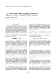
Egypt, J. Plast. Reconstr. Surg., Vol. 36, No. 2, July: 181-186, 2012<br />
<strong>De</strong>-<strong>Epithelialized</strong> <strong>Biceps</strong> <strong>Femoris</strong> <strong>Myocutaneous</strong> V-Y Advancement<br />
Flap for Ischial Bed Sore Reconstruction<br />
AHMED M. ALBARAH, M.D.<br />
The <strong>De</strong>partment of Plastic and Reconstructive Surgery, Faculty of Medicine, Menoufia University<br />
ABSTRACT<br />
Ischial bed sore is the most common location for pressure<br />
sores in paraplegic patients who are confined to wheelchairs.<br />
It is the most difficult pressure sores to treat with very high<br />
recurrence. <strong>Biceps</strong> femoris muscle long head provides the<br />
basis for several reliable flaps for ischial pressure ulcer<br />
reconstruction. One of these is the biceps femoris myocutaneous<br />
V-Y advancement flap. The Aim of this study is to<br />
evaluate the results of ischial bed sores reconstruction by <strong>De</strong>epithelialized<br />
biceps femoris myocutaneous V-Y advancement<br />
flaps. This modified flap was used for 11 ischial bed ulcers<br />
reconstruction. All ulcers were classified as grade 4; seven<br />
of them were recurrent and ranged in size from 5x5 cm to<br />
9x7 cm. After an average follow-up period of 9 months (range<br />
from 6 to 24 months), Eight ulcers (72.7%) healed with no<br />
further surgical intervention and three ulcers (27.3%) had<br />
Local recurrence that managed by re-advancement of the same<br />
flap. This study concluded that de-epithelialized biceps femoris<br />
myocutaneous V-Y advancement flap can be used efficiently<br />
to reconstruct recurrent and difficult ischial pressure sores.<br />
<strong>De</strong>-epithelialization provides the flap with more advantages<br />
and improves the results.<br />
INTRODUCTION<br />
A pressure ulcer is an area of localized damage<br />
to the skin and underlying tissue caused by pressure,<br />
shear, friction, and/or a combination of these [1].<br />
Ischial bed sore is the most common location for<br />
pressure sores in paraplegic patients who are confined<br />
to wheelchairs [2].<br />
Historically, ischial sores have been the most<br />
difficult pressure sores to treat. Motion over the<br />
ischial area, which is greater than with other pressure<br />
sore sites, and the pressure exerted on the<br />
region during sitting may help to explain why [3].<br />
Conway and Griffith [4] reported a recurrence rate<br />
of 75% to 77% in ischial pressure sore patients<br />
management regardless the type of treatment they<br />
received (operative or non operative). For this<br />
reason, patients with ischial pressure sores may<br />
require several flap surgeries during their lifetime<br />
181<br />
[5]. To treat these patients, it is important to adopt<br />
a surgical strategy in which the vascular pedicles<br />
for future flaps are not injured during the initial<br />
flap procedure [6].<br />
There are various types of flaps available to<br />
reconstruct an ischial pressure ulcer. They include<br />
gluteal thigh rotation flap, hamstring musculocutaneous<br />
flap, biceps femoris musculocutaneous<br />
flap, tensor fascia lata (TFL) flap, gracilis musculocutaneous<br />
flap, gluteal musculocutaneous flap,<br />
inferior gluteal artery perforator flap, adductor<br />
muscle perforator flap, and medially based posterior<br />
thigh skin flap with or without biceps femoris [7].<br />
The biceps femoris muscle long head provides<br />
the basis for several reliable flaps for ischial pressure<br />
ulcer reconstruction. These flaps include<br />
musculocutaneous advancement, hamstring V-Y<br />
and turnover muscle flaps [8].<br />
The biceps femoris V-Y advancement flap has<br />
several advantages: It easily covers large ischial<br />
defects; it is highly reliable; a maximum number<br />
of reconstructive options are preserved for recurrent<br />
cases, in contrast to other local flaps; and it transfers<br />
both muscle and skin over the ischium, providing<br />
greater bulk that protects the membranous urethra<br />
and better balances the pelvis in sitting [9]. In<br />
addition to, the muscle component provides greater<br />
reliability in contaminated wounds, presumably<br />
because of better vascular perfusion of the muscle<br />
[10].<br />
In this study, the biceps femoris myocutaneous<br />
flap was used as a V-Y advancement flap for ischial<br />
bed sore reconstruction. <strong>De</strong>-epithelialization for a<br />
suitable segment of the proximal part of the flap<br />
was done for all cases. The effect of this technique<br />
will be discussed.
182 Vol. 36, No. 2 / <strong>De</strong>-<strong>Epithelialized</strong> <strong>Biceps</strong> <strong>Femoris</strong> <strong>Myocutaneous</strong><br />
PATIENTS AND METHODS<br />
Between April 2007 and April 2011, <strong>De</strong>epithelialized<br />
biceps femoris myocutaneous V-Y<br />
advancement flaps were used for 11 ischial bed<br />
ulcers reconstruction. Ten paraplegic patients were<br />
included in this study; 5 males and 5 females (one<br />
female had bilateral ulcers); age ranged from 19<br />
to 62 years (average 28.4 years). The ulcers were<br />
classified as grade 4. Seven ulcers were recurrent<br />
and four were primary. Three of the recurrent cases<br />
were managed before by gluteal musculocutaneous<br />
flaps. The causes of paraplegia were spinal cord<br />
injury due to trauma in 9 patients, vascular malformation<br />
in one patient, and complication of spinal<br />
surgery in one patient.<br />
Operative Procedure:<br />
Systemic intravenous sedation was used in most<br />
of our cases; only in 2 patients with spastic hyperreflexia<br />
spinal anesthesia was used. Patients were<br />
placed in jackknifed flexed position for accurate<br />
assessment to the ulcer size and to avoid tension<br />
over the flap during flexion of the hip postoperatively.<br />
Marking of the ulcer to be excised was done<br />
at the healthy skin; the flap was outlined on the<br />
posterior thigh centralized on the long head of<br />
biceps femoris muscle. The flap was designed as<br />
large as possible to achieve tension-free closure.<br />
Subcutaneous adrenaline injection (1:200,000) was<br />
used.<br />
Radical debridement of the ulcer was done. All<br />
necrotic and devitalized tissues and bursa were<br />
excised (en bloc) with great care to avoid injury<br />
of the near by structures. Total or subtotal ischieec-<br />
Case (1): a Case (1): b Case (1): c<br />
tomy was avoided but bony prominences were<br />
excised to leave a smooth non-prominent surface<br />
of the ischium that reduces local pressure postoperatively.<br />
The base of the V is usually incised during the<br />
excision of the ulcer. The rest of the incisions were<br />
made medially and laterally to the apex and continued<br />
through the skin, subcutaneous tissues and<br />
deep fascia to the underlying muscles.<br />
The biceps femoris muscle was dissected laterally<br />
from the vastus lateralis and medially from<br />
the semimembranosus and semitendinosus muscles.<br />
Both the origin and insertion of the long head were<br />
divided. If more advancement of the flap was<br />
needed the deep surface was dissected and vascular<br />
pedicles were identified and mobilized without<br />
division.<br />
The proximal end of the skin Island was deepithelialized<br />
for a distance of 5 to 10cm according<br />
to the ulcer size. This part was introduced into the<br />
ulcer cavity and deep sutures were taken.<br />
Two closed suction drains were used; one was<br />
placed in the ulcer bed and the other under the<br />
flap. <strong>De</strong>ep fascia was closed in a separate layer<br />
followed by the subcutaneous tissue and skin.<br />
Actually, during closure of the donor site, the<br />
vertical limb of the y shaped wound was short or<br />
absent in many cases. The first drain was left in<br />
place up to 10 days.<br />
Sleeping or seating on the flap sites was prohibited<br />
for 1 month. Education of the patient and<br />
relatives for pressure relief was done before discharge.<br />
Figs. (1-4) represent four clinical cases.<br />
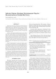
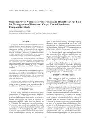
Fig. (1): Female patient, 28 years, paraplegic due to surgical spinal cord injury ten years ago, had recurrent right ischial bed<br />
sore managed before by Gluteus maximus musculocutaneous rotational flap. (a) The recurrent ischial bed sore and<br />
the scar of previous surgery. (b) Intra operative view shows the flap and the de-epithelialized part. (c) Two years<br />
postoperative view shows good healing.
Egypt, J. Plast. Reconstr. Surg., July 2012 183<br />
Case (2): a Case (2): b Case (2):c<br />
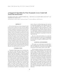
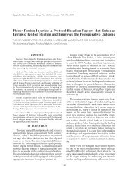
Fig. (2): Female patient, 37 years, paraplegic due to traumatic spinal cord injury ten years ago, had recurrent right ischial<br />
bed sore managed before by Inferior gluteal artery perforator flap. (a) Recurrent ischial bed sore and the scare of<br />
previous surgery. (b) The de-epithelialized part of the flap introduced into the ulcer cavity. (c) Nine months<br />
postoperative view shows good result.<br />
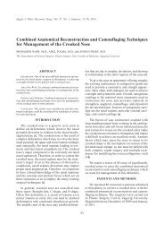
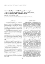
Case (3): a Case (3): b Case (3): c<br />
Fig. (3): Female patient, 48 years old, paraplegic due to traumatic spinal cord injury twenty five years ago, had bilateral<br />
recurrent ischial bed sores. (a) Bilateral recurrent ischial bed sores. (b) Three months postoperative, the left one<br />
was reconstructed with good healing. (c) Two years postoperative picture shows survival of both flaps and good<br />
healing of both ulcers.<br />
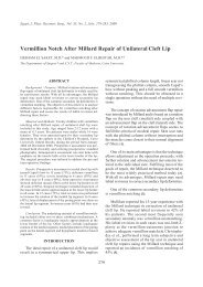
Case (4): a<br />
Case (4): b<br />
Case (4): c<br />
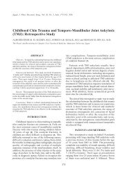
Fig. (4): Male patient, 28 years old, paraplegic due to traumatic spinal cord injury 5 years ago, had recurrent left ischial bed<br />
sore. (a) Pre operative view of the ulcer which had small opening but large cavity. (b) Intra operative view demonstrates<br />
the de-epithelialized part of the flap, the divided origin and insertion of the muscle and one of the major proximal<br />
vascular pedicles. (c) Post operative picture Shows complete survival of the flap and good healing.
184 Vol. 36, No. 2 / <strong>De</strong>-<strong>Epithelialized</strong> <strong>Biceps</strong> <strong>Femoris</strong> <strong>Myocutaneous</strong><br />
RESULTS<br />
All cases were treated in a single stage (debridement<br />
and flap reconstruction were done in one<br />
stage). Major complications such as total flap<br />
necrosis were not recorded. Eight flaps 72.7%<br />
healed with no further surgical intervention, six of<br />
them healed uneventfully without any complications<br />
and two had mild wound dehiscence (less<br />
than 1cm in width) that healed by conservative<br />
measures for less than one month. After an average<br />
follow-up period of 9 months (range from 6 to 24<br />
months), all flaps were viable and intact. Three<br />
ulcers 27.3% had Local recurrence of the bed sores<br />
and managed by re-advancement of the same flap<br />
without deep dissection. Grade 2 pressure sores<br />
appearances in new sites occurred in two patients<br />
and healed with conservative treatment.<br />
DISCUSSION<br />
Ischial pressure ulcers remain a frequent complication<br />
in hospitalized patients, geriatric population,<br />
and paraplegics. <strong>De</strong>spite high initial success<br />
rates of surgical closure, the incidence of early and<br />
late recurrence remains challenging [11].<br />
Flap selection for ischial bed sore reconstruction<br />
should take into consideration the possibility of<br />
ulcer recurrence and the need for future reconstruction.<br />
Interruption of vascular sources of adjacent<br />
flap territories during flap dissection and undermining<br />
for donor-site closure should be avoided<br />
[12].<br />
Due to the significant changes in the tension<br />
exerted across the ischial area with different lower<br />
limb positions, flaps based on the immobile trunk<br />
or pelvis have better outcome than those based on<br />
the more mobile lower extremity [13].<br />
Traditional V- Y advancement fasciocutaneous<br />
flap use for ischial pressure ulcer reconstruction<br />
was described by many authors, [14,15]. Preservation<br />
of underlying muscles, restoration of ambulatory<br />
function and capability for pressure resistance are<br />
the main advantages of this flap but its limited<br />
mobility and size shortage are the main disadvantages.<br />
A revolution occurred in the closure of pressure<br />
ulcers with the development of musculocutaneous<br />
flaps in reconstructive surgery in the late 1970s.<br />
This enabled the greatly improved blood supply,<br />
mobility and cavity filling abilities of these flaps<br />
to be used resulting in superior flap reliability and<br />
reduced recurrence rate [16].<br />
From its initial description by McCraw et al.,<br />
in 1977, the biceps femoris musculocutaneous flap<br />
has become an invaluable tool in the armoury of<br />
the reconstructive surgeon [17]. Tobin et al., introduced<br />
the notion of using the biceps femoris musculocutaneous<br />
flap as an advancement flap [8].<br />
In this study, all cases were managed in one<br />
stage. Excision of sequestrated bone and contouring<br />
of the ischium with was done for all cases. Total<br />
or subtotal ischiectomy was avoided as it may be<br />
associated with the risk of contralateral ischial bed<br />
sore occurrence and with the formation of perineal<br />
ulceration that can be complicated by the formation<br />
of urethral fistula [18].<br />
The V-Y advancement biceps femoris myocutaneous<br />
flap has a limited upward mobility, which<br />
presents a problem in a large ischial defects. The<br />
mobility can be improved by dividing origin and<br />
insertion of the muscle. This division leaves no<br />
functional loss and may relieve knee flexion spasticity<br />
or contracture [8]. More mobility can be<br />
gained by dissection of the muscle deep surface<br />
with mobilization of Neurovascular pedicles without<br />
division. The donor sites were primarily closed<br />
in all cases.<br />
In this study, three ulcers recurred despite of<br />
their flap survival. The recurrent ulcers were managed<br />
by re-advancement of the same flap. Technically,<br />
re-advancement could be performed easily<br />
by mobilizing the proximal musculocutaneous<br />
border without detailed dissection of the perforating<br />
vasculature that already performed in the initial<br />
operation.<br />
Frequently, ischial pressure ulcers are characterized<br />
by small skin defects but with a large<br />
penetrating cavity underneath [7]. So, it may seem<br />
easy to close the defect by local skin flaps or even<br />
by direct closure, but it has a very high recurrence<br />
rate. For bed sores reconstruction especially the<br />
ischial one, it is not only important to get skin to<br />
skin attachment without tension but also to obliterate<br />
the dead space with a good soft tissue volume.<br />
Without de-epithelialization of the upper part of<br />
the flap, skin to skin attachment may prevent the<br />
underlying tissues (subcutaneous fat, fascia and<br />
muscle) from packing into the ulcer cavity.<br />
So, de-epithelialization of the proximal part of<br />
the flap was done for all cases. It was made for a<br />
distance of 5 to 10cm according to the ulcer cavity;<br />
this de-epithelialization facilitates the entrance of<br />
the flap into the ulcer cavity to obliterate the ulcer<br />
dead space, form a cushion over the bone, facilitate<br />
the deep sutures that will obviate the tension over
Egypt, J. Plast. Reconstr. Surg., July 2012 185<br />
the skin, and allow more muscle bulk with high<br />
Vascularity to get inside the ulcer directly over the<br />
bone. This idea can be performed for other flaps<br />
used to reconstruct the ischial bed sores and may<br />
be a factor that reducing the recurrence rate.<br />
Long-term follow-up for ischial bed sores reconstruction<br />
demonstrated wide variability in recurrence<br />
rates. Foster et al. [3] had one of the largest<br />
series of ischial pressure sores reconstruction and<br />
compared the efficacy of one flap to another. This<br />
study reported that inferior gluteus maximus island<br />
flap and inferior gluteal thigh flap, had the highest<br />
success rates, 94% and 93%, respectively, this is<br />
followed by the gracilis flap, the V-Y hamstring<br />
flap 58% and the tensor fascia lata flap 50%.<br />
As regard flap survival, in this study, all flaps<br />
survived completely even in the three patients with<br />
recurrent ulcers. Similar results were obtained by<br />
Tobin et al. [8] who had experience with more than<br />
20 clinical flaps with no partial or complete loss;<br />
he attributed these results to the preservation of<br />
the distal segmental vascular pedicles. The same<br />
technique was used in this study as the vascular<br />
pedicles were identified and mobilized without<br />
division of anyone. No need for distal vascular<br />
pedicles division to increase the upward mobility<br />
of the flap as described by Hurteau et al. [19]. This<br />
flap survival allows the surgeon to re-advance of<br />
the same flap for recurrent cases.<br />
As regard the recurrence of ischial bed sores,<br />
Tavakoli et al. [16] had a study on 37 ulcers operated<br />
by V-Y advancement hamstring musculocutaneous<br />
island flap. At the initial follow-up with a mean<br />
of 20 months, 33% of patients had recurrent ulcers<br />
and 14.8% of them underwent re-advancements.<br />
At his second follow-up with a mean of 62 months<br />
the rate of ulcer recurrence increased to 41.4%.<br />
<strong>De</strong>spite this, 89.5% of his patients had intact flaps<br />
at the time of follow-up. Foster et al. [3] had a<br />
recurrence rate of 42% after an average follow-up<br />
period of 10 months. In this study, 3 ulcers 27.3%<br />
recurred after an average follow-up of 10 months<br />
and all were managed by advancement of the<br />
survived flaps.<br />
Conclusion:<br />
<strong>De</strong>-epithelialized biceps femoris myocutaneous<br />
V-Y advancement flap can be used efficiently to<br />
reconstruct recurrent and difficult ischial pressure<br />
sores. It carries all the advantages of the V-Y<br />
fasciocutaneous and musculocutaneous flaps. <strong>De</strong>epithelialization<br />
of the proximal part of the flap<br />
facilitates the entrance of the flap into the ulcer<br />
cavity to obliterate the dead space, form a cushion<br />
over the bone, facilitate deep sutures that will<br />
obviate the tension over the skin, and allow more<br />
muscle bulk with high vascularity to get inside the<br />
ulcer directly over the bone. Re-advancement of<br />
the same flap can be done for recurrent ulcer<br />
without deep dissection. This technique can be<br />
considered as an addendum to other flaps used for<br />
ischial bed sores reconstruction and may be a factor<br />
that reducing the recurrence rate.<br />
REFERENCES<br />
1- European Pressure Ulcer Advisory Panel (EPUAP).<br />
www.epuap.org, 2008.<br />
2- Leigh I.H. and Bennet G.: Pressure ulcers prevalence,<br />
etiology and treatment modalities. Am. J. Surg., 167: 25S,<br />
1994.<br />
3- Foster R.D., Anthony J.P., Mathes S.J. and Hoffman W.Y.:<br />
Ischial pressure sore coverage: A rationale for flap selection.<br />
British Journal of Plastic Surgery, 50: 37k-379, 1997.<br />
4- Conway H. and Griffith B.H.: Plastic surgery for closure<br />
of decubitus ulcers in patients with paraplegia. Am. J.<br />
Surg., 91: 946, 1956.<br />
5- Kim Y.S., Lew D.H., Roh T.S., Yoo W.M., Lee W.J.* and<br />
Tark K.C.: Inferior gluteal artery perforator flap: A viable<br />
alternative for ischial pressure sores. Journal of Plastic,<br />
Reconstructive & Aesthetic Surgery, 62: 1347-e1354,<br />
2009.<br />
6- Foster R.D.: Pressure sores. In: Mathes S.J., editor. Plastic<br />
surgery. 2 nd ed., Vol 6. Philadelphia: Saunders/Elsevier,<br />
p 1321, 2006.<br />
7- Lefemine V., Enoch S. and Boyce D.E.: Surgical and<br />
reconstructive management of pressure ulcers. Eur. J.<br />
Plast. Surg., 32: 63-75, 2009.<br />
8- Tobin G.R., Pompi-Sanders B., Man D. and Weiner L.J.:<br />
The biceps femoris myocutaneous advancement flap: A<br />
useful modification for ischial pressure ulcer reconstruction.<br />
Ann. Plast. Surg., 6: 396-401, 1981.<br />
9- Tobin G.R.: <strong>Biceps</strong> femoris flaps. Grabb’s encyclopedia<br />
of flaps, 3 rd ed., Vol 5, chapter 460, Lippincott Williams<br />
& Wilkins, a wolters Kluwer business, p1329, 2009.<br />
10- Michaelis L., ed.: Orthopedic surgery of the limbs in<br />
paraplegia. Berlin: Springer-Verlage, 8012, 1965.<br />
11- Kierney P.C., Engrav L.H., Isik F.F., Esselman P.C.,<br />
Cardenas D.D. and Rand R.P.: Results of 268 pressure<br />
sores in 158 patients managed by plastic surgery and<br />
rehabilitation medicine. Plast. Reconstr. Surg., 102: 765,<br />
1998.<br />
12- Scheufler O., Farhadi J. and Kovach S.J.: Anatomical<br />
basis and clinical application of the infragluteal perforator<br />
flap. Plast. Reconstr. Surg., 118: 1389-1400, 2006.<br />
13- Ichioka S., Okabe K., Tsuji S., et al.: Triple coverage of<br />
ischial ulcers with adipofascial turnover and fasciocutaneous<br />
flaps. Plast. Reconstr. Surg., 114 (4): 901-905,<br />
2004.<br />
14- Rubin J.A., Whetzel T.P. and Stevenson T.R.: The posterior<br />
thigh fasciocutaneous flap: Vascular anatomy and clinical<br />
application. Plast. Reconstr. Surg., 95: 1228-39, 1995.
186 Vol. 36, No. 2 / <strong>De</strong>-<strong>Epithelialized</strong> <strong>Biceps</strong> <strong>Femoris</strong> <strong>Myocutaneous</strong><br />
15- Tuncbilek G., Nasir S., Ozkan O., et al.: Partially deepithelialised<br />
and buried V-Y advancement flap for reconstruction<br />
of sacrococcygeal and ischial defects. Scand J.<br />
Plast. Reconstr. Surg. Hand Surg., 38 (2): 94-99, 2004.<br />
16- Tavakoli K., Rutkowski S.*, Cope C., Hassall M., Barnett<br />
R., Richards M. and Vandervord J.: Recurrence rates of<br />
ischial sores in para- and tetraplegics treated with hamstring<br />
flaps: An 8-year study. British Journal of Plastic<br />
Surgery, 52: 476–479, 1999.<br />
17- McCraw J.B., Dibbell D.G. and Carraway J.H.: Clinical<br />
definition of independent myocutaneous vascular territories.<br />
Plast. Reconstr. Surg., 60: 341-52, 1977.<br />
18- Comarr A.E. and Bors E.: Perineal urethral diverticulum.<br />
Complications of removal of ischium. JAMA, 168, 2000,<br />
1958.<br />
19- Hurteau L.E., Bostwick J., Nahai F., Hester R. and Jurkiewicz<br />
M.J.: V-Y advancement of hamstring musculocutaneous<br />
flap for coverage of ischial pressure sores. Plast.<br />
Reconstr. Surg., 68: 539-44, 1981.