Post-ablative Head and Neck Reconstruction with Free Flaps - ESPRS
Post-ablative Head and Neck Reconstruction with Free Flaps - ESPRS
Post-ablative Head and Neck Reconstruction with Free Flaps - ESPRS

Create successful ePaper yourself
Turn your PDF publications into a flip-book with our unique Google optimized e-Paper software.
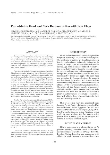
Egypt, J. Plast. Reconstr. Surg., Vol. 37, No. 1, January: 81-88, 2013<br />
<strong>Post</strong>-<strong>ablative</strong> <strong>Head</strong> <strong>and</strong> <strong>Neck</strong> <strong>Reconstruction</strong> <strong>with</strong> <strong>Free</strong> <strong>Flaps</strong><br />
AHMED M. TOHAMY, M.Sc.; MOHAMMED M. EL-SHAZLY, M.D.; MOHAMED M. FATA, M.D.;<br />
MARK MC GURK M., M.D., F.R.C.S. <strong>and</strong> MAHMOUD A. EL-OTEIFY, M.D.<br />
The Departments of Plastic Surgery, Faculty of Medicine, Assiut University, Assiut, Egypt; Oral & Maxillofacial Surgery,<br />
Faculty of Dentistry, Alex<strong>and</strong>ria University, Alex<strong>and</strong>ria, Egypt <strong>and</strong> Oral & Maxillofacial Surgery, Guy’s Hospital,<br />
King’s College University, London, UK<br />
ABSTRACT<br />
Background: Tissue defects in the head <strong>and</strong> neck region<br />
have long been a challenge for reconstructive surgeons. The<br />
ability of free flaps to transfer a large panel of tissue containing<br />
skin, mucosa, muscle or well-vascularized bone has allowed<br />
considerable progress <strong>and</strong> refinement of this type of reconstruction,<br />
<strong>with</strong> a higher level of rehabilitation for head <strong>and</strong><br />
neck cancer patients.<br />
Patients <strong>and</strong> Methods: Prospective study conducted on<br />
54 patients presenting <strong>with</strong> either oral cavity cancer or osseoradionecrosis<br />
secondary to radiotherapy treatment for head<br />
<strong>and</strong> neck cancer <strong>and</strong> all requiring bony, soft tissue reconstruction<br />
or both by using microvascular free flaps. Their age<br />
ranged at the time of operation from 32.8-85.7 years; 36 were<br />
men <strong>and</strong> 18 women. The initial disease leading to <strong>ablative</strong><br />
surgery was a malignant tumour in 42 patients including 38<br />
cases of squamous cell carcinoma (SCC) of the upper aerodigestive<br />
tract. The surgical defect involved different anatomical<br />
structures including mucosa, bone <strong>and</strong> skin. Various free flaps<br />
were used to repair these types of defects. Functional results<br />
were evaluated for the 38 patients <strong>with</strong> a SCC of the upper<br />
aerodigestive tract by the same clinician.<br />
Results: The overall success rate of free flaps was 92.6%.<br />
There were 4 free flap failures (total flap necrosis), including<br />
2 radial forearm flaps <strong>and</strong> 1 fibular flaps <strong>and</strong> 1 ALT flap.<br />
Surgical non-thrombogenic complications at the recipient site<br />
occurred in 13 patients, giving a local complication rate of<br />
24.1%. At the donor site, postoperative complications occurred<br />
in 8 cases including 3 infections (fibular flap), 1 hematoma<br />
(radial forearm flap) <strong>and</strong> 4 split thickness skin graft partial<br />
losses (radial forearm flap). Thus, the overall donor site<br />
complication rate was 14.8%. Functional <strong>and</strong> aesthetic outcomes<br />
were evaluated for patients <strong>with</strong> a SCC of the upper<br />
aero-digestive tract. They were very satisfactory in group 1<br />
(oral surgery <strong>with</strong>out m<strong>and</strong>ibulectomy). The results were<br />
worse in group 2 (oral surgery <strong>with</strong> m<strong>and</strong>ibulectomy) than in<br />
group 1.<br />
Conclusion: <strong>Free</strong> tissue transfers have proven to be very<br />
reliable to repair various types of defects in the head <strong>and</strong> neck<br />
area, <strong>with</strong> a low incidence of free flap failure <strong>and</strong> an acceptable<br />
level of complications.<br />
81<br />
INTRODUCTION<br />
Tissue defects in the head <strong>and</strong> neck region have<br />
long been a challenge for reconstructive surgeons.<br />
The goals <strong>and</strong> principles are to achieve adequate<br />
function <strong>and</strong> aesthetics <strong>and</strong> thereby to improve the<br />
quality of life [1]. <strong>Free</strong> tissue transfer has become<br />
increasingly popular for head <strong>and</strong> neck reconstruction<br />
in the last 20 years. It greatly exp<strong>and</strong>s options<br />
for patients <strong>and</strong> surgeons, <strong>and</strong> does appear to result<br />
in improved patient outcomes compared <strong>with</strong> other<br />
reconstructive techniques applied to many head<br />
<strong>and</strong> neck sites [2]. The complexity of the anatomy<br />
<strong>and</strong> function of the head <strong>and</strong> neck region explains<br />
the disappointing functional <strong>and</strong> aesthetic results<br />
obtained <strong>with</strong> conventional myocutaneous flaps.<br />
The ability of free flaps to transfer a large panel<br />
of tissue containing skin, mucosa, muscle or wellvascularized<br />
bone has allowed considerable<br />
progress <strong>and</strong> refinement of this type of reconstruction,<br />
<strong>with</strong> a higher level of rehabilitation for head<br />
<strong>and</strong> neck cancer patients [3].<br />
This prospective study is a conjoined work<br />
between Plastic Surgery Department, Assiut University<br />
Hospital, Assiut, Egypt <strong>and</strong> Oral <strong>and</strong> Maxillofacial<br />
Department, Guy’s hospital, King’s College<br />
University, London, United Kingdom. In this<br />
work, we have tried the free flap reconstruction<br />
possibilities in many types of pathologies <strong>and</strong><br />
defects in the head <strong>and</strong> neck region.<br />
PATIENTS AND METHODS<br />
This is a prospective study conducted on 54<br />
patients, attending outpatient cancer clinic of Oral<br />
& Maxillofacial Department of Guy's Hospital,<br />
London, United Kingdom <strong>and</strong> presenting <strong>with</strong><br />
either oral cavity cancer or osseoradionecrosis
82 Vol. 37, No. 1 / <strong>Post</strong>-<strong>ablative</strong> <strong>Head</strong> & <strong>Neck</strong> <strong>Reconstruction</strong><br />
secondary to radiotherapy treatment for head <strong>and</strong><br />
neck cancer <strong>and</strong> all requiring bony, soft tissue<br />
reconstruction or both by using microvascular free<br />
flaps between April 2010 <strong>and</strong> December 2011.<br />
Inclusion criteria: Any patient having soft tissue<br />
or bony reconstruction or both of the head <strong>and</strong><br />
neck region by using microvascular free flaps.<br />
Exclusion criteria: Nothing specified.<br />
Informed written consent was signed by all<br />
patients under the study.<br />
The age of the patients ranged at the time of<br />
operation from 32.8-85.7 years (mean age ± SD,<br />
61.3±12.5 years); 36 were men (66.7%) <strong>and</strong> 18<br />
women (33.3%). Immediate reconstruction was<br />
performed in 53 cases (98.1%) <strong>and</strong> secondary<br />
reconstruction in just one case (1.9%). For primary<br />
reconstructions, the initial disease leading to <strong>ablative</strong><br />
surgery was a malignant tumour in 42 patients<br />
including 38 cases of squamous cell carcinoma<br />
(SCC) of the upper aerodigestive tract <strong>with</strong> a<br />
percentage of 71.7%. In these cases of primary<br />
reconstruction, the initial disease of the patients<br />
(n=53) is given in Table (1). Patients <strong>with</strong> SCC of<br />
the upper aerodigestive tract had a primary cancer<br />
(untreated tumor) in 34 cases (89.5%) <strong>and</strong> a recurrent<br />
cancer in 4 cases (10.5%). For the primary<br />
cancers, 26 patients (76.5%) had T3 or T4 disease<br />
<strong>and</strong> 18 patients (52.9%) had clinically positive<br />
lymph nodes (6 N1, 3 N2a, 8 N2b, <strong>and</strong> 1 N2c).<br />
The characteristics of the 54 patients included in<br />
this study are presented in Table (2).<br />
The surgical defect involved different anatomical<br />
structures including mucosa, bone <strong>and</strong> skin.<br />
When these three types of tissue were included in<br />
the surgical defect, the defect was considered to<br />
be through <strong>and</strong> through. The bone defect was<br />
defined <strong>with</strong> the “HCL classification” as described<br />
by Jewer et al., [4]. The exact nature of the surgical<br />
defects is shown in Table (3).<br />
Various free flaps were used during this period<br />
to repair different types of defects, including radial<br />
forearm fasciocutaneous or osseocutaneous flaps,<br />
fibular osteocutaneous flaps, scapular osteocutaneous<br />
flap, DCIA flaps, ALT flaps <strong>and</strong> VRAM<br />
flaps. Fibular flaps were used as osteocutaneous<br />
flaps in 18 cases <strong>and</strong> as osteomyocutanous flap in<br />
2 cases. Scapular flaps were used as osseocutaneous<br />
flap in 1 case. The types of free flaps used in this<br />
study <strong>with</strong> their percentages are shown in Fig. (1).<br />
<strong>Free</strong> flap harvest was performed at the time of<br />
the <strong>ablative</strong> procedure, employing a two-team<br />
approach, except for the scapular flap. All flaps<br />
were generally fixed in their definitive position<br />
before performing the microvascular anastomoses<br />
to prevent traction on the vascular pedicle. Microvascular<br />
anastomoses were performed <strong>with</strong> an<br />
operative microscope (Zeiss, Germany) <strong>and</strong><br />
polypropylene sutures (Prolene 9.0, Ethicon,<br />
U.S.A.). The recipient vessels were identified <strong>and</strong><br />
divided during cervical lymphadenectomy when<br />
indicated.<br />
Functional results were evaluated for the 38<br />
patients <strong>with</strong> a SCC of the upper aerodigestive<br />
tract by the same clinician. The following data<br />
were recorded for all patients: Quality of oral diet,<br />
speech intelligibility, mouth opening. Results were<br />
scored from 0 to 2, as follows:<br />
Oral diet:<br />
• 2 normal.<br />
• 1 moderately impaired, restricted diet, soft diet.<br />
• 0 severely impaired or impossible, requiring<br />
maintenance of an entral feeding tube.<br />
Speech intelligibility:<br />
• 2 normal, easily intelligible.<br />
• 1 moderately altered, intelligible <strong>with</strong> effort.<br />
• 0 severely altered or impossible, patient unintelligible<br />
for the listener.<br />
Mouth opening:<br />
• 2 normal, greater than two fingerbreadths.<br />
• 1 moderately limited, between 1 <strong>and</strong> 2 fingerbreadths.<br />
• 0 severely limited, less than one fingerbreadth<br />
(Table 4).<br />
Table (1): Initial diseases leading to <strong>ablative</strong> surgery for<br />
primary (immediate) reconstructions.<br />
Initial disease<br />
Malignant tumour<br />
SCC of oral cavity<br />
Adenocystic carcinoma<br />
Mucoepidermoid carcinoma<br />
Osteosarcoma<br />
Odontogenic carcinoma<br />
Non-malignant disease<br />
Osseoradionecrosis<br />
Ameloblastoma<br />
Number<br />
of cases<br />
42<br />
38<br />
1<br />
1<br />
1<br />
1<br />
11<br />
8<br />
3<br />
Rate<br />
(%)<br />
79.2<br />
71.7<br />
1.9<br />
1.9<br />
1.9<br />
1.9<br />
20.8<br />
15.1<br />
5.6
Egypt, J. Plast. Reconstr. Surg., January 2013 83<br />
Table (2): Characteristics of the 54 patients.<br />
Patients’ characteristics<br />
Sex:<br />
Male<br />
Female<br />
Previous radiotherapy:<br />
No<br />
Yes<br />
Tumour status:<br />
Primary<br />
Secondary or recurrent<br />
T stage:<br />
T1 or T2<br />
T3 or T4<br />
N stage:<br />
N0<br />
N1, 2 or 3<br />
Table (3): Types of post-<strong>ablative</strong> surgical defects.<br />
Defect type:<br />
Mucosa alone<br />
Bone + mucosa<br />
Bone + mucosa + skin<br />
Type of bone defect:<br />
C<br />
L<br />
H<br />
LC<br />
LCL<br />
HC<br />
Table (4): Functional outcomes for the 38 patients <strong>with</strong> a SCC<br />
of the upper aerodigestive tract.<br />
Score<br />
Oral diet:<br />
2<br />
1<br />
0<br />
Speech<br />
intelligibility:<br />
2<br />
1<br />
0<br />
Mouth<br />
opening:<br />
2<br />
1<br />
0<br />
All patients<br />
n = 38 (%)<br />
20 (52.6)<br />
11 (28.9)<br />
7 (18.4)<br />
14 (36.8)<br />
13 (34.2)<br />
11 (28.9)<br />
22 (57.9)<br />
8 (21.1)<br />
8 (21.1)<br />
Number of cases<br />
36<br />
18<br />
36<br />
2<br />
34<br />
4<br />
8<br />
26<br />
16<br />
18<br />
Number of cases<br />
17<br />
36<br />
1<br />
1<br />
15<br />
2<br />
8<br />
2<br />
4<br />
For the 54 patients.<br />
For the 32 patients <strong>with</strong> a m<strong>and</strong>ibular defect, classification of<br />
Jewer et al., 1989: Type C central defect, type L lateral defect,<br />
type H lateral defect including the condyle.<br />
Oral surgery<br />
<strong>with</strong>out<br />
m<strong>and</strong>ibulectomy<br />
n = 15 (%)<br />
11 (73.3)<br />
3 (20)<br />
1 (6.7)<br />
8 (53.3)<br />
3 (20)<br />
4 (26.7)<br />
12 (80)<br />
1 (6.7)<br />
2 (13.3)<br />
Rate (%)<br />
66.7<br />
33.3<br />
94.7<br />
5.3<br />
89.5<br />
10.5<br />
23.5<br />
76.5<br />
47.1<br />
52.9<br />
For the 38 patients <strong>with</strong> a SCC of the upper aerodigestive tract.<br />
For the 34 patients <strong>with</strong> a primary (untreated) SCC of the upper<br />
aerodigestive tract.<br />
Rate (%)<br />
31.5<br />
66.7<br />
1.8<br />
2.9<br />
47.1<br />
20.6<br />
20.6<br />
5.8<br />
2.9<br />
Oral surgery<br />
<strong>with</strong><br />
m<strong>and</strong>ibulectomy<br />
n = 23 (%)<br />
9 (39.1)<br />
8 (34.8)<br />
6 (20.1)<br />
6 (26.1)<br />
10 (43.5)<br />
7 (30.4)<br />
10 (43.5)<br />
7 (30.4)<br />
6 (26.1)<br />
RESULTS<br />
Of the 54 free flaps performed for head <strong>and</strong><br />
neck reconstruction, 50 were successful, <strong>with</strong> an<br />
overall success rate of 92.6%. There were 4 free<br />
flap failures (total flap necrosis), including 2 radial<br />
forearm flaps <strong>and</strong> 1 fibular flaps <strong>and</strong> 1 ALT flap.<br />
Hence, the free flap success rates for radial forearm<br />
flap, fibular flap, DCIA flap, scapular flap <strong>and</strong><br />
VRAM flap, were, respectively, 92, 95.2,100, 100,<br />
100%. The difference between the osseous <strong>and</strong> the<br />
non-osseous free flap success rates was not statistically<br />
significant (p=0.6).<br />
Of the free flaps, 3 (5.4%) required a return to<br />
the operating theatre to control the patency of the<br />
vascular anastomoses, including 1 radial forearm<br />
flaps, 2 fibular flaps. A venous thrombosis in 2<br />
flaps, an arteriovenous thrombosis in 1 flaps <strong>and</strong><br />
good patency of the anastomoses in all the three<br />
flaps, giving an overall successful salvage rate of<br />
100%. Among the radial forearm flaps, there were<br />
4 cases (2.8%) of partial flap necrosis (
84 Vol. 37, No. 1 / <strong>Post</strong>-<strong>ablative</strong> <strong>Head</strong> & <strong>Neck</strong> <strong>Reconstruction</strong><br />
Case [1]:<br />
19%<br />
7%<br />
24%<br />
6%<br />
2% 2%<br />
Fig. (1): Types of free vascualrized flaps used <strong>and</strong> their percentages.<br />
Fig. (2): 65y old patient <strong>with</strong> recurrent SCC<br />
of reconstructed m<strong>and</strong>ible.<br />
40%<br />
Clinical Casees<br />
Fibula osseoucutaneous flap<br />
Radial forearm fasciocutaneous flap<br />
Radial forearm osseocutaneous flap<br />
DCIA flap<br />
VRAM flap<br />
ALT flap<br />
Scapular osseocutaneous flap<br />
Fig. (3): Design of scapular <strong>and</strong> parascapular flap.<br />
Fig. (4): Elevation of the flap. Fig. (5): Identification of the pedicle.<br />
Fig. (6):<strong>Post</strong>-operative intraoral view of skin paddle. Fig. (7): <strong>Post</strong>-operative extraoral view.
Egypt, J. Plast. Reconstr. Surg., January 2013 85<br />
Fig. (8): 54y old <strong>with</strong> osseoradionecrosis of left side of<br />
m<strong>and</strong>ible secondary to radiotherapy of S.C.C.<br />
tongue.<br />
Fig. (10): MR angiography of both lower limbs demonstrating<br />
patent leg arteries.<br />
Fig. (12): Intra-operative fixation of the osteotomized bony<br />
flap <strong>with</strong> a 2.5 mm reconstruction plate.<br />
Case [2]<br />
Fig. (9): Intra-operative view after bone excision.<br />
Fig. (11): Fibula osseocutaneous flap elevated but still attached<br />
to the pedicle.<br />
Fig. (13): Intra-operative view after suturing of skin paddle<br />
<strong>and</strong> completing the anastomosis.
86 Vol. 37, No. 1 / <strong>Post</strong>-<strong>ablative</strong> <strong>Head</strong> & <strong>Neck</strong> <strong>Reconstruction</strong><br />
DISCUSSION<br />
The free flap success rate of 92.8% in this study<br />
confirms the reliability of these complex reconstruction<br />
procedures in the head <strong>and</strong> neck area.<br />
Over the last 15 years, the free flap success rate<br />
has increased from 80% in the first studies to 95%<br />
in the most recent publications [5,6].<br />
All free flap failures occurred in the group of<br />
patients <strong>with</strong> SCC of the upper aerodigestive tract.<br />
In this group, the failure rate of osseous free flaps<br />
Case [3]<br />
Fig. (14): 55y old male patient <strong>with</strong> SCC of the alveolar ridge. Fig. (15): MR angiography of lower limb vessels detecting<br />
complete patency.<br />
Fig. (16): Intra-operative view of the excised specimen (segmental<br />
m<strong>and</strong>ibulectomy + selective neck dissection levels I-<br />
IV).<br />
Fig. (18): Intra-oral view of the fibula osseocutaneous flap<br />
being fixed to the resection margins.<br />
Fig. (17): Fibula free flap being osteotomized <strong>and</strong> fixed<br />
to the pre-contoured 2.3 mm plate just separated<br />
<strong>and</strong> ready for transfer.<br />
was higher than in soft tissue free flaps (15 <strong>and</strong><br />
5.5%, respectively), although this difference was<br />
not statistically significant. Several studies confirm<br />
the higher risk of free flap failure in m<strong>and</strong>ibular<br />
reconstruction compared to soft tissue reconstruction<br />
[7,8].<br />
The rate of surgical re-exploration to check<br />
microvascular anastomoses was 5.4% in this study.<br />
It is interesting to note that all cases requiring<br />
verification led to a successful free flap outcome.<br />
Identical results are given in the literature. This<br />
highlights the importance of early surveillance<br />
focused on the viability of the flap <strong>and</strong> on checking,<br />
at the least suspicion, the patency of the vascular<br />
anastomoses [6].<br />
In the context of composite osteocutaneous free<br />
flaps, vascularization of the skin paddle does not<br />
totally reflect vascularization of the osseous component<br />
of the flap. For the fibular flaps, good<br />
vascularization of the skin paddle habitually reflects<br />
good vascularization of the bone, but ischemia of<br />
the skin paddle does not necessarily correlate to<br />
defective perfusion of the bone [9]. Thus, in our<br />
study, all cases of necrosis of the bone component
Egypt, J. Plast. Reconstr. Surg., January 2013 87<br />
of fibular flaps were accompanied by necrosis of<br />
the skin paddle, while in three cases; complete<br />
necrosis of the skin paddle arose despite good<br />
viability of the bone component.<br />
In this study, partial free flap necrosis in the<br />
form of partial or complete necrosis of the flap<br />
skin paddle <strong>with</strong>out necrosis of the bone component<br />
occurred in six cases including one case of radial<br />
forearm flap, four cases of fibular flaps, one case<br />
of scapular flap. Partial free flap necrosis is a<br />
relatively rare event (11.1% of cases in this series),<br />
confirming the findings reported in the literature.<br />
In recent studies, the rates of free flap <strong>and</strong> pedicle<br />
flap failures are comparable, while partial necrosis<br />
is significantly more frequent <strong>with</strong> regional pedicle<br />
flaps [7].<br />
The radial forearm free flap is considered as<br />
the flap of choice for oral soft-tissue reconstruction.<br />
Our study confirms the excellent outcomes habitually<br />
reported after free flap reconstruction of oral<br />
mucosal defects. The radial forearm flap provides<br />
a thin <strong>and</strong> pliable skin paddle <strong>and</strong> a long vascular<br />
pedicle, which is very appreciated in this type of<br />
reconstruction [10,11].<br />
<strong>Free</strong>, vascularized bone-containing flaps have<br />
become the method of choice for reconstructing<br />
segmental defects of the m<strong>and</strong>ible <strong>and</strong> the mucosal<br />
lining. In our series, this flap was the method of<br />
choice in most cases requiring bony reconstruction<br />
especially when the native bony height is reasonable,<br />
extensive bony defects <strong>and</strong> small to moderate<br />
soft tissue defects, <strong>and</strong> when complex reshaping<br />
of the reconstructed m<strong>and</strong>ible is required <strong>with</strong><br />
multiple osteotomies reaching up to 4 osteotomies<br />
in our study.<br />
The level of non-thrombogenic complications<br />
at the recipient site in our series was 24.1% (excluding<br />
free flap failures) <strong>and</strong> similar levels are<br />
often reported in published series. For us, the most<br />
frequent local complications were infections at the<br />
recipient site (7.4% of cases), which can be<br />
favoured by other local complications (loss of free<br />
flap, fistulas <strong>and</strong> hematoma) [7,12].<br />
Functional <strong>and</strong> aesthetic outcomes were evaluated<br />
for patients <strong>with</strong> a SCC of the upper aerodigestive<br />
tract. They were very satisfactory in<br />
group 1 (oral surgery <strong>with</strong>out m<strong>and</strong>ibulectomy).<br />
The main complaint in this group concerned elocution<br />
<strong>and</strong> intelligibility of speech. This could be<br />
explained by the importance of tongue <strong>and</strong> velopalatal<br />
resection [13,14]. The results were worse in<br />
group 2 (oral surgery <strong>with</strong> m<strong>and</strong>ibulectomy) than<br />
in group 1 because group 2 patients presented more<br />
extensive defects. However, the outcomes of group<br />
2 patients were encouraging as their values were<br />
not far behind group 1 <strong>and</strong> most patients in this<br />
group fell in the category of either 2 or 1 regarding<br />
oral diet, mouth opening, <strong>and</strong> speech intelligibility.<br />
This could be explained by the quality of m<strong>and</strong>ible<br />
reconstruction. Similar results are commonly reported<br />
in the literature [11,15].<br />
Conclusion:<br />
<strong>Free</strong> tissue transfers have proven to be very<br />
reliable to repair various types of defects in the<br />
head <strong>and</strong> neck area, <strong>with</strong> a low incidence of free<br />
flap failure <strong>and</strong> an acceptable level of complications.<br />
Careful preoperative assessment, particularly<br />
concerning patient co-morbidity <strong>and</strong> history of<br />
surgery or radiotherapy, can help to identify patients<br />
<strong>with</strong> a high risk of postoperative complications.<br />
Radial forearm free flap <strong>and</strong> fibular flap were the<br />
most used flaps for soft tissue <strong>and</strong> m<strong>and</strong>ible reconstruction,<br />
<strong>with</strong> a percentage of 76.5% <strong>and</strong> 59.5%<br />
respectively.<br />
REFERENCES<br />
1- Schusterman M.A., Miller M.J., Rece G.P., Kroll S.,<br />
Marchi M. <strong>and</strong> Goepfert H.: A single centre’s experience<br />
<strong>with</strong> 308 free flaps for repair of head <strong>and</strong> neck cancer<br />
defects. Plast. Reconstr. Surg., 93: 472-478, 1994.<br />
2- Smith R.B., Sniezek J.C. <strong>and</strong> Weed D.T.: Microvascular<br />
Surgery. Subcommittee of the American Academy of<br />
Otolaryngology-<strong>Head</strong> <strong>and</strong> <strong>Neck</strong> Surgery. Utilization of<br />
free tissue transfer in head <strong>and</strong> neck surgery. Otolaryngol<br />
<strong>Head</strong> <strong>Neck</strong> Surg., 137: 182-91, 2007.<br />
3- Palme C. <strong>and</strong> Gullane P.: Principles <strong>and</strong> history of oral<br />
cavity reconstruction. In: Oral cavity reconstruction. Terry<br />
A.D. <strong>and</strong> Douglas A.G. (eds). Taylor & Francis Group,<br />
New York, 1-3, 2006.<br />
4- Jewer D.D., Boyd J.B., Manktelow R.T., Zuker R.M.,<br />
Rosen I.B., Gullane P.J., Rotstein L.E. <strong>and</strong> <strong>Free</strong>man J.E.:<br />
Orofacial <strong>and</strong> m<strong>and</strong>ibular reconstruction <strong>with</strong> the iliac<br />
crest free Xap: A review of 60 cases <strong>and</strong> a new method<br />
of classification. Plast. Reconstr. Surg., 84: 391-403,<br />
discussion 404-395, 1989.<br />
5- Hidalgo D.A.: Aesthetic improvements in free-flap m<strong>and</strong>ible<br />
reconstruction. Plast. Reconstr. Surg., 88: 574-585,<br />
discussion 586-577, 1991.<br />
6- Disa J.J., Pusic A.L., Hidalgo D.H. <strong>and</strong> Cordeiro P.G.:<br />
Simplifying microvascular head <strong>and</strong> neck reconstruction:<br />
A rational approach to donor site selection. Ann. Plast.<br />
Surg., 47: 385-389, 2001.<br />
7- Suh J.D., Sercarz J.A., Abemayor E., Calcaterra T.C.,<br />
Rawnsley J.D., Alam D. <strong>and</strong> Blackwell K.E.: Analysis of<br />
outcome <strong>and</strong> complications in 400 cases of microvascular<br />
head <strong>and</strong> neck reconstruction. Arch. Otolaryngol. <strong>Head</strong><br />
<strong>Neck</strong> Surg., 130: 962-966, 2004.<br />
8- O’Brien C.J., Lee K.K., Stern H.S., Traynor S.J., Bron<br />
L., Tew P.J. <strong>and</strong> Haghighi K.S.: Evaluation of 250 freeflap<br />
reconstructions after resection of tumours of the head<br />
<strong>and</strong> neck. Aust. N. Z. J. Surg., 68: 698-701, 1998.
88 Vol. 37, No. 1 / <strong>Post</strong>-<strong>ablative</strong> <strong>Head</strong> & <strong>Neck</strong> <strong>Reconstruction</strong><br />
9- Takushima A., Harii K., Asato H., Nakatsuka T. <strong>and</strong> Kimata<br />
Y.: M<strong>and</strong>ibular reconstruction using microvascular free<br />
flaps: A statistical analysis of 178 cases. Plast. Reconstr.<br />
Surg., 108:1555–1563, 2001.<br />
10- Shibahara T., Mohammed A.F., Katakura A. <strong>and</strong> Nomura<br />
T.: Long-term results of free radial forearm flap used for<br />
oral reconstruction: Functional <strong>and</strong> histological evaluation.<br />
J. Oral Maxillofac. Surg., 64: 1255-1260, 2006.<br />
11- Rieger J.M., Zalmanowitz J.G., Li S.Y., Sytsanko A.,<br />
Harris J., Williams D. <strong>and</strong> Seikaly H.: Functional outcomes<br />
after surgical reconstruction of the base of tongue using<br />
the radial forearm free flap in patients <strong>with</strong> oropharyngeal<br />
carcinoma. <strong>Head</strong> <strong>Neck</strong>, 58: 121-130, 2007.<br />
12- Haughey B.H., Wilson E., Kluwe L., Piccirillo J., Fredrickson<br />
J., Sessions D. <strong>and</strong> Spector G.: <strong>Free</strong> flap recon-<br />
struction of the head <strong>and</strong> neck: Analysis of 241 cases.<br />
Otolaryngol. <strong>Head</strong> <strong>Neck</strong> Surg., 125: 10-17, 2001.<br />
13- Thone M., Karengera D., Siciliano S. <strong>and</strong> Reychler H.:<br />
<strong>Reconstruction</strong> of the mobile tongue malignant tumor<br />
excision: Quality qualityof-life assessment in 19 patients.<br />
Rev. Stomatol. Chir. Maxillofac., 104: 19-24, 2003.<br />
14- Seikaly H., Rieger J., Wolfaardt J., Moysa G., Harris J.<br />
<strong>and</strong> Jha N.: Functional outcomes after primary oropharyngeal<br />
cancer resection <strong>and</strong> reconstruction <strong>with</strong> the<br />
radial forearm free flap. Laryngoscope, 113: 897-904,<br />
2003.<br />
15- Cordeiro P.G., Disa J.J., Hidalgo D.A. <strong>and</strong> Hu Q.Y.:<br />
<strong>Reconstruction</strong> of the m<strong>and</strong>ible <strong>with</strong> osseous free flaps:<br />
A 10-year experience <strong>with</strong> 150 consecutive patients. Plast.<br />
Reconstr. Surg., 104: 1314-1320, 1999.