Vertical Breast Reduction: Superomedial Pedicle - ESPRS
Vertical Breast Reduction: Superomedial Pedicle - ESPRS
Vertical Breast Reduction: Superomedial Pedicle - ESPRS

Create successful ePaper yourself
Turn your PDF publications into a flip-book with our unique Google optimized e-Paper software.
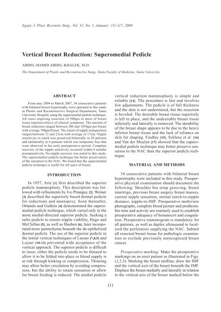
Egypt, J. Plast. Reconstr. Surg., Vol. 32, No. 1, January: 111-117, 2008<br />
<strong>Vertical</strong> <strong>Breast</strong> <strong>Reduction</strong>: <strong>Superomedial</strong> <strong>Pedicle</strong><br />
ABDEL-HAMID ABDEL-KHALEK, M.D.<br />
The Department of Plastic and Reconstructive Surgy, Tanta Faculty of Medicine, Tanta University.<br />
ABSTRACT<br />
From may 2004 to March 2007, 34 consecutive patients<br />
with bilateral breast hypertrophy were operated in this study<br />
in Plastic and Reconstructive Surgical Department, Tanta<br />
University Hospital, using the superomedial pedicle technique.<br />
All cases requiring resection of 500gm or more of breast<br />
tissue reported relieve of clinical symptoms. The amount of<br />
breast reduction ranged between 280 and 1850gm per breast<br />
with average 700gm/breast. The extent of nipple transposition<br />
ranged between 12 and 23cm with average of 17cm. Nipple<br />
sensitivity to touch was preserved bilaterally in 28 patients<br />
and unilaterally in 6 patients which was temporary loss that<br />
were observed in the early postoperative period. Complete<br />
recovery of the nipple sensitivity occurred within 6 months<br />
postoperatively. No nipple necrosis was noted in this study.<br />
The superomedial pedicle technique has better preservation<br />
of the sensation to the NAC. We found that the superomedial<br />
pedicle technique is useful for all types of breast.<br />
INTRODUCTION<br />
In 1957, Arie [1] first described the superior<br />
pedicle mammoplasty. This description was followed<br />
with refinements by Ivo Pitanguy [2]. Weiner<br />
[3] described the superiorly based dermal pedicle<br />
for reductions and mastopexy. Soon thereafter,<br />
Orlando and Guthrie [4] demonstrated the superomedial<br />
pedicle technique, which varied only in the<br />
more medial-directed superior pedicle. Seeking a<br />
safer pedicle to ensure nipple viability, Hugo and<br />
McClellan [5], as well as Hauben [6], later incorporated<br />
more parenchyma beneath the de-epithelized<br />
dermal pedicle. The use of the superior pedicle in<br />
the initial vertical techniques of Lassus [7,8,9] and<br />
Lejour [10-13] prevented wide acceptance of the<br />
vertical approach. The superior pedicle is difficult<br />
to inset; either the pedicle needs to be thinned to<br />
allow it to be folded into place or blood supply is<br />
at risk through kinking or compression. Thinning<br />
may allow better circulation by avoiding compression,<br />
but the ability to retain sensation or allow<br />
for breast feeding is reduced. The medial pedicle<br />
111<br />
vertical reduction mammoplasty is simple and<br />
reliable [14]. The procedure is fast and involves<br />
few adjustments. The pedicle is of full thickness<br />
and the skin is not undermined, but the resection<br />
is beveled. The desirable breast tissue superiorly<br />
is left in place, and the undesirable breast tissue<br />
inferiorly and laterally is removed. The durability<br />
of the breast shape appears to be due to the heavy<br />
inferior breast tissue and the lack of reliance on<br />
skin for shaping. Findlay [15], Schlenz et al. [16]<br />
and Van der Meulen [17] showed that the superomedial<br />
pedicle technique may better preserve sensation<br />
to the NAC than the superior pedicle technique.<br />
MATERIAL AND METHODS<br />
34 consecutive patients with bilateral breast<br />
hypertrophy were included in this study. Preoperative<br />
physical examination should document the<br />
following: Shoulder bra strap grooving, breast<br />
intertrigo, previous breast surgery breast masses,<br />
current nipple sensation, sternal notch-to-nipple<br />
distance, nipple-to-IMF. Preoperative multiview<br />
photographs, complete blood picture and prothrombin<br />
time and activity are routinely used to establish<br />
preoperative adequacy of hematocrit and coagulation.<br />
Preoperative mammogram is mandatory for<br />
all patients, as well as duplex ultrasound to localized<br />
the perforators supplying the NAC. Submit<br />
all resected breast tissue for pathologic examination<br />
to exclude previously unrecognized breast<br />
cancer.<br />
Preoperative marking: Make the preoperative<br />
markings on an erect patient as illustrated in Figs.<br />
(1,2,3). Marking the breast midline, draw the IMF<br />
and the vertical axis of the breast beneath the IMF.<br />
Displace the breast medially and laterally in relation<br />
to the vertical axis of the breast marked below the
112 Vol. 32, No. 1 / <strong>Vertical</strong> <strong>Breast</strong> <strong>Reduction</strong><br />
IMF. This medial and lateral displacement determines<br />
the margins of skin resection. Special mention<br />
is made of a superior displacement while<br />
marking to provide a final conical breast shape.<br />
Connect the medial and lateral margins by a gently<br />
curving line 2-4cm above the IMF. Determine the<br />
new nipple location and make the curvilinear<br />
mosque dome periareolar marking 2cm above the<br />
future nipple location. The length of the dome<br />
shape periareolar marking varies depending on<br />
breast size but usually should not exceed 16cm.<br />
The upper border of the superomedial pedicle<br />
should obliquely divide the intended NAC inset<br />
position between its inferomedial one third and<br />
the superolateral two thirds. The lower border is<br />
an oblique line, drawn within the planned skin<br />
resection margins, that respects and reflects the<br />
oblique traverse of the parasternal perforators into<br />
the pedicle. The ideal width of the pedicle base<br />
measures 8-12cm according to the extent of reduction,<br />
half of the base lies in the NAC, and half lies<br />
in the medial vertical limb.<br />
Intraoperative details: The superomedial pedicled<br />
mammoplasty begins by de-epithelization of<br />
the pedicle and circumscription of the NAC. The<br />
pedicle is developed by incision along markings<br />
straight down to but not through the loose areolar<br />
connective tissue plane directly above the superficial<br />
pectoralis major muscle fascia. The breast<br />
tissue to be excised is beveled outward, especially<br />
laterally and inferiorly. The flap is at least 1cm<br />
thick at the margins, and the beveling is performed<br />
as needed to resect the necessary breast tissue.<br />
Exposure of the pectoralis fascia is not necessary;<br />
retaining some of the tissue just superficial to the<br />
pectoralis may account for the retention of sensation<br />
with the medial pedicle (Figs. 4,5,6).<br />
<strong>Pedicle</strong> insetting: After resection, the approximate<br />
4-o’clock position of the NAC is affixed to<br />
the apex of the mosque-shaped keyhole inset site.<br />
A dermal back cut at the most inferior portion of<br />
the de-epithelialized pedicle may be required to<br />
achieve an adequate arc of rotation. This is particularly<br />
true in pedicles that are relatively short and<br />
wide. Fixation of the pedicle to the pectoral fascia<br />
by 2/0 vicryl sutures to give more projection of<br />
the breast tissue. The pedicle soft tissue bulk is<br />
transposed superomedially and secured in position<br />
by intra-parenchymal suturing to unify the medial<br />
and lateral pillars. The positional interrelation of<br />
the pillars can be manipulated by the surgeon to<br />
achieve the desired breast shape. The pillar positioning<br />
that is selected should reflect a tension-<br />
free NAC inset the base of the areolar opening is<br />
closed with a 3-0 polydioxanone (PDS) suture. No<br />
undermining of the base of the pedicle is needed<br />
for this suture (Figs. 7,8,9).<br />
Shaping: The medial and lateral pillars of breast<br />
tissue are then sutured together with 3-0 PDS. This<br />
shaping causes coning of the breast tissue, and<br />
more or less projection can be achieved as desired.<br />
If the pedicle is very large and/or long, as with a<br />
larger breast reduction (e.g., 1500g), suturing some<br />
of the pedicle to the chest wall may be indicated.<br />
Skin closure: The skin is closed with interrupted<br />
deep dermal sutures with 3-0 PDS (Fig. 10).<br />
The skin is then closed with subcuticular 3-0<br />
Monocryl, gathering the skin somewhat with closure.<br />
Minimal gathering is used superiorly, but<br />
some of the skin is gathered inferiorly. Although<br />
gathering the skin is believed to reduce the length<br />
of the vertical scar, this does not appear to make<br />
a significant difference, except at the lower end<br />
where the skin is quite redundant. Also, this skin<br />
is recognized to stretch with time because of the<br />
weight of the breast inferiorly (Fig. 11). Drains<br />
are left for 48h.<br />
Postoperative details: A gauze bandage is lightly<br />
placed over the incisions and the skin is covered<br />
with elastoplast. A surgical brassiere is then used<br />
for comfort and to hold the breast in place. Patients<br />
are advised to use the surgical bra continuously<br />
for approximately 2 weeks and then to progress to<br />
a sports bra day and night for 2 months.<br />
RESULTS<br />
34 patients, 68 breasts underwent bilateral<br />
vertical breast reduction using superomedial pedicle<br />
technique. The results were acceptable by the<br />
patients from both aesthetic and clinical points of<br />
view. All cases requiring resection of 500gm or<br />
more of breast tissue reported relieve of clinical<br />
symptoms. The amount of breast reduction ranged<br />
between 280 and 1850gm per breast with average<br />
700gm/breast. The extent of nipple transposition<br />
ranged between 12 and 23cm with average of 17cm.<br />
Nipple sensitivity to touch was preserved bilaterally<br />
in 28 patients and unilaterally in 6 patients which<br />
was temporary loss that were observed in the early<br />
postoperative period. Complete recovery of the<br />
nipple sensitivity occurred within 6 months post<br />
operatively. No nipple necrosis was noted in this<br />
study.
Egypt, J. Plast. Reconstr. Surg., January 2008 113<br />
Fig. (1): Preoperative marking. Fig. (2): Medial and lateral displacement of the breast to determine<br />
the extent of skin and glandular resction.<br />
Fig. (3): Medial and lateral displacement of the breast to determine<br />
the extent of skin and glandular resction.<br />
Fig. (5): The pedicle is developed by incision along markings straight<br />
down to but not through the loose areolar connective tissue plane<br />
directly above the superficial pectoralis major muscle fascia.<br />
Fig. (4): De-epithelization of the pedicle and circumscription of the<br />
NAC.<br />
Fig. (6): The breast tissue to be excised is beveled outward, especially<br />
laterally and inferiorly. The flap is at least 1cm thick at the<br />
margins.<br />
Fig. (7): C-shaped resceted dermo-glandular part. Fig. (8): Fixation of the pedicle to the pectoral fascia by 2/0 vicryl<br />
sutures to give more projection of the breast tissue.
114 Vol. 32, No. 1 / <strong>Vertical</strong> <strong>Breast</strong> <strong>Reduction</strong><br />
Fig. (9): After resection, the approximate 4-o’clock position of the NAC<br />
is affixed to the apex of the mosque-shaped keyhole inset site.<br />
Fig. (10): Intraoperative appearance of the vertical limb.<br />
Fig. (11): Two months post-operatively. Fig. (12): Preoperative.<br />
Fig. (13): Postoperative. Fig. (14): Preoperative.<br />
Fig. (15): Postoperative. Fig. (16): Preoperative.
Egypt, J. Plast. Reconstr. Surg., January 2008 115<br />
Fig. (17): Postoperative. Fig. (18): Preoperative.<br />
Fig. (19): Postoperative. Fig. (20): Postoperative.<br />
DISCUSSION<br />
<strong>Breast</strong> reduction has become well established<br />
with respect to safety and predictable aesthetic<br />
results. Recently limited scar techniques such as<br />
pure circumareolar scar method [21] and vertical<br />
scar mammaplasty [9,12] have become more popular.<br />
The major advantages of vertical mammaplasty<br />
are aesthetically acceptable breast shape and excellent<br />
contour projection [9,14]. As confirmed in<br />
our study it gives the breast a good seat upon the<br />
chest wall, scars are confined to the breast without<br />
inframammary involvement, as well as stability of<br />
breast shape depends on glandular suturing and<br />
not on skin tension. Van der Meulen [16] showed<br />
that the superomedial pedicle technique may better<br />
preserve sensation to the NAC than the superior<br />
pedicle technique. The author has found the superomedial<br />
pedicle technique useful for all breast<br />
types, not restricted to use on nonsmokers or patients<br />
with soft and mobile breasts. The superomedial<br />
pedicle technique can be safely used for resections<br />
of up to 2000g and cephalic NAC<br />
transpositions of 5-15cm with reliable preservation<br />
of viability and sensibility. Finger et al. [19] illustrated<br />
the safety of the superomedial pedicle tech-<br />
nique in reduction of up to 4,100gm and with<br />
pedicle lengths of up to 30cm. No cases of nippleareola<br />
necrosis were encountered in their series as<br />
the ratio between the width and length of the flap<br />
was respected in addition to careful monitoring of<br />
the thickness of the flap and degree of tension<br />
during closure. Ceydeli and Gamboa [25], believed<br />
that the dermafascial fixation suture a concept<br />
more than a technique. It incorporates the two<br />
strongest structures, the dermis and the fascia, to<br />
achieve more durable projection with short-scar<br />
vertical reduction mammaplasty. As regards nipple<br />
areola sensation, anatomical studies [22,23] have<br />
shown the fourth intercostals nerve as the main<br />
supply to the nipple-areola complex. The nerve<br />
pierces the serratus anterior muscle at the midaxillary<br />
line of the fourth intercostal space, then<br />
travels within the serratus fascia to the lateral<br />
border of the pectoralis muscle and then turns<br />
anteriorly at a right angle to enter the mammary<br />
gland. The nerve remains deep within the breast<br />
until halfway to the nipple-areola complex where<br />
it becomes superficial. However, Jaspars et al. [24]<br />
could not find this unusual course of the lateral<br />
cutaneous branches with S-shaped to the skin and<br />
areola. In addition they found a constant bilateral
116 Vol. 32, No. 1 / <strong>Vertical</strong> <strong>Breast</strong> <strong>Reduction</strong><br />
innervation of the areola by the lateral and anterior<br />
branches of the sixth intercostals nerve, therefore,<br />
they favored a superomedial pedicle for reduction<br />
mammaplasty. Schlenz et al. [17] demonstrated that<br />
the lateral intercostal nerve runs across the breast<br />
just above the level of the pectoralis fascia until<br />
the mid portion of the breast, where it then runs<br />
superiorly toward the nipple. This assessment of<br />
anatomy explains why the medial pedicle technique<br />
maintains as good sensation as the lateral pedicle<br />
technique. Hauben [18] reported his experience and<br />
refinements with the supero-medial pedicle vertical<br />
breast reduction on 212 patients; in 1 patient total<br />
loss of the NAC occurred (the first time he used<br />
this technique, the pedicle length was 26cm, and<br />
the patient was a heavy smoker). In addition, he<br />
had another partial loss of the NAC. Decreased<br />
sensation of the NAC was present, but no incidence<br />
was quoted; nipple retraction was present in 2%<br />
of patients and hematoma in 2.26%. No transfusion<br />
was necessary. Finger et al. [19] reported only 2<br />
partial losses of the NAC (
Egypt, J. Plast. Reconstr. Surg., January 2008 117<br />
10- Lejour M., Abboud M., Declety A. and Kertesz P.: <strong>Reduction</strong><br />
of mammaplasty scars: From a short inframammary<br />
scar to a vertical scar. Ann. Chir. Plast. Esthet., 35 (5):<br />
369-79, 1990.<br />
11- Lejour M.: <strong>Vertical</strong> mammaplasty and liposuction of the<br />
breast. St. Louis, Mo: Quality Medical Publishing, 1993.<br />
12- Lejour M.: <strong>Vertical</strong> mammaplasty and liposuction of the<br />
breast. Plast. Reconstr. Surg., 94 (1): 100-14, 1994.<br />
13- Lejour M. and Abboud M.: <strong>Vertical</strong> mammaplasty without<br />
inframammary scar and with breast liposuction. Plast.<br />
Reconstr. Surg., 4: 67-90, 1996.<br />
14- Hall-Findlay E.J.: A simplified vertical reduction mammaplasty:<br />
Shortening the learning curve. plast. Reconstr.<br />
Surg., 104 (3): 748-59, 1999.<br />
15- Hall-Findlay E.J.: <strong>Breast</strong> reduction, simplified vertical.<br />
Plast. Reconstr. Surg., 104 (1): 748-59, 2003.<br />
16- Van der Meulen J.C.: <strong>Superomedial</strong> pedicle technique of<br />
reduction mammaplasty. Plast. Reconstr. Surg., 84 (6):<br />
1005, 1989.<br />
17- Schlenz I., Kuzbari R., Gruber H. and Holle J.: The<br />
sensitivity of the nipple-areola complex: An anatomic<br />
study. Plast. Reconstr. Surg., 105 (3): 905-9, 2000.<br />
18- Hauben D.J.: Experience and refinements with the superomedial<br />
dermal pedicle for nipple-areola transposition in<br />
reduction mammaplasty. Aesthetic Plast. Surg., 8 (3):<br />
189-94, 1984.<br />
19- Finger R.E., Vasquez B., Drew G.S. and Given K.S.:<br />
<strong>Superomedial</strong> pedicle technique of reduction mammaplasty.<br />
Plast. Reconstr. Surg., 83 (3): 471-80, 1989.<br />
20- Hugo N.E. and McClellan R.M.: <strong>Reduction</strong> mammaplasty<br />
with a single superiorly-based pedicle. Plast. Reconstr.<br />
Surg., 63 (2): 230-4, 1979.<br />
21- Perbeck L., Alveryd A., Mattanen H. and Wallberg H.:<br />
Skin circulation in the nipple after reduction mammaplasty<br />
by upper and lower glandular resection. Scand. J. Plast.<br />
Surg., 22: 237-40, 1988.<br />
22- Courtiss E.H. and Goldwyn R.N.: <strong>Breast</strong> sensation before<br />
and after plastic surgery. Plast. Reconstr. Surg., 58: 1-3,<br />
1976.<br />
23- Farina M.A., Newby B.G. and Alani H.A.: Innervation<br />
of the nipple areola complex. Plast. Reconstr. Surg., 66:<br />
497-501, 1980.<br />
24- Jaspars J.J.P., Posma A.N., Van Immerseel A.A.H. and<br />
Gittenberger-de Goot A.C.: The cutaneous innervations<br />
of the femal breast and nipple-areola complex:<br />
Implantation for Surgery. Br. J. Plast. Surg., 50: 249-59,<br />
1977.<br />
25- Ceydeli A. and Gamboa M.: Dermafascial fixation suture:<br />
A technique for a more durable projection with short-scar<br />
vertical reduction mammaplasty. Aesthetic Plast. Surg.,<br />
30 (5): 592-4, 2006.<br />
26- Lista F. and Ahmed J.: <strong>Vertical</strong> scar reduction mammaplasty:<br />
A 15-year experience including a review of 250<br />
consecutive cases. Plast. Reconstr. Surg., 117 (7): 2152-<br />
65, 2006.<br />
27- Kreithen J., Caffee H., Rosenberg J., Chin G., Clayman<br />
M., Lawson M. and Seagle M.B.: A comparison of the<br />
Lejour and Wise pattern methods of breast reduction.<br />
Ann. Plast. Surg., 54 (3): 236-41, 2005.<br />
28- Cruz-Korchin N. and Korchin L.: <strong>Breast</strong>-feeding after<br />
vertical mammaplasty with medial pedicle. Plast. Reconstr.<br />
Surg., 15; 114 (4): 890, 2004.<br />
29- Chen C.M., White C., Warren S.M., Cole J. and Isik F.F.:<br />
Simplifying the vertical reduction mammaplasty. Plast.<br />
Reconstr. Surg., 114 (6): 1673, 2004.<br />
30- Karp N.S.: Medial pedicle/vertical breast reduction made<br />
easy: The importance of complete inferior glandular<br />
resection. Ann. Plast. Surg., 52 (5): 458-64, 2004.