Case of spontaneous regression of carotid body tumor in a SDHD ...
Case of spontaneous regression of carotid body tumor in a SDHD ...
Case of spontaneous regression of carotid body tumor in a SDHD ...

You also want an ePaper? Increase the reach of your titles
YUMPU automatically turns print PDFs into web optimized ePapers that Google loves.
Hammer et al. World Journal <strong>of</strong> Surgical Oncology 2012, 10:218<br />
http://www.wjso.com/content/10/1/218<br />
CASE REPORT Open Access<br />
<strong>Case</strong> <strong>of</strong> <strong>spontaneous</strong> <strong>regression</strong> <strong>of</strong> <strong>carotid</strong> <strong>body</strong><br />
<strong>tumor</strong> <strong>in</strong> a <strong>SDHD</strong> mutant: a discussion on<br />
potential mechanisms based on a review <strong>of</strong><br />
the literature<br />
Sebastiaan Hammer 1* , Jeroen C Jansen 2 , Eleonora PM van der Kleij-Corssmit 3 , Frederik J Hes 4 and Mark C Kruit 1<br />
Abstract<br />
Background: Head and neck paragangliomas are <strong>tumor</strong>s associated with the parasympathetic nerve system and<br />
typically show an <strong>in</strong>dolent growth pattern. Therefore a conservative management strategy is considered <strong>in</strong> selected<br />
cases.<br />
Methods and results: We present a case <strong>of</strong> a female patient who presented <strong>in</strong> 2003 with bilateral <strong>carotid</strong> <strong>body</strong><br />
<strong>tumor</strong>s and a tympanic <strong>tumor</strong>, associated with a mutation <strong>in</strong> the succ<strong>in</strong>ate dehydrogenase -sub-unit-D (<strong>SDHD</strong>). She<br />
was operated on the right <strong>carotid</strong> <strong>body</strong> <strong>tumor</strong> and the tympanic <strong>tumor</strong>. Thereafter the follow-up was performed<br />
with MR exam<strong>in</strong>ations at 2-year <strong>in</strong>tervals. After an <strong>in</strong>itial stable phase, over the last 3 years a <strong>spontaneous</strong> near-total<br />
<strong>regression</strong> <strong>of</strong> the contralateral <strong>carotid</strong> <strong>body</strong> <strong>tumor</strong> was observed, with only subtle rest-abnormalities visible <strong>in</strong> 2011.<br />
Conclusions: The present case underl<strong>in</strong>es the <strong>in</strong>dolent growth pattern <strong>of</strong> head and neck paragangliomas and for<br />
the first time describes a rare manifestation <strong>of</strong> <strong>spontaneous</strong> <strong>regression</strong> <strong>of</strong> a <strong>carotid</strong> <strong>body</strong> <strong>tumor</strong>. The literature was<br />
reviewed to discuss this phenomenon.<br />
Keywords: Tumor <strong>regression</strong>, Head and neck paragangliomas, Spontaneous <strong>in</strong>volution, Surgical resection<br />
Background<br />
Head and neck paragangliomas (HNPGL) are usually benign,<br />
slow-grow<strong>in</strong>g <strong>tumor</strong>s associated with the parasympathetic<br />
nerve system. Common sites <strong>in</strong>clude the <strong>carotid</strong><br />
<strong>body</strong>, the temporal bone, and the vagal <strong>body</strong> [1]. The<br />
majority <strong>of</strong> patients presents as apparently sporadic<br />
patients, whereas 10% to 20% <strong>of</strong> patients report a positive<br />
family history [2,3]. Multiple paragangliomas may occur<br />
<strong>in</strong> up to 40% <strong>of</strong> patients [4]. In the Netherlands the<br />
majority <strong>of</strong> cases are related to mutations <strong>in</strong> the gene<br />
encod<strong>in</strong>g for the oxidative cha<strong>in</strong> prote<strong>in</strong> succ<strong>in</strong>ate<br />
dehydrogenase-sub-unit-D (<strong>SDHD</strong>) [5]. In the neck, a<br />
paraganglioma presents as a non-tender mass or as a cause<br />
<strong>of</strong> lower cranial nerve palsy due to local compression. In<br />
the temporal bone, the first symptom <strong>of</strong> paragangliomas is<br />
* Correspondence: S.Hammer@lumc.nl<br />
1<br />
Departments <strong>of</strong> Radiology, Leiden University Medical Center, Alb<strong>in</strong>usdreef 2,<br />
Leiden, ZA 2333, The Netherlands<br />
Full list <strong>of</strong> author <strong>in</strong>formation is available at the end <strong>of</strong> the article<br />
WORLD JOURNAL OF<br />
SURGICAL ONCOLOGY<br />
usually pulsat<strong>in</strong>g t<strong>in</strong>nitus, due to the hypervascular nature<br />
<strong>of</strong> the <strong>tumor</strong>. In follow-up about 60% <strong>of</strong> the HNPGL do<br />
not exhibit growth, and if they do it typically is <strong>in</strong> an <strong>in</strong>dolent<br />
growth pattern with a median <strong>tumor</strong> double time<br />
<strong>of</strong> 4.2 years [6]. Diagnosis is generally made through a<br />
comb<strong>in</strong>ation <strong>of</strong> cl<strong>in</strong>ical f<strong>in</strong>d<strong>in</strong>gs and magnetic resonance<br />
imag<strong>in</strong>g (MRI) studies. Treatment considerations <strong>in</strong>clude<br />
the nature <strong>of</strong> the <strong>tumor</strong> (malignant or benign), the location,<br />
vasculature encasement, the extent, and growth rate<br />
[6,7]. Treatment modalities <strong>in</strong>clude surgery or radiotherapy/surgery;<br />
treatment <strong>of</strong> choice for <strong>carotid</strong> <strong>body</strong> <strong>tumor</strong>s<br />
is surgical resection. However, because <strong>of</strong> the slow growth<br />
rate and potential treatment-related <strong>in</strong>jury to the neighbor<strong>in</strong>g<br />
vessels and nerves, a conservative management<br />
strategy (wait-and-scan policy) should be considered [6].<br />
<strong>Case</strong> presentation<br />
In 2003 a 32-year-old female patient was referred to our<br />
<strong>in</strong>stitution after magnetic resonance imag<strong>in</strong>g (MRI)<br />
© 2012 Hammer et al.; licensee BioMed Central Ltd. This is an Open Access article distributed under the terms <strong>of</strong> the Creative<br />
Commons Attribution License (http://creativecommons.org/licenses/by/2.0), which permits unrestricted use, distribution, and<br />
reproduction <strong>in</strong> any medium, provided the orig<strong>in</strong>al work is properly cited.
Hammer et al. World Journal <strong>of</strong> Surgical Oncology 2012, 10:218 Page 2 <strong>of</strong> 4<br />
http://www.wjso.com/content/10/1/218<br />
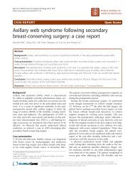
Figure 1 Transverse gadol<strong>in</strong>ium-enhanced 3D time <strong>of</strong> flight<br />
magnetic resonance angiography images at basel<strong>in</strong>e <strong>in</strong> 2004<br />
show the bilateral <strong>carotid</strong> <strong>body</strong> <strong>tumor</strong>s (arrows).<br />
exam<strong>in</strong>ation <strong>in</strong> a regional hospital had revealed bilateral<br />
<strong>carotid</strong> <strong>body</strong> <strong>tumor</strong>s. She had discovered a swell<strong>in</strong>g <strong>in</strong><br />
the right neck s<strong>in</strong>ce 6 months before, and only after<br />
question<strong>in</strong>g mentioned a long-exist<strong>in</strong>g pulsatile t<strong>in</strong>nitus<br />
on the right side. The family history for paragangliomas<br />
was positive: her father and uncle were affected.<br />
Physical exam<strong>in</strong>ation revealed a small reddish <strong>tumor</strong><br />
<strong>in</strong> the right middle ear, with positive Brown’s sign and a<br />
<strong>tumor</strong> <strong>in</strong> the right neck at the level <strong>of</strong> the hyoid bone;<br />
on the left side there was no palpable lesion. The lower<br />
cranial nerves were <strong>in</strong>tact and the hear<strong>in</strong>g was normal.<br />
In general exam<strong>in</strong>ation, the patient was all-over obese<br />
(weight 121 kg, length 1.75 m, <strong>body</strong> mass <strong>in</strong>dex 39.5),<br />
blood pressure was 130/70 mm Hg, pulse rate 80/m<strong>in</strong>.<br />
There were no other abnormalities. Ur<strong>in</strong>ary excretion<br />
(24-h sample) <strong>of</strong> catecholam<strong>in</strong>es was repeatedly normal.<br />
Germl<strong>in</strong>e mutation analysis <strong>in</strong> DNA from a blood sample<br />
demonstrated a mutation <strong>in</strong> <strong>SDHD</strong> (P-Asp92Tyr).<br />
MRI <strong>in</strong>clud<strong>in</strong>g a gadol<strong>in</strong>ium-enhanced 3D time-<strong>of</strong>flight<br />
MR angiography sequence [8] was acquired at our<br />
<strong>in</strong>stitution <strong>in</strong> April 2004, and revealed hypervascular enhanc<strong>in</strong>g<br />
lesions <strong>in</strong> the <strong>carotid</strong> bifurcation bilaterally,<br />
consistent with <strong>carotid</strong> <strong>body</strong> <strong>tumor</strong>s. The maximum<br />
transverse diameter was 23 mm on the right and 12 mm<br />
on the left (Figure 1). A tympanic <strong>tumor</strong> was identified<br />
on the right side.<br />
The right <strong>carotid</strong> <strong>body</strong> <strong>tumor</strong> was surgically resected<br />
<strong>in</strong> 2004, followed by extirpation <strong>of</strong> the tympanic <strong>tumor</strong><br />
<strong>in</strong> 2005. No radiotherapy was applied. There were no<br />
complications from the surgical procedures. In the<br />
postoperative phase a severe obstructive sleep apnea<br />
syndrome (OSAS) was diagnosed, for which she started<br />
cont<strong>in</strong>uous positive airway pressure (CPAP) therapy.<br />
In 2005 she started thyroid hormone suppletion for<br />
Hashimoto disease. Between 2005 and 2008, the patient<br />
<strong>in</strong>tentionally lost weight from 136 kg to 93 kg, because<br />
<strong>of</strong> a pregnancy wish which was eventually unsuccessful.<br />
Dur<strong>in</strong>g follow-up the patient rega<strong>in</strong>ed <strong>body</strong>weight <strong>in</strong><br />
2010 to 129 kg. In 2011 she suffered anamnestically<br />
from a transient ischemic attack (with no abnormalities<br />
on imag<strong>in</strong>g studies), after which she started stat<strong>in</strong><br />
treatment and therapy with carbaselate calcium and<br />
dipyridamole.<br />
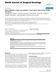
Follow-up MRI was performed with 2-year <strong>in</strong>tervals<br />
(Figure 2), and showed no evidence <strong>of</strong> recurrence on the<br />
operated sites. Between 2004 and 2008 there were no<br />
changes <strong>in</strong> size and enhancement pattern <strong>of</strong> the left<br />
<strong>carotid</strong> <strong>body</strong> <strong>tumor</strong> and therefore surgical resection was<br />
considered unnecessary. Unexpectedly, <strong>in</strong> 2010, the<br />
<strong>tumor</strong> was significantly smaller, and showed reduced<br />
enhancement <strong>in</strong> the center <strong>of</strong> the lesion, consistent<br />
with necrosis. Follow-up exam<strong>in</strong>ations <strong>in</strong> June 2011 and<br />
September 2011 showed nearly complete <strong>regression</strong>,<br />
Figure 2 Transverse gadol<strong>in</strong>ium-enhanced 3D time-<strong>of</strong>-flight magnetic resonance angiography images at subsequent <strong>in</strong>tervals show<strong>in</strong>g<br />
the <strong>carotid</strong> <strong>body</strong> <strong>tumor</strong> on the left <strong>in</strong> 2006 (A), unchanged <strong>in</strong> 2008 (B), <strong>in</strong>homogeneous r<strong>in</strong>g-like enhancement <strong>in</strong> 2010 (C), and further<br />
<strong>regression</strong> <strong>in</strong> 2011 (D). On the image only a subtle l<strong>in</strong>ear enhancement is noted, no evidence <strong>of</strong> an enhanc<strong>in</strong>g mass could be detected.
Hammer et al. World Journal <strong>of</strong> Surgical Oncology 2012, 10:218 Page 3 <strong>of</strong> 4<br />
http://www.wjso.com/content/10/1/218<br />
leav<strong>in</strong>g only m<strong>in</strong>imal rest-abnormalities at the site <strong>of</strong><br />
the previous <strong>tumor</strong>. Other affected family members<br />
showed no signs <strong>of</strong> <strong>regression</strong> dur<strong>in</strong>g follow-up.<br />
Discussion<br />
We describe a patient with hereditary bilateral head and<br />
neck paragangliomas who underwent surgical <strong>tumor</strong> removal<br />
<strong>of</strong> a right <strong>carotid</strong> <strong>body</strong> <strong>tumor</strong> and a right tympanic<br />
<strong>tumor</strong>. Dur<strong>in</strong>g the 7-year follow-up, <strong>spontaneous</strong><br />
<strong>regression</strong> <strong>of</strong> the left <strong>carotid</strong> <strong>body</strong> <strong>tumor</strong> was noted,<br />
after an <strong>in</strong>itial period without any change <strong>in</strong> <strong>tumor</strong> size.<br />
To the best <strong>of</strong> our knowledge, <strong>spontaneous</strong> <strong>in</strong>volution <strong>of</strong><br />
head and neck paragangliomas has not been reported<br />
before. In our referral center for paragangliomas, the<br />
present case with <strong>spontaneous</strong> <strong>in</strong>volution represents the<br />
first from an estimated total <strong>of</strong> more than 400 followed<br />
cases.<br />
Spontaneous <strong>regression</strong> <strong>of</strong> <strong>tumor</strong>s <strong>in</strong> general is a rare<br />
phenomenon, although numerous cases and case series<br />
can be found <strong>in</strong> the literature. Spontaneous remission <strong>of</strong><br />
<strong>tumor</strong>s with a common neural crest embryological<br />
orig<strong>in</strong> as paragangliomas has been described as well.<br />
Regression <strong>of</strong> pheochromocytomas after <strong>in</strong>itial presentation<br />
with hypertensive crises or shock has been reported<br />
[9-11], as well as <strong>spontaneous</strong> <strong>regression</strong> [12], Another<br />
known entity is <strong>spontaneous</strong> <strong>regression</strong> <strong>in</strong> a subset <strong>of</strong><br />
pediatric neuroblastomas, especially those detected with<br />
mass screen<strong>in</strong>g and with biologically favorable characteristics<br />
[13,14].<br />
Mechanism<br />
The mechanism that accounts for the <strong>regression</strong> <strong>in</strong> the<br />
present case can only be speculated upon. Below we discuss<br />
the general <strong>tumor</strong> <strong>regression</strong> hypotheses, <strong>in</strong>clud<strong>in</strong>g<br />
biological (genetic, immunological), hormonal (such as<br />
contraceptive use), vascular (vascular <strong>in</strong>sufficiency/<strong>tumor</strong><br />
necrosis/<strong>spontaneous</strong> <strong>in</strong>tra<strong>tumor</strong>al vascular thrombosis),<br />
and operative mechanisms [15].<br />
Biological mechanisms related to <strong>tumor</strong> <strong>regression</strong> <strong>in</strong>clude<br />
genetic <strong>in</strong>stability (telomerase <strong>in</strong>hibition) and programmed<br />
cell death, which has been described for<br />
neuroblastomas [16,17]. The specific <strong>SDHD</strong> mutation <strong>in</strong><br />
the present case (p.Asp92Tyr), however, is relatively common<br />
- as it is found <strong>in</strong> almost 70% <strong>of</strong> 690 Dutch SDHgene<br />
mutation carriers [5] and has not been reported<br />
to be associated with <strong>spontaneous</strong> <strong>tumor</strong> <strong>regression</strong><br />
before.<br />
Although changes <strong>in</strong> <strong>body</strong> weight may go with changes<br />
<strong>in</strong> catecholam<strong>in</strong>e levels, we found no evidence for such<br />
changes <strong>in</strong> our patient, neither after the start <strong>of</strong> CPAP<br />
treatment for OSAS. Therefore a hormonal mechanism<br />
for <strong>tumor</strong> <strong>regression</strong> seems unlikely.<br />
Vascular mechanisms may be due to changes <strong>in</strong> <strong>tumor</strong><br />
angiostructure, and have been reported <strong>in</strong> <strong>spontaneous</strong><br />
regressed (biologically favorable) neuroblastomas [18].<br />
Such vascular mechanism could have expla<strong>in</strong>ed the <strong>regression</strong><br />
<strong>in</strong> the present case, although we have no supportive<br />
biomarkers for this hypothesis.<br />
Operative mechanisms <strong>in</strong>clude <strong>regression</strong> <strong>of</strong> a <strong>tumor</strong><br />
after biopsy, which has been described <strong>in</strong> patients with<br />
Merkel cell <strong>tumor</strong>s [19,20], and <strong>in</strong>volution <strong>of</strong> metastases<br />
after resection <strong>of</strong> a primary <strong>tumor</strong>, recently described <strong>in</strong><br />
a patient with lung metastases <strong>of</strong> hepatocellular carc<strong>in</strong>oma<br />
[21]. Another example comes from a patient with<br />
three hemangioblastomas (not related to Von Hippel-<br />
L<strong>in</strong>dau disease), <strong>of</strong> which two regressed completely 6<br />
months after surgical resection <strong>of</strong> the first <strong>tumor</strong> [22].<br />
The authors hypothesized that the resected <strong>tumor</strong> may<br />
have been supportive for the existence <strong>of</strong> the other two.<br />
Furthermore, <strong>in</strong> two patients with neur<strong>of</strong>ibromatosis<br />
type 2 and bilateral vestibular schwannomas, <strong>spontaneous</strong><br />
<strong>regression</strong> after resection <strong>of</strong> the contralateral <strong>tumor</strong><br />
was reported [23]. In this paper, one patient showed an<br />
<strong>in</strong>itial <strong>in</strong>crease <strong>in</strong> <strong>tumor</strong> size, directly after resection <strong>of</strong><br />
the contralateral schwannoma. Afterwards, a gradual<br />
decrease <strong>in</strong> <strong>tumor</strong> size was objectified from 6 months<br />
postoperatively. Tumor size <strong>in</strong>creased <strong>in</strong>itially <strong>in</strong> the<br />
second patient as well, whereas a decrease <strong>in</strong> <strong>tumor</strong><br />
size was noted from approximately 60 months [23]. A<br />
similar mechanism may expla<strong>in</strong> the <strong>regression</strong> <strong>of</strong> the<br />
left <strong>carotid</strong> <strong>body</strong> <strong>tumor</strong> after <strong>in</strong>itial resection <strong>of</strong> the<br />
<strong>carotid</strong> <strong>body</strong> <strong>tumor</strong> on the right <strong>in</strong> our case.<br />
Although the present case describes <strong>spontaneous</strong> <strong>in</strong>volution,<br />
surgical resection is the favored treatment <strong>in</strong> <strong>carotid</strong><br />
<strong>body</strong> <strong>tumor</strong>s. Nevertheless, resection is associated<br />
with complications such as cranial nerve impairment,<br />
stroke, or partial scarification <strong>of</strong> the <strong>carotid</strong> arteries [24].<br />
Conclusions<br />
In conclusion, the present case underl<strong>in</strong>es the <strong>in</strong>dolent<br />
growth pattern <strong>of</strong> head and neck paragangliomas and<br />
for the first time describes a rare manifestation <strong>of</strong> <strong>in</strong>volution<br />
<strong>of</strong> a <strong>carotid</strong> <strong>body</strong> <strong>tumor</strong>.<br />
Consent<br />
Written <strong>in</strong>formed consent was obta<strong>in</strong>ed from the patient<br />
for publication <strong>of</strong> this case report and any accompany<strong>in</strong>g<br />
images. A copy <strong>of</strong> the written consent is available for<br />
review by the Editor-<strong>in</strong>-Chief <strong>of</strong> this journal.<br />
Abbreviations<br />
CPAP: Cont<strong>in</strong>uous positive airway pressure; HNPGL: Head and neck<br />
paragangliomas; MRI: Magnetic resonance imag<strong>in</strong>g; OSAS: Obstructive sleep<br />
apnea syndrome; <strong>SDHD</strong>: Succ<strong>in</strong>ate dehydrogenase-sub-unit-D.<br />
Compet<strong>in</strong>g <strong>in</strong>terest<br />
All authors declare no compet<strong>in</strong>g <strong>in</strong>terest.<br />
Authors’ contributions<br />
SH and MK carried out the MR exam<strong>in</strong>ations and drafted the manuscript. FH<br />
performed genetic analysis and drafted the manuscript. JJ and EC performed
Hammer et al. World Journal <strong>of</strong> Surgical Oncology 2012, 10:218 Page 4 <strong>of</strong> 4<br />
http://www.wjso.com/content/10/1/218<br />
physical exam<strong>in</strong>ations and patient follow-up and drafted the manuscript. All<br />
authors read and approved the f<strong>in</strong>al manuscript.<br />
Author details<br />
1 Departments <strong>of</strong> Radiology, Leiden University Medical Center, Alb<strong>in</strong>usdreef 2,<br />
Leiden, ZA 2333, The Netherlands. 2 Departments <strong>of</strong> Head and Neck Surgery,<br />
Leiden University Medical Center, Alb<strong>in</strong>usdreef 2, Leiden, ZA 2333, The<br />
Netherlands. 3 Departments <strong>of</strong> Endocr<strong>in</strong>ology, Leiden University Medical<br />
Center, Alb<strong>in</strong>usdreef 2, Leiden, ZA 2333, The Netherlands. 4 Departments <strong>of</strong><br />
Human and Cl<strong>in</strong>ical Genetics, Leiden University Medical Center, Alb<strong>in</strong>usdreef<br />
2, Leiden, ZA 2333, The Netherlands.<br />
Received: 7 August 2012 Accepted: 14 October 2012<br />
Published: 19 October 2012<br />
References<br />
1. Pellitteri PK, R<strong>in</strong>aldo A, Myssiorek D, Gary JC, Bradley PJ, Devaney KO,<br />
Shaha AR, Netterville JL, Manni JJ, Ferlito A (2004) Paragangliomas <strong>of</strong> the<br />
head and neck. Oral Oncol 40:563–575<br />
2. Burnichon N, Vescovo L, Amar L, Libe R, deReynies A, Venisse A, Jouanno E,<br />
Laurendeau I, Parfait B, Bertherat J, Plou<strong>in</strong> PF, Jeunemaitre X, Favier J,<br />
Gimenez-Roqueplo AP (2011) Integrative genomic analysis reveals somatic<br />
mutations <strong>in</strong> pheochromocytoma and paraganglioma. Hum Mol Genet<br />
20:3974–3985<br />
3. Gimenez-Roqueplo AP, Burnichon N, Amar L, Favier J, Jeunemaitre X,<br />
Plou<strong>in</strong> PF (2008) Recent advances <strong>in</strong> the genetics <strong>of</strong> phaeochromocytoma<br />
and functional paraganglioma. Cl<strong>in</strong> Exp Pharmacol Physiol 35:376–379<br />
4. Bikhazi PH, Roeder E, Attaie A, Lalwani AK (1999) Familial paragangliomas:<br />
the emerg<strong>in</strong>g impact <strong>of</strong> molecular genetics on evaluation and<br />
management. Am J Otol 20:639–643<br />
5. Hensen EF, Siemers MD, Jansen JC, Corssmit EP, Romijn JA, Tops CM,<br />
van der Mey AG, Devilee P, Cornelisse CJ, Bayley JP, Vriends AH (2011)<br />
Mutations <strong>in</strong> <strong>SDHD</strong> are the major determ<strong>in</strong>ants <strong>of</strong> the cl<strong>in</strong>ical characteristics<br />
<strong>of</strong> Dutch head and neck paraganglioma patients. Cl<strong>in</strong> Endocr<strong>in</strong>ol (Oxf)<br />
75:650–655<br />
6. Jansen JC, van den Berg R, Kuiper A, van der Mey AG, Zw<strong>in</strong>derman AH,<br />
Cornelisse CJ (2000) Estimation <strong>of</strong> growth rate <strong>in</strong> patients with head<br />
and neck paragangliomas <strong>in</strong>fluences the treatment proposal. Cancer<br />
88:2811–2816<br />
7. Mendenhall WM, Amdur RJ, Vaysberg M, Mendenhall CM, Wern<strong>in</strong>g JW<br />
(2011) Head and neck paragangliomas. Head Neck 33:1530–1534<br />
8. van den Berg R, Verbist BM, Mertens BJ, van der Mey AG, van Buchem MA<br />
(2004) Head and neck paragangliomas: improved <strong>tumor</strong> detection us<strong>in</strong>g<br />
contrast-enhanced 3D time-<strong>of</strong>-flight MR angiography as compared with<br />
fat-suppressed MR imag<strong>in</strong>g techniques. AJNR Am J Neuroradiol 25:863–870<br />
9. Delaney JP, Paritzky AZ (1969) Necrosis <strong>of</strong> a pheochromocytoma with<br />
shock. N Engl J Med 280:1394–1395<br />
10. Terai A, Terachi T, Yoshida S, Kadota K (1989) Pheochromocytoma<br />
present<strong>in</strong>g as shock and followed by <strong>spontaneous</strong> remission. Urol Int<br />
44:58–60<br />
11. Suzuki T, Mori C, Asakage H, Akaza H, Kawabe K, Ueno A, Koiso K, Niijima T<br />
(1984) Pheochromocytoma with remission follow<strong>in</strong>g phentolam<strong>in</strong>e-<strong>in</strong>duced<br />
shock. Urology 23:582–584<br />
12. Zan<strong>in</strong> L, Rossi G, Poletti A, Piotto A, Chiesura-Corona M, Pess<strong>in</strong>a AC (1993)<br />
Necrosis <strong>of</strong> a phaeochromocytoma associated with <strong>spontaneous</strong> remission<br />
<strong>of</strong> diabetes and hypertension. Cl<strong>in</strong> Endocr<strong>in</strong>ol (Oxf) 39:613–617<br />
13. Maris JM, Hogarty MD, Bagatell R, Cohn SL (2007) Neuroblastoma. Lancet<br />
369:2106–2120<br />
14. Yamamoto K, Hanada R, Kikuchi A, Ichikawa M, Aihara T, Oguma E,<br />
Moritani T, Shimanuki Y, Tanimura M, Hayashi Y (1998) Spontaneous<br />
<strong>regression</strong> <strong>of</strong> localized neuroblastoma detected by mass screen<strong>in</strong>g. J Cl<strong>in</strong><br />
Oncol 16:1265–1269<br />
15. Challis GB, Stam HJ (1990) The <strong>spontaneous</strong> <strong>regression</strong> <strong>of</strong> cancer. A review<br />
<strong>of</strong> cases from 1900 to 1987. Acta Oncol 29:545–550<br />
16. Kim NW (1997) Cl<strong>in</strong>ical implications <strong>of</strong> telomerase <strong>in</strong> cancer. Eur J Cancer<br />
33:781–786<br />
17. Nakagawara A, Nakamura Y, Ikeda H, Hiwasa T, Kuida K, Su MS, Zhao H,<br />
Cnaan A, Sakiyama S (1997) High levels <strong>of</strong> expression and nuclear<br />
localization <strong>of</strong> <strong>in</strong>terleuk<strong>in</strong>-1 beta convert<strong>in</strong>g enzyme (ICE) and CPP32 <strong>in</strong><br />
favorable human neuroblastomas. Cancer Res 57:4578–4584<br />
18. Eggert A, Grotzer MA, Ikegaki N, Liu XG, Evans AE, Brodeur GM (2002)<br />
Expression <strong>of</strong> the neurotroph<strong>in</strong> receptor TrkA down-regulates expression<br />
and function <strong>of</strong> angiogenic stimulators <strong>in</strong> SH-SY5Y neuroblastoma cells.<br />
Cancer Res 62:1802–1808<br />
19. Val-Bernal JF, Garcia-Castano A, Garcia-Barredo R, Landeras R, De JA,<br />
Garijo MF (2011) Spontaneous complete <strong>regression</strong> <strong>in</strong> merkel cell carc<strong>in</strong>oma<br />
after biopsy. Adv Anat Pathol 18:174–177<br />
20. Yagi Y, Fujisawa A, Makiura M, Morita K (2009) Spontaneous <strong>regression</strong> <strong>of</strong><br />
Merkel cell carc<strong>in</strong>oma after biopsy. J Dermatol 36:312–313<br />
21. Harimoto N, Shirabe K, Kajiyama K, Gion T, Takenaka M, Nagaie T, Maehara Y<br />
(2012) Spontaneous <strong>regression</strong> <strong>of</strong> multiple pulmonary recurrences <strong>of</strong><br />
hepatocellular carc<strong>in</strong>oma after hepatectomy: report <strong>of</strong> a case. Surg Today<br />
42:475–478<br />
22. L<strong>in</strong>dvall P, Brannstrom T (2008) Spontaneous <strong>regression</strong> <strong>of</strong> two putative<br />
supratentorial haemangioblastomas <strong>in</strong> one patient. Acta Neurochir (Wien )<br />
150:73–76<br />
23. von Eckardste<strong>in</strong> KL, Beatty CW, Driscoll CL, L<strong>in</strong>k MJ (2010) Spontaneous<br />
<strong>regression</strong> <strong>of</strong> vestibular schwannomas after resection <strong>of</strong> contralateral <strong>tumor</strong><br />
<strong>in</strong> neur<strong>of</strong>ibromatosis Type 2. J Neurosurg 112:158–162<br />
24. Lim JY, Kim J, Kim SH, Lee S, Lim YC, Kim JW, Choi EC (2010) Surgical<br />
treatment <strong>of</strong> <strong>carotid</strong> <strong>body</strong> paragangliomas: outcomes and complications<br />
accord<strong>in</strong>g to the shambl<strong>in</strong> classification. Cl<strong>in</strong> Exp Otorh<strong>in</strong>olaryngol 3:91–95<br />
doi:10.1186/1477-7819-10-218<br />
Cite this article as: Hammer et al.: <strong>Case</strong> <strong>of</strong> <strong>spontaneous</strong> <strong>regression</strong> <strong>of</strong><br />
<strong>carotid</strong> <strong>body</strong> <strong>tumor</strong> <strong>in</strong> a <strong>SDHD</strong> mutant: a discussion on potential<br />
mechanisms based on a review <strong>of</strong> the literature. World Journal <strong>of</strong> Surgical<br />
Oncology 2012 10:218.<br />
Submit your next manuscript to BioMed Central<br />
and take full advantage <strong>of</strong>:<br />
• Convenient onl<strong>in</strong>e submission<br />
• Thorough peer review<br />
• No space constra<strong>in</strong>ts or color figure charges<br />
• Immediate publication on acceptance<br />
• Inclusion <strong>in</strong> PubMed, CAS, Scopus and Google Scholar<br />
• Research which is freely available for redistribution<br />
Submit your manuscript at<br />
www.biomedcentral.com/submit