Axillary web syndrome following secondary breast-conserving surgery
Axillary web syndrome following secondary breast-conserving surgery
Axillary web syndrome following secondary breast-conserving surgery

Create successful ePaper yourself
Turn your PDF publications into a flip-book with our unique Google optimized e-Paper software.
Wei et al. World Journal of Surgical Oncology 2013, 11:8http://www.wjso.com/content/11/1/8WORLD JOURNAL OFSURGICAL ONCOLOGYCASE REPORTOpen Access<strong>Axillary</strong> <strong>web</strong> <strong>syndrome</strong> <strong>following</strong> <strong>secondary</strong><strong>breast</strong>-<strong>conserving</strong> <strong>surgery</strong>: a case reportPanmei Wei † , Liling Zhu † , Kai Chen, Weijuan Jia, Yue Hu and Fengxi Su *AbstractBackground: <strong>Axillary</strong> <strong>web</strong> <strong>syndrome</strong> is a cause of significant morbidity in the early postoperative period afteraxillary <strong>surgery</strong>.Case presentation: A patient developed axillary <strong>web</strong> <strong>syndrome</strong> after <strong>secondary</strong> <strong>breast</strong> <strong>surgery</strong> and recovered in 3weeks through physical therapy and using Aescuven Forte.Discussion: The pathogenesis of axillary <strong>web</strong> <strong>syndrome</strong> is not clear. It is reported that axillary <strong>surgery</strong> is the maincause. The presented case indicates that tissue injury might be an important cause of axillary <strong>web</strong> <strong>syndrome</strong>.Though axillary <strong>web</strong> <strong>syndrome</strong> is self-limiting, special physical therapy and Aescuven Forte can shorten the naturalduration.Conclusion: Secondary <strong>breast</strong> <strong>surgery</strong> could cause axillary <strong>web</strong> <strong>syndrome</strong>. Physical therapy and Aescuven Fortecould shorten the duration of the self-limited morbidity.Keywords: <strong>Axillary</strong> <strong>web</strong> <strong>syndrome</strong>, Breast cancer, Sentinel lymph node biopsy, ThrombophlebitisBackground<strong>Axillary</strong> <strong>web</strong> <strong>syndrome</strong> (AWS), which is characterizedby visible or palpable cord-like subcutaneous tissue, primarilydevelops under the axilla but can extend into themedial arm and even down to the antecubital fossa andwrist. It is a cause of significant morbidity in the earlypostoperative period after axillary <strong>surgery</strong>. It limits therange of motion (ROM) of the shoulder, causing numbness,pain and tightness. The pathogenesis of AWS isnot clear, and a few case reports and studies on this subjecthave demonstrated that AWS is a self-limiting benignoccurrence; no treatments have been proven to beeffective. To illustrate this poorly understood clinical<strong>syndrome</strong>, we present a case of AWS developing after<strong>secondary</strong> <strong>breast</strong>-<strong>conserving</strong> <strong>surgery</strong>.Case presentationA 39-year-old otherwise healthy woman was diagnosedwith <strong>breast</strong> cancer by mammography and underwent<strong>breast</strong>-<strong>conserving</strong> <strong>surgery</strong> and axillary lymph node biopsyfor node-negative <strong>breast</strong> cancer in December 2011.* Correspondence: fengxisu@vip.163.com† Equal contributorsDepartment of Breast Surgery, Sun Yat-sen Memorial Hospital, Sun Yat-senUniversity, Guangzhou 510120, PR ChinaThe patient recovered without postoperative sequelae viaconventional treatment, including antibiotics and exercise,during the postoperative period.During the <strong>breast</strong>-<strong>conserving</strong> <strong>surgery</strong>, we performedcavity margin assessments to achieve margin clearance[1]. However, on the 17 th day after the first <strong>surgery</strong>, thepatient had to undergo <strong>secondary</strong> <strong>breast</strong> <strong>surgery</strong> on onlythe <strong>breast</strong>, without lymph-node dissection, to achievenegative margins by excising seven margins. This wasbecause the postoperative pathology report indicated adiagnosis of ductal carcinoma in situ; however, only twoof the five margins excised in the first <strong>surgery</strong> were diagnosedwith ductal carcinoma in situ, whereas two werediagnosed with severe atypical hyperplasia. Three daysafter the <strong>secondary</strong> <strong>breast</strong> <strong>surgery</strong>, the patient complainedof severe pain in the ipsilateral axilla and interioraspect of the arm after shoulder abduction and a subcutaneouscord-like structure in the axilla.A physical examination found visible and palpablecords (string-like) extending from the axilla to the medialarm on ipsilateral shoulder abduction (Figure 1A).No erythema, warmth or any other inflammatory signswere observed in the patient. The patient complained ofsevere pain when abducting her left shoulder and her© 2013 Wei et al.; licensee BioMed Central Ltd. This is an Open Access article distributed under the terms of the CreativeCommons Attribution License (http://creativecommons.org/licenses/by/2.0), which permits unrestricted use, distribution, andreproduction in any medium, provided the original work is properly cited.
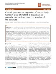
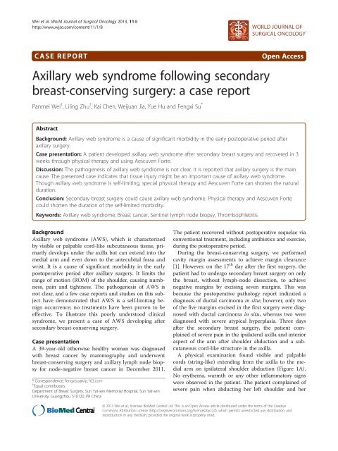
Wei et al. World Journal of Surgical Oncology 2013, 11:8 Page 2 of 4http://www.wjso.com/content/11/1/8Figure 1 Physical examination of patient with axillary <strong>web</strong> <strong>syndrome</strong>. (A) Patient demonstrates axillary <strong>web</strong> <strong>syndrome</strong> in the left arm. Tautcords extending from the axilla to the medial arm are observed. The patient is unable to fully abduct her left shoulder. (B) Taut cords radiate tothe upper outer quadrant of the <strong>breast</strong>. (C) Taut cords radiate to the thoracoabdominal wall.shoulder ROM was reduced from 175° (preoperative) to90°, as measured by a goniometer. She gave her pain ascore of 7 using the Visual Analogue Scale, which is quick,reliable and valid for measuring pain and pain relief [2,3].During follow-up, the patient complained that the cordshad extended to the elbow and the ipsilateral upper outerquadrant of the <strong>breast</strong>, and her thoracoabdominal wallwas also involved (Figure 1B,C). Numbness and tightnesswere present during the entire follow-up. B-type ultrasonographyindicated that the subcutaneous cords thatradiated from the axilla to the medial arm presented anobvious blood flow signal on color Doppler flow imaging,which proved that the cord-like structure was a blood vessel(Figure 2A-D), whereas the cords that radiated to the<strong>breast</strong> and thoracoabdominal wall presented the sameecho of subcutaneous tissue as the contralateral thoracoabdominalwall without cords.Because the patient complained of severe pain andlimited shoulder ROM, we treated her with 300 mg ofAescuven Forte twice a day for one week and advisedher to perform shoulder exercises, including abductingthe shoulder and massaging the cord-like structure for30 minutes every day in the morning and at bedtime assoon as the AWS occurred. After two weeks of followup,the patient presented with pain-free shoulder abduction,a ROM of 150°, and cords that had become smallerand not as significant. Three weeks later, the patientreturned for a physical examination and could abducther shoulder, with a ROM of 170°, without any pain.However, numbness and tightness were still present butsomewhat diminished. The cords were invisible andnon-palpable.DiscussionAWS can develop during many conditions and is consideredto be a type of Mondor’s disease, characterized by athick-wall blood vessel or lymphatic vessel [4,5]. It hasbeen attributed to prior trauma or <strong>breast</strong> <strong>surgery</strong>, such as<strong>breast</strong> reduction, mammoplasty or lumpectomy [6].Moskovitzet al. [7] coined the term AWS and proposed
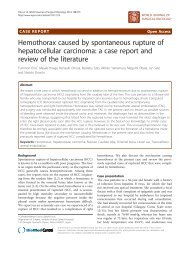
Wei et al. World Journal of Surgical Oncology 2013, 11:8 Page 3 of 4http://www.wjso.com/content/11/1/8Figure 2 B-type ultrasonography of the cords extending from the axilla to the medial arm. (A) The cords under the axilla present Eco of atube structure. (B) The cords in the medial arm present the echo of a tube structure. (C, D) Blood is observed in the tube structures of (A) and(B), respectively.that the pathogenesis is lymphovenous damage, hypercoagulation,superficial venous and lymphatic stasis, and disordersand injuries of tissues as a result of the disruptionof superficial lymphatic and blood vessels during axillary<strong>surgery</strong>. Our patient developed AWS after <strong>secondary</strong><strong>breast</strong> <strong>surgery</strong> without lymph-node dissection to achievenegative margins, which suggests that it was the <strong>secondary</strong><strong>breast</strong> <strong>surgery</strong> that promoted development of AWS ratherthan the original axillar lymph-node biopsy. This differedfrom previous studies, in which stress from the axillarlymph-node dissection or axillar lymph-node biopsy causedAWS[8,9].Weproposethatthetissueinjurycausedbythe<strong>secondary</strong> <strong>surgery</strong> led to the release of inflammatory factors,which caused phlebitis via intravasation in the axilla,medial arm, <strong>breast</strong> and thoracoabdominal wall throughmultiple mechanisms. The hypothesis is consistent with theB-type ultrasonography findings, but was not definitive dueto the lack of a histopathology report. Most of theinvestigators were inclined to define the cord-like structureas a lymph vessel [7,8,10,11].AWS is a self-limiting cause of morbidity in the earlypostoperative period <strong>following</strong> axillary <strong>surgery</strong> [7,8].There are no standard therapeutic methods reported forAWS. Previous studies have indicated that physical therapycan shorten the natural course of AWS to 6 to 8weeks [7,8,12]. In this case, we recommended that thepatient perform physical therapy to alleviate the symptomsand treated her with Aescuven Forte, which is usedto treat phlebitis in the clinic. After the interventions,the duration of AWS in this case was shortened to 3weeks, which suggests that Aescuven Forte might be effectivein improving AWS in combination with physicaltherapy. However, we cannot determine exactly whichtherapeutic method had the better effect on the alleviationof AWS. Therefore, more clinical data should becollected to study the therapy of AWS.