Post-radiation sciatic neuropathy: a case report and review of the ...
Post-radiation sciatic neuropathy: a case report and review of the ...
Post-radiation sciatic neuropathy: a case report and review of the ...

You also want an ePaper? Increase the reach of your titles
YUMPU automatically turns print PDFs into web optimized ePapers that Google loves.
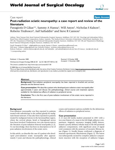
World Journal <strong>of</strong> Surgical OncologyBioMed CentralCase <strong>report</strong><strong>Post</strong>-<strong>radiation</strong> <strong>sciatic</strong> <strong>neuropathy</strong>: a <strong>case</strong> <strong>report</strong> <strong>and</strong> <strong>review</strong> <strong>of</strong> <strong>the</strong>literaturePanagiotis D Gikas* 1 , Sammy A Hanna 1 , Will Aston 2 , Nicholas S Kalson 3 ,Roberto Tirabosco 4 , Asif Saifuddin 5 <strong>and</strong> Steve R Cannon 1Open AccessAddress: 1 Bone Tumour Unit, Royal National Orthopaedics Hospital, Stanmore, Middlesex, HA7 4LP, UK, 2 Oncology <strong>and</strong> Arthroplasty Fellow,Royal Prince Alfred Hospital, Camperdown, Sydney, Australia, 3 The Medical School, University <strong>of</strong> Manchester, Oxford Road, Manchester, M139PT, UK, 4 Department <strong>of</strong> Pathology, Royal National Orthopaedics Hospital, Stanmore, Middlesex, HA7 4LP, UK <strong>and</strong> 5 Department <strong>of</strong> Radiology,Royal National Orthopaedics Hospital, Stanmore, Middlesex, HA7 4LP, UKEmail: Panagiotis D Gikas* - pdgikas@doctors.org.uk; Sammy A Hanna - sammyhanna@hotmail.com;Will Aston - willaston1@googlemail.com; Nicholas S Kalson - nicholas.s.kalson@stud.man.ac.uk;Roberto Tirabosco - roberto.tirabosco@rnoh.nhs.uk; Asif Saifuddin - asif.saifuddin@rnoh.nhs.uk; Steve R Cannon - ttarr@rnoh.nhs.uk* Corresponding authorPublished: 11 December 2008World Journal <strong>of</strong> Surgical Oncology 2008, 6:130 doi:10.1186/1477-7819-6-130This article is available from: http://www.wjso.com/content/6/1/130Received: 10 October 2008Accepted: 11 December 2008© 2008 Gikas et al; licensee BioMed Central Ltd.This is an Open Access article distributed under <strong>the</strong> terms <strong>of</strong> <strong>the</strong> Creative Commons Attribution License (http://creativecommons.org/licenses/by/2.0),which permits unrestricted use, distribution, <strong>and</strong> reproduction in any medium, provided <strong>the</strong> original work is properly cited.AbstractBackground: <strong>Post</strong>-<strong>radiation</strong> peripheral <strong>neuropathy</strong> has been <strong>report</strong>ed in brachial <strong>and</strong> cervicalplexuses <strong>and</strong> <strong>the</strong> femoral nerve.Case presentation: We describe a patient who developed post-<strong>radiation</strong> <strong>sciatic</strong> <strong>neuropathy</strong> afterapproximately 3 years <strong>and</strong> discuss <strong>the</strong> pathophysiology, clinical course <strong>and</strong> treatment optionsavailable for <strong>the</strong> deleterious effects <strong>of</strong> <strong>radiation</strong> to peripheral nerves.Conclusion: This is <strong>the</strong> first <strong>case</strong> <strong>of</strong> post-<strong>radiation</strong> involvement <strong>of</strong> <strong>the</strong> <strong>sciatic</strong> nerve <strong>report</strong>ed in<strong>the</strong> literature.Background<strong>Post</strong>-<strong>radiation</strong> <strong>neuropathy</strong> was first <strong>report</strong>ed in patientstreated with radio<strong>the</strong>rapy to <strong>the</strong> axillary gl<strong>and</strong>s for malignantbreast tumours. It has also been <strong>report</strong>ed in patientstreated for malignant lesions in <strong>the</strong> faciomaxillary region,where <strong>the</strong> cervical plexus, facial or hypoglossal nerveshave been involved. Fur<strong>the</strong>rmore, two <strong>case</strong> <strong>report</strong>s exist in<strong>the</strong> literature <strong>of</strong> post-<strong>radiation</strong> femoral <strong>neuropathy</strong> [1,2].To our knowledge, <strong>the</strong>re has been no description so far <strong>of</strong>post-<strong>radiation</strong> involvement <strong>of</strong> <strong>the</strong> <strong>sciatic</strong> nerve.In this article, we describe <strong>the</strong> <strong>case</strong> <strong>of</strong> a patient who developedpost-<strong>radiation</strong> <strong>sciatic</strong> <strong>neuropathy</strong> after approximately3 years <strong>and</strong> discuss <strong>the</strong> pathophysiology, clinicalcourse <strong>and</strong> treatment options available for <strong>the</strong> deleteriouseffects <strong>of</strong> <strong>radiation</strong> to peripheral nerves.Case presentationA 22 year-old media student presented in 2001 with atwo-year history <strong>of</strong> a mass in her left thigh adductor compartment.Magnetic resonance imaging (MRI) demonstrateda poorly defined, lobular mass in <strong>the</strong> left proximaladductor compartment, with significant areas <strong>of</strong> signalvoid consistent with <strong>the</strong> presence <strong>of</strong> excessive fibrous tissue(Figure 1). Needle biopsy confirmed a diagnosis <strong>of</strong>musculo-aponeurotic fibromatosis (Figure 2). The lesionwas subsequently excised by complete adductor compartmentresection with <strong>the</strong> exception <strong>of</strong> adductor longus.Page 1 <strong>of</strong> 5(page number not for citation purposes)
World Journal <strong>of</strong> Surgical Oncology 2008, 6:130http://www.wjso.com/content/6/1/130MRI Figure <strong>of</strong> <strong>the</strong> 1 left thighMRI <strong>of</strong> <strong>the</strong> left thigh. Axial T1W SE (a) <strong>and</strong> coronal STIR (b) images showing a poorly defined, lobular mass in <strong>the</strong> left adductorcompartment (arrows) showing extensive areas <strong>of</strong> signal void due to fibrous tissue. Note <strong>the</strong> location <strong>of</strong> <strong>the</strong> <strong>sciatic</strong> nerve(arrowhead).Typical microsopic features <strong>of</strong> musculoaponeurotic fibromatosisFigure 2Typical microsopic features <strong>of</strong> musculoaponeurotic fibromatosis. Interlacing bundles <strong>of</strong> uniform spindle-shaped cellswith pale oval nuclei <strong>and</strong> eosinophilic cytoplasm; <strong>the</strong>re is a prominent collagen stroma.Page 2 <strong>of</strong> 5(page number not for citation purposes)
World Journal <strong>of</strong> Surgical Oncology 2008, 6:130http://www.wjso.com/content/6/1/130<strong>Post</strong>-operatively <strong>the</strong> patient completed a course <strong>of</strong> radio<strong>the</strong>rapy,receiving a total dose <strong>of</strong> 50 Gy in 25 fractionsover five weeks.Towards <strong>the</strong> end <strong>of</strong> 2002, she developed a swelling in <strong>the</strong>postero-medial thigh, distal to <strong>the</strong> previous ir<strong>radiation</strong>field. MRI confirmed recurrence just above <strong>the</strong> level <strong>of</strong> <strong>the</strong>femoral condyles. In December 2002, she underwent fur<strong>the</strong>rresection followed by a fur<strong>the</strong>r course <strong>of</strong> radio<strong>the</strong>rapy(30 Gy in 15 fractions over 4 weeks) with an inch overlapwith <strong>the</strong> previous <strong>radiation</strong> field superiorly.In June 2003, <strong>the</strong> patient developed a fur<strong>the</strong>r proximalthigh recurrence in <strong>the</strong> previously surgically treated area,within <strong>the</strong> initial radio<strong>the</strong>rapy field. She was started onTamoxifen <strong>and</strong> fur<strong>the</strong>r excision performed.In March 2004, she started complaining <strong>of</strong> progressiveweakness <strong>of</strong> dorsiflexion <strong>of</strong> her left foot, associated withpain around <strong>the</strong> medial aspect <strong>of</strong> <strong>the</strong> foot <strong>and</strong> sole. Onclinical examination, she had normal hip flexion/extension<strong>and</strong> abduction with almost absent adduction, <strong>and</strong>normal knee flexion <strong>and</strong> extension. Foot flexion <strong>and</strong>inversion was 4/5. Foot <strong>and</strong> toe extension <strong>and</strong> eversionwere 2/5. The ankle tendon reflex was absent. There wasnormal sensation on <strong>the</strong> anterior <strong>and</strong> posterior aspects <strong>of</strong>her thigh indicating that <strong>the</strong> posterior cutaneous nerve <strong>of</strong><strong>the</strong> thigh coming from <strong>the</strong> sacral plexus was intact <strong>and</strong>hence that any lesion was distal to <strong>the</strong> sacral plexus. Shehad almost no sensation in <strong>the</strong> sole <strong>of</strong> her foot withreduced perception <strong>of</strong> touch on <strong>the</strong> dorsum <strong>of</strong> her foot.When palpating along <strong>the</strong> course <strong>of</strong> <strong>the</strong> <strong>sciatic</strong> nerve ara<strong>the</strong>r dense region <strong>of</strong> local scarring was present on <strong>the</strong>posterior aspect <strong>of</strong> <strong>the</strong> thigh approximately 10 cm from<strong>the</strong> knee.Electrophysiological assessment indicated a non localizing<strong>sciatic</strong> nerve <strong>sciatic</strong> nerve injury. Based on <strong>the</strong> clinicalfindings <strong>and</strong> investigations, a diagnosis <strong>of</strong> <strong>radiation</strong>inducedinjury to <strong>the</strong> <strong>sciatic</strong> nerve was made, affecting <strong>the</strong>common peroneal portion more that <strong>the</strong> tibial portion.Repeat MRI <strong>of</strong> <strong>the</strong> left thigh demonstrated a seroma in <strong>the</strong>left groin <strong>and</strong> diffuse oedema <strong>and</strong> swelling <strong>of</strong> <strong>the</strong> left <strong>sciatic</strong>nerve (Figure 3). A decision to perform neurolysis <strong>of</strong><strong>the</strong> <strong>sciatic</strong> nerve was made, with a view to freeing <strong>the</strong> nervefrom any associated scar tissue, <strong>the</strong>reby halting any fur<strong>the</strong>rdeterioration in function.At <strong>the</strong> most recent follow up in 2008, <strong>the</strong> patient is freefrom recurrence. There has been no fur<strong>the</strong>r deteriorationin <strong>sciatic</strong> nerve function but weakness <strong>of</strong> foot dorsiflexionpersists, necessitating use <strong>of</strong> a splint.Follow up MRI <strong>of</strong> <strong>the</strong> thighFigure 3Follow up MRI <strong>of</strong> <strong>the</strong> thigh. Coronal STIR (a) <strong>and</strong> axial fat suppressed T2W FSE (b) images showing diffuse swelling <strong>and</strong>oedema <strong>of</strong> <strong>the</strong> <strong>sciatic</strong> nerve (arrows) <strong>and</strong> a postoperative seroma in <strong>the</strong> groin (arrowhead).Page 3 <strong>of</strong> 5(page number not for citation purposes)
World Journal <strong>of</strong> Surgical Oncology 2008, 6:130http://www.wjso.com/content/6/1/130DiscussionPathophysiology <strong>and</strong> clinical courseVery little is known about <strong>the</strong> pathophysiology <strong>and</strong> <strong>the</strong>histopathological changes that occur in peripheral nervesafter <strong>the</strong>rapeutic ir<strong>radiation</strong>. Early experimental studiesindicated that <strong>the</strong> peripheral nerves are extremely radioresistant.However, <strong>the</strong> follow up time was short <strong>and</strong> it islikely that <strong>the</strong> injury did not have an opportunity todevelop [3].Today we know that post-ir<strong>radiation</strong> <strong>neuropathy</strong> occursboth directly <strong>and</strong> indirectly: directly by <strong>the</strong> harmful effect<strong>of</strong> <strong>the</strong> <strong>radiation</strong> on <strong>the</strong> nerve itself, <strong>and</strong> indirectly by <strong>the</strong>fibrosis that <strong>radiation</strong> causes in <strong>the</strong> tissue around <strong>the</strong>nerve [2].Direct effects <strong>of</strong> ir<strong>radiation</strong> on nerve include bioelectricalalterations (subnormal action potentials, altered conductiontime), enzyme changes, abnormal microtubuleassembly, altered vascular permeability <strong>and</strong> neurilemmaldamage. All <strong>of</strong> <strong>the</strong>se changes are observed experimentallywithin 2 days after ir<strong>radiation</strong> <strong>and</strong> are all dose dependent<strong>and</strong> irreversible [4-6].The secondary damage to <strong>the</strong> nerve is due to <strong>the</strong> extensivefibrosis <strong>of</strong> <strong>the</strong> connective tissue around <strong>the</strong> nerve, whichbecomes densely hyalinised. There is also a progressiveloss <strong>of</strong> elasticity <strong>and</strong> <strong>the</strong> development <strong>of</strong> contractures thatultimately consolidate <strong>the</strong> adjacent structures with <strong>the</strong>nerve. In addition, <strong>the</strong> decreased vascularity <strong>of</strong> <strong>the</strong> areamay destroy some adjacent peripheral nerves. Regeneration<strong>of</strong> <strong>the</strong> affected nerves may be impeded [2]. In a <strong>report</strong><strong>of</strong> findings at autopsy in two patients who had post-ir<strong>radiation</strong>brachial-plexus syndrome [7], varying degrees <strong>of</strong>fibrosis <strong>of</strong> <strong>the</strong> neurilemma, as well as demyelinization<strong>and</strong> fibrous replacement <strong>of</strong> <strong>the</strong> fibrils, were described.Mendes et al. in histological examination <strong>of</strong> femoral nervebranches removed during surgical decompression <strong>of</strong> <strong>the</strong>femoral nerve, in a patient with post-ir<strong>radiation</strong> femoral<strong>neuropathy</strong>, also found demyelinated nerve fibres surroundedby abundant scar tissue with areas <strong>of</strong> hyalinization[2].Peripheral nerve damage is a rare but underst<strong>and</strong>ablymajor complication <strong>of</strong> <strong>radiation</strong> <strong>the</strong>rapy associated withsignificant morbidity. The frequency <strong>of</strong> injury <strong>report</strong>edfrom some <strong>of</strong> <strong>the</strong> older studies is probably higher thanwould occur today as prior to <strong>the</strong> advent <strong>of</strong> CT <strong>and</strong> MRI,larger fields were used because <strong>of</strong> greater uncertaintyabout <strong>the</strong> dimensions <strong>of</strong> <strong>the</strong> tumour.In studies looking into post-ir<strong>radiation</strong> <strong>neuropathy</strong>involving <strong>the</strong> brachial <strong>and</strong> cervical plexuses after radio<strong>the</strong>rapyfor breast carcinoma it was found that symptomsgenerally begin within one to two years after treatment<strong>and</strong> are initially mainly sensory (e.g. burning pain, numbness,pares<strong>the</strong>sia) [7,8]. Any motor deficits that developare usually delayed for about eighteen months <strong>and</strong>include paresis <strong>of</strong> a group <strong>of</strong> muscles <strong>and</strong> complete paralysis<strong>of</strong> <strong>the</strong> arm [9]. Stoll et al. <strong>and</strong> Powell et al. have bothfound a direct relationship between <strong>the</strong> dosage <strong>of</strong> <strong>radiation</strong><strong>and</strong> <strong>the</strong> severity/time <strong>of</strong> appearance <strong>of</strong> symptoms[7,10].In a <strong>review</strong> <strong>of</strong> <strong>radiation</strong> injury to peripheral nerves publishedby Giese <strong>and</strong> Kinsella <strong>the</strong> authors conclude thatperipheral <strong>neuropathy</strong> is relatively infrequent at lowerdoses per fraction [11]. They expressed concern that c<strong>of</strong>actorssuch as radiosensitizers, chemo<strong>the</strong>rapeutic agents<strong>and</strong> surgical manipulations could possibly increase <strong>the</strong>incidence. Breast cancer patients receiving cytotoxic chemo<strong>the</strong>rapyhad a higher incidence <strong>of</strong> <strong>radiation</strong> induced brachialplexopathy compared to those having <strong>radiation</strong> onlyfollowing mastectomy [12].Any peripheral nerve may be affected by post-<strong>radiation</strong><strong>neuropathy</strong> <strong>and</strong> it is likely that <strong>the</strong> unique location <strong>of</strong> thistumour reflects 1) <strong>the</strong> special site <strong>of</strong> <strong>radiation</strong> <strong>the</strong>rapy <strong>and</strong>2) <strong>the</strong> repeated doses <strong>of</strong> <strong>radiation</strong> administered [12].Latency is an important factor to be considered when evaluatingnerve injury [12]. Stoll <strong>and</strong> Andrews did notobserve any <strong>neuropathy</strong> occurring before 5 months, witha majority occurring between 10 <strong>and</strong> 22 months after ir<strong>radiation</strong>.They also noted that <strong>the</strong> higher dose group didshow signs earlier than <strong>the</strong> lower dose group. Powell et al.did not observe any nerve injury prior to 10 months postir<strong>radiation</strong>,whereas <strong>report</strong>s exist in <strong>the</strong> literature <strong>of</strong> neuropathiesoccurring as late as 11 years after ir<strong>radiation</strong> forbreast cancer. Therefore, latency, as our <strong>case</strong> demonstrates,is a very important factor to be considered, sinceshort follow-up times may underestimate <strong>the</strong> true incidence<strong>of</strong> post-ir<strong>radiation</strong> injury to peripheral nerves.ManagementWhen considering management <strong>of</strong> post-ir<strong>radiation</strong>peripheral <strong>neuropathy</strong>, it is important to realise that anunalterable condition is <strong>the</strong> status <strong>of</strong> <strong>the</strong> patient's underlyingmalignancy prior to initiation <strong>of</strong> treatment, includingtumour size, location <strong>and</strong> structures involved/destroyed [12]. Fur<strong>the</strong>rmore, release <strong>of</strong> entrapped nervesfrom a fibrous mass can be challenging even for <strong>the</strong> mostskilled surgeon. Therefore, a short life expectancy coupledwith uncertainty <strong>of</strong> recovery from surgical interventionmake conservative management more appropriate.Also important in overall response to <strong>and</strong> recovery from<strong>the</strong>rapy is <strong>the</strong> general health <strong>of</strong> <strong>the</strong> patient <strong>and</strong>, if a child,<strong>the</strong> stage <strong>of</strong> development <strong>and</strong> growth [12]. If surgery is apart <strong>of</strong> <strong>the</strong> overall treatment, as was in our <strong>case</strong>, <strong>the</strong>n <strong>the</strong>Page 4 <strong>of</strong> 5(page number not for citation purposes)
World Journal <strong>of</strong> Surgical Oncology 2008, 6:130http://www.wjso.com/content/6/1/130extent <strong>of</strong> <strong>the</strong> surgical resection <strong>and</strong> <strong>the</strong> techniques usedare also <strong>of</strong> major importance to post-<strong>the</strong>rapy function. Inaddition, <strong>the</strong> long-term s<strong>of</strong>t tissue response to <strong>radiation</strong> isa complex function <strong>of</strong> many <strong>radiation</strong> related factors(total dose, dose volume <strong>and</strong> distribution, fraction size,dose rate, treatment interval <strong>and</strong> overall treatment time)some <strong>of</strong> which are poorly understood. Important non<strong>radiation</strong>factors that play a role in influencing <strong>the</strong> development,progression <strong>and</strong> response to treatment <strong>of</strong> post<strong>radiation</strong><strong>neuropathy</strong>, include o<strong>the</strong>r <strong>the</strong>rapies such as surgery<strong>and</strong> chemo<strong>the</strong>rapy, major organ system performance,overall activity level <strong>and</strong> chronic conditions such ashypertension, diabetes <strong>and</strong> connective tissue disorders.In patients who have a good life expectancy after tumourexcision, an increasing motor deficit <strong>and</strong>/or intolerablepain in <strong>the</strong> distribution <strong>of</strong> a peripheral nerve may presentsome years after <strong>the</strong> initial treatment. Some patients mayrequire surgical release <strong>of</strong> <strong>the</strong> <strong>radiation</strong> induced scar tissuesurrounding <strong>the</strong> nerve. However, <strong>the</strong> patient has to beaware <strong>of</strong> <strong>the</strong> uncertainty <strong>of</strong> recovery.When a decision has been made to pursue a surgical path,treatment should not be delayed as research has shownthat pathological changes in a peripheral nerve restrictedby fibrosis are progressive.AcknowledgementsWe thank <strong>the</strong> patient for <strong>the</strong>ir permission to write <strong>the</strong>ir <strong>case</strong> <strong>report</strong>.References1. Love S: An experimental study <strong>of</strong> peripheral nerve regenerationafter x-ir<strong>radiation</strong>. Brain 1983, 106(Pt 1):39-54.2. Mendes DG, Nawalkar RR, Eldar S: <strong>Post</strong>-ir<strong>radiation</strong> femoral <strong>neuropathy</strong>.A <strong>case</strong> <strong>report</strong>. J Bone Joint Surg Am 1991, 73:137-140.3. Janzen AH, Warren S: Effect <strong>of</strong> roentgen rays on <strong>the</strong> peripheralnerve <strong>of</strong> <strong>the</strong> rat. Radiology 1942, 38:333-337.4. Calvo W, Forteza-Vila J: Glycogen changes in bone marrownerves after whole-body x-ir<strong>radiation</strong>. Acta Neuropathol 1972,20:78-83.5. Coss RA, Bamburg JR, Dewey WC: The effects <strong>of</strong> X ir<strong>radiation</strong>on microtubule assembly in vitro. Radiat Res 1981, 85:99-115.6. Krayevskii NA: Studies in <strong>the</strong> pathology <strong>of</strong> <strong>radiation</strong> disease. New York1965.7. Stoll BA, Andrews JT: Radiation-induced peripheral <strong>neuropathy</strong>.British Medical Journal 1966, 1:834-837.8. Westling P, Svensson H, Hele P: Cervical plexus lesions followingpost-operative <strong>radiation</strong> <strong>the</strong>rapy <strong>of</strong> mammary carcinoma.Acta Radiol Ther Phys Biol 1972, 11:209-216.9. Suit HD, Russell WO, Martin RG: Management <strong>of</strong> patients withsarcoma <strong>of</strong> s<strong>of</strong>t tissue in an extremity. Cancer 1973,31:1247-1255.10. Powell S, Cooke J, Parsons C: Radiation-induced brachial plexusinjury: follow-up <strong>of</strong> two different fractionation schedules.Radio<strong>the</strong>r Oncol 1990, 18:213-220.11. Giese WL, Kinsella TJ: Radiation injury to peripheral <strong>and</strong> cranialnerves. In Radiation injury to <strong>the</strong> Nervous System Edited by: Gutin PH,Leibel SA, Sheline G. New York: Raven Press; 1991:282-403.12. Olsen NK, Pfeiffer P, Johannsen L, Schroder H, Rose C: Radiationinducedbrachial plexopathy: neurological follow-up in 161recurrence-free breast cancer patients. Int J Radiat Oncol BiolPhys 1993, 26:43-49.ConclusionDespite being a rare entity, post-<strong>radiation</strong> peripheral <strong>neuropathy</strong>can be associated with significant morbidity. Fur<strong>the</strong>rresearch is crucial in identifying <strong>the</strong> majorpathophysiological mechanisms, both direct <strong>and</strong> indirect,underlying damage to peripheral nerves following <strong>the</strong>rapeutic<strong>radiation</strong>. A good underst<strong>and</strong>ing <strong>of</strong> pathophysiologyat a cellular/molecular level is essential for <strong>the</strong>development, in <strong>the</strong> future, <strong>of</strong> appropriate prophylacticmeasures for people requiring radio<strong>the</strong>rapy.ConsentWritten informed consent was obtained from <strong>the</strong> patientfor publication <strong>of</strong> this <strong>case</strong> <strong>report</strong> <strong>and</strong> any accompanyingimages. A copy <strong>of</strong> <strong>the</strong> written consent is available for<strong>review</strong> by <strong>the</strong> Editor-in-Chief <strong>of</strong> this journal.Competing interestsThe authors declare that <strong>the</strong>y have no competing interests.Authors' contributionsPG, WA, SH, <strong>and</strong> NSK <strong>review</strong>ed <strong>the</strong> literature, wrote <strong>the</strong>Background <strong>and</strong> Case presentation sections, <strong>the</strong> Conclusion<strong>and</strong> edited <strong>the</strong> manuscript. RT described <strong>the</strong> histologicalfindings <strong>and</strong> confirmed <strong>and</strong> edited <strong>the</strong> manuscript. ASdescribed <strong>the</strong> radiological findings <strong>and</strong> confirmed <strong>and</strong>edited <strong>the</strong> manuscript. SRC conceived <strong>the</strong> <strong>case</strong> <strong>report</strong> <strong>and</strong>helped draft <strong>the</strong> manuscript.Publish with BioMed Central <strong>and</strong> everyscientist can read your work free <strong>of</strong> charge"BioMed Central will be <strong>the</strong> most significant development fordisseminating <strong>the</strong> results <strong>of</strong> biomedical research in our lifetime."Sir Paul Nurse, Cancer Research UKYour research papers will be:available free <strong>of</strong> charge to <strong>the</strong> entire biomedical communitypeer <strong>review</strong>ed <strong>and</strong> published immediately upon acceptancecited in PubMed <strong>and</strong> archived on PubMed Centralyours — you keep <strong>the</strong> copyrightBioMedcentralSubmit your manuscript here:http://www.biomedcentral.com/info/publishing_adv.aspPage 5 <strong>of</strong> 5(page number not for citation purposes)