Protein Folding, Structure and Function - Fox Chase Cancer Center
Protein Folding, Structure and Function - Fox Chase Cancer Center
Protein Folding, Structure and Function - Fox Chase Cancer Center

You also want an ePaper? Increase the reach of your titles
YUMPU automatically turns print PDFs into web optimized ePapers that Google loves.
<strong>Protein</strong> <strong>Folding</strong>, <strong>Structure</strong> <strong>and</strong> <strong>Function</strong><br />
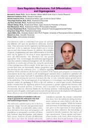
Heinrich Roder, Ph.D., Senior Member; Adjunct Professor of Biochemistry <strong>and</strong> Biophysics, <strong>and</strong> Member<br />
of the Graduate Group in Biochemistry <strong>and</strong> Molecular Biophysics, University of Pennsylvania<br />
Hong Cheng, Ph.D., Staff Scientist<br />
Harvey H. Hensley, Ph.D., Staff Scientist<br />
Dharmaraj Samuel, Ph.D., Postdoctoral Fellow<br />
Pavel Elistratov, * Graduate Student, Moscow State University,<br />
Moscow, Russia<br />
Paul W. Riley, B.S., Graduate Student, Temple University,<br />
Philadelphia, PA<br />
Colin M. Hayden, * Student Assistant, La Salle University,<br />
Philadelphia, PA<br />
Lisa Wang, * Student Assistant, Germantown Academy,<br />
Fort Washington, PA<br />
Much of our research is aimed at underst<strong>and</strong>ing the mechanism<br />
of protein folding, focusing primarily on early structural<br />
events in the transition from the unfolded to the native<br />
(folded) state of a protein. Many proteins encounter partially organized structures during early<br />
stages of folding, <strong>and</strong> these are thought to be important for directing a protein toward its unique<br />
native conformation. However, important questions remain to be answered concerning the nature<br />
<strong>and</strong> origin of the kinetic barriers encountered during early stages of folding <strong>and</strong> the structural properties<br />
of the intermediate states. Are compact partially folded states the result of a non-specific chain<br />
collapse or more specific folding events? What is the relative importance of local vs. long-range interactions?<br />
Do intermediates contain native-like tertiary interactions? For some proteins, intermediates<br />
are populated even at equilibrium, <strong>and</strong> this raises further questions concerning the origin of structural<br />
cooperativity, the relationship between kinetic <strong>and</strong> thermodynamic intermediates, <strong>and</strong> any<br />
residual interactions remaining in the denatured state.<br />
We have addressed these fundamental questions by coupling advanced mixing techniques for<br />
rapid initiation of folding reactions with a variety of detection methods, including intrinsic <strong>and</strong><br />
extrinsic fluorescence probes <strong>and</strong> H/D exchange labeling experiments with NMR detection. We use<br />
these approaches, often in combination with protein engineering, to gain detailed insight into the<br />
folding mechanism of a diverse set of proteins (for a recent comprehensive review, see (1)).<br />
Studies of protein folding in vitro provide the necessary framework for investigating the folding of<br />
proteins in their cellular environments, protein trafficking <strong>and</strong> degradation. <strong>Protein</strong> stability <strong>and</strong><br />
folding also play a central role in our underst<strong>and</strong>ing of the biological consequences of mutations<br />
<strong>and</strong> in de novo design of proteins. Moreover, insight into the properties of protein-folding intermediates<br />
is critical for a mechanistic underst<strong>and</strong>ing <strong>and</strong> treatment of a wide range of diseases that<br />
can be linked to the aggregation of partially denatured or misfolded forms of proteins.<br />
We are especially interested in elucidating the structural basis for formation of amyloid fibrils,<br />
using fragments of the human prion as a test case. By coupling NMR with quenched hydrogen<br />
exchange techniques, we can observe the degree of solvent protection in the fibrillar state of an<br />
amyloidogenic peptide or protein. Together with molecular dynamics simulations (with<br />
R.L. Dunbrack § ), the data provide unique insight into the structural basis of prion protein fibril formation.<br />
We also rely on rapid mixing methods to explore the folding mechanism of the human<br />
prion protein in order to gain insight into the role of partially folded intermediates in the conversion<br />
of the normal cellular form of the protein into its cytotoxic <strong>and</strong> infectious aggregates (in collaboration<br />
with A.C. Apetri a <strong>and</strong> W.K. Surewicz a ).<br />
In other ongoing projects we make extensive use of multidimensional NMR methods to investigate<br />
the structure, dynamics <strong>and</strong> molecular interactions of various proteins of biological <strong>and</strong>/or clinical<br />
interest, including blood coagulation factor XI (with P.N. Walsh b ), domain 5 of high molecular weight<br />
kininogen <strong>and</strong> its role in angiogenesis (with R.W. Colman b ) <strong>and</strong> protein modules involved in signal<br />
transduction, such as CARD domains (with Y.C. Park § ) <strong>and</strong> PDZ domains (with Z. Bu § ).<br />
<strong>Fox</strong> <strong>Chase</strong> <strong>Cancer</strong> <strong>Center</strong> 2005 Scientific Report 1
Kinetics of loop formation <strong>and</strong> breakage in<br />
unfolded cytochrome c.<br />
Roder, in collaboration<br />
with Kurchan, d Bowlerd<br />
The formation of loops is among the earliest<br />
events in protein folding. By measuring the rate<br />
of formation for different loops in unfolded<br />
proteins, we can gain insight into the dynamic<br />
properties of the denatured state <strong>and</strong> detect any<br />
residual tertiary interactions. Conversely, the<br />
rate of loop breakage is related to the persistence<br />
of nascent structures during initial stages<br />
of folding. Cytochrome c offers a unique opportunity<br />
to study these dynamic events because of<br />
the strong affinity of the heme iron for deprotonated<br />
histidines, (see Figure 1a).<br />
His18 remains<br />
bound to the heme under both native <strong>and</strong> denaturing<br />
conditions while additional histidines<br />
elsewhere in the sequence (in blue in Figure 1a)<br />
have a strong tendency to form non-native<br />
lig<strong>and</strong> interactions in the denatured state.<br />
Rates of loop formation <strong>and</strong> breakage were<br />
measured by monitoring the heme absorbance<br />
changes associated with pH jumps in either<br />
direction across the histidine titration range<br />
(2). The use of continuous- <strong>and</strong> stopped-flow<br />
mixing methods allowed us to measure the pHdependent<br />
rates for a series of variants of yeast<br />
cytochrome c containing engineered His residues<br />
at several positions designed to form<br />
loops ranging in size from 16 to 83 residues.<br />
Figure 1b<br />
shows representative kinetic results<br />
for the formation (red symbols) <strong>and</strong> breakage<br />
(black symbols) of a 56-residue loop involving<br />
a His at position 73. A simple two-step mechanism<br />
allowed us to extract the elementary rates<br />
of loop formation, kf,<br />
<strong>and</strong> breakage, kb,<br />
as a<br />
function of loop size. The magnitude <strong>and</strong> range<br />
of kf<br />
values (300–7000 s–1)<br />
measured for different<br />
variants ( Figure 1c)<br />
are consistent with loop<br />
formation being reaction-limited rather than<br />
diffusion-limited. The observed decrease in kf<br />
with increasing loop size yields a scaling factor of<br />
–1.8. This is consistent with the values expected<br />
for a r<strong>and</strong>om coil with excluded volume. The<br />
rate of loop breakage, kb,<br />
is lower than the rate<br />
of dissociation of free histidine from the metal<br />
ion<strong>and</strong> goes through a minimum at an intermediate<br />
loop size of 37 residues. Our conclusion<br />
that a substantial amount of residual tertiary<br />
structure persists in the denatured state is<br />
Figure 1. a) Schematic illustration of loop formation in unfolded cytochrome c mediated by histidine-heme ligation.<br />
Since only the neural form of His can ligate the positively charged heme iron, loop formation is coupled with<br />
deprotonation of the His side chain, <strong>and</strong> vice versa. b) Plot of the observed rate of His73 linkage to the heme in<br />
iso-1 cytochrome c vs. pH. Rates of loop formation (kf, squares) <strong>and</strong> breakage (kb, circles) were measured in<br />
upward <strong>and</strong> downward pH jump experiments, respectively, using stopped-flow (red) or continuous-flow (black)<br />
methods. c) Logarithmic plot of the rate of loop formation, kf vs. loop size, n. The observed slope (–1.8) is consistent<br />
with the loop closure dynamics of a Gaussian (r<strong>and</strong>om) chain with excluded volume.<br />
<strong>Fox</strong> <strong>Chase</strong> <strong>Cancer</strong> <strong>Center</strong> 2005 Scientific Report<br />
2
further supported by the observed variation in<br />
loop breakage rates for different histidines.<br />
Structural analysis of partially unfolded states<br />
of cytochrome c by NMR. Latypov, Cheng, Wang,<br />
Roder<br />
In contrast to other small proteins, cytochrome<br />
c is found to populate partially structured intermediate<br />
states not only in kinetic experiments,<br />
but also under certain equilibrium conditions.<br />
To gain further insight into the structure <strong>and</strong><br />
stability of such equilibrium intermediates, we<br />
monitored the reversible denaturant-induced<br />
unfolding transition of oxidized horse cytochrome<br />
c using a variety of spectroscopic<br />
methods (3), such as, fluorescence, circular<br />
dichroism (CD) <strong>and</strong> heme absorbance b<strong>and</strong>s.<br />
The normalized transition curves for different<br />
probes diverge between 1 <strong>and</strong> 2.5 M GuHCl,<br />
indicating that a partially unfolded intermediate<br />
state is populated under mildly denaturing<br />
conditions. The use of global fitting techniques<br />
allowed us to simultaneously fit a three-state<br />
equilibrium model to the combined data, <strong>and</strong><br />
thus revealed the spectroscopic properties of<br />
the equilibrium intermediate. In particular, a<br />
global three-state fit of the heme absorbance<br />
data in the Soret region (350–450 nm) made it<br />
possible to deconvolute the heme absorbance<br />
spectrum of the intermediate. To rule out the<br />
possibility that heme misligation plays a role in<br />
stabilizing intermediates, we showed that a cyt<br />
c variant in which the predominant non-native<br />
lig<strong>and</strong>, His33, is replaced by Asn shows identi-<br />
cal three-state equilibrium behavior as the<br />
wild-type protein.<br />
The fact that the cytochrome c intermediate<br />
is well populated within the equilibrium<br />
unfolding transition (e.g., it reaches nearly 50%<br />
of the total population in 4.3 M urea) makes it<br />
possible to study its structural properties in<br />
more detail by the use of NMR (3). By recording<br />
a series of 2D 1H-15N<br />
correlation NMR<br />
spectra vs. urea concentration, we were able to<br />
follow the changes in peak intensity for individual<br />
backbone amide groups ( Figure 2).<br />
Some residues show greatly diminished intensity<br />
already at low urea concentrations (blue in<br />
Figure 2),<br />
while others, especially those in<br />
α-helical<br />
regions (red), remain at their native<br />
level prior to the main unfolding transition.<br />
The main unfolding transition gives rise to a<br />
new set of sharp peaks at r<strong>and</strong>om-coil positions<br />
(green). The intensities of the blue peaks track<br />
the population of the native state (N) predicted<br />
by the optical data (solid line) while the red<br />
peaks follow the urea dependence expected for<br />
the combined population of native <strong>and</strong> intermediate<br />
states (I). The NMR data thus provide<br />
striking evidence for the formation of a partially<br />
structured state at intermediate urea concentrations.<br />
While many peaks approach the limiting<br />
cases indicated by the blue <strong>and</strong> red lines in<br />
Figure2a,<br />
others fall in between, indicating<br />
partial disruption or dynamic contributions. In<br />
Figure 2b<br />
the data are parametrized in terms of<br />
a fractional contribution of the intermediate<br />
to the intensity of each assigned HSQC peak.<br />
Figure 2. NMR analysis of an equilibrium-folding intermediate in cytochrome c.<br />
a) 15N-1H<br />
cross peak intensities<br />
vs. urea concentrations, including residues in locally unfolded (blue) <strong>and</strong> structured (red) regions of the intermediate<br />
sate. Representative peaks assigned to the fully unfolded form are shown in green. b) Fractional contribution<br />
of intermediate population to N-state peak intensity. Values near 1 indicated regions that retain native-like structure<br />
<strong>and</strong> values near 0 indicate locally unfolded segments.<br />
<strong>Fox</strong> <strong>Chase</strong> <strong>Cancer</strong> <strong>Center</strong> 2005 Scientific Report<br />
3
Values near 1 indicating ordered regions are<br />
seen mainly for the relatively hydrophobic<br />
segments of the cytochrome c sequence,<br />
including the three α-helices<br />
<strong>and</strong> a cluster of<br />
apolar residues in the 30s. Together with the<br />
optical studies (3), the results indicate that the<br />
partially unfolded form of cytochrome c popu-<br />
Publications<br />
1.<br />
2.<br />
3.<br />
lated at low denaturant concentrations contains<br />
many native-like features, including a cluster of<br />
stable α-helices<br />
<strong>and</strong> a partially buried heme<br />
group. On the other h<strong>and</strong>, the native Met80iron<br />
linkage <strong>and</strong> other specific tertiary interactions<br />
are disrupted in the intermediate, <strong>and</strong><br />
most of the loop regions are disordered.<br />
Roder, H., Maki, K., Cheng, H. Early events in protein folding explored by rapid mixing methods. Chem. Rev.<br />
(in press).<br />
Kurchan, E., Roder, H., Bowler, B.E. Kinetics of loop formation <strong>and</strong> breakage in the denatured state of iso-1-cytochrome<br />
c. J. Mol. Biol. 353:730-743,<br />
2005.<br />
Latypov, R.F., Cheng, H., Roder, N.A., Zhang, J., Roder, H. Structural characterization of an equilibrium unfolding<br />
intermediate in cytochrome c.<br />
J. Mol. Biol. 357:1009-1025,<br />
2006.<br />
§ <strong>Fox</strong> <strong>Chase</strong> researcher<br />
* Personnel left <strong>Fox</strong> <strong>Chase</strong><br />
a A.C. Apetri, W.K. Surewicz: Case Western Reserve University, Clevel<strong>and</strong>, OH<br />
b P.N. Walsh, R.W. Colman: Temple University, Philadelphia, PA<br />
c E.W. Kurchan, B.E. Bowler: University of Denver, Denver, CO<br />
<strong>Fox</strong> <strong>Chase</strong> <strong>Cancer</strong> <strong>Center</strong> 2005 Scientific Report<br />
4