

Understanding Hair Transplants and Hair Loss - Pacific Hair
Understanding Hair Transplants and Hair Loss - Pacific Hair
Understanding Hair Transplants and Hair Loss - Pacific Hair

You also want an ePaper? Increase the reach of your titles
YUMPU automatically turns print PDFs into web optimized ePapers that Google loves.
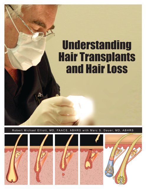
<strong>Underst<strong>and</strong>ing</strong><br />
<strong>Hair</strong> <strong>Transplants</strong><br />
<strong>and</strong> <strong>Hair</strong> <strong>Loss</strong><br />
R o b e r t M i c h a e l E l l i o t t , M D , FA A C S , A B H R S w i t h M a r c S . D a u e r, M D , A B H R S
Contents<br />
What is a <strong>Hair</strong> Transplant? . . . . . . . . . . . . . . . . . . . . . . . . . . . . . . . . . . . . 4<br />
Proper Diagnosis of <strong>Hair</strong> <strong>Loss</strong> . . . . . . . . . . . . . . . . . . . . . . . . . . . . . . . . . . 4<br />
Pattern <strong>Hair</strong> <strong>Loss</strong> . . . . . . . . . . . . . . . . . . . . . . . . . . . . . . . . . . . . . . . . . 5<br />
Non-pattern <strong>Hair</strong> <strong>Loss</strong> . . . . . . . . . . . . . . . . . . . . . . . . . . . . . . . . . . . . . . 6<br />
<strong>Hair</strong> Shedding . . . . . . . . . . . . . . . . . . . . . . . . . . . . . . . . . . . . . . . . . . 6<br />
Causes of <strong>Hair</strong> Shedding . . . . . . . . . . . . . . . . . . . . . . . . . . . . . . . . . . . . . 6<br />
Focal Non-Scarring Alopecia . . . . . . . . . . . . . . . . . . . . . . . . . . . . . . . . . . . 7<br />
Scarring Alopecias . . . . . . . . . . . . . . . . . . . . . . . . . . . . . . . . . . . . . . . . 7<br />
<strong>Hair</strong> Breakage, Causes . . . . . . . . . . . . . . . . . . . . . . . . . . . . . . . . . . . . . 7<br />
Pattern Baldness Versus Generalized Diffuse <strong>Hair</strong> <strong>Loss</strong> . . . . . . . . . . . . . . . . . . . . 8<br />
Comprehensive Medical Workup for Diffuse Thinning <strong>and</strong> <strong>Hair</strong> <strong>Loss</strong> . . . . . . . . . . . . . . 8<br />
Medical Treatment – Male Pattern <strong>Hair</strong> <strong>Loss</strong> . . . . . . . . . . . . . . . . . . . . . . . . . . 8<br />
Medical Treatment – Female Pattern <strong>Hair</strong> <strong>Loss</strong> . . . . . . . . . . . . . . . . . . . . . . . . . 9<br />
Laser Treatment of <strong>Hair</strong> <strong>Loss</strong> . . . . . . . . . . . . . . . . . . . . . . . . . . . . . . . . . 10<br />
<strong>Hair</strong> Restoration after Mohs or Cosmetic Surgery . . . . . . . . . . . . . . . . . . . . . . . 11<br />
History of <strong>Hair</strong> Transplantation . . . . . . . . . . . . . . . . . . . . . . . . . . . . . . . . . 11<br />
Anatomy of the <strong>Hair</strong> Follicle <strong>and</strong> Follicular Unit . . . . . . . . . . . . . . . . . . . . . . . . 12<br />
Why Transplanted <strong>Hair</strong>s Grow at Three Months . . . . . . . . . . . . . . . . . . . . . . . . 16<br />
Time Lapse Photo History of Scotty . . . . . . . . . . . . . . . . . . . . . . . . . . . . . . 16<br />
How to Encourage Grafts to Grow Before Three Months . . . . . . . . . . . . . . . . . . . 16<br />
Follicular Unit Transplantation (FUT) Versus Follicular Unit Extraction (FUE) . . . . . . . . . 17<br />
Evaluation for Follicular Unit Transplantation (FUT) . . . . . . . . . . . . . . . . . . . . . . 17<br />
Evaluation for Follicular Unit Extraction (FUE) . . . . . . . . . . . . . . . . . . . . . . . . . 18<br />
Treatment of Various Patterns . . . . . . . . . . . . . . . . . . . . . . . . . . . . . . . . . 18<br />
Progression of <strong>Hair</strong> <strong>Loss</strong> . . . . . . . . . . . . . . . . . . . . . . . . . . . . . . . . . . . . 19<br />
Younger Men . . . . . . . . . . . . . . . . . . . . . . . . . . . . . . . . . . . . . . . . . . 19<br />
Case Presentations . . . . . . . . . . . . . . . . . . . . . . . . . . . . . . . . . . . . . . 20<br />
<strong>Hair</strong>line Design . . . . . . . . . . . . . . . . . . . . . . . . . . . . . . . . . . . . . . . . . 26<br />
Process of <strong>Hair</strong> Transplantation at <strong>Pacific</strong> <strong>Hair</strong> Institute . . . . . . . . . . . . . . . . . . . . 27<br />
Revising Old Plugs . . . . . . . . . . . . . . . . . . . . . . . . . . . . . . . . . . . . . . . 31<br />
Eyebrow <strong>Hair</strong> Restoration . . . . . . . . . . . . . . . . . . . . . . . . . . . . . . . . . . . 32<br />
How to Select a <strong>Hair</strong> Transplant Surgeon . . . . . . . . . . . . . . . . . . . . . . . . . . . 33<br />
Human <strong>Hair</strong> Follicle Cloning . . . . . . . . . . . . . . . . . . . . . . . . . . . . . . . . . . 35<br />
Appendix . . . . . . . . . . . . . . . . . . . . . . . . . . . . . . . . . . . . . . . . . . . . 48<br />
About the Authors . . . . . . . . . . . . . . . . . . . . . . . . . . . . . . . . . . . . . . . 56
Introduction<br />
I have put together this book in order for the non-physician public to underst<strong>and</strong> the pro-<br />
cess of hair loss <strong>and</strong> hair transplants . Many people think that hair loss generally is the<br />
common male <strong>and</strong> female pattern baldness. In reality, they are close to being right, as<br />
almost all cases are either male or female pattern baldness .<br />
Nevertheless, there are some rare endocrine abnormalities, scarring alopecias from a<br />
variety of causes, <strong>and</strong> other reasons why a person may have hair loss . That is why it is<br />
very important to consult an experienced physician for proper diagnosis before deciding<br />
what you should do .<br />
In this book, we first walk you through the diagnostic process for each type of hair loss,<br />
<strong>and</strong> then describe the treatments available for the most common types of hair loss, the<br />
pattern baldnesses .<br />
Following that, we discuss in detail the process of hair transplantation <strong>and</strong>, finally, bring<br />
you a brief synopsis of some of the latest research regarding hair loss .<br />
I hope you enjoy it.<br />
ROBERT MICHAEL ELLIOTT, M.D.
What is a <strong>Hair</strong> Transplant?<br />
<strong>Hair</strong> transplants are minor dermatologic surgical procedure in which hair follicles are<br />
transferred from the permanent <strong>and</strong> thick donor area around the sides <strong>and</strong> back of the<br />
head to areas of thinning or balding generally found on the front, top, <strong>and</strong> crown of the<br />
head, as well as eyebrows, beard areas, <strong>and</strong> sometimes even chest. In rare cases, even<br />
body hair can be used as donor, if it is very thick <strong>and</strong> luxurious in areas such as the chest .<br />
Proper Diagnosis of <strong>Hair</strong> <strong>Loss</strong><br />
However, before one can decide if they might need a hair transplant, a proper diagnosis<br />
of their hair loss condition should be made by a dermatologist or hair loss specialist . <strong>Hair</strong><br />
loss may be pattern hair loss (male or female) or non-pattern hair loss .<br />
Most hair loss is male or female pattern hair loss.<br />
HAIR SHAFT DIAMETER<br />
Normal O O O Miniaturized o o o<br />
In your consultation, your doctor must consider:<br />
• Actual hair loss versus hair breakage<br />
• Focal hair loss versus diffuse hair loss<br />
• <strong>Hair</strong> thinning versus hair shedding<br />
• Scarring hair loss versus non-scarring hair loss<br />
• <strong>Hair</strong> shaft miniaturization versus reduced density<br />
U n d e r s t a n d i n g H a i r T r a n s p l a n t s a n d H a i r L o s s<br />
4
Pattern <strong>Hair</strong> <strong>Loss</strong><br />
Miniaturized or missing hairs in a distinct male or female pattern occurs in the pattern<br />
illustrated below .<br />
Figure #1<br />
Norwood: Male Pattern <strong>Hair</strong> <strong>Loss</strong><br />
2 3A 4a 5V<br />
2A 3V 5 6<br />
3 4 5A 7<br />
Nugget<br />
The great majority of all hair loss is male or female pattern<br />
baldness. The treatment for these is chemical <strong>and</strong>/or surgical<br />
therapy.<br />
Ludwig: Female Pattern <strong>Hair</strong> <strong>Loss</strong><br />
1 2 3 4<br />
U n d e r s t a n d i n g H a i r T r a n s p l a n t s a n d H a i r L o s s<br />
1 Christmas Tree Pattern<br />
2 Ludwig Pattern I<br />
3 Ludwig Pattern II<br />
4 Ludwig Pattern III<br />
5
Non-Pattern <strong>Hair</strong> <strong>Loss</strong><br />
<strong>Hair</strong> loss (or alopecia) that is not in a genetic male or female pattern is divided into:<br />
1) <strong>Hair</strong> shedding .<br />
2) Scarring alopecia .<br />
3) Focal non-scarring alopecia .<br />
4) Telogen effluvium.<br />
5) <strong>Hair</strong> breakage problems .<br />
6) Diffuse thinning .<br />
A discussion of each follows .<br />
<strong>Hair</strong> Shedding<br />
Sometimes generalized hair thinning is caused by hair shedding . More than 100 hairs<br />
per day are significant – it usually is a telogen effluvium (hairs which have entered the<br />
resting or telogen phase of the growth cycle – <strong>and</strong> are thus falling out) . When hair follicles<br />
enter the telogen phase, the hairs held firmly in those follicles become loose <strong>and</strong><br />
fall out . Certain severe toxins can cause anagen effluvium – where hairs are shed during<br />
the anagen (growth) phase of the cycle – as the follicles are destroyed by a toxin . A<br />
telogen effluvium usually occurs about three months after the precipitating event, whereas<br />
anagen effluvium occurs closer to the toxic event. (See fig, 5 on page 16.)<br />
Causes of <strong>Hair</strong> Shedding (telogen or anagen effluvium)<br />
Telogen Effluvium Common Drugs That Can Anagen Effluvium –<br />
Common Precipitating Events Cause Telogen Effluvium Common Precipitating Events<br />
Childbirth ACE inhibitors Chemotherapy<br />
Drug-induced Androgens Early alopecia areata<br />
General anesthesia Anticholesterol agents Loose anagen syndrome<br />
High fever Beta blockers Radiation<br />
Hormonal changes Cimetidine Toxins<br />
Protein-deficient diet Coumadin, Heparin<br />
Starting or stopping OCAs Lithium<br />
Stress Oral contraceptives (OCAs)<br />
Sudden weight loss<br />
Systemic disease<br />
Vitamin A<br />
U n d e r s t a n d i n g H a i r T r a n s p l a n t s a n d H a i r L o s s<br />
6
Focal Non-Scarring Alopecia<br />
Entity Distinguishing features<br />
Patchy alopecia areata History, exclamation point hairs, hair pull test, depigmented hairs<br />
Secondary syphilis Serology for syphilis (contagious)<br />
Tinea capitis (ringworm) Broken hairs, scaling, erythema, positive smear <strong>and</strong> culture<br />
(contagious)<br />
Traction alopecia Typical pattern from traction<br />
Triangular alopecia Pattern, configuration <strong>and</strong> history on temple<br />
Trichoterlomania Shaved hairs<br />
Trichotillomania Broken hairs present from manipulation, hairs of various lengths<br />
Scarring Alopecias<br />
Generally, scarring alopecias present with a smooth, shiny scalp without pores, because<br />
the hair follicles have been destroyed by the scarring process . Usually a biopsy is required<br />
in these cases, as well as lab tests . There may be redness <strong>and</strong> scaling at the active<br />
borders . They may usually be transplanted after they have been “burned out” (inactive)<br />
for one year<br />
Scarring Alopecia Entities<br />
Discoid lupus Lichen planopilaris Folliculitis decalvans<br />
Morphea Pseudopelade Infection – Pseudofolliculitis barbae<br />
Sarcoidosis Fibrosing alopecia in a pattern distribution<br />
Follicular degeneration syndrome (hot-comb alopecia) (CCCA)<br />
<strong>Hair</strong> Breakage, Causes:<br />
1) Chemical or Physical Damage<br />
2) Trichotillomonia<br />
3) Anagen Effluvium<br />
4) <strong>Hair</strong> Shaft Anomalies:<br />
Monilethrix (beaded hair)<br />
Pili torti (twisted hair)<br />
Trichorrhexis invaginata (bamboo hair)<br />
Pili annulati (ringed hair)<br />
Bubble hair (damage from heat of hair dryers, curling irons, etc .)<br />
Trichorrhexis nodosa (nodes on hair)<br />
Trichonodosis (knotted hair)<br />
Trichoptilosis (split ends)<br />
Trichoschisis<br />
U n d e r s t a n d i n g H a i r T r a n s p l a n t s a n d H a i r L o s s<br />
7
Pattern Baldness Versus Generalized Diffuse <strong>Hair</strong> <strong>Loss</strong><br />
Note that male <strong>and</strong> female pattern baldness are just that, hair loss in a pattern area,<br />
generally on the top, sides, <strong>and</strong> back of the head, but sparing a thick donor area . Other<br />
types of systemic problems such as low thyroid, iron deficiency, collogen disorder,<br />
growth or sex hormone deficiency, secondary syphillus all may cause diffuse hair thinning.<br />
If you have generalized hair thinning, you need a complete medical workup for the<br />
various causes . Also note that some people have both a pattern hair loss as well as a<br />
diffuse or generalized decrease in density . These people may well have both conditions<br />
simultaneously but still require a complete medical workup, normally with lab tests <strong>and</strong><br />
biopsy .<br />
Comprehensive Medical Workup<br />
for Diffuse Thinning <strong>and</strong> <strong>Hair</strong> <strong>Loss</strong><br />
A) LAB: CBC, Free T3 <strong>and</strong> T4 (thyroid), Ferritin, Total <strong>and</strong> Free Testosterone, SHGB,<br />
<strong>and</strong> Estradiol, DHEAS, Prolactin, RPR, TSH, IGF-1, DHT, Progesterone, ANA<br />
B) SCALP BIOPSIES: Vertical <strong>and</strong> horizontal sections<br />
C) OFFICE TESTS: <strong>Hair</strong>-pull test, hair window, KOH prep, bacterial <strong>and</strong> fungal culture<br />
<strong>and</strong> sensitivity<br />
Medical Treatment – Male Pattern <strong>Hair</strong> <strong>Loss</strong><br />
For optimal chemical therapy for male pattern hair loss, the combination of Rogaine<br />
Foam <strong>and</strong> Propecia (1 mg per day) is the place to start for men with early thinning <strong>and</strong><br />
miniaturization of their hair (in a pattern as described in the chart) . When you see your<br />
dermatologist or hair restoration specialist, he will prescribe these items for you . These<br />
are generally tried for several months, following which a second set of detailed photographs<br />
are compared with the ones taken at your initial evaluation. If you have either<br />
stayed the same or improved, that is a win for the medical therapy .<br />
You may very well need hair transplants in addition (to restore your hair), but you may<br />
have stopped the progression of the balding process with the medical therapy . For<br />
example, sometimes younger men have their hairline transplanted where it has receded<br />
in the front, but the medical therapy keeps the back from falling out for many years . The<br />
nuances of this should be discussed with your physician . Remember that Rogaine must<br />
be used twice a day to be effective .<br />
U n d e r s t a n d i n g H a i r T r a n s p l a n t s a n d H a i r L o s s<br />
8
Propecia’s generic name is finasteride, <strong>and</strong> it blocks conversion of testosterone to<br />
dihydrotestosterone in the hair follicle to the extent of about 70% . Another prescription<br />
drug which has not been fully studied in hair loss is Dutasteride, <strong>and</strong> it may well block<br />
the levels of DHT in hair follicles down about 90% . However, with Dutasteride, you are<br />
blocking both isoenzymes of 5-alpha reductase, <strong>and</strong> the systemic side effects have not<br />
been worked out . As men get older, side effects from these medications may present<br />
that were not present in their younger years . You need to speak to your physician about<br />
the details of this .<br />
Men may suffer generalized hair thinning (sometimes with pattern loss also) from low<br />
thyroid or iron, as do women. In older men, low testosterone can cause thinning of the<br />
donor area on the sides <strong>and</strong> back of the head . The donor, like beard <strong>and</strong> body hair,<br />
needs testosterone . See the discussion in the appendix .<br />
Medical Treatment – Female Pattern <strong>Hair</strong> <strong>Loss</strong><br />
For female pattern baldness, the true causes have not been worked out well . We believe<br />
most commonly that it is caused by the small amount of male hormone, or testosterone,<br />
in women, but there probably are other causes . After menopause, it is common<br />
for women to show generalized hair thinning due to lower levels of female hormones,<br />
as well as the loss of the ability to convert T4 to T3 in the tissues, which results in a low<br />
thyroid-type of thinning hair, as well as thinning of the outer third of the eyebrows .<br />
T4 is the principal type of thyroid hormone produced in the thyroid gl<strong>and</strong> . T3 is the more<br />
active form, which is converted from T4 in the tissues . As we age, the ability to convert<br />
T4 to T3 in the tissues diminishes so that many people that are in excess of 50 years<br />
old have a normal T4 level in their blood, but have diminished T3 in the tissues . This is<br />
determined by measuring the free T3 hormone blood level . Even if you are at the lower<br />
end of the normal range, your hair may well benefit from additional thyroid supplement,<br />
which will bring you up to the high end of the normal range . This also results in greater<br />
energy, higher metabolism, <strong>and</strong> usually some degree of weight loss . Blood workup is<br />
required, as well as a thorough evaluation by your physician . Other causes of diffuse<br />
thinning are iron deficiency, collagen disease, infectious disease, <strong>and</strong> other hormone<br />
deficiencies.<br />
The principal medical treatment for female pattern hair loss (the three Ludwig patterns<br />
as well as the Christmas tree pattern in Fig . 1) is female Rogaine used twice a day,<br />
with some improvement in about 30% of cases . Generally, assuming there is a good<br />
U n d e r s t a n d i n g H a i r T r a n s p l a n t s a n d H a i r L o s s<br />
9
<strong>and</strong> thick area of donor hair in the back of the head, women can be transplanted one<br />
to three times in the area of thinning to recover decent density . Sometimes the thinning<br />
goes over the sides <strong>and</strong> back of the head so that there is insufficient donor hair to do all<br />
of it . Generally, in these cases, if one starts at the hairline <strong>and</strong> transplants back to the<br />
apex (or highest point of the head), a drastic improvement in appearance will be had by<br />
adding several thous<strong>and</strong> hairs to the top of the head. It is not necessary or possible to<br />
transplant the entire thinning area in many women .<br />
Laser Treatment of <strong>Hair</strong> <strong>Loss</strong><br />
In the last few years, “so-called” lasers (actually a group of red light diodes) have been<br />
introduced to the hair loss treatment market as a means of encouraging hair growth,<br />
much like minoxidil solution might. Our offices have had the large type of cold laser for<br />
three years <strong>and</strong> we offer it to our patients without charge .<br />
Another type of laser is a h<strong>and</strong>-held, “comb” style. There is no scientific evidence that<br />
these facilitate hair growth .<br />
There is evidence of physiologic changes in the hair follicle . Many people have anecdotal<br />
stories that these may help. Our experience has not proven any benefit, but also<br />
no detriment . They are used by some doctors after transplant to supposedly encourage<br />
hair growth .<br />
U n d e r s t a n d i n g H a i r T r a n s p l a n t s a n d H a i r L o s s<br />
10
Graft<br />
Location<br />
<strong>Hair</strong> Restoration after Mohs or Cosmetic Surgery<br />
Before transplant:<br />
Patient hair loss due to<br />
Mohs surgery for skin<br />
cancer two years prior<br />
After transplant: one session of 2050 grafts covers scarring<br />
from earlier Mohs surgery<br />
U n d e r s t a n d i n g H a i r T r a n s p l a n t s a n d H a i r L o s s<br />
11
History of <strong>Hair</strong> Transplantation<br />
The history of hair transplantation began in 1931 in France, where French surgeon Passot<br />
moved some hair from a thicker area to a bald area, creating what is thought to be the first<br />
hair transplant .<br />
In 1939, Japanese hair researcher, Dr. Okuda published his results in a Japanese<br />
medical journal regarding his technique of inserting small hair-bearing grafts into<br />
needle-stick recipients to fill in defects in eyebrows. This publication was lost during<br />
the Second World War <strong>and</strong> later discovered in the 1970s .<br />
Dr . Norman Orentrich, a prominent New York dermatologist <strong>and</strong> researcher,<br />
reinvented the process of hair transplantation in 1956 . At that time, he was doing<br />
some experimental work on skin grafts, <strong>and</strong> noticed that the hair in the grafts<br />
grew after the grafts had been transplanted . He developed a theory of donor<br />
dominance, which states that the thicker hair from the donor area will remain thick<br />
once it has been moved to the formerly bald area. In the 1970s, many physicians<br />
began doing punch graft hair transplants using 4-mm round plugs taken from<br />
the thicker donor area in the back of the head <strong>and</strong> transplanting it to the balding<br />
area on the top of the head . The author began this in 1971, while a dermatology<br />
resident . At that time, a great deal of skill was required to get these plugs to look<br />
natural . Unfortunately, many physicians tended to plant them more or less like<br />
trees so that you could easily see the plugs sticking up on the top of the head<br />
<strong>and</strong>, of course, this produced an unnatural appearance . The correct way to place<br />
plugs was to have them exit the skin at about a 30-degree angle so the hairs<br />
overlapped each other like the shingles on a roof. In this way, when the hair was<br />
combed, it looked good, but not as good as using much smaller plugs for the hairline.<br />
In 1981, the author custom ordered a number of sizes of very small punches<br />
down to 1 .0 mm . These punches could harvest down to one or two individual<br />
hairs <strong>and</strong>, at that point, it was possible to create a natural-looking head of hair using<br />
the plug technique, properly angled, <strong>and</strong> properly designed . At right are some<br />
photographs of a case from 1985, showing this technique on one of the author’s<br />
patients .<br />
In the 1990s, the method of donor harvesting (various sized, round plug grafts)<br />
evolved into using a strip, which was closed in a thin line . The donor strip was then<br />
carefully dissected under microscopes <strong>and</strong>/or high-power magnification to create<br />
small follicular unit grafts containing one to four hairs in one follicular unit .<br />
U n d e r s t a n d i n g H a i r T r a n s p l a n t s a n d H a i r L o s s<br />
12<br />
Micro-Plug Dr. E<br />
<strong>Hair</strong> Transplant - 1985<br />
Before<br />
1/2 done<br />
1/2 done<br />
After<br />
After
Epidermis<br />
Sebaceous gl<strong>and</strong><br />
Arrector pili<br />
muscle<br />
Anatomy of the<br />
<strong>Hair</strong> Follicle <strong>and</strong><br />
Follicular Unit<br />
<strong>Hair</strong> shaft<br />
The hair follicle is a complex but small organ, which contains nerve fibers <strong>and</strong> blood<br />
vessels around the actual hair follicle. (fig. 2) About 80% of hair follicles are paired (or<br />
come in clusters of two) . The rest are either singles, triples or an occasional quad . These<br />
clusters are called follicular units because they share a common blood <strong>and</strong> nerve supply<br />
. When doing hair transplantation <strong>and</strong> dissecting the donor area, it is important not to<br />
cut these follicular units apart because this generally results in miniaturized transplanted<br />
hairs rather than the full-sized hairs that are desired .<br />
U n d e r s t a n d i n g H a i r T r a n s p l a n t s a n d H a i r L o s s<br />
<strong>Hair</strong> Bulb<br />
Figure #2<br />
13
Figure #3<br />
Scalp <strong>Hair</strong> : Types<br />
Vellus hair<br />
(1mm long)<br />
Terminal hair<br />
(up to 3 feet<br />
long)<br />
Figure #4 shows the gradual miniaturization of hair follicles<br />
in male or female pattern baldness . Note that this process<br />
takes a few to several years .<br />
U n d e r s t a n d i n g H a i r T r a n s p l a n t s a n d H a i r L o s s<br />
Figure #3 shows the two general types of<br />
scalp hair, the full-growing, long terminal<br />
hair, which is what is desired in hair transplantation,<br />
<strong>and</strong> the velus (or miniaturized)<br />
hair, which is not long enough to really be<br />
of any benefit in solving a hair loss issue.<br />
Figure #4 Time-lapse : Miniturization of hair follicles in baldness<br />
14
Human <strong>Hair</strong> : Growth Cycle<br />
Anagen Catagen Telogen<br />
ACTIVE<br />
GROWTH PHASE<br />
2-6 Years<br />
Figure #5<br />
Club <strong>Hair</strong><br />
TRANSITION<br />
PHASE<br />
1 - 2 Weeks<br />
Figure #5 illustrates the four growth cycles of a human hair . Note that human hairs<br />
generally grow for two to six years in the anagen phase, <strong>and</strong> then shift into the catagen<br />
<strong>and</strong> subsequently telogen phase . The telogen phase usually lasts about three months .<br />
During telogen, the hair becomes loose <strong>and</strong> falls out, <strong>and</strong> the hair follicle withers up <strong>and</strong><br />
virtually disappears . This is shown in the illustration of return to anagen . Miraculously,<br />
after about three months, the hair follicle regenerates<br />
<strong>Hair</strong> Fiber : Characteristics<br />
(from the interaction of two types of stem cells, those<br />
from the bulge area around the sebaceous gl<strong>and</strong>, <strong>and</strong><br />
others from the epithelial layer .) Following this, the regenerated<br />
hair follicle generates a new hair, which then<br />
grows for two to six years .<br />
Figure #6 illustrates the differences in hair shaft characteristics<br />
between straight hair, curly hair, <strong>and</strong> very<br />
curly to wooly hair . Note it is simply a difference in<br />
the shape of the cross-sectional area of the actual<br />
hair shaft. In other words, round hairs generally grow<br />
straight, <strong>and</strong> oval hairs grow in various degrees of<br />
curliness .<br />
Figure #6<br />
U n d e r s t a n d i n g H a i r T r a n s p l a n t s a n d H a i r L o s s<br />
Dermal<br />
papilla<br />
RESTING<br />
PHASE<br />
5-6 Weeks<br />
Secondary<br />
germ cells<br />
RETURN TO<br />
Anagen<br />
<strong>Hair</strong> Matrix<br />
forming new hair<br />
RESTING<br />
PHASE<br />
5-6 Weeks<br />
15
Why Transplanted <strong>Hair</strong>s Grow at Three Months<br />
Now you have learned from Figure #5 above the details of the hair growth cycle . You<br />
have learned that the resting or telogen phase generally lasts about three months . When<br />
hairs are transplanted in a hair transplantation procedure, the small hairs in the hair<br />
grafts (which are about 1-2 mm long), will generally fall out within the first two weeks<br />
following the transplant . Those hair follicles then go into the resting or telogen phase for<br />
about three months . At the end of three months, the hairs will grow out .<br />
Before<br />
After 5 months – Two months of growth<br />
After two procedures - Two years out<br />
How to Encourage Grafts to Grow Before Three Months<br />
Currently our offices are in experimental trial with a thyroid (T-3) spray-on solution. This<br />
solution is applied after transplant surgery . Several cases have begun to grow at one<br />
month post-op . These are preliminary results . Some physicians recommend the cold laser<br />
for this .<br />
U n d e r s t a n d i n g H a i r T r a n s p l a n t s a n d H a i r L o s s<br />
16
Follicular Unit Transplantation (FUT)<br />
Versus Follicular Unit Extraction (FUE)<br />
In general, hair transplants in 2010 are done in either follicular unit transplantation or<br />
follicular unit extraction, otherwise known as FUT <strong>and</strong> FUE . Follicular unit transplantation<br />
is the method whereby a strip of donor hair is removed from the back <strong>and</strong>/or sides of the<br />
head, usually about the width of your little finger, with the length determining the number<br />
of grafts . For example, a 1-cm wide strip will usually generate about 100 grafts per running<br />
centimeter. If 1000 grafts are needed, the strip would need to be 1 cm wide <strong>and</strong> 10<br />
cm long, etc . The donor strips are taken from the nuchal ridge, around the sides <strong>and</strong> over<br />
the ears in the very thickest <strong>and</strong> best permanent donor hair area . The experience of the<br />
physician determines where this should be, neither too high nor too low, so that the fine<br />
line scar is hidden regardless of future progression of pattern hair loss . Typically 3000-<br />
5000 grafts are harvested in one session .<br />
FUE is a method where 1mm plugs are extracted <strong>and</strong> used as transplant grafts . Some<br />
physicians claim that there is less donor scarring with FUE versus FUT . This is incorrect .<br />
FUT 2500 grafts taken twice FUE 400 1mm round scars<br />
tesulting in a donor scar resulting in donor scars<br />
2mm wide x 350mm long .5 x .5 x 3 .14 x 4000<br />
= 700 mm2 for 4000 grafts = 3140mm2 for 4000 grafts<br />
The scar area is much larger with FUE than FUT .<br />
Evaluation for Follicular Unit Transplantation (FUT)<br />
When you are seen by your hair restoration physician, he will evaluate the size <strong>and</strong><br />
progress of your hair loss . You may be in an early stage in which you will require several<br />
treatments over many years, or in a late stage where you are nearly fully bald <strong>and</strong><br />
require one or two treatments over one to two years . Generally, the number of follicular<br />
units that can be transplanted per session vary from about 20 to 40 follicular units per<br />
square centimeter of baldness . A practical method which allows for transplanting most<br />
of the head in most cases is one in which the physician’s objective is to install about<br />
20 follicular units per square centimeter of baldness on the first session, with a second<br />
session of similar density about one year later . The one-year delay is necessary to allow<br />
the donor area to relax <strong>and</strong> loosen up so that a similar strip can be taken in the same<br />
place as the previous one, thus removing the old scar <strong>and</strong> leaving only one fine-line scar.<br />
With good surgical technique, it is frequently possible to even do a third procedure in the<br />
same area, still leaving only one fine-line scar.<br />
U n d e r s t a n d i n g H a i r T r a n s p l a n t s a n d H a i r L o s s<br />
17
Evaluation for Follicular Unit Extraction (FUE)<br />
Follicular unit extraction is a method whereby small individual 1-mm plugs containing<br />
one hair follicle are extracted from the scalp in a scattered area throughout the donor<br />
area . This is the advanced version of the original plug technique . These individual<br />
rounded single-hair or single-follicular unit grafts are then inserted into very small incisions<br />
on the top of the head . This process is much slower than follicular unit transplantation,<br />
<strong>and</strong> generally physicians charge at least 50 to 100% more due to the excessive<br />
time involved. The limitation is around 500 to 800 grafts per session, so several sessions<br />
would be required to do a substantial area in transplantation. Initially, the proponents<br />
of this method thought that the small extracted round grafts would heal with no<br />
scarring . Unfortunately, that turned out not to be correct, <strong>and</strong> thus the result is a donor<br />
area peppered with small 1-mm round scars . The sum of the area of the scars with this<br />
method is far more than the sum of the area of a fine-line strip scar, so most authors<br />
believe that follicular unit transplantation using the strip donor method is the first choice,<br />
with the FUE method as a backup selection in cases where people have had substantial<br />
old scarring in their donor area in years gone by, or for other reasons in which the donor<br />
might not be suitable for strip excision .<br />
In very extreme cases where there is insufficient good terminal donor hair on the head<br />
<strong>and</strong> it is necessary to resort to using body hair as the donor, the FUE method is the best<br />
choice . Note that these cases are extremely rare <strong>and</strong> not optimal .<br />
Treatment of Various Patterns<br />
Generally, there are two main groups of hair loss by age . One group is more severe<br />
hair loss which usually begins in the twenties <strong>and</strong> frequently progresses to a 6 or 7 . The<br />
second group generally begins in the forties or fifties, <strong>and</strong> progresses to a 5 to 6 by the<br />
time life ends . Dr . Otar Norwood, an early researcher <strong>and</strong> hair transplant specialist/dermatologist<br />
from Oklahoma City, believes that all men would eventually go to a Class 6 if<br />
they lived long enough. In between the two main groups, there are other individuals who<br />
fall into the other age groups. A particularly difficult group is boys who begin to lose their<br />
hair in their mid-teenage years <strong>and</strong> become bald by their early twenties . These individuals<br />
are a different genetic makeup <strong>and</strong> frequently have early heart disease as well . Persons<br />
presenting with this picture should be evaluated for the various cardiac risk factors<br />
to help them in their future life .<br />
U n d e r s t a n d i n g H a i r T r a n s p l a n t s a n d H a i r L o s s<br />
18
Progression of <strong>Hair</strong> <strong>Loss</strong><br />
Generally, a young man who is a Class 4 by age 21, will probably be a Class 6 by age<br />
30 . Sometimes there is not enough donor hair to transplant the entire head once one<br />
projects the size of the future balding area. Nevertheless, there is usually enough donor<br />
hair to do at least the hairline <strong>and</strong> the entire top of the head, leaving only a round spot in<br />
the back if there is not sufficient donor hair. If years go by <strong>and</strong> it turns out there is sufficient<br />
donor hair <strong>and</strong> the pattern is not as rapidly advancing as was suspected, at least a<br />
modest coating of hair can be put in the crown or vertex of the head, which will present a<br />
very nice appearance .<br />
Younger Men<br />
When beginning transplanting on young men, it is best to decide where to put the hairline<br />
based on previous pictures, family pictures, <strong>and</strong> with the sense of what will generally<br />
make the person look the best. It is not necessary to create a receded hairline look<br />
in young men . There are plenty of men who have non-receded hairlines well into their<br />
sixties, the author being one. Note the pictures of the author’s gr<strong>and</strong>father, Dr. J.T.<br />
Waggener, from age 28 to age 100. Dr. Waggener’s hair was quite thin at age 100, but<br />
with very little hairline recession present . Note that this thinning of old age is what is<br />
termed senile alopecia <strong>and</strong> probably results from the falloff in thyroid hormones <strong>and</strong><br />
growth hormones in later life. It is not male pattern hair loss. If a young man has lost his<br />
hairline, it is not too early to put him on medical therapy <strong>and</strong> rebuild his hairline with a<br />
hair transplant .<br />
John Todd Waggener, MD<br />
Age 28 Age 62 Age 85 Age 100<br />
U n d e r s t a n d i n g H a i r T r a n s p l a n t s a n d H a i r L o s s<br />
19
Case Presentations<br />
Before Before<br />
After After<br />
U n d e r s t a n d i n g H a i r T r a n s p l a n t s a n d H a i r L o s s<br />
Case Study: Pattern 5<br />
Patient : J.D.<br />
This patient has had two full hair transplant<br />
sessions plus one touch up procedure<br />
totalling 3,488 grafts<br />
20
Case Presentations<br />
Before Before<br />
After<br />
After<br />
U n d e r s t a n d i n g H a i r T r a n s p l a n t s a n d H a i r L o s s<br />
Case Study: Pattern 5 (cont’d)<br />
Patient : J.D.<br />
This patient has had two full hair transplant<br />
sessions plus one touch up procedure<br />
totalling 3,488 grafts<br />
21
Case Presentations<br />
Before (top & bottom) After (top & bottom) one hair transplant of<br />
2,405 grafts<br />
Case Study: Density<br />
Patient : J.S.<br />
The patient had two hair transplants of<br />
4,894 grafts. The photo (right) was taken<br />
14 months after second surgery; hair has<br />
reached maximum density .<br />
U n d e r s t a n d i n g H a i r T r a n s p l a n t s a n d H a i r L o s s<br />
22
Case Presentations<br />
Before After 2100 grafts<br />
Before After 2100 grafts<br />
Before After 2100 grafts<br />
Case Study: Pattern 5-1/2<br />
Patient : R .A .<br />
U n d e r s t a n d i n g H a i r T r a n s p l a n t s a n d H a i r L o s s<br />
Patient before (left) <strong>and</strong> after (right) one hair transplant at<br />
six months . This patient had one session of<br />
approximately 2,100 grafts .<br />
23
Case Presentations<br />
After 4200 grafts<br />
After 4200 grafts After 4200 grafts<br />
Case Study: Pattern 5-1/2<br />
Patient : R .A .<br />
Patient after 4200 grafts<br />
U n d e r s t a n d i n g H a i r T r a n s p l a n t s a n d H a i r L o s s<br />
24
Case Presentations<br />
After 4200 grafts<br />
After 4200 grafts<br />
Case Study: Pattern 5-1/2<br />
Patient : R .A .<br />
Patient after 4200 grafts<br />
U n d e r s t a n d i n g H a i r T r a n s p l a n t s a n d H a i r L o s s<br />
After 4200 grafts<br />
25
<strong>Hair</strong>line Design<br />
Probably the most important part of the hair transplantation process is the design of the<br />
hairline. The objective is to make the person look as good <strong>and</strong> as natural as possible,<br />
whether male or female . A hairline that is slightly lower in the center of the forehead <strong>and</strong><br />
slightly rises towards the corners will give the most attractive appearance for most men<br />
with oval heads. For men with wider brows <strong>and</strong> a more flat forehead, a more straightacross<br />
hairline will generally look the best <strong>and</strong> is commonly found in some Asian <strong>and</strong><br />
Hispanic head shapes. This is frequently seen in Japanese <strong>and</strong> Korean persons, but<br />
note that Chinese <strong>and</strong> Vietnamese head shapes are more oval <strong>and</strong> similar to the typical<br />
Caucasian head shape . African head shapes can approach either the oval or the more<br />
flat brow appearance. Generally a female blush with rounded corners of the hairline is<br />
created when replacing a female hairline . This is especially true in women who have lost<br />
hair in the typical male 3 pattern, frequently occurring after menopause .<br />
U n d e r s t a n d i n g H a i r T r a n s p l a n t s a n d H a i r L o s s<br />
Patient R .A .<br />
26
Process of <strong>Hair</strong> Transplantation at <strong>Pacific</strong> <strong>Hair</strong> Institute<br />
The process <strong>and</strong> methods used for hair transplantation vary amongst physicians. <strong>Pacific</strong><br />
<strong>Hair</strong> Institute Physicians, (including the author for 38 years), have developed certain<br />
techniques which they feel to be the best . The following are the steps in the author’s hair<br />
transplant process:<br />
1) The first thing to do when you present to the office for a hair transplantation is to<br />
go through the paperwork with your physician, including the postop instructions so<br />
that you have a thorough underst<strong>and</strong>ing of what is going to happen . This includes<br />
deciding what prescriptions you would like after the procedure, sending a copy of<br />
your medical insurance card to the drugstore along with the prescriptions for delivery<br />
later in the day <strong>and</strong>, of course, reviewing the consent form <strong>and</strong> settling your<br />
account .<br />
2) The second step is to review what old photographs you have brought with you,<br />
<strong>and</strong> design the hairline . The doctor will take considerable time working with you to<br />
come up with the perfect hairline design that will please you the most . This is done<br />
by drawing with eyebrow pencil on your forehead where you would like to see the<br />
hairline, <strong>and</strong> then reviewing with a mirror. Adjustments are then made until perfect.<br />
3) Since the hair transplant process is done in a comfortable chair similar to an<br />
airplane first-class chair, in a reclining position, you will need to pick out a couple<br />
of movies from the selection at the office (or decide you will read a book, listen to<br />
music, watch satellite TV, or bring your own movies) . The procedure rooms have<br />
large plasma screens for your entertainment .<br />
4) The next step is to take a small amount of preop medication such an antibiotic <strong>and</strong><br />
Valium, which will relax you <strong>and</strong> counteract the agitative effects of the anesthetic<br />
you will receive . Time is then allowed for the preop medications to begin working .<br />
5) The next step is for the doctor to select the proper donor area, which will provide<br />
a sufficient number of grafts for your proposed transplant. Note that the number of<br />
grafts required have already been worked out at your previous consultation . Please<br />
remember that it is important to have a thorough consultation with your hair transplantation<br />
physician at least one week prior to the actual surgery. In this way, a<br />
complete history can be taken, you can be taken off whatever medicines you might<br />
be on which might cause difficulties in the transplants (such as bleeding from aspirin),<br />
<strong>and</strong> the design <strong>and</strong> measurements of the area of balding can be taken so that<br />
U n d e r s t a n d i n g H a i r T r a n s p l a n t s a n d H a i r L o s s<br />
27
the number of grafts can be projected. Do not expect to have a consultation <strong>and</strong> a<br />
hair transplant on the same day, as this is not a practical or prudent idea .<br />
6) Once the selected donor area for the strip harvest has been shaved <strong>and</strong> painted<br />
with Betadine or other antiseptic, your physician will anesthetize the area. In the<br />
author’s offices, this is done with four to six nerve blocks along the length of the donor<br />
area where the principal nerves lie . A vibrator is used to block the feeling while<br />
a very small injection is placed. After that, several minutes elapse while the injections<br />
soak in to the nerve areas, thus anesthetizing the donor area . Once the donor<br />
area is asleep, a little additional anesthesia is put in, another 10 minutes or so are<br />
allowed to elapse, <strong>and</strong> the donor area is ready for harvesting .<br />
7) The next step is to place the patient in a prone position (on stomach) on the operating<br />
chair which has now been made flat, with the headrest to hold the forehead.<br />
Once you are in a comfortable position, the donor process can begin. If the donor<br />
strip is just on the back of the head, the harvesting will be done with your head<br />
centered, but if back <strong>and</strong> both sides are necessary, the doctor will turn the head to<br />
one side <strong>and</strong> harvest the right side <strong>and</strong> back first. Once this has been harvested<br />
<strong>and</strong> sewn up with a careful plastic closure (which will result in a very small scar),<br />
the head is rotated to the other side <strong>and</strong> the left side is harvested . All of this takes<br />
about half an hour . There is absolutely no discomfort or feeling as the process goes<br />
on because the area has already been 100% anesthetized . There is also not any<br />
significant bleeding because the infrared cautery is used to eliminate any bleeders<br />
that are encountered. Once all bleeders have been eliminated, the dry field is sutured.<br />
If there was a previous donor scar, it is removed. In cases of severe widened<br />
old scarring from transplants done in other offices, your doctor can remove those<br />
widened older scars as part of the new strip, thus resulting in much less scarring<br />
when done . Of course, when removing old scars, it is not possible to harvest as<br />
many grafts . The secret to success for small scars in donor harvesting is a long but<br />
narrow strip being removed, <strong>and</strong> closed under no tension . A short but wide strip will<br />
almost always result in a widened scar, thus the design of the actual strip layout is,<br />
as in the hairline, extremely important .<br />
8) Once the donor has been harvested, the strip will go to the cutting area where it<br />
will be dissected into follicular unit grafts under microscopic <strong>and</strong> high-power magnification.<br />
Two to six well-trained surgical technicians will perform this cutting task,<br />
depending on how large the case is . For example, a case of about 2000 grafts<br />
would probably require three technicians . The dissection process will take one to<br />
U n d e r s t a n d i n g H a i r T r a n s p l a n t s a n d H a i r L o s s<br />
28
two hours . During this time, lunch will be ordered in for you <strong>and</strong> the staff . You will<br />
select what you wish to eat from a variety of menus, <strong>and</strong> have about a one <strong>and</strong><br />
one-half hour hiatus before additional procedures are done . During this time, you<br />
can walk around, watch television, go to the restroom, or whatever you wish to do .<br />
9) After your hiatus, your physician will place anesthesia in the hairline design (which<br />
you have previously drawn out working with the physician) . Again, the vibrator will<br />
be used to block the feeling while the anesthesia is put in, in about four spots to<br />
block the front of the head . Note that once the donor area <strong>and</strong> the hairline <strong>and</strong> front<br />
of the head are blocked, you have a ring block going all the way around the head .<br />
This ring block will completely block all the feeling in the top of the head . Once in<br />
a great while there is a small perforator nerve that comes through the skull to the<br />
center or top of the head . This is taken care of by the anti-swelling solution .<br />
10) Once the anesthesia is fully instilled <strong>and</strong> the recipient area is anesthetized, the<br />
physician will instill the anti-swelling solution. This consists of an anti-inflammatory<br />
steroid, a small amount of local anesthetic, <strong>and</strong> in some cases, a small amount of<br />
epinephrine . This will puff up the scalp for a few minutes . After the anti-swelling<br />
solution has been absorbed, it is time to make the recipient sites .<br />
11) Using high-power magnification, your hair transplant surgeon will next make the<br />
recipient sites using custom-cut blades of 0 .6 mm to 1 .2 mm in size . While he does<br />
this, an assistant will count using a special electronic counter . Obviously one must<br />
have the same number of recipient sites as grafts. The final graft count will be<br />
determined at the end of the dissection process, <strong>and</strong> if there are extra grafts, as<br />
there usually are, the physician will make extra recipient sites to receive them . The<br />
recipient sites are made by the physician in a precise pattern, which will determine<br />
the direction that the hair grows out (once it begins growing) . This is an extremely<br />
important part of the process, as the hair direction is constantly turning on the<br />
top of the head . The author has seen some hair transplants where inexperienced<br />
physicians placed all of the hairs pointing forward, <strong>and</strong> this will result in an unnatural<br />
appearance . <strong>Hair</strong> direction must match up in one corner of the hairline with the<br />
existing hairs on the side of the head <strong>and</strong> gradually rotate to match up with the hair<br />
direction on the other corner on the other side of the head . Similarly, the hair in the<br />
whorl (back of the head) must follow the original radial pattern, or a newly-created<br />
radial pattern in cases of severe <strong>and</strong> complete baldness. It usually takes about an<br />
hour for the hair transplant physician to make all of the recipient sites in a typical<br />
case of 2000-3000 grafts . As the recipient sites are being made with one h<strong>and</strong>, the<br />
U n d e r s t a n d i n g H a i r T r a n s p l a n t s a n d H a i r L o s s<br />
29
surgeon is placing pressure on the other areas previously done to control bleeding .<br />
It is not necessary to cut the hair short, as the left h<strong>and</strong> can move the hairs out of<br />
the way as the right h<strong>and</strong> makes the recipient sites. It is important that the surgeon<br />
be ambidextrous for this process .<br />
12) Following the surgeon’s making of the recipient incisions, it is time for the installation<br />
of the grafts . The surgical techs have been cutting the grafts for about two to<br />
three hours now, <strong>and</strong> they should be about finished. Having placed hundreds of<br />
thous<strong>and</strong>s of grafts each, they are now ready to install the grafts with one person<br />
working in the back of the head, <strong>and</strong> two others working on the right <strong>and</strong> left sides .<br />
Simultaneously working as described, the surgical techs, under the supervision of<br />
the surgeon, can install 2000-3000 grafts in about 2-3 hours . Of note is that a soft<br />
scalp can have its grafts installed more quickly than a firm scalp, <strong>and</strong> there are<br />
other variables .<br />
13) Once all the grafts are in place <strong>and</strong> adjusted to the proper position, (which is slightly<br />
puffy <strong>and</strong> not completely flat), the doctor will review the overall placement <strong>and</strong> review<br />
the postop instructions . The postop instructions will also be put on the plasma<br />
screen in front of you for your review. If you want to view them now, you may go to<br />
the <strong>Pacific</strong> <strong>Hair</strong> Skin & Laser Institute website <strong>and</strong> click on “Postop Instructions” on<br />
the video selections .<br />
Patient ready for donor harvesting<br />
U n d e r s t a n d i n g H a i r T r a n s p l a n t s a n d H a i r L o s s<br />
30
Revising Old Plugs<br />
Patient J.A. after five hair transplants of 200 round “plug” grafts done elsewhere (top) <strong>and</strong><br />
after four transplants of 558 grafts by Dr. Elliott to refine the “plug” look (bottom).<br />
If you have had old transplant plug grafts which are not angled well <strong>and</strong> are readily apparent,<br />
<strong>and</strong> assuming you want more density because you may have lost more hair over<br />
the years, <strong>and</strong> you would like a properly designed natural hairline, this usually can be<br />
done (with sufficient donor hair). Most of the time, it is possible to harvest a strip through<br />
the little white scars of the old plug donor areas . We simply dissect the white scars out<br />
of the strip once we have it removed. It is usually most important to build a new hairline<br />
slightly in front of the old plugs so they are hidden <strong>and</strong> no longer seen . Similarly, it is frequently<br />
necessary to fill in along the part lines or in the crown of the head with the new<br />
smaller grafts to hide the appearance of old plugs from the back .<br />
U n d e r s t a n d i n g H a i r T r a n s p l a n t s a n d H a i r L o s s<br />
31
Eyebrow <strong>Hair</strong> Restoration<br />
Marc Dauer, M .D ., ABHRS<br />
Eyebrows are one of the most important defining characteristics of the face. Often you<br />
don’t even realize the full impact that eyebrows make until you see a person without<br />
them . With the Follicular Unit Transplantation, it is now possible to restore natural looking<br />
eyebrows that will last a lifetime .<br />
Eyebrow hair loss can occur for several reasons .<br />
Physical trauma (such as burns or lacerations),<br />
medical treatments (such as chemotherapy or<br />
radiation therapy), excessive plucking, <strong>and</strong> even<br />
menopause, can all contribute to eyebrow hair<br />
loss. In the past some people opted for eyebrow<br />
tattoos to recreate lost eyebrow hair . Eyebrow<br />
transplants can be implanted over eyebrow tattoos<br />
to recreate natural looking eyebrows .<br />
The hair to be transplanted into the eyebrows<br />
is usually harvested from either the the back of<br />
the head in the middle of the scalp or just behind<br />
the ear. In both cases the hair in these areas is<br />
of finer quality, thus more accurately resembling<br />
natural eyebrow hair . The hair may be harvested<br />
After<br />
with the traditional “Strip Method” or by Follicular<br />
Unit Extraction, which involves removing the donor hairs individually one at a time .<br />
With an artistic eye, <strong>and</strong> keen attention paid to the individuals facial characteristics, the<br />
boundaries of the new eyebrows are drawn in so that the patient can see the shape of<br />
their new eyebrows . Once the design is completed, the area to be transplanted is anesthetized<br />
with local anesthetic . Small recipient sites are then made with custom blades<br />
measuring approximately .5mm . Only single hair grafts are placed in the eyebrows <strong>and</strong><br />
special care is taken to angle the eyebrow hairs as flat as possible to the skin surface<br />
<strong>and</strong> in the proper direction based on the location of the eyebrow hair . Since eyebrow<br />
hairs change directions acutely depending on where in the eyebrow you are located, this<br />
process requires a strong attention to detail to be performed correctly .<br />
After the eyebrow hair grafts are placed, within 24 hours they form small scabs that appear<br />
like tiny grains of s<strong>and</strong> . Within one week most of these scabs fall off . Rarely there<br />
U n d e r s t a n d i n g H a i r T r a n s p l a n t s a n d H a i r L o s s<br />
Before<br />
32
Before After<br />
is a small amount of swelling around the eyes which usually resolves in 2-3 days . Some<br />
of the grafts may remain <strong>and</strong> start to grow, but usually most of the grafts fall out within 4<br />
weeks <strong>and</strong> then begin to grow again in 8-12 weeks. Since the donor hair comes from the<br />
scalp, the hair will grow longer than st<strong>and</strong>ard eyebrow hair <strong>and</strong> will require periodic trimming<br />
. For most people this is a minor inconvenience given the prospect of having new<br />
eyebrows .<br />
U n d e r s t a n d i n g H a i r T r a n s p l a n t s a n d H a i r L o s s<br />
33
How to Select a <strong>Hair</strong> Transplant Surgeon<br />
The first step is to go to the website of the American Board of <strong>Hair</strong> Restoration Surgery<br />
<strong>and</strong> select from the many diplomates of the board . Once you have decided on one or<br />
two surgeons, schedule a consultation <strong>and</strong> let the doctor evaluate your case <strong>and</strong> make<br />
recommendations .<br />
Generally you will find that a Class 2 requires about 2000 grafts, a Class 4 about 5000,<br />
<strong>and</strong> a Class 6 around 8000 total grafts. Obviously the numbers depend on the size of<br />
the balding area <strong>and</strong> the availability of donor hair. In some severe cases of baldness, it<br />
may be necessary to leave a small bald spot or a thinly covered bald spot in the back of<br />
the head .<br />
The doctor will be able to design something that will make you look good <strong>and</strong> fulfill<br />
most of your wishes for having a head of hair . Fees generally run in the range of $3 .00<br />
to $8.00 per graft, depending on the number of grafts <strong>and</strong> the location <strong>and</strong> individual<br />
charging practices of the medical group . Remember, however, the best physicians are<br />
not necessarily the ones that charge the most .<br />
Do not be afraid to seek a thorough evaluation with the best physicians you can find.<br />
This is true not just in hair transplantation but in almost every field of cosmetic surgery<br />
or cosmetic medicine .<br />
Today there is also long-term financing available through three or four different financial<br />
institutions for almost every type of cosmetic surgical procedure . Your doctor will be<br />
able to point you in the right direction for that . Otherwise, you can simply pay with cash,<br />
check, or credit card .<br />
Lastly, remember that it will take six to ten months for your hair to grow out <strong>and</strong> look<br />
good. As you remember, the hair will not grow for the first three months, so at six<br />
months, you have three months of hair growth . <strong>Hair</strong> grows on the average at about<br />
one-half inch per month, so expect about 1 ½” of hair after six months . You can do the<br />
math on the growth <strong>and</strong>, of course, there is variation depending on individuals . When<br />
transplanting into areas of old scar, it may take six or eight months for the hair to begin<br />
growing due to the limited blood supply .<br />
U n d e r s t a n d i n g H a i r T r a n s p l a n t s a n d H a i r L o s s<br />
34
Signal Molecules <strong>and</strong> Mineral Ascorbate<br />
Containing Media for Human <strong>Hair</strong> Follicle Cloning<br />
R.C. Dana, D. Kong, M. Elliott**, J. Yang, <strong>and</strong> E. Suponeva-Dana Committee for<br />
World Health, Foothill Ranch, CA 92610 <strong>and</strong> **Elliott <strong>and</strong> True, Newport Beach, CA<br />
rdana@cworldhealth.org<br />
Abstract<br />
<strong>Hair</strong> follicle cells continuously cycle in the skin <strong>and</strong> are rapidly renewed from plastic<br />
stem cells . The mechanism for the formation of a hair follicle is not understood, however,<br />
signaling molecules <strong>and</strong> growth conditions which include mineral ascorbates have<br />
recently been identified which together control stem cell migration, follicle development,<br />
<strong>and</strong> maintenance . We are developing an in vitro system to better underst<strong>and</strong> how the<br />
different signaling molecules work synergistically to produce a hair follicle . We recently<br />
found that we can increase the life-span of hair follicle cells by adding repeated telomeric<br />
DNA sequences to chromosome endings <strong>and</strong> are starting to determine how the<br />
signal molecules regulate cell proliferation <strong>and</strong> differentiation . Using both surgicallyremoved<br />
follicles <strong>and</strong> anagen hair follicle sheath, papilla, fibroblast, <strong>and</strong> bulge regions,<br />
we are transfecting cells with different constructs, including hTERT (Geron) which has<br />
been engineered with a TET on switch (Clonetech) . The goal of these studies is to have<br />
a sufficient number of cells for regulation studies <strong>and</strong> to better define conditions for gene<br />
delivery <strong>and</strong> gene regulation, <strong>and</strong> produce cells which can be used therapeutically to<br />
restore hair follicles .<br />
Introduction<br />
We have been introducing inducible plasmid constructs (Geron Corp;Clonetech) into hair<br />
follicle cells which can be turned on with tetracycline (tet) . The induction of telomerase<br />
synthesis following the administration of a tet inducer increases the length of the ends of<br />
the chromosomes <strong>and</strong> then the tet inducer is removed, by replacing the media with tetracycline-free<br />
media, to stop the process . Without this ability to shut down the telomerase<br />
gene, there will be no control over the cells <strong>and</strong> cancerous growth cycles would proceed .<br />
Therefore, we walk a fine line between creating immortality <strong>and</strong> starting a process which<br />
leads to cancer. It has been reported by Shay (1998), that high levels of telomerase activity<br />
is found in 90% of malignant tumors <strong>and</strong> absent in all normal body tissues .<br />
U n d e r s t a n d i n g H a i r T r a n s p l a n t s a n d H a i r L o s s<br />
35
Increasing the length of the telomere restores the cells to an earlier state of develop-<br />
ment, at least from the st<strong>and</strong>point of chromosome length, as young cells have longer<br />
chromosomes . This now permits us to exp<strong>and</strong> the number of different types of stem<br />
cells, since there appears to be a heterogeneous population (Ghazizadeh <strong>and</strong> Taichman,<br />
2001) . Furthermore, we can increase our underst<strong>and</strong>ing of the signaling factors which<br />
are required to grow cells for hair follicle replacement <strong>and</strong> possible also for replacing the<br />
heart, brain, liver, pancreas, <strong>and</strong> essentially all organs of the body .<br />
The New Revolution: Plastic Cells<br />
A major advance in biology has just occurred; dynamic Plastic Cells throughout the body<br />
have been discovered . And as we work on creating immortal stem cells, we must integrate<br />
the plastic cell concept into our research programs . Plastic cells can be anything<br />
that they want to be . The view of the adult stem cell is rapidly changing . Adult bone marrow<br />
cells are one form of plastic cells that can not only restore the immune cells of the<br />
blood but can also repopulate brain, blood vessels, heart, liver <strong>and</strong> muscle (Blau et al .,<br />
2001) . Muscle cells <strong>and</strong> brain cells may also be a source of blood cells .<br />
It appears that cells can move from one organ to another <strong>and</strong> differentiate in an environment-dependent<br />
manner . Therefore, a cell can change its morphology as required <strong>and</strong><br />
start performing the function as its neighbors dictate . Human hair follicle stem cells appear<br />
to move around to different parts of the follicle (Akiyama, et al ., 2000), although it is<br />
possible that these cells provide evidence for the properties of mobile Plastic Cells . The<br />
signal molecules which determine the fate of these hair follicle Plastic Cells appear to be<br />
beta-1 integrin <strong>and</strong> epidermal growth factor <strong>and</strong> the cells have keratin 19 markers . These<br />
findings are very unsettling because it can set the entire practice of medicine upside<br />
down . New therapeutic procedures which use a patient’s own stem cells have already<br />
been performed. In fact, Modex, a Swiss firm established in June 2000, has a produced<br />
called EpiDex, which is a skin product that the company makes from a patient’s own<br />
outer root sheath hair follicle cells . Repair of damaged heart muscles has been accomplished<br />
after intravascular injection of bone-marrow-derived stem cells (Jackson et al.,<br />
2001)<br />
Mineral Ascorbates <strong>and</strong> the Growth of Plastic Cells<br />
One view that has not been turned upside down is the fundamental need of all cell types<br />
to have the proper amount of mineral ascorbates within their cells . A plastic cell requires<br />
U n d e r s t a n d i n g H a i r T r a n s p l a n t s a n d H a i r L o s s<br />
36
mineral ascorbates for survival <strong>and</strong> may indeed respond better with higher amounts of<br />
ascorbate present . The brain has as much as 10 mM ascorbate inside of the cells <strong>and</strong><br />
has been found to have plastic cells throughout life <strong>and</strong> even after death (Gage, 2000) .<br />
Therefore, mineral ascorbates surely play a role to ensure that these plastic cells can be<br />
mobilized to act either locally in the organs where they normally are found, or move to<br />
distant tissues of another type to play a role in regeneration .<br />
It is amazing that cells may change from a complex differentiated cell like a neuron or a<br />
striated muscle cell, that is multinucleated with actin <strong>and</strong> myosin proteins, to an undifferentiated<br />
plastic cell . The factors which induce these changes in cells are starting to be<br />
determined .<br />
Control of Differentiation<br />
Adults have 5 million hair follicles, 1 million on the head, <strong>and</strong> 100,000 in the scalp (Szabo<br />
1958). A human hair follicle will survive for only three years before they atrophy <strong>and</strong><br />
a new follicle develops . Only the palms of the h<strong>and</strong>s <strong>and</strong> soles of the feet are devoid of<br />
hair follicles . Akiyama et al ., (2000) reported that putative stem cells in human hair follicles<br />
had high levels of beta-1 integrin <strong>and</strong> low levels of beta <strong>and</strong> gamma-catenin .<br />
There are about 100 other factors which direct the differentiation of the plastic hair follicle<br />
stem cells, including: axin, beta-catenin, beta-integrins, Cerberus, Dickkopf-1, Dishevelled<br />
2 (DVL2) protein, Frizzled-related proteins, Lef1, Sonic Hedgehog, Stem cell factor,<br />
TCf3, <strong>and</strong> Wnt signaling proteins . The Wnt signaling protein family induce both embryonic<br />
<strong>and</strong> adult skin cells to make hair follicles (Kishimoto et al ., 2000) <strong>and</strong> work synergistically<br />
with Lef1, Tcf3, <strong>and</strong> Sonic Hedgehog proteins. In addition to Wnt signaling, Lef1<br />
requires stabilized beta-catenin to express the hair specific keratin genes <strong>and</strong> control<br />
hair differentiation (Merrill et al ., 2001) . Tcf3 regulates the formation of the outer sheath<br />
<strong>and</strong> also the bulge region of the hair follicle which is made up of multipotent Plastic Cells .<br />
Millar et al ., (1999), has been working on the characterization of the molecular pathways<br />
controlling differentiation <strong>and</strong> proliferation in mammalian hair follicles . The protooncogene<br />
Wnt3, which encodes a paracrine signaling molecule, is expressed in developing<br />
<strong>and</strong> mature hair follicles . The report that a short-hair phenotype is due to altered<br />
differentiation of hair shaft precursor cells, <strong>and</strong> cyclical balding resulting from hair shaft<br />
structural defects <strong>and</strong> associated with an abnormal profile of protein expression in the<br />
hair shaft . Dishevelled 2 (DVL2) protein is a c<strong>and</strong>idate cystoplasmic effector molecule for<br />
WNT3 signaling, that is normally present at high levels in a subset of cells in the outer<br />
U n d e r s t a n d i n g H a i r T r a n s p l a n t s a n d H a i r L o s s<br />
37
oot sheath <strong>and</strong> in precursor cells of the hair shaft cortex <strong>and</strong> cuticle which lie immedi-<br />
ately adjacent to Wnt3-expressing cells.<br />
Millar found that overexpression of Dvl2 in the outer root sheath mimics the short-hair<br />
phenotype produced by overexpression of Wnt3 . This supports the hypothesis that<br />
Wnt3 <strong>and</strong> Dvl2 have the potential to act in the same pathway in the regulation of hair<br />
growth .<br />
These experiments demonstrate a previously unrecognized role for WNT signaling in<br />
the control of hair growth <strong>and</strong> structure, as well as presenting the first example of a<br />
mammalian phenotype resulting from overexpression of a Dvl gene <strong>and</strong> providing an<br />
accessible in vivo system for analysis of mammalian WNT signaling pathways .<br />
Bafico et al., (2001), report that Wnt signaling has an important role in hair follicle<br />
fate determination, tissue patterning <strong>and</strong> tumorigenesis . Secreted antagonists of Wnt<br />
include Frizzled (Fz)-related proteins (FRPs), Cerberus, Wnt inhibitory factor (WIF) <strong>and</strong><br />
Dickkopf (Dkk). FRPs, Cerberus <strong>and</strong> WIF have all been shown to act by binding <strong>and</strong><br />
sequestering Wnt . We report a novel mechanism of Wnt-signaling inhibition by human<br />
Dkk-1 . Dkk-1 demonstrated no interaction with Wnt but bound a single cell surface site<br />
with high affinity (KD=0.39 nM). Its receptor was detectable in a complex with a relative<br />
molecular mass of 240,000 (Mr 240K) with [125l] Dkk-1 by covalent affinity cross-linking<br />
. Wnt signaling through beta-catenin is mediated by the Fz receptor <strong>and</strong> a recently<br />
identified low-density-lipoprotein-receptor-related co-receptor, LRP6/Arrow. Overproduction<br />
of the 200K LRP6 protein, but not of Fz, strikingly increased Dkk-1 binding as<br />
well as the amount of the 240K cross-linked complex, which was shown to be composed<br />
of Dkk-1 <strong>and</strong> LRP6 . Moreover, Dkk-1 function was completely independent of Fz<br />
but LRP6 dramatically interfered with the Dkk-1 inhibition of Wnt signaling . Thus, unlike<br />
Wnt antagonists, which exert their effects by molecular mimicry of Fz or Wnt sequestration<br />
through other mechanisms, Dkk-1 specifically inhibits canonical Wnt signaling by<br />
binding to the LRP6 component of the receptor complex .<br />
S100A6 calcium binding protein is expressed in the epithelial sac of the hair follicle during<br />
regenerative processes (Ito <strong>and</strong> Kizawa, 2001). This protein is present in the bulge<br />
area where the stem cells are located during the catagen-telogen-anagen transition<br />
periods .<br />
Lu et al ., report that cytokeratin K19 is expressed in epithelial cells, basal cells of nonkeratinized<br />
stratified squamous epithelium, epidermal cells during the embryonic stage,<br />
<strong>and</strong> squamous carcinoma cells, but it is not expressed in adult epidermis. Interestingly,<br />
U n d e r s t a n d i n g H a i r T r a n s p l a n t s a n d H a i r L o s s<br />
38
when epidermal cells are cultured in vitro, K19 is re-expressed in the supra-basal layer .<br />
K19 expression was used as a marker for epidermal cell growth <strong>and</strong> differentiation. In<br />
order to clarify the temporal <strong>and</strong> spatial sequential expression in cultured keratinocyte,<br />
two-stage human keratinocyte culture systems were used to examine K19 expression<br />
in keratinocytes in a proliferation <strong>and</strong> differentiation stages through immunoblotting <strong>and</strong><br />
immunohistochemistry assay . According to their results, K19 was not expressed in<br />
cultured human keratinocytes in the proliferation stage but was re-expressed in keratinocytes<br />
three days after the cultured medium was changed to a differentiation medium .<br />
Immunohistochemical observation revealed that K19 was persistently expressed in the<br />
supra-basal layer of cultured keratinocytes during the first three weeks of culturing, but<br />
none was detectable in the basal cell layer . When keratinocytes were cultured with an<br />
“inserted cultured dish”, K19 was persistently expressed in all layers of keratinocytes<br />
nourished by medium both from an inner chamber <strong>and</strong> an outer chamber . The different<br />
expression of K19 in these two different culture systems seemed to indicate that down<br />
regulation of K19 expression in keratinocyte was related to the direction of medium supply<br />
.<br />
R<strong>and</strong>all et al ., (2001), reported that <strong>and</strong>rogens regulate many aspects of human hair<br />
growth in both sexes . After puberty, they transformed tiny vellus follicles in many areas,<br />
e.g., the face, to terminal ones producing long, thick, pigmented hairs. In genetically<br />
predisposed individuals, <strong>and</strong>rogens also caused the reverse transformation of terminal<br />
scalp follicles into vellus ones, causing balding. In the current hypothesis for <strong>and</strong>rogen<br />
action, <strong>and</strong>rogens control most follicular cells indirectly acting via the mesenchymederived<br />
dermal papilla which regulates many aspects of follicular activity. In this model,<br />
<strong>and</strong>rogens binding to <strong>and</strong>rogen receptors in dermal papilla cells alter their production<br />
of regulatory molecules which influence other follicular components; these molecules<br />
may be soluble paracrine factors <strong>and</strong>/or extracellular matrix proteins . This hypothesis is<br />
supported by immunohistochemical localization of <strong>and</strong>rogen receptors in dermal papilla<br />
cell nuclei <strong>and</strong> the demonstrations that <strong>and</strong>rogen receptor content <strong>and</strong> testosterone<br />
metabolism patterns of cultured dermal papilla cells from various body sites reflect hair<br />
growth in <strong>and</strong>rogen-insensitivity syndromes . The next question is whether <strong>and</strong>rogens<br />
alter the paracrine factors secreted by dermal papilla cells . Cultured dermal papilla cells<br />
do release soluble, proteinaceous factors into their media which stimulate the growth of<br />
keratinocytes <strong>and</strong> other dermal papilla cells. Importantly, testosterone in vitro stimulates<br />
the mitogenic potential of beard cells, but in contrast inhibits production by balding scalp<br />
cells reflecting their in vivo <strong>and</strong>rogenic responses. Since <strong>and</strong>rogens in vitro do alter the<br />
secretion of paracrine factors, the current focus lies in identifying specific factors pro-<br />
U n d e r s t a n d i n g H a i r T r a n s p l a n t s a n d H a i r L o s s<br />
39
duced, e.g. IGF-I <strong>and</strong> stem cell factor (SCF), using ELISA <strong>and</strong> RT-PCR, <strong>and</strong> comparing<br />
their expression in cells from follicles to <strong>and</strong>rogens .<br />
<strong>Hair</strong> follicles have a life cycle which is broken down into three main stages: catagen,<br />
telogen, <strong>and</strong> anagen . During catagen cells cycling segment, epithelial cells die, <strong>and</strong> their<br />
dermal papilla moves up to the bulge. The hair falls out during telogen. In the anagen<br />
stage, signals from the dermal papilla, including the Wnt proteins, stimulate the growth of<br />
new hair sheath cells <strong>and</strong> hair growth .<br />
Detmar et al ., (1993), reported on a variety of in vitro models for the cultivation of hair<br />
follicles <strong>and</strong> their constituents . Outer root sheath (ORS) keratinocytes (KC) have been<br />
mainly studied in explants cultures, planted on bovine eye lens capsules, collagen substrata,<br />
3T3 cell feeder layers, or dermal equivalents, yielding outgrowth if a multilayered<br />
stratified epithelium with some biochemical <strong>and</strong> ultrastructural characteristics of keratinocytic<br />
differentiation . More recently, ORS KC cultures have also been initiated from single<br />
cell suspensions, <strong>and</strong> organotypic cultures have been obtained by recombination with<br />
dermal cells, inducing a higher degree of epidermal differentiation . <strong>Hair</strong> matrix cells have<br />
been isolated from plucked anagen hair follicles <strong>and</strong> have been successfully propagated<br />
on 3T3 cell or normal human fibroblast feeder layers, giving rise to multilayered stratified<br />
KC cultures. In contrast, only preliminary data exist concerning the cultivation of bulge<br />
cells that have been suggested to represent follicular stem cells . ORS KC <strong>and</strong> hair matrix<br />
cell cultures that have increased our knowledge on the regulation of the human hair<br />
cycle by soluble factors <strong>and</strong> dermal-epidermal interactions .<br />
Moll (1995), reported that he “examined colony-forming ability, localization of colonyforming<br />
cells, <strong>and</strong> in vitro life spans of outer root sheath keratinocytes of different fragments<br />
of adult human plucked hair follicles . These were shown by immunohistochemical<br />
staining for cytokeratins <strong>and</strong> integrins to contain a preserved basal cell layer . By microdissection,<br />
five fragments of the outer root sheath (B1, B2, B3-1, B3-2, B4) were separated,<br />
dispersed by trypsin into single cell suspensions, <strong>and</strong> grown on human feeder<br />
fibroblasts. All fragments gave rise to at least some colonies, but colony-forming ability<br />
was mostly marked in the intermediate part (B2) <strong>and</strong> the lower half of the central part<br />
(B3-1); approximately 60% of colony-forming cells of a hair follicle localized to the fragment<br />
B3-1 <strong>and</strong> 28% to the fragment B3-2 (upper half of the central part, including bulge).<br />
To compare the in vitro life spans of cells from the various fragments, we subcultured<br />
isolated keratinocytes under identical conditions . The longest was found in the fragment<br />
B3-2 <strong>and</strong> the shortest in the fragment B1 (bulb) . Moreover, the differentiation state of the<br />
native cells <strong>and</strong> the cells of all cultures were studied during their whole life spans by im-<br />
U n d e r s t a n d i n g H a i r T r a n s p l a n t s a n d H a i r L o s s<br />
40
munocytochemical analysis of various proliferation <strong>and</strong> differentiation markers . Surpris-<br />
ingly, keratinocytes of all fragments, as shown by expression of high-molecular-weight<br />
cytokeratins <strong>and</strong> filaggrin, were capable of terminal differentiation. These data indicate<br />
that cells with long life spans are localized in central parts of the outer root sheath close<br />
to the bulge area <strong>and</strong> that cells with high colony-forming ability are localized in the lower<br />
central parts . The latter are usually removed by plucking <strong>and</strong> may therefore not represent<br />
stem cells but rather cells important for hair growth during a single cycle . Cells with<br />
long life spans—also included in plucked hair follicles—may be immediate progeny of<br />
stem cells that will be segregated in the bulge area . Finally, our results are important<br />
for gene transfer <strong>and</strong> stem cell gene therapy in genodermatoses, because plucked hair<br />
follicles are easily available <strong>and</strong> keratinocytes close to the bulge area should be used<br />
selectively .<br />
Yang (1993), reported that corneal keratinocyte stem cells can proliferate in vitro better<br />
than their progeny cells. In this paper, we applied this approach to the identification of<br />
hair follicular stem cells . When human scalp hair follicles were placed in explant culture,<br />
the bulge area yielded best outgrowths. In another experiment, we isolated different<br />
subpopulations of human follicular keratinocytes by micro-dissection, dispersed them<br />
by trypsin/EDTA into single cells, <strong>and</strong> grew them in the presence of 3T3 feeder cells .<br />
The keratinocytes were then subcultured under identical conditions to compare their in<br />
vitro life span . Our results indicate that the life span of keratinocytes of the upper follicle<br />
(containing mainly the isthmus area)>sebaceous gl<strong>and</strong>> lower follicle (between<br />
the bulge <strong>and</strong> bulb) > bulb (containing the matrix cells) . The cultured upper follicular<br />
keratinocytes tend to be small <strong>and</strong> relatively uniform in size . The poor in vitro growth of<br />
matrix cells may reflect their non-stem cell nature <strong>and</strong>/or special growth requirement(s)<br />
satisfied in vivo by the neighboring dermal papilla cells. Unexpectedly, we found that<br />
the upper follicular keratinocytes grow even better than epidermal keratinocytes . The<br />
existence of a subpopulation of keratinocytes with an in vitro growth potential superior<br />
than other known keratinocytes of the skin supports the hypothesis that follicular stem<br />
cells reside in the upper follicle . Our data also raise the possibility that putative follicular<br />
stem cells are involved not only in forming the follicle, but also in the long-term maintenance<br />
of the epidermis . Finally, we discuss the possibility that keratinocyte stem cells,<br />
as defined by their in vivo slow-cycling nature, are absent in culture.<br />
Hung et al ., (2001), reported that human Frizzled-3 is important to developing hair follicle<br />
<strong>and</strong> identified the mRNA for human Frizzled-3 in epidermal keratinocytes <strong>and</strong> in<br />
the HaCaT keratinocyte cell line . Human Frizzled-3 mRNA encodes a 666 amino acid<br />
U n d e r s t a n d i n g H a i r T r a n s p l a n t s a n d H a i r L o s s<br />
41
protein maps to the short arm of chromosome 8 between the markers WI-1172 <strong>and</strong> WI-<br />
8496 near the loci for the Hypotrichosis of Marie Unna <strong>and</strong> <strong>Hair</strong>less genes.<br />
Signal molecules that control hair growth<br />
SUBSTANCE SITE OF ACTION EFFECT ON<br />
HAIR GROWTH<br />
Basic fibroblast growth factor (bFGF) Dermal papilla cells increase (H)<br />
Platelet-derived growth factor (PDGF) Dermal papilla cells increase (H)<br />
Transforming growth factor beta (TGF-$) Dermal papilla cells decrease (H)<br />
Interleukin 1-alpha (IL-l-”) <strong>Hair</strong> matrix cells decrease (H)<br />
Fibroblast growth factor type 5 (FGF5) <strong>Hair</strong> matrix cells decrease (H)<br />
Epidermal growth factor (EGF) <strong>Hair</strong> matrix cells decrease (H)<br />
Keratinocyte growth factor (KGF) <strong>Hair</strong> matrix cells increase (R)<br />
Insulin-like growth factor I (IFG-I) <strong>Hair</strong> matrix cells increase (H)<br />
Substance P Unknown increase (M)<br />
Parathyroid hormone (PTH) Unknown decrease (M)<br />
S100A4 Epithelial sac increase (M)<br />
Table 1. Signal molecules that control hair growth. The species studied is noted in parentheses<br />
adjacent to the effect: H = human, R = rat, M = mouse. It should be noted that there<br />
are vast differences between animal models <strong>and</strong> human hair follicles.<br />
New Stem Cell Differentiation Concepts: Sonic Hedgehog<br />
Hooper is currently doing research at the University of Colorado on two secreted proteins,<br />
Wingless <strong>and</strong> Hedgehog . Wingless (Wg) is expressed by the most posterior cells<br />
in each parasegment; Hedgehog (Hh) is expressed in the most anterior cells of the next<br />
parasegment. Immediately after gastrulation, the two cell types are mutually dependent<br />
. Local Wg signaling stabilized Hh expression <strong>and</strong> local Hh signaling stabilizes Wg<br />
expression . Direct Wg autoregulation (autocrine signaling) is masked by its paracrine<br />
role in maintaining Hh, which in turn maintains Wg. I have used zeste-white 3 (zw3) <strong>and</strong><br />
patched (ptc) mutant backgrounds to uncouple genetically this positive-feedback loop<br />
<strong>and</strong> to study autocrine Wg signaling. I report here that direct Wg autoregulation differs<br />
from Wg signaling to adjacent cells in the importance of fused (fu), smoothened (smo)<br />
U n d e r s t a n d i n g H a i r T r a n s p l a n t s a n d H a i r L o s s<br />
42
<strong>and</strong> cubitus interruptus (ci) relative to zw3 <strong>and</strong> armadillo (arm). I also find that Wg auto-<br />
regulation during this early Hh-dependent phase differs from later Wg autoregulation by<br />
lack of gooseberry (gsb) participation .<br />
Zhu et al ., (1999), has stated that although Sonic Hedgehog (Shh) plays a critical role in<br />
brain development, its actions on neural progenitor cell proliferation <strong>and</strong> differentiation<br />
have not been clearly defined. Transcripts for the putative Shh-receptor genes patched<br />
(Ptc) <strong>and</strong> smoothened (Smo) are expressed by embryonic, postnatal, <strong>and</strong> adult progenitor<br />
cells, suggesting that Shh can act directly on these cells . The recombinant human<br />
amino-terminal fragment of Shh protein (Shh-N) alone did not support the survival of<br />
cultured progenitor cells, but treatment with Shh-N in the presence of bFGF increased<br />
progenitor cell proliferation . Furthermore, treatment of embryonic rat progenitor cells<br />
propagated either in primary culture or after mitogen expansion significantly increased<br />
the proportions of both beta-tubulin- (neuronal marker) <strong>and</strong> O4- (oligodendroglial marker)<br />
immunoreactive cells <strong>and</strong> reduced the proportion of nestin- (uncommitted neural<br />
progenitor cell marker) immunoreactive cells . By contrast, Shh-N had no effect on the<br />
elaboration of GFAP – (astroglial marker) immunoreactive cells . Cotreatment with Shh-N<br />
<strong>and</strong> bone morphogenetic protein-2 (BMP2) inhibited the anti-proliferative, astroglialinductive,<br />
<strong>and</strong> oligodendroglial-suppressive effects of BMP2 . Our observations suggest<br />
that Shh-N selectively promotes the elaboration of both neuronal <strong>and</strong> oligodendroglial<br />
lineage species <strong>and</strong> inhibits the effects of BMP2 on progenitor cell proliferation <strong>and</strong> astroglial<br />
differentiation .<br />
In a report entitled “Hedgehog signal transduction: from flies to vertebrates,” by Murone<br />
et al ., it was recently shown that the patterning <strong>and</strong> morphogenesis of multicellular<br />
organisms require a complex interplay of inductive signals which control proliferation,<br />
growth arrest, <strong>and</strong> differentiation of different cell types . A number of such signaling<br />
molecules have been identified in vertebrates <strong>and</strong> invertebrates. The molecular dissection<br />
of these pathways demonstrated that in vertebrates, mutations or abnormal function<br />
of these signaling pathways were often associated with developmental disorders <strong>and</strong><br />
cancer formation . The Hedgehog (Hh) family of secreted proteins provides a perfect<br />
example of such signaling proteins. In the following review, we will not discuss in detail<br />
the role of Hh as a morphogen, but rather focus on its signal transduction pathway <strong>and</strong><br />
its role in various human disorders .<br />
The Hedgehog induces opposite changes in turnover <strong>and</strong> subcellular localization of<br />
patched <strong>and</strong> smoothened. It was demonstrated by Denef et al., (2001), from Heidel-<br />
U n d e r s t a n d i n g H a i r T r a n s p l a n t s a n d H a i r L o s s<br />
43
erg, Germany, that secreted signaling proteins of the Hedgehog family organize spatial<br />
pattern during animal development . Two integral membrane proteins have been identified<br />
with distinct roles in Hedgehog signaling. Patched functions in Hedgehog binding,<br />
<strong>and</strong> Smoothened functions in transducing the signal . Current models view Patched <strong>and</strong><br />
Smoothened as a preformed receptor complex that is activated by Hedgehog binding<br />
. Here we present evidence that Patched destabilizes Smoothened in the absence<br />
of Hedgehog. Hedgehog binding causes removal of Patched from the cell surface. In<br />
contrast, Hedgehog causes phosphorylation, stabilization, <strong>and</strong> accumulation of Smoothened<br />
at the cell surface. These findings raise the possibility that Patched acts indirectly<br />
to regulate Smoothened activity .<br />
Kalderon has reported that the sonic Hedgehog receptor patched associates with caveolin-1<br />
in cholesterol-rich microdomains of the plasma membrane <strong>and</strong> Karpen et al .,<br />
(2001), reported that the Hedgehog signaling pathway is involved in early embryonic<br />
patterning as well as in cancer; however, little is known about the subcellular localization<br />
of the Hedgehog receptor complex of Patched <strong>and</strong> Smoothened . Since Hh has been<br />
found in lipid rafts in Drosophila, we hypothesized that Patched <strong>and</strong> Smoothened might<br />
also be found in these cholesterol-rich microdomains. In this study, we demonstrate that<br />
both Smoothened <strong>and</strong> Patched are in caveolin-1-enriched/raft microdomains. Immunoprecipitation<br />
studies show that Patched specifically interacts with caveoloin-1, whereas<br />
Smoothened does not . Fractionation studies show that Patched <strong>and</strong> caveolin-1 can be<br />
co-isolated from buoyant density fractions that represent caveolae/raft microdomains<br />
<strong>and</strong> that Patched <strong>and</strong> caveolin-1 co-localize by confocal microscopy . Glutathione Stransferase<br />
fusion protein experiments show that the interaction between Patched <strong>and</strong><br />
caveolin-1 involves the caveolin-1 scaffolding domain <strong>and</strong> a Patched consensus binding<br />
site. Immunochemistry data <strong>and</strong> fractionation studies also show that Patched seems to<br />
be required for transport of Smoothened to the membrane . Depletion of plasmalemmal<br />
cholesterol influences the distribution of the Hh receptor complex in the caveolinenriched/raft<br />
microdomains . These data suggest that caveolin-1 may be integral for<br />
sequestering the Hh receptor complex in these caveolin-enriched microdomains, which<br />
act as a scaffold for the interactions with the Hh protein .<br />
Martin from Madrid has the following report, “The Hedgehog (Hh) family of signaling molecules<br />
function as organizers in many morphogenetic processes . Hh signaling requires<br />
cholesterol in both signal-generating <strong>and</strong> –receiving cells, <strong>and</strong> it requires the tumor suppressor<br />
Patched (Ptc) in receiving cells in which it plays a negative role . Ptc both blocks<br />
the Hh pathway <strong>and</strong> limits the spread of Hh . Sequence analysis suggests that it has 12<br />
U n d e r s t a n d i n g H a i r T r a n s p l a n t s a n d H a i r L o s s<br />
44
transmembrane segments, 5 of which are homologous to a conserved region that has<br />
been identified in several proteins involved in cholesterol homeostasis <strong>and</strong> has been<br />
designated the sterol-sensing domain (SSD). In the present study, we show that a Ptc<br />
mutant with a single amino acid substitution in the SSD induces target gene activation<br />
in a lig<strong>and</strong>-independent manner . This mutant Ptc(SSD) protein shows dominant-negative<br />
activity in blocking Hh signaling by preventing the downregulation of Smoothened<br />
(Smo), a positive effector of the Hh pathway . Despite its dominant-negative activity, the<br />
mutant Ptc protein functioned like the wild-type protein in sequestering <strong>and</strong> internalizing<br />
Hh. In addition, we show that Ptc(SSD) preferentially accumulates in endosomes of the<br />
endocytic compartment . All these results suggest a role of the SSD of Ptc in mediating<br />
the vesicular trafficking of Ptc to regulate Smo activity.” Strutt et al., MRC Intercellular<br />
Signaling Group, Centre for Developmental Genetics, Department of Biomedical Science,<br />
University of Sheffield, Sheffield, United Kingdom, “The tumor suppressor gene<br />
patched (ptc) encodes an approximately 140 kDa polytopic transmembrane protein<br />
that binds members of the Hedgehog (Hh) family of signaling proteins <strong>and</strong> regulates<br />
the activity of Smoothened (Smo), a G protein-coupled receptor-like protein essential<br />
for Hh signal transduction . Ptc contains a sterol-sensing domain (SSD), a motif found<br />
in proteins implicated in the intracellular trafficking of cholesterol, <strong>and</strong>/or other cargoes.<br />
Cholesterol plays a critical role in Hedgehog (Hh) signaling by facilitating the regulated<br />
secretion <strong>and</strong> sequestration of the Hh protein, to which it is covalently coupled. In addition,<br />
cholesterol synthesis inhibitors block the ability of cells to respond to Hh, <strong>and</strong> this<br />
finding points to an additional requirement for the lipid in regulating downstream components<br />
of the Hh signaling pathway . Although the SSD of Ptc has been linked to both the<br />
sequestration of, an the cellular response to Hh, definitive evidence for its function has<br />
so far been lacking. Here we describe the identification <strong>and</strong> characterization of two missense<br />
mutations in the SSD of Drosophila Ptc; strikingly, while both mutations abolish<br />
Smo repression, neither affects the ability of Ptc to interact with Hh . We speculate that<br />
Ptc may control Smo activity by regulating an intracellular trafficking process dependent<br />
upon the integrity of the SSD .<br />
In Vitro Growth of <strong>Hair</strong> Follicle Cells<br />
Mass-Szabowski et al ., (2001), reported that organotypic cocultures of keratinocytes<br />
<strong>and</strong> fibroblasts generate normal epithelial cells. These results were obtained with fibroblasts<br />
from any species of any tissue type . Further, they report that the “use of mouse<br />
fibroblasts <strong>and</strong> human keratinocytes facilitates the identification of the origin of compounds<br />
involved in epidermal tissue reconstitution <strong>and</strong> growth regulation . Moreover, the<br />
U n d e r s t a n d i n g H a i r T r a n s p l a n t s a n d H a i r L o s s<br />
45
functional significance for the keratinocyte phenotype of genetically modified fibroblasts<br />
from transgenic or knockout mice, even those exhibiting an embryonic lethal phenotype,<br />
can be studied in such heterologous in vitro tissue equivalents . Here we communicate<br />
results of such studies revealing the antagonistic function of mouse fibroblasts defective<br />
in the AP-1 constituents c-June <strong>and</strong> JunB, respectively, on human keratinocyte<br />
growth <strong>and</strong> differentiation . Furthermore, the hematopoietic growth factor granulocyte<br />
macrophage-colony stimulating factor has been identified as a novel regulator of keratinocyte<br />
growth <strong>and</strong> differentiation . As will be reported in detail elsewhere, both granulocyte<br />
macrophage-colony stimulating factor <strong>and</strong> keratinocyte growth factor have been<br />
identified as major mediators of fibroblast-keratinocyte interactions <strong>and</strong> their expression<br />
is induced via AP-1 by interleukin-1 released by the epithelial cells . Thus, these heterologous<br />
cocultures provide a novel promising tool for elucidating molecular mechanisms of<br />
epithelial-mesenchymal interactions <strong>and</strong> their consequences on epithelial cell proliferation<br />
<strong>and</strong> differentiation .”<br />
Identification of Plastic Cells<br />
The tissue source or site of Plastic Cells which form the human hair follicle may not<br />
be known, but there is some evidence supporting the hair follicle bulge cells as being<br />
part of the cascade of development <strong>and</strong> having properties which make them biochemically<br />
distinct. Lyle et al., (1999) reported that the C8/144B monoclonal antibody, raised<br />
against a CD8 peptide sequence, immunostains the human hair follicle bulge <strong>and</strong> recognizes<br />
cytokeratins 15 (K15) in keratinocytes, <strong>and</strong> that K15-positive bulge cells possess<br />
a slowly cycling nature, proliferate the onset of new hair follicle growth, <strong>and</strong> have a high<br />
level of beta1 integrin expression . These results suggest that human hair follicle stem<br />
cells are localized to the bulge <strong>and</strong> that K15 is preferentially expressed in epithelial stem<br />
cells. A line of experiments which uses the c8/144B monoclonal antibody as a marker for<br />
Plastic Cell isolation might therefore prove fruitful . A recent observation that keratinocyte<br />
stem cells have p63 transcriptions factors should also be considered during the characterization<br />
of plastic stem cells (Pellegrini et al ., 2001) . P63 is a transcription factor which<br />
is essential for regenerative proliferation of keratinocytes . The marker may have clinical<br />
applications because of its usefulness in identifying cells with proliferative potential <strong>and</strong><br />
then separating them by monoclonal antibodies attached to magnetic beads .<br />
U n d e r s t a n d i n g H a i r T r a n s p l a n t s a n d H a i r L o s s<br />
46
Transfecting of Human <strong>Hair</strong> Follicle Cells<br />
Gupta el al ., (2001), reported that the human hair follicle consists of plastic cells for hair<br />
follicle cycling <strong>and</strong> for epidermal keratinocytes, melanocytes, <strong>and</strong> Langerhans cells .<br />
They found that a combination of liposomes <strong>and</strong> vector DNA can be used to target hair<br />
follicle cells in human scalp xenografts . The liposome composition <strong>and</strong> stage of the hair<br />
cycle were found to be important parameters influencing transfection of human hair<br />
follicles. Transfection was only successful during anagen onset. Hoffman (1998) has<br />
reported that liposomes may also be used topically to deliver genes to hair follicles .<br />
Conclusion<br />
It is important to have cells which survive long enough to permit us to study their properties<br />
<strong>and</strong> which signal molecules control their differentiation . Adult human hair follicle cells<br />
do not proliferate <strong>and</strong> die in culture after several weeks . After transfection with hTERT,<br />
we now have the ability to increase their lifespan <strong>and</strong> study their differentiation under the<br />
control of signal peptides .<br />
It is not clear how hair follicle morphogenesis is controlled, but it appears that many signal<br />
proteins are involved. Mesenchymal <strong>and</strong> epithelial cells use fibroblast growth factor<br />
<strong>and</strong> bone morphogenic proteins to provide the critical signals to the stem cells .<br />
The use of mineral ascorbates in modern medicine has the ability to improve many of<br />
the above reviewed processes. It is imperative the researchers work together <strong>and</strong> use<br />
mineral ascorbates in every procedure to validate their beneficial effects. Every cell<br />
process requires mineral ascorbates in one way or another. It is the job of the research<br />
community to serve the consumer <strong>and</strong> devote substantial research efforts to the mineral<br />
ascorbates which the public has already started to consume in great quantities .<br />
Contact Dr. Elliott for a list of references.<br />
U n d e r s t a n d i n g H a i r T r a n s p l a n t s a n d H a i r L o s s<br />
47
Appendix<br />
RECENT SCIENTIFIC WORK ON HAIR GROWTH, WORLD WIDE<br />
1) Hormone replacement therapy is frequently used as a technique for anti-aging in<br />
Southern California <strong>and</strong> other locations . People are usually prescribed various<br />
vitamins, supplements, thyroid hormone (in the form of Armour thyroid, which is T3<br />
<strong>and</strong> T4), growth hormone, <strong>and</strong> sex hormones such as Testosterone or Estradiol .<br />
The theory goes that as we age, the ability to convert T4 into T3 (which is done in<br />
the tissues) diminishes so that persons over 55 are frequently deficient in free T3,<br />
even though they may have sufficient T4.<br />
2) It has been observed for several years that grey hair tends to darken up on these<br />
therapies. It was previously speculated that this was largely due to the growth hormone<br />
. The article by Redondo, et al ., in the Forum, March/April 2008, shows two<br />
mice whose hair darkens up on thyroid hormone . More interestingly, the follicles of<br />
the “thyroid solution stimulated mouse” entered Anagen phase at day 6 compared<br />
to day 10 or 16 for the mouse without thyroid solution applied . Similar results were<br />
found with follicular units in a thyroid solution . The thyroid solution is tri iodothyronine<br />
(T3) .<br />
3) About five years ago, the author transplanted a 50-year-old man, who postoperatively<br />
had relatively sparse results from his hair transplant . After his hair transplant,<br />
his donor hair began to thin, so that some sort of diffuse thinning process was<br />
suspected . A hormonal workup was advised, but he declined at that time . A year<br />
or two later, he had a hormonal workup from his internist, who then put him on<br />
AndroGel (testosterone) . Within a few months of being on AndroGel, the patient<br />
reported that all of his hair, both transplanted <strong>and</strong> original donor hair, thickened up<br />
<strong>and</strong> developed a luxurious quality that he had had when he was much younger . He<br />
is now ready for a second hair transplant .<br />
We have known for a long time that body hair <strong>and</strong> beard hair requires the support<br />
of testosterone . We have also all assumed that scalp hair is negatively affected by<br />
DHT .<br />
Based on the above observations in this patient, it would appear that donor hair<br />
requires testosterone to support it, as well as body hair <strong>and</strong> beard hair . This may<br />
explain why some late-middle-aged men have diffuse thinning of their hair . Perhaps<br />
all should have an endocrine panel <strong>and</strong> then be treated with testosterone, as<br />
well as T3, <strong>and</strong> other hormones as indicated by lab evaluation .<br />
U n d e r s t a n d i n g H a i r T r a n s p l a n t s a n d H a i r L o s s<br />
48
4) Another observation by Dr . Neal Rouzier in Palm Springs, CA, who has a large<br />
anti-aging practice, relates the two following facts . 1) A very common cause of depression<br />
in postmenopausal women is low free T3 . Women who are at the low end<br />
of the normal range are frequently treated by Dr . Rouzier to bring them up to the<br />
high end of the normal range of free T3 or even a little higher . By titrating free T3,<br />
he is able to reverse the depression, reverse the loss of energy, observe darkening<br />
of white hair, <strong>and</strong> thickening of hair overall . He also has communicated to me<br />
that generally postmenopausal women have immeasurable levels of <strong>and</strong>rogens .<br />
That being the case, perhaps some of the diffuse hair thinning commonly seen in<br />
postmenopausal women is related to loss of <strong>and</strong>rogen support for their donor hair .<br />
Perhaps treatment with testosterone for both postmenopausal women <strong>and</strong> for aging<br />
men will be a principal factor, along with thyroid hormone, in reversing the DPA<br />
or diffuse thinning situations that are commonly seen .<br />
5) In looking at the article on gene expression by Drs. Kim <strong>and</strong> Kim of Korea in the<br />
July/August Forum, they observed that DHT in frontal cells of the scalp induced<br />
DKK-1, which inhibits hair growth . Contrarily, in beard dermal papilla cells, DHT<br />
stimulated the EDA pathway which caused hair growth of the beard cells. Is it likely<br />
that occipital <strong>and</strong> parietal donor area cells are stimulated by DHT (<strong>and</strong>/or testosterone)<br />
to activate the EDA pathway, rather than the DKK-1 pathway? It would seem<br />
that this is probable, based on the observations .<br />
In general, activation of the Wnt pathway in hair follicles <strong>and</strong> insulin-like growth<br />
factors (IGFs) are involved in <strong>and</strong>rogen induction, <strong>and</strong> TGF-β pathways are related<br />
to catagen progression . Frontal DP cells also more predominantly produced many<br />
stress proteins including heat shock proteins . Thus, frontal DP cells are, before<br />
<strong>and</strong>rogen stimulation, more severely stressed cells .<br />
DKK-1 is induced by DHT in frontal DP cells <strong>and</strong> inhibits hair growth .<br />
Ectodysplasin A (EDA) pathway is activated in beard DP cells by DHT stimulation<br />
<strong>and</strong> induces anagen hair growth .<br />
In beard DP cells, DKK-1 was not induced by DHT, as it was in frontal balding DP<br />
cells . DHT rapidly increased the EDA2R pathway stimulating hair growth in beard<br />
DP cells .<br />
6) Nilofer Farjo writes in her “<strong>Hair</strong> Sciences” column in the March/April issue of the<br />
Forum that the addition of IGF-1 or 2 (insulin growth factor) maintains antigen<br />
U n d e r s t a n d i n g H a i r T r a n s p l a n t s a n d H a i r L o s s<br />
49
growth in vitro in dermal papilla cells. IGF-1 is essentially growth hormone, <strong>and</strong> so<br />
the question presents itself whether or not using perhaps a double dose of growth<br />
hormone on patients for two or three months after a hair transplant would get the<br />
hairs growing a lot sooner . Perhaps it would eliminate the hair shock <strong>and</strong> three<br />
months of telogen, which is commonly experienced . That therapy could be systemic<br />
or topical . Research will need to be done with this .<br />
7) Similarly, topical T3 solution postop may also contribute to a shortening of the telogen<br />
phase or its elimination . Another recent report has suggested that there may<br />
be little or no type 2 5-alpha reductase located in occipital (donor) dermal papilla<br />
cells . Of course this would explain, if true, why occipital dermal papilla cells are<br />
relatively impervious to the effects of DHT, since most DHT is located intracellular<br />
<strong>and</strong> not in the circulation. It may be that there are very low levels of DHT in these<br />
cells due to the lack of 5-alpha reductase type 2. It may also be that testosterone,<br />
not DHT, is the stimulator for the growth factors in these cells .<br />
8) Dr. Perez- Mesa, et al. found that growth factor activity appeared as follows following<br />
hair transplantation:<br />
TGFA appeared on day 1, others appeared on day 3 . Growth factors increased on<br />
days 7 through 21. By day 28, EGF <strong>and</strong> BEGF were the only ones present.<br />
Three phases of revascularization of hair grafts were seen:<br />
1) Plasmatic imbibition at days 1 to 3 .<br />
2) Primary inosculation <strong>and</strong> growth of blood vessels .<br />
3) Secondary inosculation/neovascularization with changes in the vessels<br />
including the lymphatics . These changes took place simultaneously with<br />
the increase in growth factors .<br />
The study showed that after the hair graft takes <strong>and</strong> survives, the process<br />
may continue with one of the following scenarios:<br />
a) The hair will continue to grow in anagen phase from day one with<br />
no hair loss .<br />
b) The hair will fall out from one to six weeks postop (anagen effluvium)<br />
with new hairs beginning to grow two to four weeks later .<br />
c) The hair will fall out from seven to twelve weeks postop (telogen<br />
U n d e r s t a n d i n g H a i r T r a n s p l a n t s a n d H a i r L o s s<br />
50
effluvium) with new hairs beginning to grow two to four weeks<br />
later . <strong>Hair</strong>s with individual follicular units do not grow at the same<br />
rate <strong>and</strong> may take any of the above three alternatives . They<br />
conclude that maintaining a healthy blood supply to the scalp <strong>and</strong><br />
minimizing hypoxia <strong>and</strong> ischemia of the hair graft during transplant<br />
surgery <strong>and</strong> for five days postop should increase survival<br />
rate .<br />
9) Dr. Cooley has found that ischemia reperfusion injury results in 600% increase in<br />
free radicals within the hair follicles . This can be decreased by:<br />
1) Giving the patient anti-oxidants such as vitamin E or melatonin .<br />
2) Corticosteroids .<br />
3) Additives to graft holding solutions . Hypothermasol contains two potent anti-oxidants,<br />
glutathione <strong>and</strong> a synthetic analog of vitamin E .<br />
Dr . Cooley’s current practice is to keep a mini-fridge in the O .R . <strong>and</strong> placed unslivered<br />
strips in Hypothermasol there . Slivers are also kept in Hypothermasol on<br />
coolers. It is important to keep Hypothermasol cold, <strong>and</strong> they switch out coolers at<br />
lunch. In this way, we keep the solution at 10°C or less. Thus all tissue has been<br />
soaked in antioxidants for theoretical protection against ischemia reperfusion injury.<br />
The dissected grafts are then kept on DMEM/HEPES culture media . DMEM storage<br />
media st<strong>and</strong>s for Dullbecco’s Modified Eagles Medium. This culture media<br />
contains glucose, vitamins, <strong>and</strong> a buffer to keep the pH steady . He keeps the grafts<br />
on DMEM-soaked Reston foam at the air liquid interface as opposed to immersion .<br />
He does not chill them . Both the Hypothermasol <strong>and</strong> DMEM have the HEPES buffer<br />
in them which keeps the pH steady .<br />
10) Dr . Cotsarelis has found that stem cells in hair follicles present as keratinocytes,<br />
which maintain a high proliferative potential <strong>and</strong> are long lived, slowly cycling, <strong>and</strong><br />
apparently immune privileged cells. Topical treatment with RU486 to these bulged<br />
stem cells resulted in hair follicles entering the anagen phase after only five days.<br />
These cells have the potential to regenerate new hair follicles at about four weeks<br />
after implantation into mouse skin .<br />
11) The regular spacing of hair follicles throughout the scalp <strong>and</strong> elsewhere is con-<br />
U n d e r s t a n d i n g H a i r T r a n s p l a n t s a n d H a i r L o s s<br />
51
trolled by an Edar/BMP activation-inhibition mechanism, which operates along side<br />
a label pre-pattern, suggesting that Edar-mediated stabilization of beta-catenin active<br />
foci is a key event in determining definitive follicle locations. The BMPs repress<br />
epidermal Edar <strong>and</strong> hence, follicle rate . Edar activation also induces connective<br />
tissue growth factor, an inhibitor of BMP signaling, allowing BMP action only at a<br />
distance from their site of synthesis . Consistent with this model, transgenic hyperactivation<br />
of Edar signaling leads to widespread overproduction of hair follicles .<br />
12) Plasma-lyte A has a pH of 7 .4 <strong>and</strong> uses an acetate buffer . DMEM used in hair<br />
studies normally contains the more expensive HEPES buffer, which works well in<br />
open-air situations .<br />
13) Dr . Klugluger of Vienna with the Mosier Clinic has found that a tissue culture medium<br />
containing inhibitors of nitric oxide has resulted in grafts with no transient hair<br />
loss that begin growing immediately after hair transplantation . A variety of additives<br />
have been included in storage solutions, most commonly energy substrates<br />
<strong>and</strong> anti-oxidants . DMEM-containing inhibitors of inducible nitric oxide synthase<br />
(iNOS) prevented post-transplant hair shedding in grafts in six or six patients . The<br />
primary inhibitor of nitric oxide synthase was amino guanidine . DMEM containing<br />
arachidonic acid inhibitors prevented graft hair shedding in five of six patients versus<br />
zero of six in controls. Both additives also demonstrated significant improvement<br />
in hair shaft elongation studies .<br />
14) Work is currently being conducted to determine the effectiveness of an inexpensive<br />
ATP supplementation preparation called Lipo-Tripolyphosphate, which would be<br />
applied topically postoperatively in hair transplantation .<br />
15) Postoperative anti-oxidants which could be administrated orally include vitamin C<br />
<strong>and</strong> vitamin K .<br />
16) Platelet-rich plasma contains growth factors such as PDGF, TGF Beta 1, <strong>and</strong><br />
VEGF . Using this for grafts <strong>and</strong> donor healing may result in a 15% increase in<br />
growth .<br />
17) Grafts will grow better if left slightly chubby with a little tissue beyond the dermal<br />
sheath <strong>and</strong> papilla .<br />
18) Dr. Rinaldi of Milan studied the twice-daily topical application of a postop solution<br />
containing adenosine sulfate 0 .1%, taurine 1 .0%, <strong>and</strong> ornithine chloride 1 .0%<br />
(called 1-3 Atodine) . Adenosine sulfate regulates vascular endothelial growth factor<br />
U n d e r s t a n d i n g H a i r T r a n s p l a n t s a n d H a i r L o s s<br />
52
(VEGF) <strong>and</strong> follicular growth factor-7 (FGF-7), while taurine <strong>and</strong> ornithine stimulate<br />
outer root sheath growth . At one month, vessel diameter <strong>and</strong> hair shaft diameter<br />
were both larger than placebo . Revascularization was quicker by nearly threefold,<br />
<strong>and</strong> the follicle growth was improved .<br />
Since the outer root sheath is more accessible to topical therapy than the dermal<br />
papilla, it may be that topical 1-3 Atodine solution might be effective .<br />
19) According to Dr . Carlos O . Uebel OF Brazil, the way to obtain platelet-derived<br />
growth factors is as follows:<br />
“The flasks are centrifuged at 1000 rpm for 10 minutes. The slow speed is important<br />
so that the platelets remain moved <strong>and</strong> redistributed into 4 other flasks for a<br />
second centrifugation of 5000 rpm for 10 minutes . The plasma supernatant is then<br />
removed, leaving only 2 cc of the concentrate, which is the platelet-rich plasma<br />
(PRP) . The PRPR contains four to six times more platelets than normal plasma<br />
<strong>and</strong> includes a high concentration of growth factors . This concentrate is then<br />
added to the FUs prior to their implantation . The PRP is kept in contact with the<br />
hair follicles for 15 minutes to allow the growth factors to attach to the stem cells<br />
located in the bulge area . Next, we add ten drops of 10% calcium chloride with the<br />
intent of transforming fibrinogen into fibrin in order to produce a plasmatic gel that<br />
will seal the micrografts with the growth factors around them .”<br />
20) According to Drs . Kim <strong>and</strong> Kim of Korea, frontal scalp <strong>and</strong> beard dermal papilla<br />
cells, respond differently to DHT . Beard dermal papilla cells respond positively<br />
through the Wnt pathway <strong>and</strong> produce hair growth stimulating signals such as<br />
EDA2R . Frontal scalp dermal papilla cells respond with the production of Wnt inhibiting<br />
signals such as dickhoff (DKK)-1, which inhibit hair growth .<br />
Circulating <strong>and</strong>rogens enter the dermal papilla through its capillaries <strong>and</strong>, after<br />
binding to <strong>and</strong>rogen receptors, activate or repress target genes . These target<br />
genes produce paracrine regulatory factors . Dermal papilla cells in the balding<br />
frontal scalp secrete transforming growth factor (TGF)-β, which inhibits the epithelial<br />
cell growth in response to <strong>and</strong>rogens . These frontal DP cells also contain more<br />
AR <strong>and</strong> type II 5-alpha reductase than non-balding occipital DP cells. They are<br />
known to secrete inhibitory autocrine factors that affect the growth of DP cells . The<br />
data suggests that <strong>and</strong>rogen-driven alteration of the autocrine <strong>and</strong> paracrine factors<br />
may be the key to the pathogenesis of <strong>and</strong>rogenetic alopecia .<br />
U n d e r s t a n d i n g H a i r T r a n s p l a n t s a n d H a i r L o s s<br />
53
21) Dr. Nilofer Farjo of Britain writes that local autocrine/paracrine factors are involved<br />
in the regulation of hair growth. She noticed that the addition of IGF-I or II (insulin<br />
growth factor) maintains anagen growth rates . When these factors are removed,<br />
the follicles go into premature catagen phase . The implication is that human growth<br />
hormone through its IGF-I factor is obviously necessary for good hair growth. The<br />
old observation that anti-aging therapies with growth hormone caused the hair to<br />
thicken up is thus validated .<br />
22) Dr. Francisco Jimenez-Acosta, M.D. of Spain writes that caffeine alone led to significant<br />
stimulation of hair follicle growth at low concentrations of .001% <strong>and</strong> .005%<br />
in vitro . Paradoxically, high concentrations of caffeine showed inhibitory effects .<br />
23) Dr . Reese of Minnesota notes that, through the use of platelet-rich plasma, there<br />
was more rapid normalization of the scalp with respect to both crusting <strong>and</strong> erythema<br />
in those cases in which grafts were bathed in PRP .<br />
24) Dr . Sinclair of Australia writes that the aromatase gene is also a c<strong>and</strong>idate for<br />
female pattern hair loss . The female balding scalp is characterized by low levels of<br />
aromatase <strong>and</strong> high levels of dihydrotestosterone . The implication of this is that the<br />
low aromatase does not produce sufficient estrogen to supply the female balding<br />
scalp. In other words, the intracellular estrogen may be low, even though the circulating<br />
estrogen could be normal . This is consistent with the loss of hair following<br />
drop in estrogens postpartum . Naturally, if there is low aromatase in female balding<br />
scalps, the testosterone levels <strong>and</strong> DHT levels would rise since testosterone is not<br />
getting converted to estrogen .<br />
25) Dr. Acosta of Spain also writes that iron deficiency has long been associated with<br />
telogen effluvium, but recently Dr. Kantor in the JID in 2003, noted that ferritin<br />
levels in female patients with <strong>and</strong>rogenetic alopecia <strong>and</strong> focal alopecia areata was<br />
significantly lower than in normals without hair loss.<br />
26) The prostagl<strong>and</strong>ins Bimatoprost <strong>and</strong> Latanoprost, which are used as eye drops to<br />
treat glaucoma, are noted by an ophthalmologist to increase the length <strong>and</strong> thickness<br />
of eyelashes .<br />
27) <strong>Hair</strong> Dx Test - <strong>and</strong>rogen receptor allele hypothesis that females with this defect<br />
may be responsive to finasteride under investigation.<br />
28) <strong>Hair</strong> or follicle cloning: Dr. Beeson Farjo <strong>and</strong> Dr. Nilofer Farjo in Engl<strong>and</strong> are working<br />
with a research company to develop the ability to clone hair folicle stem cells .<br />
U n d e r s t a n d i n g H a i r T r a n s p l a n t s a n d H a i r L o s s<br />
54
Once this is developed, the next step is to inject these cells onto the scalp of hu-<br />
man subjects (from whom the cells were derived) to see if they will generate hair<br />
follicles . After that, the challenge is to control hair direction as is currently done<br />
with hair transplants .<br />
This tecnology will be expensive <strong>and</strong> will probably be used for those with little donor<br />
hair .<br />
Please see our research paper (page 36) which was presented to the <strong>Hair</strong> <strong>and</strong><br />
Wool Society in 2002<br />
U n d e r s t a n d i n g H a i r T r a n s p l a n t s a n d H a i r L o s s<br />
55
About the Author<br />
Dr . Elliott grew up in Omaha, Nebraska, the fourth genera-<br />
tion in medicine. He attended John Hopkins University<br />
Undergraduate, University of Nebraska College of Medicine<br />
for medical school, <strong>and</strong> University of Iowa Hospitals, Department<br />
of Dermatology for Residency . He was trained in<br />
hair transplantation in 1971, while a Dermatology Resident .<br />
Over the years, he invented many things which advanced<br />
the process of hair transplantation . He has always been at<br />
the forefront of the latest technology . You can rest assured<br />
that the techniques used at the <strong>Pacific</strong> <strong>Hair</strong> Institute have been <strong>and</strong> will continue to be<br />
the latest technology available . He is a Diplomate of the American Board of <strong>Hair</strong> Restoration<br />
Surgery. He lives in Newport Beach <strong>and</strong> is founder of <strong>Pacific</strong> <strong>Hair</strong> Skin & Laser<br />
Institute, APMC. (800-990-HAIR). If you wish a complimentary video, call John Peters at<br />
949-263-0800<br />
About the Co-author<br />
Dr . Dauer graduated with honors from New York Medical<br />
College <strong>and</strong> trained in the Department of Head <strong>and</strong> Neck<br />
Surgery at UCLA Medical Center . He subsequently received<br />
special training in <strong>Hair</strong> Restoration <strong>and</strong> has been<br />
a published author in numerous medical publications . Dr .<br />
Dauer is also a Diplomate of the American Board of <strong>Hair</strong><br />
Restoration Surgery <strong>and</strong> a member of the International<br />
Society of <strong>Hair</strong> Restoration Surgery . He comes from a<br />
prominent Beverly Hills medical family .<br />
U n d e r s t a n d i n g H a i r T r a n s p l a n t s a n d H a i r L o s s<br />
56
FREE Consultation<br />
If you are ready to give serious consideration<br />
to the idea of a hair transplant<br />
procedure, or if you still have questions<br />
about the process, Dr. Elliott offers<br />
complimentary consultations at his<br />
offices in Newport Beach, Brentwood<br />
<strong>and</strong> San Francisco.<br />
Convenient Saturday appointments are<br />
available.<br />
To schedule your<br />
complimentary consultation<br />
Order the book:<br />
U n d e r s t a n d i n g H a i r T r a n s p l a n t s a n d H a i r L o s s<br />
BRENTWOOD<br />
11645 Wilshire Blvd<br />
Suite 1100<br />
Los Angeles 90025<br />
310-914-4000<br />
NEWPORT BEACH<br />
1901 Newport Blvd<br />
Suite 225<br />
Costa Mesa 92627<br />
949-263-0800<br />
SAN FRANCISCO<br />
500 Sutter Street<br />
Suite 503<br />
San Francisco 94102<br />
415-986-1866<br />
www.phsli.com<br />
UNDERSTANDING HAIR TRANSPLANTS AND HAIR LOSS<br />
was written by one of America’s leading hair transplant specialists to<br />
help explain, to the average reader, the kinds <strong>and</strong> causes of hair loss,<br />
hair transplant procedures <strong>and</strong> techniques, <strong>and</strong> to illustrate - with<br />
numerous before <strong>and</strong> after photos - the excellent results that can be<br />
achieved.<br />
If you would like to order another copy of this book for yourself or a<br />
friend, please call our oces at 800-990-4247<br />
ROBERT ELLIOTT, MD is one of the world's most experienced<br />
physicians in the treatment of hair loss.<br />
57 56