Parasites of mullets from two different waters
Parasites of mullets from two different waters
Parasites of mullets from two different waters

Create successful ePaper yourself
Turn your PDF publications into a flip-book with our unique Google optimized e-Paper software.
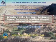
<strong>Parasites</strong> <strong>of</strong> <strong>mullets</strong> <strong>from</strong> <strong>two</strong><br />
<strong>different</strong> <strong>waters</strong><br />
Al-Bassel D.A.*, Al-Swaehly A.I.** , Abdel-Baki A. S.***, Atwa M.T.*<br />
and Al-Shawsh R.M****<br />
*Faculty <strong>of</strong> Science Fayoum University, Egypt, ** Faculty <strong>of</strong> Science<br />
El- Fateh University, Libya, *** Faculty <strong>of</strong> Science Bani Suif<br />
University , Egypt **** Faculty <strong>of</strong> Science, 7 October<br />
University , Libya
ABSTRACTS<br />
Two <strong>different</strong> localities were selected for the present<br />
investigation. Out <strong>of</strong> 200 Mugil cephalus, 63 (31.5%)<br />
were found infected . Out <strong>of</strong> 119 Mugil cephalus, 44 (36.9<br />
%) collected <strong>from</strong> Lake Qarun at Fayoum in Egypt were<br />
found infected with Trematoda , Acanthocephala ,<br />
Trichodina and Myxosporea .The incidence <strong>of</strong> infection<br />
88.6 % , 27. 2% , 95.5% and 27.3% respectively .<br />
<strong>Parasites</strong> <strong>of</strong> the Egyptian <strong>mullets</strong> includes: 4 species <strong>of</strong><br />
trematoda (Haplosplanchnus caudatus, H. pachysomus,<br />
Lecithobotrys putrescens and Dicrogaster contractus); one<br />
species <strong>of</strong> Acanthocephala (Neoechinorhynchus sp. );<br />
three species <strong>of</strong> Trichodina ( Trichodina lepsii ,T. puytoraci<br />
and T. batala ) and one species <strong>of</strong> Myxosporedia<br />
(Myxobolus. sp. ).
ABSTRACTS continue<br />
It is worth mentioned that protozoan infections were<br />
restricted in Egyptian <strong>mullets</strong> . On the other hand, out <strong>of</strong><br />
81 Mugil cephalus , 19 ( 23.45%) caught <strong>from</strong> the coastal<br />
water <strong>of</strong> the Mediterranean near Missurata in Libya were<br />
found infected with <strong>two</strong> genera <strong>of</strong> trematodes.<br />
(Vitellibaculum girelia and Lecithocladium exisum ). All<br />
parasites were figured and identified to the species level<br />
according to yamaguti,1971.
INTRODUCTION<br />
Two <strong>different</strong> saline <strong>waters</strong>, lake Qarun in Egypt and<br />
coastal water <strong>of</strong> the Mediterranean near Missurata in<br />
Libya were selected for the present study. Lake Qarun is<br />
the third largest lake in Egypt and the second most<br />
famous one after Lake Nasser in the southern part <strong>of</strong><br />
Egypt. It lies some 44 meters below sea level and<br />
occupies the lowest, northern section <strong>of</strong> the Fayoum<br />
depression. The Lake Qarun has a surface area <strong>of</strong> 214<br />
square kilometers. It has a maximum depth <strong>of</strong> just over 8<br />
meters and a volume <strong>of</strong> 800 million cubic meters. It is 42<br />
km long and 9 km wide at its broadest point (Meshal,<br />
1973).
INTRODUCTION continue<br />
The Mediterranean is a semi- enclosed Sea and almost<br />
suffering <strong>from</strong> several pollutants. In polluted Sea water,<br />
oxygen depletion, stress-induced mucus which support<br />
parasitic infestation on fishes, compounding an already<br />
stressful state .Fish parasites are nowadays considered<br />
as one <strong>of</strong> the most inviting fields <strong>of</strong> national research<br />
especially after the importance <strong>of</strong> fishes as a valuable<br />
source <strong>of</strong> protein, in which heavy infection may cause<br />
functional disturbances, retard growth, increase the<br />
susceptibility <strong>of</strong> fish for other infection and give the fish<br />
an unaesthetic appearance.
INTRODUCTION continue<br />
During the last decade, parasites <strong>of</strong> marine<br />
and fresh water fishes in Egypt have been<br />
received much attention (Al-Bassel,1997a,<br />
1997b,1999a,1999b,2000a,2000b,2001a,2<br />
001b,2002a,2002b,2003a,2003b,2004,200<br />
6a,2006b,2006c; Al-Bassel and El-<br />
Damarany,2001, Al-Bassel and<br />
Ohida,2006 and Al-Bassel et al., 2007) .
INTRODUCTION continue<br />
The <strong>mullets</strong> Mugil cephalus (Mugilidae) is considered a<br />
commercial marine fish .This species is omnivorous,<br />
feeds on plankton , thus it is more exposed to infection by<br />
trematodes than other marine fish, and is therefore<br />
selected for the present investigation. Libya was selected<br />
as the area since Mugil cephalus is a common species<br />
throughout most <strong>of</strong> the year. Although some information<br />
on the parasites <strong>of</strong> marine fishes <strong>from</strong> other areas is<br />
known, differences in the parasite fauna <strong>of</strong> a widely<br />
distributed species can be expected with <strong>different</strong><br />
geographical locations .The objective <strong>of</strong> this investigation<br />
was to study the natural parasite fauna <strong>of</strong> the <strong>mullets</strong> <strong>from</strong><br />
one region <strong>of</strong> its distribution and to extend our knowledge<br />
about the prevalence and distribution <strong>of</strong> parasites in Mugil<br />
cephalus in the Mediterranean Sea in Libya. In Egypt,<br />
research has been done on <strong>mullets</strong> and its parasites.
MATERIALS AND METHODS<br />
Smears <strong>from</strong> the skin, gill and fin surfaces are made on glass slides, air<br />
dried and fixed. Then, the whole body surface <strong>from</strong> just behind the<br />
gill cover till the end <strong>of</strong> the caudal fin was scraped using a scalpel<br />
blade or spatula. The scraped materials were then put on a glass<br />
slide, covered with a coverslip to be examined microscopically. In<br />
order to study the adhesive disc, positive smears were air-dried,<br />
then impregnated with Ag No3 and irradiated with UV (Klein's<br />
method).For studying the recorded Myxosporea, length and width <strong>of</strong><br />
cysts were measured then squashed for studying the spore<br />
morphology. Glycerin/gelatin mixture was used to preserve and<br />
examine some details <strong>of</strong> the spores. Measurements <strong>of</strong> the fresh<br />
spores were taken using calibrated ocular micrometer on Zeiss<br />
photomicroscope.
MATERIALS AND METHODS continue<br />
Organs are opened by a pair <strong>of</strong> fine scissors and<br />
left in 0.7% saline solution. A hand lens and a<br />
binocular dissecting microscope were used for<br />
the helminthological examination. The collected<br />
worms were cleaned by washing them several<br />
times with isotonic saline<br />
solution.Relaxation,fixation,staining and<br />
mounting were carried out by the usual way All<br />
measuments are in millimeter unless otherwise<br />
state.
The fish host <strong>of</strong> the present work<br />
Mugil cephalus was<br />
caught <strong>from</strong> <strong>two</strong><br />
<strong>different</strong> <strong>waters</strong> (Lake<br />
Qarun in Fayoum,<br />
Egypt and coastal<br />
water in Missurata,<br />
Libya) and<br />
investigation for<br />
parasites
RESULTS AND DISCUSSION
A) Protozoan<br />
infections
1-Trichodina lepsii Lom, 1962 (Fig. 1)<br />
S.bar = 10 µm<br />
Host: Mugil cephalus<br />
Location: gills<br />
Locality: Lake Qarun<br />
,Egypt<br />
This species was previously<br />
redescribed in details by<br />
Al-Bassel et al., 2007.<br />
This finding suggests that<br />
M. cephalus is the type<br />
host <strong>of</strong> this trichodinid<br />
and not Mugil auratus as<br />
proposed by Lom (1962).<br />
(Al-Bassel et al., 2007)
2-Trichodina puytoraci Lom, 1962 (Fig. 2)<br />
S.bar = 10 µm<br />
Host: Mugil cephalus<br />
Location: gills<br />
Locality: Lake Qarun ,<br />
Egypt<br />
This species was previously<br />
redescribed in details by<br />
Al-Bassel et al., 2007.<br />
This finding suggests that<br />
M. cephalus is the type<br />
host <strong>of</strong> this trichodinid<br />
and not Mugil auratus as<br />
proposed by Lom (1962).<br />
(Al-Bassel et al., 2007)
3-Trichodina batala Ali, 1996 (Fig. 3)<br />
S.bar = 10 µm<br />
Host: Mugil cephalus<br />
Location: gills<br />
Locality: Lake Qarun ,Egypt<br />
This species was previously<br />
redescribed in details by Al-<br />
Bassel et al., 2007. This<br />
finding suggests that M.<br />
cephalus is the type host <strong>of</strong><br />
this trichodinid. (Al-Bassel et<br />
al., 2007).<br />
The above three species were<br />
reported in the present work<br />
agreed fully with the<br />
description <strong>of</strong> Al-Bassel et al.,<br />
2007.
4 -Myxobolus sp. (Fig. 4 )<br />
(S.bar= 10 µm)<br />
Host: Mugil cephalus<br />
Location: body cavity and mesentery<br />
Locality: Lake Qarun ,Egypt<br />
Description:<br />
Only one Mugil cephalus was<br />
infected with one plasmodium <strong>of</strong><br />
this myxosporean.The cyst was<br />
embedded in the mesenteries <strong>of</strong><br />
the body cavity. Spores are oval to<br />
subspherical in frontal view. The<br />
polar capsules are pyriform,<br />
almost equal. Sporoplasm is<br />
binucleate and fills the all<br />
extracapsular space.
4 -Myxobolus sp.<br />
Discussion:<br />
Review <strong>of</strong> myxosporean infecting the <strong>mullets</strong><br />
indicated that 17 Myxobolus species is<br />
infecting this group <strong>of</strong> fishes. Only five<br />
Myxobolus species were found close in<br />
general shape to the present parasite.<br />
However, we refrain <strong>from</strong> final allocation <strong>of</strong><br />
this parasite till further examination <strong>of</strong><br />
mature spores.
B) Trematode<br />
infections
5-Haplosplanchnus Caudatus (Srivastava, 1939) (Fig. 5 )<br />
(S .bar =0.4 mm)<br />
Host: Mugil cephalus<br />
Location: Intestine<br />
Locality: Lake Qarun ,Fayoum,Egypt<br />
Description :<br />
The body is Y-Shaped, with unequal arms, one end<br />
is the oral sucker, the second end is the<br />
protruded ventral sucker and the third end<br />
formed <strong>of</strong> the curved posterior end <strong>of</strong> the body<br />
which looks like a tail like structure. The oral<br />
sucker is oval, subterminal. The ventral sucker<br />
is large club shaped found usually projected<br />
<strong>from</strong> the body but in some cases may be<br />
retracted into the body parenchyma. The<br />
pharynx is well developed, spherical in shape.<br />
The esophagus and the single simple straight<br />
intestinal caeca extend posteriorly to a short<br />
distance behind the level <strong>of</strong> the ventral sucker.<br />
The single testis is oval in shape lying at the<br />
posterid third <strong>of</strong> the body. genital pore opens<br />
between the oral and the ventral sucker.<br />
The ovary is oval in shape and lies at the right<br />
side, just anterior to testis. The vitelline follicles<br />
are few in number located in 3-4 groups in the<br />
lateral field surrounding the ovary. The uterus<br />
extends <strong>from</strong> the anterior margin <strong>of</strong> the testes<br />
up to the level <strong>of</strong> the ventral sucker and it is full<br />
<strong>of</strong> mature, large and yellow colored eggs.
Discussion<br />
H. caudatus recorded in the present work was<br />
closely similar in all its morphological aspects<br />
mentioned by Al-Bassel (1987) except that the<br />
eggs in the present study were slightly larger in<br />
size. Specimens identified by Ezz El-Dien (1990)<br />
<strong>from</strong> Mugil species caught <strong>from</strong> Red Sea and<br />
Raef (1990) <strong>from</strong> Mugil species obtained <strong>from</strong><br />
Mediterranean Sea were collectively larger in all<br />
measurements.
RESULTS<br />
6-Haplosplanchuns pachysomus (Eysenhardt,1829) Looss,1902<br />
( Fig. 6)<br />
Host: Mugil cephalus<br />
Location: Intestine<br />
Locality: Lake Qarun,Fayoum,Egypt<br />
Description:<br />
The worms are orange in color when fresh. They are fleshy parasites, fairly large.<br />
The oral sucker is triangular, muscular and subterminal .The ventral sucker is long,<br />
club – shaped, may be retracted into body parenchyma or projects prominently. The<br />
prepharynx is very short. The pharynx is well developed. The esophagus cannot be<br />
distinguished <strong>from</strong> the intestinal caecum which is a simple straight structure<br />
extending nearly to the middle <strong>of</strong> the body.<br />
The testis is single, globular in shape.<br />
The genital atrium is tubular .The genital pore lies between the oral and ventral<br />
sucker, and opens on a prominent papilla found between the <strong>two</strong> suckers. The ovary<br />
is spherical in shape and lies anterior to the testis. The vitelline follicles lies laterally<br />
near the ovary. The uterus is full <strong>of</strong> large and elongate eggs, which containing<br />
developing miracidia. The excretory vesicle is present and ends by a terminal<br />
execratory pore.
6-Haplosplanchuns pachysomus (Eysenhardt,1829) Looss,1902<br />
(S .bar 0.4 mm)<br />
Host: Mugil cephalus<br />
Location: Intestine<br />
Locality: Lake Qarun,Fayoum,Egypt<br />
Description:<br />
The worms are orange in color when fresh.<br />
They are fleshy parasites, fairly large. The<br />
oral sucker is triangular, muscular and<br />
subterminal .The ventral sucker is long, club<br />
– shaped, may be retracted into body<br />
parenchyma or projects prominently. The<br />
prepharynx is very short. The pharynx is well<br />
developed. The esophagus cannot be<br />
distinguished <strong>from</strong> the intestinal caecum<br />
which is a simple straight structure extending<br />
nearly to the middle <strong>of</strong> the body.<br />
The testis is single, globular in shape.<br />
The genital atrium is tubular .The genital pore<br />
lies between the oral and ventral sucker, and<br />
opens on a prominent papilla found between<br />
the <strong>two</strong> suckers. The ovary is spherical in<br />
shape and lies anterior to the testis. The<br />
vitelline follicles lies laterally near the ovary.<br />
The uterus is full <strong>of</strong> large and elongate eggs,<br />
which containing developing miracidia. The<br />
excretory vesicle is present and ends by a<br />
terminal execratory pore.
Discussion<br />
H. pachysomus was described <strong>from</strong> Mugil cephalus and<br />
M. cheloat by Looss, 1902.In Egypt, H. pachysomus was<br />
first described <strong>from</strong> Mugil cephalus and M ramada by<br />
Fischthal and Kuntz (1963). Then, it was redescribed<br />
<strong>from</strong> M. cephalus, Liza ramada, M. chelo and M. saliens<br />
<strong>from</strong> Lake Qarun by Al-Bassel (1987). Also, Al-Bassel<br />
(1990) described the same parasite in M. cephalus and<br />
L. ramada <strong>from</strong> Lake Edku.<br />
The present material is similar to that described by<br />
Fishthal and Kuntz (1963), Al-Bassel (1987) and Eid<br />
(1997) in their main characteristics, but there are certain<br />
minor differences in body length and measurements <strong>of</strong><br />
some internal organs.
RESULTS<br />
7-Dicrogaster contractus (Looss, 1902) (Fig. 7 )<br />
Host: Mugil cephalus<br />
Lacation: Intestine<br />
Locality: Lake Qarun<br />
Description:<br />
It is an oval worm. The body is mostly covered with spines. The oral sucker is sub<br />
teeminal, oval in shape. A well developed muscular pharynx is present; it is globular<br />
in shape. The esophagus is long. The intestinal caeca is saccular in shape extends to<br />
the middle <strong>of</strong> the worm .The ventral sucker is oval in shape<br />
The testis is globular in shape, located at the middle <strong>of</strong> he posterior half <strong>of</strong> the body. The<br />
cirrus pouch is well developed, pre-acetabular in position. The cirrus pouch contains<br />
prostatic gland cells, a hermaphroditic duct and internal seminal vesicle, the external<br />
seminal vesicle is oval in shape. The ovary is nearly spherical, smaller than testes<br />
and postacetabular in position .The vitelline glands are in 2 groups mostly at one<br />
lateral side between the testis and the caecal end. The excretory vesicle is spherical<br />
in shape and opens at the posterior end with a terminal excretory pore. The eggs are<br />
oval in shape
7-Dicrogaster contractus (Looss, 1902) (Fig. 7 )<br />
(S .bar 0.2 mm)<br />
Host: Mugil cephalus<br />
Lacation: Intestine<br />
Locality: Lake Qarun<br />
Description:<br />
It is an oval worm. The body is mostly covered<br />
with spines. The oral sucker is sub teeminal,<br />
oval in shape. A well developed muscular<br />
pharynx is present; it is globular in shape.<br />
The esophagus is long. The intestinal caeca<br />
is saccular in shape extends to the middle <strong>of</strong><br />
the worm .The ventral sucker is oval in<br />
shape<br />
The testis is globular in shape, located at the<br />
middle <strong>of</strong> he posterior half <strong>of</strong> the body. The<br />
cirrus pouch is well developed, preacetabular<br />
in position. The cirrus pouch<br />
contains prostatic gland cells, a<br />
hermaphroditic duct and internal seminal<br />
vesicle, the external seminal vesicle is oval<br />
in shape. The ovary is nearly spherical,<br />
smaller than testes and postacetabular in<br />
position .The vitelline glands are in 2 groups<br />
mostly at one lateral side between the testis<br />
and the caecal end. The excretory vesicle is<br />
spherical in shape and opens at the<br />
posterior end with a terminal excretory pore.<br />
The eggs are oval in shape
Discussion<br />
In the present work, D. contractus was identical in its morphological aspects<br />
to Looss (1902) specimens except for minor differences in certain<br />
measurements. In the redescription recorded by Fares and Maillard (1974),<br />
the excretory vesicle was y-shaped while in the present material it was<br />
spherical, a feature which has been recorded by Looss (1902). Al-Bassel<br />
(1987) recorded the parasite but his measurements were smaller (total body<br />
length 0.49-0.65) than that recorded in the present work (0.738-0.838)<br />
hence, other body organs were also smaller than that obtained in the<br />
present work, including prepharynx and intestinal caeca. It was important to<br />
noticed that the genus Dicrogaster has been originally reported <strong>from</strong> fishes<br />
in the Mediterranean sea (Looss, 1902), this genus has apparently reached<br />
Fayoum area with Mugil spp. Which have been transferred to water bodies<br />
in Fayoum (Khalil, 1978).Al-Bassel (1987) recorded the parasite <strong>from</strong> Lake<br />
Wadi Al-Rayan in Fayoum Province and not <strong>from</strong> Lake Qarun, although<br />
specimens <strong>of</strong>. D. contractus collected <strong>from</strong> Mugil cephalus in the present<br />
work, collected <strong>from</strong> Lake Qarun.
RESULTS
8-Lecithobotrys putrescens Looss, 1902 ( Fig. 8)<br />
( S. bar 0.3 mm<br />
Host:<br />
Mugil cephalus<br />
Location: intestine<br />
Locality: Lake Qarun ,Egypt<br />
Description:<br />
The worms are grayish white in color, elongated<br />
and tapering anteriorly. The whole body<br />
surface <strong>of</strong> the worm is covered by spines<br />
which are easily shed. The oral sucker is subterminal<br />
and relatively larger than the ventral<br />
sucker. The pharynx is large, elongated in<br />
shape. The prepharynx is short . The<br />
intestinal caeca are tubular in shape and<br />
extending back far beyond the ventral sucker<br />
to the posterior end <strong>of</strong> testis. The bifurcation<br />
<strong>of</strong> the intestine occurs dorsal to the ventral<br />
sucker.<br />
The testis is located not far behind the ventral<br />
sucker and it is nearly rounded . The cirrus<br />
pouch is egg-shaped. The ovary is median,<br />
spherical in shape, lying behind the testis.<br />
The vitelline glands are formed <strong>of</strong> <strong>two</strong><br />
irregular shaped follicles in each laterally.<br />
The eggs are numerous .The excretory<br />
vesicle is large and saccular opened by the<br />
main excretory pore which is sub-terminal<br />
and lies in a large depression in the posterior<br />
extremity <strong>of</strong> the worm.
Discussion<br />
The morphological characters <strong>of</strong> L.<br />
putrescence under investigation were<br />
agreed with that described by Al-Bassel<br />
(1987) although the measurements <strong>of</strong> the<br />
ovary and testis in the present specimens<br />
were smaller in size, a feature which may<br />
be due to incompletely grown parasite<br />
obtained in the present work.
9- Vitellibaculum girelia Montgomery , 1957 (Fig. 9)<br />
(S .bar 0.5 mm)<br />
Host : Mugil cephalus<br />
Location : intestine<br />
Locality : Missurata , Libya<br />
Description:<br />
The entire worm oval. The oral sucker is<br />
terminal . Prepharynx is long. A well<br />
developed muscular pharynx is present; it is<br />
globular in shape. The esophagus absent .<br />
The intestinal caeca is tubular in shape .The<br />
ventral sucker is larger than the oral suker.<br />
The testes are tandom , located at the middle <strong>of</strong><br />
he posterior half <strong>of</strong> the body. The cirrus<br />
pouch is well developed, pre-acetabular in<br />
position. The ovary is nearly spherical,<br />
smaller than testes and anterior to the<br />
testes. The vitelline glands are scattered in<br />
posterior half <strong>of</strong> the body. The excretory<br />
vesicle is y- shaped and opens at the<br />
posterior end with a terminal excretory pore.<br />
The eggs are oval in shape<br />
9
Discussion<br />
This species was originally described by<br />
Montgomery ,1957 <strong>from</strong> Girelia uigricans<br />
fish <strong>from</strong> California . The present work<br />
agreed fully with the original description<br />
,but the present description represent new<br />
host and new locality recored
RESULTS AND DISCUSSION<br />
Host : Mugil cephalus<br />
Location : intestine<br />
Locality : Missurata , Libya<br />
Description:<br />
The entire worm elongate with tail like structure. The oral sucker is funelshaped.<br />
Pharynx is well developed. The esophagus is short . The intestinal<br />
caeca is tubular in shape .<br />
The testes are small , located at the middle <strong>of</strong> the body .The ovary is nearly<br />
spherical, larger than testes and posterior to testes. The vitelline glands<br />
tubular and consists <strong>of</strong> 7 covoluted posterior to ovary. The excretory vesicle<br />
is elongate, tubular in shaped and opens at the posterior end with a<br />
terminal excretory pore. The eggs are small.<br />
Discussion : •<br />
This species was originally described by (Rud, 1819) Luhe, 1901 <strong>from</strong><br />
Scombrus scombrus fish <strong>from</strong> the Mediterranian . The present work agreed<br />
fully with the original description ,but the present description represent new<br />
host and new locality recored.
10- Lecithocladium excisum (Rud , 1819) Luhe , 1901(Fig. 10 )<br />
(S .bar 0.3 mm)<br />
10<br />
Host : Mugil cephalus<br />
Location : intestine<br />
Locality : Missurata , Libya<br />
Description:<br />
The entire worm elongate with tail<br />
like structure. The oral sucker is<br />
funel-shaped. Pharynx is well<br />
developed. The esophagus is<br />
short . The intestinal caeca is<br />
tubular in shape .<br />
The testes are small , located at the<br />
middle <strong>of</strong> the body .The ovary is<br />
nearly spherical, larger than testes<br />
and posterior to testes. The<br />
vitelline glands tubular and<br />
consists <strong>of</strong> 7 covoluted posterior to<br />
ovary. The excretory vesicle is<br />
elongate, tubular in shaped and<br />
opens at the posterior end with a<br />
terminal excretory pore. The eggs<br />
are small.
Discussion :<br />
This species was originally described by (Rud, 1819)<br />
Luhe, 1901 <strong>from</strong> Scombrus scombrus fish <strong>from</strong> the<br />
Mediterranian . The present work agreed fully with<br />
the original description ,but the present description<br />
represent new host and new locality recored.
C) Acanthocephala<br />
infections
11- Neoechinorhynchus sp. (Figs. 11,12 ,13 )<br />
(S .bar 5 mm)<br />
11<br />
Host: Mugil cephalus<br />
Location: Intestine<br />
Locality: Lake Qarun, Egypt .<br />
Description:<br />
The body <strong>of</strong> the worm is elongated and<br />
cylindrical with truncate ends . The<br />
body is covered with a thin tegument.<br />
The proboscis is small and globular.<br />
The proboscis has 18 hooks arranged<br />
in three spiral rows, each row has six<br />
hooks, each hook is provided with root<br />
processes .The hooks <strong>of</strong> the first row<br />
are the largest. The proboscis is<br />
keeping in the proboscis receptacle<br />
when be invaginated. Three or four<br />
giant nuclei are present in the dorsal<br />
side while four are present on the<br />
ventral side. The <strong>two</strong> lemnisci are<br />
unequal. There is a few number <strong>of</strong><br />
ovarian balls represented the ovary .<br />
The uterine ball is oval ,followed by a<br />
short uterus opens into vagina and<br />
finally opens posteriorly by the female<br />
gono-pore.
Neoechinorhynchus sp. continue<br />
13 (S. bar 0.5mm ) 12( S.bar 0.4mm)<br />
13
Discussion<br />
The worm described above is evidently belonging<br />
to the genus Neoechinorhychus Hamann,<br />
1892.The <strong>two</strong> related species are the<br />
Neoechinorhynchus ichthyobori Saoud et aI.,<br />
1974 which was described <strong>from</strong> fresh water fish<br />
Ichthyoborus besse caught <strong>from</strong> the white Nile at<br />
Jebel Al-Awliya in Sudan and also<br />
Neoechinorhynchus sp, which was redescribed<br />
by Al-Bassel 1990 <strong>from</strong> Mugil cephalus caught<br />
<strong>from</strong> lake Edku. It is worth to mention that the<br />
present material was reported in a new locality.
Thank you