Superdex High-performance Columns
Superdex High-performance Columns
Superdex High-performance Columns

Create successful ePaper yourself
Turn your PDF publications into a flip-book with our unique Google optimized e-Paper software.
GE Healthcare<br />
Data file 18-1163-79 AD<br />
Gel filtration<br />
<strong>Superdex</strong> <strong>High</strong>-<strong>performance</strong><br />
<strong>Columns</strong><br />
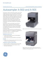
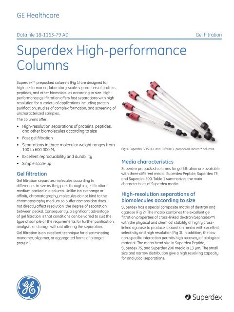
<strong>Superdex</strong> prepacked columns (Fig 1) are designed for<br />
high-<strong>performance</strong>, laboratory-scale separations of proteins,<br />
peptides, and other biomolecules according to size. <strong>High</strong><strong>performance</strong><br />
gel filtration offers fast separations with high<br />
resolution for a variety of applications including protein<br />
purification, studies of complex formation, and screening of<br />
uncharacterized samples.<br />
The columns offer:<br />
• <strong>High</strong>-resolution separations of proteins, peptides,<br />
and other biomolecules according to size<br />
• Fast gel filtration<br />
• Separations in three molecular weight ranges from<br />
100 to 600 000 M r<br />
• Excellent reproducibility and durability<br />
• Simple scale-up<br />
Gel filtration<br />
Gel filtration separates molecules according to<br />
differences in size as they pass through a gel filtration<br />
medium packed in a column. Unlike ion exchange or<br />
affinity chromatography, molecules do not bind to the<br />
chromatography medium so buffer composition does<br />
not directly affect resolution (the degree of separation<br />
between peaks). Consequently, a significant advantage<br />
of gel filtration is that conditions can be varied to suit the<br />
type of sample or the requirements for further purification,<br />
analysis, or storage without altering the separation.<br />
Gel filtration is an excellent technique for discriminating<br />
monomer, oligomer, or aggregated forms of a target<br />
protein.<br />
Fig 1. <strong>Superdex</strong> 5/150 GL and 10/300 GL prepacked Tricorn columns.<br />
Media characteristics<br />
<strong>Superdex</strong> prepacked columns for gel filtration are available<br />
with three different media: <strong>Superdex</strong> Peptide, <strong>Superdex</strong> 75,<br />
and <strong>Superdex</strong> 200. Table 1 summarizes the main<br />
characteristics of <strong>Superdex</strong> media.<br />
<strong>High</strong>-resolution separations of<br />
biomolecules according to size<br />
<strong>Superdex</strong> has a special composite matrix of dextran and<br />
agarose (Fig 2). The matrix combines the excellent gel<br />
filtration properties of cross-linked dextran (Sephadex)<br />
with the physical and chemical stability of highly crosslinked<br />
agarose to produce separation media with excellent<br />
selectivity and high resolution (Fig 3). In addition, the low<br />
non-specific interaction permits high recovery of biological<br />
material. The mean bead size in <strong>Superdex</strong> Peptide,<br />
<strong>Superdex</strong> 75, and <strong>Superdex</strong> 200 media is 13 μm. The small<br />
size and narrow distribution give a high resolving capacity<br />
for analytical separations.<br />
imagination at work<br />
<strong>Superdex</strong>
Table 1. The main characteristics of <strong>Superdex</strong> media<br />
<strong>Superdex</strong> Peptide <strong>Superdex</strong> 75 <strong>Superdex</strong> 200<br />
Separation range (M r) 100 to 7 000 (peptides) 3 000 to 70 000 (globular proteins) 10 000 to 600 000 (globular proteins)<br />
Exclusion limit (M r) 20 000 100 000 (globular proteins) 1 300 000 (globular proteins)<br />
pH stability 1 to 14 (long- and short-term) 3 to 12 (long-term),<br />
1 to 14 (short-term)<br />
3 to 12 (long-term),<br />
1 to 14 (short-term)<br />
Temperature stability 4°C to 40°C 4°C to 40°C 4°C to 40°C<br />
Working and storage<br />
temperature<br />
Matrix<br />
4°C to 30°C 4°C to 30°C 4°C to 30°C<br />
Spherical composite of crosslinked<br />
agarose and dextran<br />
Spherical composite of crosslinked<br />
agarose and dextran<br />
Average particle size 13 μm 13 μm 13 μm<br />
Spherical composite of crosslinked<br />
agarose and dextran<br />
Dextran<br />
Cross-linked agarose<br />
K av<br />
1.00<br />
<strong>Superdex</strong> Peptide<br />
0.75<br />
<strong>Superdex</strong> 75<br />
<strong>Superdex</strong> 200<br />
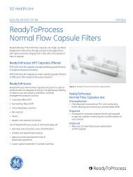
Fig 2. Schematic view through a section of a bead of <strong>Superdex</strong>. The average<br />
particle size is 13 μm. The composite dextran/cross-linked agarose matrix<br />
gives steep selectivity and high stability.<br />
0.50<br />
<strong>High</strong> speed gel filtration<br />
<strong>Superdex</strong> is a composite medium with high chemical<br />
and physical stabilities. The rigid matrix withstands high<br />
pressures and allows high flow rates to be used, which<br />
will give short separation times with retained function and<br />
stability. Even shorter cycles times are made possible with<br />
<strong>Superdex</strong> 75 and 200 5/150 GL, with bed heights of 150 mm,<br />
a natural column choice for screening experiments.<br />
0.25<br />
10<br />
100<br />
1000<br />
10000<br />
100000<br />
1000000<br />
Log M r<br />
Simple scale-up<br />
Scale-up to industrial production is simple and predictable,<br />
as <strong>Superdex</strong> 30, 75, and 200 prep grade media (mean bead<br />
size 34 μm) have similar selectivities as <strong>Superdex</strong> Peptide,<br />
75 and 200, respectively. <strong>Superdex</strong> 30, 75, and 200 prep<br />
grade media are available prepacked in HiLoad 16/60 and<br />
HiLoad 26/60 columns.<br />
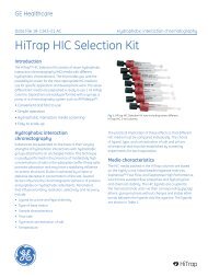
Figure 4 shows the scale-up of a concentrated mouse<br />
monoclonal IgG cell supernatant from a <strong>Superdex</strong> 200 high<strong>performance</strong><br />
column, 10 mm × 300 mm (i.d. × bed height)<br />
(Fig 4A) to HiLoad 16/60 <strong>Superdex</strong> 200 prep grade (Fig 4B).<br />
Sample volume was increased five-fold yet the separation<br />
was almost identical.<br />
Fig 3. Selectivity curves for <strong>Superdex</strong> Peptide, 75, and 200. The steep<br />
selectivity gives high-resolution separations in the molecular weight range<br />
of 100 to 600 000.<br />
Chemical stability<br />
<strong>Superdex</strong> Peptide<br />
<strong>Superdex</strong> Peptide has outstanding chemical stability and<br />
is stable in the pH range of 1 to 14. Polar organic solvents,<br />
such as 70% acetonitrile in water and methanol, and<br />
chaotropic agents such as 6 M guanidine hydrochloride<br />
or 8 M urea can also be used. The high pH stability<br />
allows separation of very hydrophobic peptides in 70%<br />
formic acid, as well as use of alkaline buffers. Exposure<br />
to repeated 48-h cycles of 1.0 M NaOH, 0.1 M HCl, 10%<br />
TFA, 70% acetonitrile, and 70% formic acid has no effect<br />
on selectivity. The recommended cleaning procedure,<br />
described in the instructions, includes the use of 0.5 M<br />
NaOH or 0.1 M HCl. Hydrophobic contaminants can be<br />
removed with 30–70% acetonitrile.<br />
<br />
Data file 18-1163-79 AD
A<br />
A 280<br />
0.60<br />
0.40<br />
0.20<br />
V O<br />
lgG 1<br />
V C<br />
5.0 15.0 25.0<br />
Vol. (ml)<br />
A 280<br />
0.60<br />
0.40<br />
0.20<br />
lgG 1<br />
50 100 150<br />
Vol. (ml)<br />
Fig 4. Simple and predictable five-fold scale up of a mouse monoclonal IgG 1<br />
separation from a <strong>Superdex</strong> 200 high-<strong>performance</strong> column, 10 mm ×<br />
300 mm (i.d. × bed height) (A) to a HiLoad 16/60 <strong>Superdex</strong> 200 prep grade<br />
(B). <strong>Superdex</strong> 200 prep grade medium (mean particle size 34 μm) has a<br />
similar selectivity to <strong>Superdex</strong> 200 (mean particle size 13 μm).<br />
B<br />
V O<br />
V C<br />
<strong>Superdex</strong> 75 and <strong>Superdex</strong> 200<br />
<strong>Superdex</strong> 75 and <strong>Superdex</strong> 200 are stable in aqueous<br />
solutions over the pH range 3 to 12. Chaotropic agents<br />
(6 M guanidine hydrochloride, 8 M urea) and detergents<br />
such as SDS (up to 2%) can also be used, as well as polar<br />
organic solvents such as 30% acetonitrile. <strong>Superdex</strong> 75 and<br />
200 withstand the conditions used for cleaning-in-place<br />
(CIP) over the pH range 1 to 14. Studies have shown that<br />
short cycles (approx. 3 h) with 0.1 M HCl or 1.0 M NaOH have<br />
no significant influence on the chromatographic behavior.<br />
However, the columns should not be stored in these<br />
solutions.<br />
Column characteristics<br />
<strong>Superdex</strong> media are prepacked in high-<strong>performance</strong><br />
glass columns. Both column types, Tricorn and Precision<br />
(PC), are made of glass, which allows visual inspection of<br />
the medium bed. Tricorn columns have fittings for simple<br />
connections to ÄKTAdesign and other high-<strong>performance</strong><br />
systems. Precision <strong>Columns</strong> were designed for SMART<br />
systems, but the Precision Column Holder allows use of the<br />
columns with a variety of high-<strong>performance</strong> systems. All<br />
parts of the columns are biocompatible. Table 2<br />
summarizes the main characteristics of Tricorn and<br />
Precision columns prepacked with <strong>Superdex</strong>.<br />
Table 2. Characteristics of Tricorn and Precision columns packed with <strong>Superdex</strong><br />
Tricorn <strong>Columns</strong><br />
<strong>Superdex</strong><br />
Peptide<br />
10/300 GL<br />
<strong>Superdex</strong> 75<br />
10/300 GL<br />
<strong>Superdex</strong> 200<br />
10/300 GL<br />
<strong>Superdex</strong> 75<br />
5/150 GL<br />
<strong>Superdex</strong> 200<br />
5/150 GL<br />
Precision <strong>Columns</strong><br />
<strong>Superdex</strong><br />
Peptide PC<br />
3.2/30<br />
<strong>Superdex</strong> 75<br />
PC 3.2/30<br />
<strong>Superdex</strong> 200<br />
PC 3.2/30<br />
Bed<br />
dimensions<br />
(i.d. × height)<br />
Bed<br />
volume<br />
(ml)<br />
Recommended<br />
sample volume<br />
(μl)<br />
Theoretical<br />
plates<br />
(N/m)<br />
Recommended<br />
flow rate 1<br />
(ml/min)<br />
Max. flow<br />
rate 1<br />
(ml/min)<br />
Max. pressure<br />
over column<br />
10 × 300 mm 24 25 to 250 > 30 000 0.2 to 1.0 1.2 18 bar<br />
(261 psi, 1.8 MPa)<br />
10 × 300 mm 24 25 to 250 > 30 000 0.5 to 1.0 1.5 18 bar<br />
(261 psi, 1.8 MPa)<br />
10 × 300 mm 24 25 to 250 > 30 000 0.25 to 0.75 1.0 15 bar<br />
(217 psi, 1.5 MPa)<br />
5 × 150 mm<br />
5 × 150 mm<br />
3<br />
3<br />
4 to 50<br />
4 to 50<br />
> 25 000<br />
> 25 000<br />
0.15 to 0.6<br />
0.15 to 0.6<br />
0.7<br />
0.8<br />
18 bar<br />
(261 psi, 1.8 MPa)<br />
15 bar<br />
(217 psi, 1.5 MPa)<br />
3.2 × 300 mm 2.4 2 to 25 > 30 000 0.01 to 0.15 0.15 20 bar<br />
(290 psi, 2 MPa)<br />
3.2 × 300 mm 2.4 2 to 25 > 30 000 0.01 to 0.10 0.10 24 bar<br />
(348 psi, 2.4 MPa)<br />
3.2 × 300 mm 2.4 2 to 25 > 30 000 0.01 to 0.10 0.10 15 bar<br />
(217 psi, 1.5 MPa)<br />
1<br />
H 2 O at 25°C<br />
Data file 18-1163-79 AD
Column sizes<br />
<strong>Superdex</strong> columns come in two bed heights, 150 and<br />
300 mm, thereby allowing greater flexibility to suit<br />
different analytical needs. The 150-mm prepacked Tricorn<br />
columns are designed for rapid size analysis of proteins,<br />
peptides and other biomolecules. Short cycle times<br />
together with small sample volumes and low consumption<br />
of buffers makes the column a natural choice for<br />
screening experiments, such as quick analyses of protein<br />
homogeneity. The range of 300-mm <strong>Superdex</strong> columns<br />
(see Table 2) is designed for studies where high-resolution<br />
separation of biomolecules is critical.<br />
Excellent reproducibility and durability<br />
Reproducible results are essential in all research. The<br />
long working life and high reproducibility of <strong>Superdex</strong><br />
prepacked columns are the result of optimized design, the<br />
stable nature of the medium, and controlled production<br />
procedures. Figure 5 shows chromatograms of the test<br />
separation runs 1 and 210 on <strong>Superdex</strong> 75 10/300 GL.<br />
Column:<br />
Sample:<br />
Sample volume: 500 μl<br />
<strong>Superdex</strong> 75 10/300 GL<br />
1. BSA (Mr 67 000) 8 mg/ml<br />
2. Ovalbumin (Mr 43 000) 2.5 mg/ml,<br />
3. Ribonuclease A (Mr 13 700) 5 mg/ml,<br />
4. Aprotinin (Mr 6512) 2 mg/ml,<br />
5. Vitamin B12 (Mr 1355) 0.1 mg/ml<br />
Buffer: 0.05 M phosphate buffer, 0.15 M NaCl, pH 7.0<br />
Flow rate:<br />
System:<br />
A 280 mAU<br />
250<br />
200<br />
150<br />
100<br />
50<br />
0<br />
A 280 mAU<br />
200<br />
150<br />
100<br />
50<br />
0<br />
0.4 ml/min, room temperature<br />
ÄKTAfplc<br />
Test separation, run 1<br />
0.0 5.0 10.0 15.0 20.0 ml<br />
Test separation, run 210<br />
1 2 3 4<br />
1 2 3 4<br />
0.0 5.0 10.0 15.0 20.0 ml<br />
Fig 5. Comparison of the test separation runs 1 and 210 after repeated<br />
injection of a test sample on <strong>Superdex</strong> 75 10/300 GL. After 210 runs, no<br />
difference in chromatographic <strong>performance</strong> was observed.<br />
5<br />
5<br />
Operation<br />
Choice of eluents<br />
Eluents can be chosen freely to improve recovery from<br />
separations of crude sample or to overcome solubility<br />
problems. For example, when using <strong>Superdex</strong> Peptide the<br />
sample can be made up and run in up to 70% formic acid<br />
or acetonitrile/TFA, as well as aqueous buffer solutions in<br />
the pH range 1 to 14, as demanded by the solubility of the<br />
peptides being separated. Similarly, chaotropic agents<br />
and detergents can be used to improve the solubility<br />
of membrane proteins when working with prepacked<br />
<strong>Superdex</strong> 75 or <strong>Superdex</strong> 200 columns. To avoid pHdependent,<br />
nonionic interactions with the matrix, 0.15 M<br />
NaCl, or a buffer with equivalent ionic strength, is<br />
recommended.<br />
Sample volumes and flow rates<br />
The quality of a gel filtration separation is largely independent<br />
of sample concentration, but to achieve high resolution,<br />
the sample volume should be less than 5% of the<br />
total column volume. Sample volumes between 0.1% and<br />
1.0% of the bed volume give the highest resolution. By using<br />
lower flow rates a higher resolution result can be achieved<br />
from a given column. For more information see Table 2.<br />
Applications using <strong>Superdex</strong> Peptide<br />
<strong>Superdex</strong> Peptide provides a powerful complement to<br />
traditional reversed-phase chromatography for separating<br />
small peptides. The excellent physical and chemical<br />
stabilities permit a wide choice of elution conditions, which<br />
extends the user’s ability to separate peptides that are<br />
only soluble under special conditions. The mobile phase<br />
can also be selected to meet the requirements of detection<br />
techniques such as mass spectrometry or surface plasmon<br />
resonance.<br />
Detecting peptide fragments<br />
Biologically active neuropeptides in the brain are cleaved<br />
by various peptidases, causing the formation of different<br />
fragments with other biological effects. Previously,<br />
radioactive substrates or immunological techniques<br />
have been used to detect the products formed, but these<br />
methodologies lack structural specificity and are timeconsuming.<br />
Gel filtration on <strong>Superdex</strong> Peptide using a<br />
SMART system, in combination with mass spectrometry,<br />
allows these studies to be carried out without such<br />
drawbacks, and all peptide fragments can be detected<br />
in a single analysis (Silberring, J., 1994, personal<br />
communication).<br />
Separation of standard peptides<br />
Figure 6 shows a separation of standard peptides on<br />
<strong>Superdex</strong> Peptide 10/300 GL.<br />
<br />
Data file 18-1163-79 AD
Column: <strong>Superdex</strong> Peptide 10/300 GL<br />
Sample: 1. Cytochrome c (M r 12 384) 1 mg/ml<br />
2. Aprotinin (M r 6 512) 2 mg/ml<br />
<strong>Superdex</strong> 3. Peptide Vitamin 10/300 BGL<br />
12 (M r 1 355) 0.1 mg/ml<br />
1. Cytochrome 4. (Gly)3 c (M r 12 (M384) r 189) 1 mg/ml 0.1 mg/ml<br />
2. Aprotinin (M<br />
5. Gly r 6 512) 2 mg/ml<br />
(M r 75) 7.8 mg/ml<br />
3. Vitamin B 12 (M r 1 355) 0.1 mg/ml<br />
Flow rate: 4. (Gly)3 (M0.5 r 189) ml/min, 0.1 mg/mlroom temperature<br />
5. Gly (M r 75) 7.8 mg/ml<br />
Sample volume:<br />
0.5 ml/min,<br />
50<br />
room<br />
μl<br />
temperature<br />
Eluent: 0.05 M phosphate, 0.15 M NaCl, pH 7.0<br />
System: ÄKTAexplorer 100<br />
Column:<br />
Sample:<br />
Flow rate:<br />
Sample volume: 50 μl<br />
Eluent: 0.05 M phosphate, 0.15 M NaCl, pH 7.0<br />
System: ÄKTAexplorer 100<br />
mAU<br />
mAU<br />
600<br />
A 215 nm<br />
500<br />
600<br />
A 215 nm<br />
3<br />
Conductivity<br />
mS/cm<br />
45.0<br />
3<br />
Conductivity<br />
mS/cm<br />
45.0<br />
Column:<br />
<strong>Superdex</strong> 200 10/300 GL<br />
Sample:<br />
Monoclonal antibody<br />
Sample volume (load): 100 μl<br />
Elution buffer: 0.02 M Tris HCl, pH 7.5, 0.15 M NaCl<br />
Flow rate:<br />
0.25 ml/min<br />
System: ÄKTAexplorer 100<br />
A 280 mAU<br />
150<br />
100<br />
50<br />
0<br />
Dimer<br />
Monomer<br />
0.0 5.0 10.0 15.0 20.0 25.0 30.0 ml<br />
Fig 7. Separation of the monomer and dimer of a monoclonal antibody on<br />
<strong>Superdex</strong> 200 10/300 GL.<br />
400<br />
500<br />
40.0<br />
300<br />
200<br />
400<br />
1<br />
2<br />
4<br />
5<br />
35.0<br />
30.0<br />
4<br />
40.0<br />
Column:<br />
<strong>Superdex</strong> 75 10/300 GL<br />
Sample:<br />
recCys-prot<br />
Sample volume (load): 200 μl<br />
Elution buffer: 0.05 M Tris HCl, 1 mM EDTA, 0.15 M NaCl, pH 8.4<br />
Flow rate:<br />
0.5 ml/min<br />
System: ÄKTAexplorer 100<br />
100<br />
0<br />
300<br />
200<br />
V 0<br />
0 5.0 10.0 15.0 20.0 25.0 ml<br />
1<br />
2<br />
5<br />
35.0<br />
30.0<br />
Fig 6. A separation of standard peptides on <strong>Superdex</strong> Peptide 10/300 GL.<br />
Vt<br />
25.0<br />
A<br />
B<br />
A 280 mAU<br />
1500<br />
1000<br />
500<br />
0<br />
0.0 5.0 10.0 15.0 20.0 25.0 ml<br />
A 280 mAU<br />
Dimer<br />
Monomer<br />
Applications 100 using <strong>Superdex</strong> 75<br />
and 200 10/300 GL<br />
<strong>Superdex</strong> 75 10/300 GL and <strong>Superdex</strong> 200 10/300 GL give<br />
high <strong>performance</strong> gel Vfiltration 0<br />
of peptides and Vt<br />
proteins<br />
within<br />
0<br />
the recommended separation ranges. They can be<br />
used for any application of gel filtration, such as monitoring<br />
0 5.0 10.0 15.0 20.0 25.0 ml<br />
changes in molecular size, determining molecular weight, or<br />
as a polishing step in a purification scheme.<br />
Separation of the monomer and dimer of a<br />
monoclonal antibody<br />
Figure 7 shows separation of the monomer and dimer of a<br />
monoclonal antibody on <strong>Superdex</strong> 200 10/300 GL.<br />
Dimer-monomer separation of a recombinant<br />
cystein-containing protein<br />
A cystein-containing protein spontaneously forms dimers<br />
via a disulfide bridge. These dimers can be cleaved by<br />
adding the reducing agent dithioerythritol (DTE). The<br />
chromatogram from the <strong>Superdex</strong> 75 10/300 GL run reflects<br />
that there is a baseline separation between the monomer<br />
and dimer (Fig 8a) and that it is possible to reduce the<br />
dimers into monomers by addition of DTE (Figs 8b and 8c).<br />
25.0<br />
C<br />
250<br />
200<br />
150<br />
100<br />
50<br />
0<br />
Monomer<br />
0.0 5.0 10.0 15.0 20.0 25.0 ml<br />
M r 20 000<br />
Fig 8. (A) Dimer-monomer separation of a recombinant cystein-containingprotein<br />
(recCys-prot.) on <strong>Superdex</strong> 75 10/300 GL. (B) purification of the dimer<br />
fraction reduced with DTE. (C) shows a Coomassie stained SDS-PAGE gel.<br />
Lane S is LMW-SDS Marker Kit (17-0446-01), lane 1 is the original dimermonomer<br />
sample; the dimer content is high, which also is reflected in the<br />
chromatogram. Lane 2 is the dimer fraction and lane 3 corresponds to the<br />
monomer fraction from A, respectively. Lane 4 shows the monomer peak<br />
from B. Lanes 1 to 4 were run under non-reducing conditions.<br />
Data file 18-1163-79 AD
Applications using <strong>Superdex</strong> 5/150 GL<br />
<strong>Superdex</strong> 75 and 200 5/150 GL columns have small bed<br />
volumes which is useful for a range of applications such<br />
as screening solubilization conditions for membrane<br />
purification and analyses of protein-protein interactions.<br />
Shorter <strong>Superdex</strong> columns use less buffer and sample than<br />
longer columns and are thus an excellent choice when<br />
time and sample buffer consumption are more critical than<br />
achieving high resolution.<br />
Reduced cycle time using <strong>Superdex</strong> 5/150 GL<br />
Figure 9 provides comparisons of analyses run on <strong>Superdex</strong><br />
200 5/150 GL and 10/300 GL (Fig 9 A-B), as well as <strong>Superdex</strong><br />
75 5/150 GL and 10/300 GL (Fig 9 C-D). Note that analyses<br />
run with <strong>Superdex</strong> 200 5/150 GL and <strong>Superdex</strong><br />
75 5/150 are 4 times faster than those run on <strong>Superdex</strong><br />
200 10/300 GL and <strong>Superdex</strong> 75 10/300, respectively. Using<br />
shorter columns that provide good resolution together with<br />
short analysis time can lead to significant cost savings.<br />
Consequently, short columns are ideal for screening. Long<br />
columns involve longer analysis time, but are preferable<br />
when high resolution analyses are required.<br />
Analysis of protein-protein interaction<br />
To evaluate the efficacy of <strong>Superdex</strong> 75 5/150 GL in<br />
analyzing protein-protein interactions, Trypsin and Aprotinin<br />
were run separately on <strong>Superdex</strong> 75 5/150 GL and then<br />
mixed and injected on the column (Fig 10). Only one peak<br />
eluted from the mixture of Trypsin and Aprotinin with an<br />
elution volume shifted toward the void volume, indicating<br />
protein complex formation.<br />
Column: <strong>Superdex</strong> 200 5/150 GL (curve A below)<br />
<strong>Superdex</strong> 200 10/300 GL (curve B below)<br />
Sample: Ferritin (Mr 440 000), Aldolase (Mr 158 000),<br />
Ovalbumin (Mr 43 000), Ribonuclease A (Mr 13 700)<br />
Sample volume: 12.5 μl (<strong>Superdex</strong> 200 5/150 GL)<br />
100 μl (<strong>Superdex</strong> 200 10/300 GL)<br />
Buffer: PBS, pH 7.4<br />
Flow rate: 0.3 ml/min, <strong>Superdex</strong> 200 5/150 GL<br />
0.6 ml/min, <strong>Superdex</strong> 200 10/300 GL<br />
Analysis time: 12 min (<strong>Superdex</strong> 200 5/150 GL)<br />
47 min (<strong>Superdex</strong> 200 10/300 GL)<br />
System: ÄKTAexplorer<br />
Column: <strong>Superdex</strong> 75 5/150 GL (curve C below)<br />
<strong>Superdex</strong> 75 10/300 GL (curve D below)<br />
Sample: Conalbumin (Mr 75 000);<br />
Carbonic Anhydrase (Mr 29 000)<br />
Ribonuclease A (Mr 13 700), Aprotinin (Mr 6 500)<br />
Sample volume: 12.5 μl (<strong>Superdex</strong> 75 5/150 GL)<br />
100 μl (<strong>Superdex</strong> 75 10/300 GL)<br />
Buffer: PBS, pH 7.4<br />
Flow rate: 0.3 ml/min, <strong>Superdex</strong> 75 5/150 GL<br />
0.6 ml/min, <strong>Superdex</strong> 75 10/300 GL<br />
Analysis time: 12 min (<strong>Superdex</strong> 75 5/150 GL)<br />
47 min (<strong>Superdex</strong> 75 10/300 GL)<br />
System: ETTAN LC<br />
A 280 mAU<br />
300<br />
A <strong>Superdex</strong> 200 5/150 GL<br />
B <strong>Superdex</strong> 200 10/300 GL<br />
mAU<br />
400<br />
C <strong>Superdex</strong> 75 5/150 GL<br />
D <strong>Superdex</strong> 75 10/300 GL<br />
350<br />
250<br />
300<br />
200<br />
250<br />
150<br />
200<br />
150<br />
100<br />
100<br />
50<br />
50<br />
0<br />
0.0 2.0 4.0 6.0 8.0 10.0 min<br />
0.0 10.0 20.0 30.0 40.0 min<br />
0<br />
0.0 2.0 4.0 6.0 8.0 10.0 min<br />
0.0 10.0 20.0 30.0 40.0 min<br />
Fig 9. Comparison of analyses run on different <strong>Superdex</strong> columns. The larger 10/300 shows excellent resolution for sample analyses, while the short 5/150<br />
column provides rapid analysis time suitable for screening experiments.<br />
<br />
Data file 18-1163-79 AD
Column: <strong>Superdex</strong> 75 5/150 GL<br />
Sample: Trypsin (Mr 23 800) 11mg/ml<br />
Aprotinin (Mr 6 500) 3 mg/ml<br />
Trypsin 11 mg/ml and Aprotinin 3mg/ml<br />
Sample volume: 10 µl<br />
Buffer: PBS, pH 7.4<br />
Flow rate: 0.3 ml/min<br />
System: ETTAN LC<br />
mAU<br />
700<br />
600<br />
500<br />
400<br />
300<br />
200<br />
100<br />
0<br />
<strong>Superdex</strong> 75 nr 2 aprotinin 002:10_UV1_280nm <strong>Superdex</strong> 75 nr 2 Trypsin001:10_UV1_280nm<br />
<strong>Superdex</strong> 75 nr 2 Trypsin Aprotinin001:10_UV1_280nm <strong>Superdex</strong> 75 nr 2 aprotinin 002:10_Inject<br />
Trypsin and<br />
Aprotinin<br />
Trypsin<br />
Aprotinin<br />
Autoprotolytic<br />
fragments<br />
0.0 0.5 1.0 1.5 2.0 2.5 3.0 ml<br />
Fig 10. Monitoring of protein complex formation between Trypsin and<br />
Aprotinin.<br />
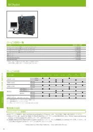
Screening of buffer conditions for a membrane<br />
protein<br />
Size homogeneity is a useful indicator of stability, since<br />
e.g. membrane proteins often oligomerize or aggregate<br />
rapidly when destabilized. Rapid gel filtration with <strong>Superdex</strong><br />
200 5/150 GL was used to screen for homogeneity under<br />
various pH and salt conditions (Fig 11).<br />
Screening with rapid gel filtration showed a symmetrical<br />
peak when the separation was performed at pH 5.2 in 0.1 M<br />
NaCl (see Fig 11 A), indicating a homogenous protein under<br />
these conditions. At somewhat higher salt concentration<br />
(Fig 11 D) a small peak appeared close to the void volume,<br />
indicating that oligomerization or aggregation appeared to<br />
a limited extent. At both pH 7.5 and pH 9.5 significant peaks<br />
were obtained close to the void volume, indicating severe<br />
oligomerization or aggregation. The complete screening<br />
procedure was achieved in only a few hours, including the<br />
time for column equilibration. Sample consumption was<br />
6 × 10 µl for the complete screen.<br />
Column:<br />
<strong>Superdex</strong> 200 5/150 GL<br />
Sample:<br />
Integral membrane protein (M r 60 000) from E. coli<br />
Sample volume: 10 µl<br />
Eluents (including 0.1 or 0.3 M NaCl): 0.02 M sodium acetate, 0.03 % dodecyl maltoside, 0.5 mM TCEP, pH 5.2<br />
0.02 M HEPES, 0.03 % dodecyl maltoside, 0.5 mM TCEP, pH 7.5<br />
0.02 M CAPSO, 0.03 % dodecyl maltoside, 0.5 mM TCEP, pH 9.5<br />
Flow rate:<br />
0.35 ml/min<br />
Detection:<br />
280 nm<br />
System:<br />
ÄKTAexplorer<br />
pH 5.2 pH 7.5 pH 9.5<br />
0.1 M NaCl<br />
20.0<br />
mAU<br />
Buffer: 20 mM Na-acetate pH 5.2 100 mM NaCl<br />
A<br />
Sample: 14 ug (10ul) EM35<br />
Buffer: 20 mM HEPES pH 7.5 100 mM NaCl<br />
mAU<br />
12.0<br />
10.0<br />
B<br />
15.0<br />
Sample: 9.4 ug (10 ul) EM35<br />
Buffer: 20 mM CAPSO pH 9.5 100 mM NaCl<br />
mAU<br />
C<br />
1.76<br />
15.0<br />
10.0<br />
5.0<br />
0.0<br />
0.0 0.5 1.0 1.5 2.0 2.5 3.0 ml<br />
8.0<br />
6.0<br />
4.0<br />
2.0<br />
0.0<br />
-2.0<br />
0.0 0.5 1.0 1.5 2.0 2.5 3.0 ml<br />
10.0<br />
5.0<br />
0.0<br />
1.29<br />
0.0 0.5 1.0 1.5 2.0 2.5 3.0 ml<br />
0.3 M NaCl<br />
mAU<br />
40.0<br />
35.0<br />
30.0<br />
25.0<br />
20.0<br />
15.0<br />
10.0<br />
5.0<br />
Sample: 16 ug (10 ul) EM35<br />
Buffer: 20 mM Na-acetate pH 5.2 300 mM NaCl<br />
D<br />
0.0<br />
0.0 0.5 1.0 1.5 2.0 2.5 3.0 ml<br />
mAU<br />
16.0<br />
14.0<br />
12.0<br />
10.0<br />
8.0<br />
6.0<br />
4.0<br />
2.0<br />
0.0<br />
Sample: 9.4 ug (10 ul) EM35<br />
Buffer: 20 mM HEPES pH 7.5 300 mM NaCl<br />
E<br />
1.29<br />
1.78<br />
0.0 0.5 1.0 1.5 2.0 2.5 3.0 ml<br />
mAU<br />
14.0<br />
12.0<br />
10.0<br />
8.0<br />
6.0<br />
4.0<br />
2.0<br />
0.0<br />
Sample: 7.2 ug (10 ul) EM35<br />
Buffer: 20 mM CAPSO pH 9.5 300 mM NaCl<br />
F<br />
1.3<br />
1.78<br />
0.0 0.5 1.0 1.5 2.0 2.5 3.0 ml<br />
Fig 11. Screening of pH and ion strength conditions for optimal homogeneity and stability of a detergent-protein complex. Chromatogram A–F represent<br />
the results from the different screening conditions.<br />
Data file 18-1163-79 AD
Ordering information<br />
Product Quantity Code no.<br />
<strong>Superdex</strong> Peptide 10/300 GL 1 17-5176-01<br />
<strong>Superdex</strong> Peptide PC 3.2/30 1 17-1458-01<br />
<strong>Superdex</strong> 75 10/300 GL 1 17-5174-01<br />
<strong>Superdex</strong> 75 5/150 GL 1 28-9205-04<br />
<strong>Superdex</strong> 75 PC 3.2/30 1 17-0771-01<br />
<strong>Superdex</strong> 200 10/300 GL 1 17-5175-01<br />
<strong>Superdex</strong> 200 5/150 GL 1 28-9065-61<br />
<strong>Superdex</strong> 200 PC 3.2/30 1 17-1089-01<br />
Related products<br />
Product Quantity Code no.<br />
<strong>Superdex</strong> 30 prep grade 25 ml 17-0905-10<br />
<strong>Superdex</strong> 30 prep grade 150 ml 17-0905-01<br />
<strong>Superdex</strong> 75 prep grade 25 ml 17-1044-10<br />
<strong>Superdex</strong> 75 prep grade 150 ml 17-1044-01<br />
<strong>Superdex</strong> 200 prep grade 25 ml 17-1043-10<br />
<strong>Superdex</strong> 200 prep grade 150 ml 17-1043-01<br />
HiLoad 16/60 <strong>Superdex</strong> 30 prep grade 1 17-1139-01<br />
HiLoad 26/60 <strong>Superdex</strong> 30 prep grade 1 17-1140-01<br />
HiLoad 16/60 <strong>Superdex</strong> 75 prep grade 1 17-1068-01<br />
HiLoad 26/60 <strong>Superdex</strong> 75 prep grade 1 17-1070-01<br />
HiLoad 16/60 <strong>Superdex</strong> 200 prep grade 1 17-1069-01<br />
HiLoad 26/60 <strong>Superdex</strong> 200 prep grade 1 17-1071-01<br />
Accessories<br />
Product Elanders Östervåla 2007 12345 Quantity Code no.<br />
Gel Filtration LMW Calibration Kit 1 28-4038-41<br />
Gel Filtration HMW Calibration Kit 1 28-4038-42<br />
Precision Column holder 1 17-1455-01<br />
Related product literature<br />
Literature<br />
Code no.<br />
Data File: HiLoad <strong>Superdex</strong> 30/75/200 prep grade 18-1100-52<br />
Data File: <strong>Superdex</strong> 30, 75, & 200 prep grade<br />
Bioprocess Media 18-1020-92<br />
Data File: Empty Tricorn <strong>Columns</strong> 18-1147-36<br />
Gel Filtration Handbook 18-1022-18<br />
Gel Filtration Selection Guide 18-1124-19<br />
Purifying Challenging Proteins Handbook 28-9095-31<br />
Acknowledgements<br />
Elanders Östervåla 2007 12345<br />
Elanders Östervåla 2007<br />
Elanders Östervåla 2007<br />
We thank Dr. Said Eshaghi, Karolinska Institute, Stockholm, Sweden, for<br />
providing unpublished data.<br />
Elanders Östervåla 2007 12345<br />
Elanders Östervåla 2007 12345<br />
Elanders Östervåla 2007<br />
For contact information for your local office,<br />
please visit, www.gelifesciences.com/contact<br />
www.gelifesciences.com/protein-purification<br />
GE Healthcare Bio-Sciences AB<br />
Björkgatan 30<br />
751 84 Uppsala<br />
Sweden<br />
imagination at work<br />
GE, imagination at work, and GE monogram are trademarks of General Electric Company.<br />
ÄKTAdesign, ÄKTAfplc, ÄKTAexplorer, Drop Design, HiLoad, Sephadex, <strong>Superdex</strong>, and<br />
Tricorn are trademarks of GE Healthcare companies.<br />
The Tricorn column and components are protected by US design patents<br />
USD500856, USD506261, USD500555, USD495060 and their equivalents in other<br />
countries.<br />
All third party trademarks are the property of their respective owners.<br />
© 2002–2007 General Electric Company – All rights reserved.<br />
First published 2002.<br />
All goods and services are sold subject to the terms and conditions of sale of the<br />
company within GE Healthcare which supplies them. A copy of these terms and<br />
conditions is available on request. Contact your local GE Healthcare representative<br />
for the most current information<br />
GE Healthcare Limited, Amersham Place, Little Chalfont, Buckinghamshire,<br />
HP7 9NA, UK<br />
GE Healthcare Bio-Sciences Corp., 800 Centennial Avenue, P.O. Box 1327, Piscataway,<br />
NJ 08855-1327, USA<br />
GE Healthcare Europe GmbH, Munzinger Strasse 5, D-79111 Freiburg, Germany<br />
GE Healthcare Bio-Sciences KK, Sanken Bldg., 3-25-1, Hyakunincho, Shinjuku-ku,<br />
Tokyo, 169-0073 Japan<br />
18-1163-79 AD 06/2007
![[PDF] マニュアル GradiFrac](https://img.yumpu.com/22037825/1/190x253/pdf-gradifrac.jpg?quality=85)
![[PDF] Sample preparation for analysis of protein, peptides and ...](https://img.yumpu.com/21549715/1/190x257/pdf-sample-preparation-for-analysis-of-protein-peptides-and-.jpg?quality=85)
![[PDF] Data File: rProtein A Sepharose Fast Flow](https://img.yumpu.com/21549316/1/190x253/pdf-data-file-rprotein-a-sepharose-fast-flow.jpg?quality=85)
![[PDF] MBP-tagged protein purification](https://img.yumpu.com/21548507/1/184x260/pdf-mbp-tagged-protein-purification.jpg?quality=85)

![[PDF] AKTA ready system Data file](https://img.yumpu.com/21540925/1/190x253/pdf-akta-ready-system-data-file.jpg?quality=85)
![[PDF] Data File - rProtein A/Protein G GraviTrap](https://img.yumpu.com/21539052/1/190x253/pdf-data-file-rprotein-a-protein-g-gravitrap.jpg?quality=85)