Structure of the Scales of Dermophis and Microcaecika - Integrative ...
Structure of the Scales of Dermophis and Microcaecika - Integrative ...
Structure of the Scales of Dermophis and Microcaecika - Integrative ...

You also want an ePaper? Increase the reach of your titles
YUMPU automatically turns print PDFs into web optimized ePapers that Google loves.
. I I<br />
’.i /’ . ,<br />
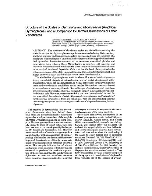
JOURNAL OF MORPHOLOGY 2062-43 (1990)<br />
<strong>Structure</strong> <strong>of</strong> <strong>the</strong> <strong>Scales</strong> <strong>of</strong> <strong>Dermophis</strong> <strong>and</strong> <strong>Microcaecika</strong> (Amphibia:<br />
Gymnophiona), <strong>and</strong> a Comparison to Dermal Ossifications <strong>of</strong> O<strong>the</strong>r<br />
Vertebrates<br />
LOUISE ZYLBERBERG AND MARVALEE H. WAKE<br />
CNRS UA 161 <strong>and</strong> Laboratoire d’Anatomie Comparhe, Universith de Paris VII,<br />
Paris 75251, France (L.Z.); Department <strong>of</strong> <strong>Integrative</strong> Biology <strong>and</strong> Museum <strong>of</strong><br />
Vertebrate Zoology, University <strong>of</strong> California, Berkeley, California 94720<br />
ABSTRACT The structures <strong>of</strong> <strong>the</strong> dermal scales <strong>and</strong> <strong>the</strong> cells surrounding <strong>the</strong><br />
scales in two species <strong>of</strong> gymnophione amphibians were studied using histochemistry<br />
<strong>and</strong> light, scanning <strong>and</strong> transmission electron microscopy. <strong>Scales</strong> are composed <strong>of</strong> a<br />
basal plate <strong>of</strong> several layers <strong>of</strong> unmineralized collagenous fibers topped with mineralized<br />
squamulae. Squamulae are composed <strong>of</strong> numerous mineralized globules <strong>and</strong><br />
mineralized, thick collagen fibers. Mineralization is <strong>the</strong>refore both spheritic <strong>and</strong><br />
inotropic. Isolated flattened cells lie on <strong>the</strong> outer surface <strong>of</strong> <strong>the</strong> squamulae <strong>and</strong> seem<br />
to be involved in mineral deposition. Cells that line <strong>the</strong> basal plate syn<strong>the</strong>size <strong>the</strong><br />
collagenous stroma <strong>of</strong> <strong>the</strong> plate. Each scale lies in a thin connective tissue pocket, <strong>and</strong><br />
a large connective tissue pouch includes several scales in each annulus.<br />
The similarities <strong>of</strong> gymnophione scales to elasmoid scales <strong>of</strong> osteichthyans are<br />
largely superficial. Aspects <strong>of</strong> mineralization <strong>and</strong> <strong>of</strong> pocket development differ<br />
considerably. There are also similarities, as well as differences, in <strong>the</strong> gymnophione<br />
scales <strong>and</strong> osteoderms <strong>of</strong> amphibians <strong>and</strong> <strong>of</strong> reptiles. We consider that such dermal<br />
structures have arisen many times in diverse lineages <strong>of</strong> vertebrates, <strong>and</strong> that <strong>the</strong>se<br />
are expressions <strong>of</strong> properties <strong>of</strong> dermal collagen to support mineralization by specialized<br />
dermal cells. However, we recommend that <strong>the</strong> term “dermal scale” be used for<br />
<strong>the</strong> mineralized dermal units <strong>of</strong> osteichthyans <strong>and</strong> gymnophiones, <strong>and</strong> “osteoderm”<br />
for <strong>the</strong> dermal structures <strong>of</strong> frogs <strong>and</strong> squamates, with <strong>the</strong> underst<strong>and</strong>ing that <strong>the</strong><br />
terminology recognizes certain convergent attributes <strong>of</strong> shape <strong>and</strong> structure, but not<br />
<strong>of</strong> process.<br />
The presence <strong>of</strong> dermal scales that are composed<br />
<strong>of</strong> an unmineralized base plate <strong>of</strong> collagenous<br />
fibers <strong>and</strong> a superficial layer <strong>of</strong> mineralized<br />
squamulae is unique to members <strong>of</strong> <strong>the</strong> amphibian<br />
Order Gymnophiona among all living tetrapods.<br />
Because gymnophione scales are structurally<br />
similar to those <strong>of</strong> teleost fishes, <strong>the</strong>y have<br />
been assumed to be homologous (Zylberberg et<br />
al. ’80; Ruibal <strong>and</strong> Shoemaker, ’84). However,<br />
recent examination <strong>of</strong> new data on gymnophione<br />
scales, toge<strong>the</strong>r with an assessment <strong>of</strong> <strong>the</strong> morphology<br />
<strong>of</strong> fish scales, <strong>the</strong> dermal ossifications <strong>of</strong><br />
frogs, <strong>and</strong> <strong>the</strong> osteoderms <strong>of</strong> lizards, causes us to<br />
question <strong>the</strong> assumption <strong>of</strong> homology <strong>of</strong> dermal<br />
scales. This analysis indicates that all dermal<br />
ossifications share a number <strong>of</strong> features that are<br />
structural properties <strong>of</strong> <strong>the</strong> particular organization<br />
<strong>of</strong> <strong>the</strong> dermis, <strong>and</strong> that <strong>the</strong> similarities <strong>and</strong><br />
differences in scale <strong>and</strong> osteoderm structure<br />
found in various lineages constitute evidence for<br />
convergent evolution, in response to <strong>the</strong> structural<br />
constraints <strong>of</strong> <strong>the</strong> tissue involved.<br />
There have been several descriptions <strong>of</strong> <strong>the</strong><br />
scales <strong>of</strong> gymnophione taxa in <strong>the</strong> last 150 years.<br />
Mayer (1829a,b), Rathke (1852), <strong>and</strong> Leydig<br />
(1853) commented briefly on scale morphology.<br />
Sarasin <strong>and</strong> Sarasin (1887-90) described <strong>the</strong><br />
scales <strong>of</strong> Ichthyophis glutinosus from Sri Lanka,<br />
<strong>and</strong> several subsequent analyses have corroborated<br />
<strong>the</strong>ir conclusions about <strong>the</strong> relationship <strong>of</strong><br />
<strong>the</strong> scales to o<strong>the</strong>r dermal <strong>and</strong> to epidermal<br />
structures. Cockerell (’11, ’12), Phisalix (’10, ’12),<br />
Datz (’23), Ochotorena (’32), Marcus (’34), Gabe<br />
(’71a,b), Casey <strong>and</strong> Lawson (’79), Zylberberg et<br />
al. (’80), Perret (’82), <strong>and</strong> Fox (’83) described <strong>the</strong><br />
scales <strong>of</strong> diverse species, usually Ichthyophis or<br />
Hypogeophis, at various levels <strong>of</strong> resolution. Taylor<br />
(’72) assembled an “atlas <strong>of</strong> scales” composed<br />
<strong>of</strong> photographs <strong>and</strong> descriptions <strong>of</strong> single scales<br />
from virtually all <strong>of</strong> <strong>the</strong> species <strong>of</strong> gymno-<br />
o 1990 WILEY-LISS, INC.
26 L. ZYLBERBERG AND M.H. WAKE<br />
phiones. He indicated that features such as scale<br />
size, shape, numbers <strong>and</strong> distribution, <strong>and</strong> pattern<br />
<strong>of</strong> mineralization might be useful for systematic<br />
analysis. However, Wake <strong>and</strong> Nygren ('87)<br />
demonstrated marked individual, ontogenetic,<br />
<strong>and</strong> sexual variation in nearly all scale parameters<br />
for <strong>Dermophis</strong> mexicanus from a single<br />
population, <strong>and</strong> suggested that scales are <strong>of</strong> little<br />
use as taxonomic characters to diagnose gymnophione<br />
species. Although Feuer ('62) proposed<br />
that scales might provide information about ages<br />
<strong>of</strong> individuals, Zylberberg et al. ('80) presented<br />
evidence to <strong>the</strong> contrary.<br />
The greatest numbers <strong>of</strong> scales occur in taxa<br />
<strong>of</strong> gymnophiones considered primitive based on<br />
o<strong>the</strong>r characters (osteology, myology, reproductive<br />
biology; Ichthyophis, Caudacaecilia, Rhinatrerna,<br />
Epicrionops-see Taylor, '68, '72; Nussbaum<br />
<strong>and</strong> Wilkinson, '89). <strong>Scales</strong> are embedded<br />
in virtually all <strong>of</strong> <strong>the</strong> annuli (i.e., body rings) <strong>of</strong><br />
primitive species, <strong>and</strong> <strong>the</strong>se taxa have several<br />
annuli per body segment. In gymnophiones, <strong>the</strong>re<br />
is a trend toward <strong>the</strong> reduction <strong>of</strong> numbers <strong>of</strong><br />
annuli <strong>and</strong>, concomitantly, <strong>of</strong> scales. Taxa considered<br />
highly derived based on o<strong>the</strong>r characters<br />
(e.g., typhlonectids <strong>and</strong> scolecomorphids) characteristically<br />
lack secondary annuli <strong>and</strong> scales<br />
(though Wake, ['75] <strong>and</strong> Moodie, ['78] found<br />
scales in some Typhlonectes).<br />
However, <strong>the</strong> scales <strong>of</strong> few taxa have been<br />
examined in detail with comparison to outgroups<br />
in mind. We present new data on <strong>the</strong><br />
morphology <strong>of</strong> scales <strong>of</strong> <strong>Dermophis</strong> mexicanus<br />
<strong>and</strong> Microcaecilia unicolor (both Gymnophiona:<br />
Caeciliaidae wde Nussbaum <strong>and</strong> Wilkinson, '891)<br />
to augment <strong>the</strong> detailed data available for Ichthyophis<br />
kohtaoensis (Gabe,'71a,b; Zylberberg<br />
et al., '80; Fox, '83) <strong>and</strong> Hypogeophis rostratus<br />
(Casey <strong>and</strong> Lawson, '79; Zylberberg et al., '80).<br />
The morphology <strong>of</strong> <strong>the</strong> scales <strong>of</strong> D. mexicanus<br />
<strong>and</strong> M. unicolor is compared with that <strong>of</strong> those<br />
scales described previously, <strong>and</strong> with that <strong>of</strong><br />
dermal ossifications in o<strong>the</strong>r vertebrates. Our<br />
comparative morphological approach elucidates<br />
<strong>the</strong> similarities <strong>and</strong> differences in mineralization<br />
patterns <strong>of</strong> such dermal derivatives as scales <strong>and</strong><br />
osteoderms <strong>and</strong> allows assessment <strong>of</strong> homology<br />
versus convergence <strong>of</strong> teleost scales, caecilian<br />
scales, <strong>and</strong> anuran <strong>and</strong> reptilian osteoderms. The<br />
study also provides a basis for continued research<br />
into <strong>the</strong> questions <strong>of</strong> development <strong>and</strong><br />
homologies <strong>of</strong> dermal structures in vertebrates.<br />
MATERIALS AND METHODS<br />
<strong>Scales</strong> <strong>of</strong> <strong>Dermophis</strong> mexicanus from San<br />
Marcos Prov., Guatemala, <strong>and</strong> Chiapas, Mexico,<br />
<strong>and</strong> Microcaecilia unicolor from Guyana were<br />
processed for analysis by several techniques. The<br />
specimens <strong>of</strong> <strong>Dermophis</strong> mexicanus from which<br />
scales were taken are deposited in <strong>the</strong> collections<br />
<strong>of</strong> <strong>the</strong> Museum <strong>of</strong> Vertebrate Zoology, University<br />
<strong>of</strong> California, Berkeley, <strong>and</strong> <strong>the</strong> M. unicolor<br />
in <strong>the</strong> Museum National d'Histoire naturelle,<br />
Paris. <strong>Scales</strong> were stripped from <strong>the</strong> annuli <strong>of</strong><br />
several formalin-ked, alcohol-preserved specimens<br />
<strong>of</strong> different sizes (<strong>and</strong> presumably ages)<br />
<strong>and</strong> sexes; all scales per annulus were stained<br />
with alizarin red-S (see Wake <strong>and</strong> Nygren, '87,<br />
for details). Quadrants <strong>of</strong> skin including several<br />
annuli were fixed in neutral buffered formalin or<br />
in Bouin's mixture, embedded in paraffin, <strong>and</strong><br />
sectioned sagittally or frontally. Stains <strong>and</strong> histochemical<br />
reactions are summarized in Tables 1<br />
<strong>and</strong> 2. <strong>Scales</strong> were prepared for scanning electron<br />
microscopy by excising, mounting on stubs,<br />
critical-point drying, <strong>and</strong> sputter-coating with<br />
gold or gold-palladium alloy. <strong>Scales</strong> were examined<br />
<strong>and</strong> photographed with an ISI-DS-130 dualstage<br />
scanning electron microscope or with a<br />
JEOL 8s M 35 SEM at an operating voltage <strong>of</strong><br />
25 kV.<br />
For transmission electron microscopy, small<br />
pieces <strong>of</strong> skin were removed from several regions<br />
<strong>of</strong> <strong>the</strong> body, fixed in a mixture <strong>of</strong> 2.5 % glutaraldehyde<br />
<strong>and</strong> 2 % paraformaldehyde in 0.1 M cacodylate<br />
buffer (pH 7.4) for 2 or 3 hr at room<br />
temperature; specimens were washed in 0.1 M<br />
cacodylate buffer containing 10% sucrose, <strong>and</strong><br />
postfixed in 1% osmium tetroxide in 0.1 M cacodylate<br />
buffer. Some samples were decalcified<br />
in <strong>the</strong> fixative with 0.1 M ethylene-diamino-tetraacetic<br />
acid (EDTA) added for 1 or 2 days at 4"C,<br />
<strong>and</strong> <strong>the</strong>n washed <strong>and</strong> postfixed. Ru<strong>the</strong>nium red<br />
(Martino et al., '79) was used as an electron<br />
microscopic stain for extracellular polyanions<br />
(Luft, '71a,b) such as acid mucosubstances <strong>and</strong><br />
acid phospholipids. Ru<strong>the</strong>nium red was added<br />
to <strong>the</strong> fixative containing EDTA, <strong>the</strong> wash buffer,<br />
<strong>and</strong> <strong>the</strong> osmium tetroxide solution. All <strong>the</strong> fixed<br />
samples were dehydrated in ethanol <strong>and</strong> cleared<br />
in 1-2 epoxy-propane. They were left in a 1:l<br />
epoxy-propanernpon mixture overnight or<br />
longer, <strong>and</strong> <strong>the</strong>n transferred to fresh resin. Polymerization<br />
was carried out at 60°C. Thick sections<br />
(-1 pm) were stained with toluidin blue for<br />
light microscopy. Thin sections <strong>of</strong> selected areas<br />
were cut with a Reichert ultramicrotome using a<br />
diamond knife with <strong>the</strong> tissue block oriented<br />
obliquely to <strong>the</strong> knife to improve sectioning <strong>of</strong><br />
hard fibrous tissues (AUizard <strong>and</strong> Zylberberg,<br />
'82). Sections were mounted on collodion-coated<br />
grids <strong>and</strong> stained with uranyl acetate <strong>and</strong> lead
Histological stain<br />
Azan<br />
Mallory's azan<br />
Hematoxylin & eosin<br />
One-step trichrome<br />
Histochemical reactions<br />
Proteins<br />
Danielli's coupled tetrazolium reaction<br />
Ferric ferricyanide reaction<br />
Carbohydrates<br />
Periodic acid-Sch8 (PAS)<br />
Alcian blue pH 2.5 (AB 2.5)<br />
Alcian blue pH 0.5 (AB 0.5)<br />
AB2.5 + PAS<br />
AB 0.5 + PAS<br />
Toluidine blue pH 4.2<br />
Paraldehyde-fuchsian blue<br />
pH 2.5<br />
Danielli's reaction +AB 2.5<br />
Calcium<br />
Alizarin red3<br />
GYMNOPHIONE SCALE MORPHOLOGY 27<br />
TABLE 1. Histological stains employed for analysis <strong>of</strong> scales<br />
Specificity<br />
General reaction<br />
Reducing groups<br />
Neutral mucosubstances<br />
Acid mucosubstances<br />
Sulfated mucosubstances<br />
Neutral <strong>and</strong> acid muccaubstances<br />
Neutral <strong>and</strong> sulfated mucosubstances<br />
Neutral vs. acid (carboxyl-rich <strong>and</strong> sulfated<br />
mucosubstances)<br />
Sulfated mucosubstances<br />
Protein <strong>and</strong> mucosubstances<br />
Calcium<br />
Reference<br />
Heidenhain in Gabe ('68)<br />
Humason ('79)<br />
Humason ('79)<br />
Gabe <strong>and</strong> Martoja in Gabe ('68)<br />
Reference<br />
Gabe ('68)<br />
Adams in Gabe ('68)<br />
MacManus in Gabe ('68)<br />
Mowry in Gabe ('68)<br />
Mowry in Gabe ('68)<br />
Mowry in Gabe ('68)<br />
Mowry in Gabe ('68)<br />
Lison in Gabe ('68)<br />
Gabe ('68)<br />
Lillie <strong>and</strong> Turner in Gabe ('68)<br />
Wake <strong>and</strong> Nygren ('87)<br />
TABLE 2. Histological <strong>and</strong> histochemical characteristics <strong>of</strong> scales <strong>of</strong> <strong>Dermophis</strong> mexicanus <strong>and</strong> Microcaecilia unicolor<br />
Scale<br />
Basal plate<br />
Squamulae<br />
Reaction Inner Outer Dermis<br />
Azan Red Blue Blue Blue<br />
Mallory's azan Blue Red Red Blue<br />
Hematoxylin & eosin Pink Purple Purple Pink<br />
One-step trichrome Red Green Green Green<br />
Picro-ponceau Yellow-pink Dark yellow Red Pink<br />
Danielli's reaction ++ +++ +- ++<br />
Ferric ferricyanide - - - -<br />
PAS +- +++ ++ +<br />
AB 2.5 -<br />
++ ++ -<br />
AB 0.5 - + +++ -<br />
Toluidine blue Blue Blue or purplish-blue Purplish-blue Blue<br />
PAS AB 2.5 Red Purplish-blue Blue Red<br />
PAS + AB 0.5 Red Purplish-red Purplish-blue Red<br />
Paraldehyde-fuchsin + PAS Red Reddish-purple Purple Red<br />
Danielli's reaction + AB 2.5 Purple Purple <strong>and</strong> Blue Blue Purple<br />
Alizarin red-S - Red Red -<br />
citrate (Reynolds,'63). The grids were examined<br />
in a Philips EM 300 electron microscope at an<br />
operating voltage <strong>of</strong> 80 kV with a cooled anticontamination<br />
device.<br />
RESULTS<br />
General organization <strong>of</strong> <strong>the</strong> scales<br />
The most anterior scales in <strong>Dermophis</strong> mexicanus<br />
are located in <strong>the</strong> tenth to twentieth primary<br />
annulus (counting posteriorly from <strong>the</strong><br />
head); <strong>the</strong>ir sizes <strong>and</strong> numbers increase posteri-<br />
orly in both primary <strong>and</strong> secondary annuli to<br />
mid-body, from which point numbers are relatively<br />
constant, although sizes within each annulus<br />
show much variation, until <strong>the</strong>y diminish in<br />
<strong>the</strong> most posterior annuli (see Wake <strong>and</strong> Nygren,<br />
'87, for data). In both taxa examined, <strong>the</strong><br />
scale rows are covered by a thin layer <strong>of</strong> epidermis<br />
(Figs. la, 2), hence are not exposed to <strong>the</strong><br />
environment. <strong>Scales</strong> are somewhat irregular in<br />
size <strong>and</strong> shape, <strong>and</strong> a diversity <strong>of</strong> scale sizes <strong>and</strong><br />
shapes is found in each scale row (Wake <strong>and</strong><br />
Nygren, '87). Excised scales treated with alizarin
28 L. ZYLBERBERG AND M.H. WAKE<br />
red-S have red-stained structures atop an unstained<br />
base plate, indicating specificity for calcium<br />
in <strong>the</strong> mineralized squamulae; <strong>the</strong> latter<br />
also have been called denticles. We prefer <strong>the</strong><br />
more precise term squamulae, to avoid possible<br />
confusion <strong>of</strong> <strong>the</strong>se structures with <strong>the</strong> “denticles”<br />
<strong>of</strong> sharks, for example.<br />
In parasagittal sections <strong>of</strong> skin with several<br />
annuli, <strong>the</strong> scale pouches are embedded in each<br />
annulus <strong>and</strong> form a ring that partly or completely<br />
encircles <strong>the</strong> body. Characteristically, <strong>the</strong><br />
distal end <strong>of</strong> <strong>the</strong> scale pouch lies in <strong>the</strong> dermis<br />
below <strong>the</strong> epidermis (Figs. la, 2, 3). The distal<br />
margin <strong>of</strong> <strong>the</strong> pouch may be deep to small dermal<br />
mucous gl<strong>and</strong>s; <strong>the</strong> proximal end <strong>of</strong> <strong>the</strong><br />
pouch is broader <strong>and</strong> located at <strong>the</strong> boundary<br />
between <strong>the</strong> superficial <strong>and</strong> <strong>the</strong> deep dense dermis<br />
that overlies <strong>the</strong> body wall musculature (Figs.<br />
la, 2). Each scale pouch usually is associated<br />
with large mixed granular (so-called “poison”)<br />
gl<strong>and</strong>s both dorsally <strong>and</strong> ventrally. The pouch is<br />
a thin, connective tissue structure (Figs. 2, 3).<br />
Each scale lies in its own pocket or sac within <strong>the</strong><br />
pouch (Figs. 2, p6). Fibroblasts line <strong>the</strong> inner<br />
surface <strong>of</strong> each pocket (Figs. 7, 8), <strong>and</strong>, within<br />
each pocket, <strong>the</strong> fibroblasts (scleroblasts) that<br />
form <strong>the</strong> scale are arranged in layers surrounding<br />
each scale. The scleroblasts constitute an<br />
uninterrupted layer on <strong>the</strong> bad surface <strong>of</strong> <strong>the</strong><br />
scale. At <strong>the</strong> scale margin, <strong>the</strong> collagenous stroma<br />
<strong>of</strong> <strong>the</strong> scale aligns with <strong>the</strong> connective tissue <strong>of</strong><br />
<strong>the</strong> scale pocket (Figs. Ib, 25). Numerous scales,<br />
contained in <strong>the</strong>ir pockets, overlap within a pouch<br />
(Fig. 1, 2). All scales lie similarly in <strong>the</strong> pouch<br />
with <strong>the</strong> denticulate surface facing outward.<br />
Some large scales are folded back on <strong>the</strong>mselves<br />
in <strong>the</strong> scale pockets (Fig. 4). Scale structure<br />
varies with size <strong>of</strong> scale <strong>and</strong> with region on <strong>the</strong><br />
scale (central vs. peripheral). Large scales have a<br />
thick base plate composed <strong>of</strong> as many as seven<br />
layers, or plies, <strong>of</strong> fibers. The orientation <strong>of</strong> <strong>the</strong><br />
fibers alternates regularly (Figs. IC, 5,13). Mineralized<br />
squamulae lie atop, <strong>and</strong> slightly embedded<br />
in, <strong>the</strong> upper layer <strong>of</strong> <strong>the</strong> base plate (Figs. IC,<br />
3, 13). Each squamula is a discrete structure<br />
(Figs. 3,13).<br />
The squamulae<br />
At low magnification, central squamulae on<br />
<strong>the</strong> base plate appear to be square to round<br />
shaped, whereas more peripheral squamulae are<br />
more elongate. Squamulae are arrayed in irregularly<br />
concentric circles (Figs. 9,lO) <strong>and</strong> are highly<br />
irregular in shape. The longitudinal sectional<br />
pr<strong>of</strong>iles <strong>of</strong> <strong>the</strong> squamulae vary; <strong>the</strong>y are relatively<br />
flat but bear points <strong>and</strong> ridges (Figs. 2-5).<br />
Squamulae are flattest centrally; points <strong>and</strong><br />
ridges <strong>of</strong> more peripheral squamulae are higher<br />
(Figs. 9, 10). At moderate magnification<br />
(- x 1,OOO), <strong>the</strong>y show numerous ridges <strong>and</strong> folds<br />
(Figs. 11, 12). At higher magnification (Fig. 13),<br />
it is clear that each squamula is an array <strong>of</strong><br />
round, mineralized deposits <strong>of</strong> varying heights.<br />
Some <strong>of</strong> <strong>the</strong> deposits are fused superficially or<br />
linearly; however, <strong>the</strong> bases <strong>of</strong> <strong>the</strong> mineralized<br />
structures rarely are fused.<br />
The points <strong>and</strong> ridges that form <strong>the</strong> uppermost<br />
surface <strong>of</strong> each squamula include a loose<br />
framework composed <strong>of</strong> acid mucosubstances<br />
that stain with Alcian blue (Fig. 14), whereas<br />
abundant neutral mucosubstances <strong>and</strong> proteins<br />
are located more centrally within <strong>the</strong> squamula<br />
(Figs. 14,15; Table 2). The uppermost surface <strong>of</strong><br />
a squamula is composed <strong>of</strong> numerous mineralized<br />
globules that appear as isolated structures,<br />
particularly at <strong>the</strong> periphery <strong>of</strong> <strong>the</strong> squamula<br />
(Figs. 7,24,25), or that aggregate to form large<br />
concretions in <strong>the</strong> central part <strong>of</strong> <strong>the</strong> squamula<br />
(Figs. 13, 16). The mineral deposit is composed<br />
<strong>of</strong> crystals that have a radial arrangement within<br />
<strong>the</strong> globules (Figs. 18,22). In demineralized sections,<br />
organic matrix cannot be distinguished in<br />
many globules. They appear as electron-lucent<br />
circular spaces (Fig. 17). In o<strong>the</strong>r globules, a thin<br />
fibrillar network forms a central core surrounded<br />
by an electron-lucent space (Fig. 19). An electrondense<br />
opaque material that displays high contrast<br />
with ru<strong>the</strong>nium red lines <strong>the</strong> surface <strong>of</strong> <strong>the</strong><br />
globules (Fig. 19). The central part <strong>of</strong> <strong>the</strong> squamula<br />
contains both mineralized globules <strong>and</strong> mineralized,<br />
thick collagen fibrils that arise from <strong>the</strong><br />
basal plate (Figs. 16, 23). The crystals are oriented<br />
along <strong>the</strong> collagen fibrils, <strong>and</strong> invade <strong>the</strong><br />
fibrils somewhat. At <strong>the</strong> inner surface <strong>of</strong> <strong>the</strong><br />
squamulae, mineralized globules are inserted in<br />
<strong>the</strong> network formed by <strong>the</strong> collagenous stroma<br />
(Fig. 23).<br />
Isolated, flattened cells are located on <strong>the</strong> outer<br />
surface <strong>of</strong> <strong>the</strong> squamulae (Fig. 13). They contain<br />
a voluminous central nucleus, <strong>and</strong> are more numerous<br />
in young animals than in older ones. In<br />
young animals, <strong>the</strong> cells have long cytoplasmic<br />
processes that cover <strong>the</strong> surfaces <strong>of</strong> <strong>the</strong> squamulae,<br />
<strong>and</strong> <strong>the</strong> processes are near <strong>the</strong> outer mineralized<br />
globules (Fig. 13). A fuzzy organic material<br />
is located near <strong>the</strong> plasma membrane facing <strong>the</strong><br />
squamula where <strong>the</strong> first crystal deposits appear<br />
(Fig. 20), suggesting that <strong>the</strong>se cells are involved<br />
in <strong>the</strong> production <strong>of</strong> <strong>the</strong> mineralized globules.<br />
The crystals first deposited in <strong>the</strong>se globules<br />
have no apparent orientation (Fig. 20). The radial<br />
organization <strong>of</strong> <strong>the</strong> crystals becomes apparent<br />
in globules still in <strong>the</strong> vicinity <strong>of</strong> <strong>the</strong> sclero-
GYMNOPHIONE SCALE MORPHOLOGY 29<br />
Fig. 1. Diagrammatic representation <strong>of</strong> a caecilian scale. =<strong>Scales</strong> in scale pocket relative to “poison”<br />
gl<strong>and</strong>s, mucous gl<strong>and</strong>s, <strong>and</strong> annuli. b Margin <strong>and</strong> central part <strong>of</strong> scale, showing cells lining scale <strong>and</strong> <strong>the</strong><br />
arrangement <strong>of</strong> <strong>the</strong> basal plate <strong>and</strong> <strong>the</strong> squamulae.
on<br />
L. ZYLBERBERG AND M.H. WAKE
GYMNOPHIONE SCALE MORPHOLOGY 31<br />
blast (Fig. 21). The globules subsequently<br />
aggregate to form larger concretions.<br />
The basal plate<br />
The basal plate is <strong>the</strong> most extensive part <strong>of</strong><br />
<strong>the</strong> scale, <strong>and</strong> is made up <strong>of</strong> superimposed plies<br />
<strong>of</strong> collagen fibrils. A diameter <strong>of</strong> -100 nm is<br />
achieved by most <strong>of</strong> <strong>the</strong> newly syn<strong>the</strong>sized collagen<br />
fibrils that lie in <strong>the</strong> vicinity <strong>of</strong> <strong>the</strong> plasma<br />
membrane (Figs. 28,29,30). The collagen fibrils<br />
<strong>of</strong> <strong>the</strong> basal plate are distinctly thicker than<br />
those <strong>of</strong> <strong>the</strong> dermis (i.e., 100 vs. 30-nm dia; Figs.<br />
35,36). In each ply, <strong>the</strong> collagen fibrils are packed<br />
in thick bundles, oriented in parallel (Figs. 13,<br />
26), <strong>and</strong> <strong>of</strong>ten, <strong>the</strong> periodic structure <strong>of</strong> groups <strong>of</strong><br />
fibrils is in register (Fig. 34). The direction <strong>of</strong> <strong>the</strong><br />
fibrils varies from one ply to ano<strong>the</strong>r; <strong>the</strong> angle <strong>of</strong><br />
rotation between two adjacent plies is about 90’.<br />
Thus, <strong>the</strong> plies constitute an orthogonal plywood-<br />
Fig. 2. Longitudinal section perpendicular to <strong>the</strong> surface<br />
<strong>of</strong> <strong>the</strong> skin at <strong>the</strong> level <strong>of</strong> an annulus in <strong>Dermophis</strong> mexicanus<br />
(light micrograph [LM]). The pouch(p) containsfouroverlapping<br />
scales (8) <strong>and</strong> lies below <strong>the</strong> epidermis (e). The scale<br />
pouch <strong>and</strong> <strong>the</strong> gl<strong>and</strong>s (mg = mucous gl<strong>and</strong>, pg = “poison”<br />
gl<strong>and</strong>) lie within <strong>the</strong> loose dermis (d) <strong>and</strong> do not penetrate <strong>the</strong><br />
dense dermis.<br />
Fig. 3. Enlargement <strong>of</strong> outlined area <strong>of</strong> Figure 2 showing<br />
<strong>the</strong> outer part <strong>of</strong> <strong>the</strong> pouch (p) with <strong>the</strong> four scales (8). The<br />
mucou gl<strong>and</strong>s (mg) are located in <strong>the</strong> dermis (d) between <strong>the</strong><br />
epidermis (e) <strong>and</strong> <strong>the</strong> scale pouch. A thin layer <strong>of</strong> connective<br />
tissue (ct) separates <strong>the</strong> scales in <strong>the</strong>ir pocket. The squamulae<br />
(sq) lie on <strong>the</strong> dorsal surface <strong>of</strong> <strong>the</strong> scales.<br />
Fig. 4. Longitudinal section (LM) <strong>of</strong> <strong>the</strong> inner part <strong>of</strong> a<br />
pouch containing a folded scale (8) in Dermuphis mexicanus.<br />
pg = poison gl<strong>and</strong>.<br />
Fig. 5. Enlargement <strong>of</strong> outlined area in Figure 4. The two<br />
scales (s) are inserted in <strong>the</strong>ir own pocket (sp), surrounded by<br />
connective tissue (d). The thick central part <strong>of</strong> <strong>the</strong> scale has<br />
six plies, whereas <strong>the</strong> thin, folded part has only one ply. The<br />
squamulae (sq) top <strong>the</strong> basal plate. The margin <strong>of</strong> <strong>the</strong> scale<br />
(solid arrow) is not lined by scleroblasts.<br />
Fig. 6. The thin layer <strong>of</strong> connective tissue (ct) between<br />
two scales separates <strong>the</strong> pockets (sp); transmission electron<br />
micrograph (TEM); longitudinel section (Ls). Fibroblasts<br />
(IC) line <strong>the</strong> pocket. The scale is surrounded by <strong>the</strong> scleroblasts<br />
(sc). <strong>Dermophis</strong> mexicanus. bp = basal plate, sq =<br />
squamula.<br />
Fig. 7. Detail <strong>of</strong> <strong>the</strong> dorsal part <strong>of</strong> a pocket limed by a<br />
fibroblast (IC) in Microcaeciliu unicolor (TEM LS). The<br />
scleroblast (sc) lies on <strong>the</strong> squamula (sq). p = pouch; sp =<br />
scale pocket.<br />
Fig. 8. Detail <strong>of</strong> <strong>the</strong> basal part <strong>of</strong> a pocket in <strong>Dermophis</strong><br />
mexicanus (TEM, LS). The scale pocket (sp) is lined by a<br />
fibroblast (IC). bp = basal plate; p = pouch; sc = scleroblast.<br />
like structure. The collagen fibrils <strong>of</strong> <strong>the</strong> basal<br />
plate mineralize only within <strong>the</strong> squamulae.<br />
The collagenous stroma <strong>of</strong> <strong>the</strong> basal plate is<br />
syn<strong>the</strong>sized by <strong>the</strong> cells that line <strong>the</strong> basal surface<br />
<strong>of</strong> <strong>the</strong> scale. These flattened cells have a<br />
voluminous central nucleus <strong>and</strong> form a continuous<br />
sheet that is considered a pseudoepi<strong>the</strong>lium<br />
composed <strong>of</strong> scleroblasts (Figs. 26,27). The rough<br />
endoplasmic reticulum (RER) is composed <strong>of</strong><br />
short saccules. Mitochondria are not abundant<br />
<strong>and</strong> <strong>the</strong> Gob areas are not well developed (Fig.<br />
13). Micr<strong>of</strong>ilaments are abundant; in some cells,<br />
<strong>the</strong>y appear to be aligned with microtubules <strong>and</strong><br />
with <strong>the</strong> newly syn<strong>the</strong>sized collagen fibrils (Figs.<br />
28,29), whereas in o<strong>the</strong>r cells such alignment is<br />
not observed (Fig. 30). The scleroblasts have<br />
long cytoplasmic processes that insert among<br />
<strong>the</strong> collagen fibrils, <strong>the</strong>reby separating <strong>the</strong> fibrils<br />
into distinct bundles (Figs. 13, 27). Collagen<br />
fibrils arise from <strong>the</strong>se processes perpendicularly<br />
to <strong>the</strong> collagenous plies (Fig. 13). When a new<br />
ply is formed, <strong>the</strong> orientation <strong>of</strong> <strong>the</strong> first collagen<br />
fibrils syn<strong>the</strong>sized is at a right angle to that <strong>of</strong><br />
<strong>the</strong> preceding ply (Fig. 31); <strong>the</strong>n an isolated<br />
bundle is formed (Fig. 32), <strong>and</strong> finally <strong>the</strong> whole<br />
newly syn<strong>the</strong>sized ply is formed by collagen fibrils<br />
aligned in <strong>the</strong> new direction (Fig. 33).<br />
DISCUSSION<br />
Comparative morphology <strong>of</strong> dermal<br />
ossifications<br />
The gymnophione condition<br />
The presence <strong>of</strong> dermal scales is unique to<br />
gymnophiones among extant tetrapods. Various<br />
workers (Taylor, ’72; Zylberberg et al., ’80; Perret,<br />
’82) have described <strong>the</strong> variation in scale<br />
morphology (e.g., size, shape, depth <strong>of</strong> base plate,<br />
distribution <strong>and</strong> shape <strong>of</strong> squamulae) among<br />
caecilian taxa. Certain features <strong>of</strong> scalation, such<br />
as overall distribution <strong>and</strong> presence or absence,<br />
have been used as reliable systematic characters.<br />
We describe microscopic features <strong>of</strong> scale morphology<br />
that might be taxonomically useful.<br />
Among adults, <strong>the</strong> shapes <strong>of</strong> <strong>the</strong> squamulae on<br />
<strong>the</strong> scales <strong>and</strong> <strong>the</strong> pattern <strong>of</strong> deposition <strong>of</strong> mineralized<br />
material seem to be fairly consistent. Such<br />
features are best examined by scanning electron<br />
microscopy, so that details <strong>of</strong> structure can be<br />
assessed large samples <strong>of</strong> scales have yet to be<br />
evaluated for any single taxon. A comparison <strong>of</strong><br />
<strong>the</strong> scanning electron micrographs <strong>of</strong> Hypogeophis<br />
(Casey <strong>and</strong> Lawson, ’79), Ichthyophis <strong>and</strong><br />
Hypogeophis (Zylberberg et al., ’80), Geotrypetes<br />
<strong>and</strong> Herpele (Perret, ’82), <strong>Dermophis</strong><br />
(Wake <strong>and</strong> Nygren, ’87), <strong>and</strong> those in Taylor’s<br />
atlas (’72) reveals considerable variation in squa-
32 L. ZYLBERBERG AND M.H. WAKE<br />
9<br />
Figures 9-12
mular shape <strong>and</strong> structure. The rectangular<br />
shape <strong>of</strong> peripheral squamulae in Ichthyophis<br />
(Zylberberg et al., ’80) contrasts markedly with<br />
<strong>the</strong> rounded shapes <strong>of</strong> Geotrypetes (Perret, ’82)<br />
<strong>and</strong> <strong>Dermophis</strong> (Wake <strong>and</strong> Nygren, ’87). It<br />
should be noted that <strong>the</strong>se contrasts are greatest<br />
among peripheral squamulae; examination <strong>of</strong> <strong>the</strong><br />
published photomicrographs indicates that <strong>the</strong><br />
centers <strong>of</strong> scales bear squamulae that are both<br />
rounder <strong>and</strong> more irregular in shape. A common<br />
feature <strong>of</strong> squamulae <strong>of</strong> virtually all species examined<br />
is that mineralized material is deposited in<br />
a globular manner (see photomicrographs cited<br />
above). The only exception to this pattern that<br />
we have observed is in Caecilia tentaculata, in<br />
which deposition is essentially smooth (Wake<br />
<strong>and</strong> Zylberberg, unpublished data), <strong>and</strong> we are<br />
examining this situation fur<strong>the</strong>r.<br />
Comparison with osteichthyan scales<br />
The pouch <strong>and</strong> <strong>the</strong> pocket<br />
Our data confirm that gymnophione scales<br />
have peculiarities apparently related to <strong>the</strong>ir location<br />
in <strong>the</strong> segmentally arranged annuli, <strong>and</strong><br />
that <strong>the</strong>y share more structural similarities with<br />
<strong>the</strong> scales <strong>of</strong> extant osteichthyans than with <strong>the</strong><br />
osteoderms <strong>of</strong> extant amphibians <strong>and</strong> reptiles.<br />
Because <strong>of</strong> <strong>the</strong>ir position relative to <strong>the</strong> annuli,<br />
<strong>the</strong> overlapping scales in gymnophiones occur in<br />
narrow transverse rows that partly or completely<br />
encircle <strong>the</strong> body. In osteichthyans, <strong>the</strong> imbricate<br />
scales are distributed over <strong>the</strong> body completely<br />
or in patches, without an obvious segmental<br />
association. Our data indicate that every scale<br />
in both species examined lies within its own<br />
pocket, as do <strong>the</strong> elasmoid scales <strong>of</strong> osteichthyans<br />
(Whitear et al., ’80). Recent descriptions,<br />
including those at <strong>the</strong> ultrastructural level, have<br />
mentioned that several overlapping scales lie in a<br />
“pocket” (Taylor, ’72; Gabe, ’71a; Wake, ’75;<br />
Zylberberg et al., ’80; Fox, ’83). However, as<br />
noted above, that “pocket” is actually a large<br />
connective tissue pouch associated with an annulus.<br />
The pouch contains <strong>the</strong> scales, each <strong>of</strong> which<br />
lies in a thin, connective tissue pocket, homologous<br />
to that <strong>of</strong> osteichthyans. However, <strong>the</strong> fibro-<br />
Fig. 9. R<strong>and</strong>omly selected scale <strong>of</strong> Microcaecilia unicolor<br />
stained with k in-red to show shape <strong>and</strong> arrangement <strong>of</strong><br />
squamulae.<br />
Fig. 10. R<strong>and</strong>omly selected scale <strong>of</strong> <strong>Dermophis</strong> mexicanus<br />
stained with alizarin-red.<br />
Fig. 11. Scanning electron micrograph (SEM) <strong>of</strong> aquamulae<br />
<strong>of</strong> Microcaecilia unicolor.<br />
Fig. 12. Squamulae <strong>of</strong> <strong>Dermophis</strong> mexicam (SEM).<br />
GYMNOPHIONE SCALE MORPHOLOGY 33<br />
blasts that line <strong>the</strong> scale pocket <strong>of</strong> gymnophiones<br />
do not differentiate as in fishes (see<br />
Whitear et al., ’80). In osteichthyans, <strong>the</strong> outer<br />
surface <strong>of</strong> <strong>the</strong> elasmoid scale is connected to <strong>the</strong><br />
superficial dermis by collagenous anchoring bundles<br />
that arise from <strong>the</strong> outer surface <strong>of</strong> <strong>the</strong> scale<br />
<strong>and</strong> extend through <strong>the</strong> dermis to <strong>the</strong> epidermaldermal<br />
junction (Zylberberg <strong>and</strong> Meunier, ’81;<br />
Sire, ’85). Such bundles are absent in <strong>the</strong> gymnophione<br />
scales examined. This phenomenon may<br />
be related to <strong>the</strong> location <strong>of</strong> <strong>the</strong> scale ra<strong>the</strong>r than<br />
to <strong>the</strong> structure <strong>of</strong> <strong>the</strong> squamulae. In gymnophiones,<br />
<strong>the</strong> scales are well inserted within <strong>the</strong><br />
dermis among skin gl<strong>and</strong>s, <strong>and</strong> <strong>the</strong>y are covered<br />
by a thick pleuristratified epidermis (Gabe, ’71a).<br />
Similarly, <strong>the</strong> reduced scales <strong>of</strong> <strong>the</strong> eel which are<br />
covered by a mosaic <strong>of</strong> smooth squamulae do not<br />
have anchoring bundles. These scales also are<br />
covered by a thick pleuristratified epidermis (Zylberberg<br />
et al., ’84). In fur<strong>the</strong>r contrast, <strong>the</strong> large<br />
scales <strong>of</strong> Protopterus, which are located in a very<br />
loose dermis close to <strong>the</strong> thin epidermis, have<br />
squamulae with well-developed collagenous anchoring<br />
bundles (Zylberberg, ’88).<br />
Squamulae<br />
Our observations confirm that mineralization<br />
in gymnophione scales is limited to <strong>the</strong> squamulae,<br />
which are isolated plates topping <strong>the</strong> scales;<br />
mineralization does not occur in <strong>the</strong> basal plate<br />
as described by Casey <strong>and</strong> Lawson (’79). Gymnophione<br />
squamulae have two simultaneous processes<br />
<strong>of</strong> mineralization, termed spheritic <strong>and</strong><br />
inotropic (Orvig, ’68). These two processes are<br />
<strong>the</strong> result <strong>of</strong> <strong>the</strong> heterogeneous composition <strong>and</strong><br />
organization <strong>of</strong> <strong>the</strong> extracellular matrix in <strong>the</strong><br />
squamulae. Recent reports point out <strong>the</strong> importance<br />
<strong>of</strong> <strong>the</strong> organic matrix in controlling crystal<br />
deposition (reviewed by Weiner, ’86). Where<br />
spheritic mineralization occurs, <strong>the</strong> shape <strong>of</strong> <strong>the</strong><br />
spicules depends on <strong>the</strong> radiating arrangement<br />
<strong>of</strong> <strong>the</strong> noncollagenous matrix. The crystals associated<br />
with <strong>the</strong> collagen fibrils are aligned along<br />
<strong>the</strong> fibrils so that <strong>the</strong>ir long axis (<strong>the</strong> crystallographic<br />
c axis) is parallel to <strong>the</strong> collagen fibril<br />
axis (Schmidt, ’36; Stuhler, ’38). Moreover, <strong>the</strong><br />
crystals are distributed according to <strong>the</strong> axial<br />
periodicity <strong>of</strong> <strong>the</strong> collagen fibrils as first described<br />
in bone (Hodge <strong>and</strong> Petruska, ’63; Glimcher<br />
<strong>and</strong> Krane, ’68; Glimcher, ’81; Arsenault,<br />
’88).<br />
According to Orvig (’68), spheritic mineralization<br />
may be considered <strong>the</strong> phylogenetic precursor<br />
<strong>of</strong> inotropic mineralization. He considered<br />
<strong>the</strong> latter to represent <strong>the</strong> “ultimate stages” in a<br />
phyletic process <strong>of</strong> increasing complexity <strong>of</strong> “calcification<br />
mechanisms.” Spheritic mineralization<br />
also may be considered an ontogenetic pre-
Figures 13-17
cursor <strong>of</strong> inotropic mineralization, because during<br />
dentine ontogenesis, spheritic mineralization<br />
(which forms globular dentine) is replaced by<br />
inotropic mineralization (Keil, '39; Orvig, '67;<br />
Poole, '67). Therefore, <strong>the</strong> squamulae <strong>of</strong> gymnophiones,<br />
<strong>the</strong> outer surface <strong>of</strong> osteichthyan scales<br />
(Sire, '85; Zylberberg, '88 <strong>and</strong> unpublished data),<br />
<strong>and</strong> <strong>the</strong> osteoderms <strong>of</strong> reptiles (Levrat-Calviac<br />
<strong>and</strong> Zylberberg, '86) have retained a primitive<br />
type <strong>of</strong> mineralization that occurs concomitantly<br />
with a "more advanced" mineralization process.<br />
The mineralized spherules are more abundant<br />
on <strong>the</strong> outer surface <strong>of</strong> <strong>the</strong> squamulae, but also<br />
are found on <strong>the</strong> inner surface among <strong>the</strong> collagen<br />
fibrils. These spherules, which have a radiation<br />
arrangement <strong>of</strong> crystals, do not correspond<br />
to <strong>the</strong> M<strong>and</strong>l's corpuscles characteristic <strong>of</strong> elasmoid<br />
scales. In M<strong>and</strong>l's corpuscles, <strong>the</strong> crystals<br />
are oriented by <strong>the</strong> alignment <strong>of</strong> <strong>the</strong> collagen<br />
fibrils (Schonborner et al., '81). The limit between<br />
<strong>the</strong> squamulae <strong>and</strong> <strong>the</strong> basal plate is represented<br />
by <strong>the</strong> mineralizing front, though collagen<br />
fibrils arise from <strong>the</strong> basal plate <strong>and</strong><br />
penetrate <strong>the</strong> squamulae. These fibrils are not<br />
part <strong>of</strong> <strong>the</strong> plywood-like structure <strong>of</strong> <strong>the</strong> basal<br />
plate itself. As noted by Zylberberg et al., ('80),<br />
<strong>the</strong> TEM micrographs <strong>of</strong> well-developed scales<br />
show mineral deposits both along <strong>the</strong> collagen<br />
fibrils <strong>and</strong> within <strong>the</strong> interfibrillary matrix, as<br />
well as in spherules, where <strong>the</strong>y are arranged in a<br />
radiating pattern. Therefore, <strong>the</strong> site <strong>of</strong> nucleation<br />
cannot be determined (present study; Zylberberg<br />
et al., '80). Moreover, <strong>the</strong> cells that par-<br />
Fg. 13. Scale <strong>of</strong> Microcaecih unicolor sectioned perpendicular<br />
to its surface (TEM). The fibroblasts (IC) forming <strong>the</strong><br />
scale pocket surround <strong>the</strong> scleroblasb (sc). The outer part <strong>of</strong><br />
<strong>the</strong> squamula (sq) has abundant mineralized globules. In <strong>the</strong><br />
inner part <strong>of</strong> <strong>the</strong> squamula <strong>the</strong> minerahng front (mf) is<br />
located in <strong>the</strong> basal plate (bp). The basal scleroblasts have<br />
long processes (arrows) that separate <strong>the</strong> bundles <strong>of</strong> collagen<br />
fibrils.<br />
Fig. 14. The inner part <strong>of</strong> <strong>the</strong> squamulae (sq) is PASreactive<br />
(dark color) whereas <strong>the</strong> outer part shows weak<br />
staining with alcian blue (pH 2.5) (arrows) (LM, LS). The<br />
basal plate does not react to ei<strong>the</strong>r stain. <strong>Dermophis</strong> mexicanus.<br />
Fig. 15. The inner part <strong>of</strong> <strong>the</strong> squamulae (sq) is strongly<br />
reactive to tetrazolium, though <strong>the</strong> outer part (arrows) <strong>and</strong><br />
<strong>the</strong> base plate (bp) stain weakly (LM LS). Dermophk mexicanus.<br />
Fig. 16. The outer surface <strong>of</strong> <strong>the</strong> squamula (sq) has many<br />
mineralized globules (TEM LS). <strong>Dermophis</strong> mexicam.<br />
Fig. 17. Demineralized (EDTA) scale section from<strong>Dermophis</strong><br />
mexicaw (TEM LS). Theglobulea are primaily at <strong>the</strong><br />
outer part <strong>the</strong> squamula They do not show any organic<br />
matrix. ct = connective tissue; mf = mineralizing front.<br />
GYMNOPHIONE SCALE MORPHOLOGY 35<br />
ticipate in <strong>the</strong> mineralization processes have not<br />
been identified.<br />
Basal plate<br />
In gymnophiones, <strong>the</strong> thick collagenous fibrils<br />
forming <strong>the</strong> basal plate are organized as a plywood-like<br />
structure, but it is less regularly arranged<br />
than in <strong>the</strong> elasmoid scales <strong>of</strong> teleosts<br />
<strong>and</strong> sarcopterygians. The reduction <strong>of</strong> organization<br />
<strong>of</strong> <strong>the</strong> basal plate in gymnophiones may be<br />
associated with a generalized reduction <strong>of</strong> <strong>the</strong><br />
dermal skeleton in both fishes <strong>and</strong> tetrapods.<br />
The most reduced osteichthyan scale observed<br />
occurs in <strong>the</strong> eel in which <strong>the</strong> basal plate has only<br />
one ply (Zylberberg et al., '84). The scales <strong>of</strong><br />
gymnophiones are thin <strong>and</strong> <strong>the</strong> plies are much<br />
less numerous than in fishes. However, <strong>the</strong> basal<br />
plate, which shows <strong>the</strong> usual histological <strong>and</strong><br />
histochemical characteristics <strong>of</strong> lamellar bone,<br />
does not mineralize as it does in <strong>the</strong> basal plate<br />
<strong>of</strong> osteichthyans (Meunier, '84). According to<br />
Meunier, thick basal plates made <strong>of</strong> isopedine<br />
that has lost its ability to be mineralized reflect a<br />
general trend toward reduction <strong>of</strong> <strong>the</strong> dermal<br />
skeleton.<br />
The collagen fibrils <strong>of</strong> gymnophione scales,<br />
like those <strong>of</strong> fish, are thicker than those <strong>of</strong> <strong>the</strong><br />
surrounding dermis, including <strong>the</strong> collagen fibrils<br />
that separate <strong>the</strong> bundles forming <strong>the</strong> plywoodlike<br />
structure <strong>of</strong> <strong>the</strong> base plate <strong>and</strong> those that<br />
cross <strong>the</strong> scale perpendicularly to <strong>the</strong> plies. The<br />
latter collagen fibrils differ from <strong>the</strong> TC fibers<br />
described in <strong>the</strong> elasmoid scales <strong>of</strong> Cyprinidae.<br />
They are thinner than <strong>the</strong> collagen fibrils <strong>of</strong> <strong>the</strong><br />
plywood-like structure (Onozato <strong>and</strong> Watabe,<br />
'79) <strong>and</strong> are involved in <strong>the</strong> first stages <strong>of</strong> mineralization<br />
<strong>of</strong> <strong>the</strong> basal plate (Zylberberg <strong>and</strong> Nicolas,<br />
'82). As in fish scales, <strong>the</strong> collagenous stroma<br />
as well as <strong>the</strong> noncollagenous extracellular matrix<br />
are syn<strong>the</strong>sized by <strong>the</strong> cells surrounding <strong>the</strong><br />
scales, which also control mineral deposition.<br />
Therefore, <strong>the</strong> gymnophione scale is an acellular<br />
tissue as defined by Meunier ('87); fur<strong>the</strong>r, <strong>the</strong><br />
basal plate may be considered a bony tissue<br />
which has lost its capacity to be mineralized<br />
(Meunier, '84).<br />
The scleroblasts involved in <strong>the</strong> formation <strong>of</strong><br />
<strong>the</strong> scales line each scale. They are contiguous<br />
only along <strong>the</strong> inner surface <strong>of</strong> <strong>the</strong> scale where<br />
<strong>the</strong>y form a pseudoepi<strong>the</strong>lium. Because <strong>the</strong>y syn<strong>the</strong>size<br />
<strong>the</strong> thick collagen fibrils that form <strong>the</strong><br />
basal plate, <strong>the</strong>y are assumed to control fibrillogenesis<br />
<strong>and</strong> <strong>the</strong> orientation <strong>of</strong> <strong>the</strong> collagen as has<br />
been described for fish scales (see above). The<br />
long cytoplasmic processes that penetrate deeply<br />
within <strong>the</strong> basal plate are involved in <strong>the</strong> formation<br />
<strong>of</strong> <strong>the</strong> bundles that form <strong>the</strong> superimposed<br />
plies. However, <strong>the</strong> processes seem to orient <strong>the</strong>
Figures 18-24
collagen fibrils that arise from <strong>the</strong> cell processes<br />
perpendicularly to <strong>the</strong> plies. An obvious coincidence<br />
<strong>of</strong> <strong>the</strong> orientation <strong>of</strong> cytoskeletal elements<br />
(superficial microtubules <strong>and</strong> actin filaments)<br />
with <strong>the</strong> innermost collagen fibrils has been described<br />
(Zylberberg et al., ’%), <strong>and</strong> indicates that<br />
cytoskeletal organization also is involved in <strong>the</strong><br />
orientation <strong>of</strong> collagen fibrils. Such relationships<br />
between <strong>the</strong> cytoskeleton <strong>and</strong> newly syn<strong>the</strong>sized<br />
collagen fibrils are not observed in <strong>the</strong> basal<br />
scleroblasts <strong>of</strong> gymnophione scales. Therefore<br />
<strong>the</strong> less well organized plywood-like structure <strong>of</strong><br />
<strong>the</strong> basal plate in gymnophione scales may be<br />
related to reduced involvement <strong>of</strong> <strong>the</strong> cytoskeleton<br />
in <strong>the</strong> orientation <strong>of</strong> fibrils produced by <strong>the</strong><br />
scleroblasts. The scleroblasts are connected to<br />
each o<strong>the</strong>r <strong>and</strong> are not able to modify <strong>the</strong>ir<br />
shapes, as do isolated fibroblasts (Birk <strong>and</strong> Trelstad,<br />
’84; Birk <strong>and</strong> Trelstad, ’86).<br />
Most <strong>of</strong> <strong>the</strong> cells surrounding well-developed<br />
gymnophione scales do not appear to be actively<br />
secretory, suggesting that increase in size <strong>and</strong> in<br />
thickness <strong>of</strong> developed scales is slow, if it occurs<br />
at all. In teleosts, increase <strong>of</strong> thickness <strong>of</strong> developed<br />
elasmoid scales is primarily a contribution<br />
<strong>of</strong> basal scleroblasts, which have a low rate <strong>of</strong><br />
syn<strong>the</strong>sis (Waterman, ’70; Frietsche <strong>and</strong> Bailey,<br />
’80). Increase in size results from <strong>the</strong> activity <strong>of</strong><br />
marginal scleroblasts that form a rim around <strong>the</strong><br />
Fig. 18. Partially demineralized section from Microcaecilia<br />
unicolor (TEM, LS). The globules have concentric<br />
spheres <strong>of</strong> mineralization.<br />
Fig. 19. Scale <strong>of</strong> Microcaecilia unicolor treated with<br />
EDTA <strong>and</strong> ru<strong>the</strong>nium red (TEM). The globules are lined by<br />
an electron-dense material (arrows). Some also have an electron-dense<br />
central core.<br />
Figs. 2&22. Formation <strong>of</strong> globules <strong>of</strong> mineralization in<br />
Microcaecilia unicolor (TEM).<br />
Fig. 20. The crystals (arrows) appear in globules near <strong>the</strong><br />
vicinity <strong>of</strong> a scleroblast (sc).<br />
Fig. 21. The crystals are arranged in a radiating pattern.<br />
The globules lie near <strong>the</strong> scleroblast (sc). Small globules are<br />
agprwtinp.<br />
Fig. 22. The globule has <strong>the</strong> same location <strong>and</strong> <strong>the</strong> same<br />
organization as it increases in size. sc = scleroblast.<br />
Fig. 23. Mineralizing front (mf) at <strong>the</strong> inner part <strong>of</strong> a<br />
squamula (sq) (TEM LS). Mineralized globules lie among<br />
<strong>the</strong> collagen fibrils <strong>of</strong> <strong>the</strong> basal plate (bp). <strong>Dermophis</strong> mexicanus.<br />
Fig. 24. Periphery <strong>of</strong> a squamula (sq) <strong>of</strong> <strong>Dermophis</strong> mexicanus<br />
(TEM, LS). Isolated globules are abundant (arrows)<br />
among <strong>the</strong> collagen fibrils <strong>of</strong> <strong>the</strong> base plate (bp).<br />
GYMNOPHIONE SCALE MORPHOLOGY 37<br />
scale. A similar rim <strong>of</strong> marginal cells is absent in<br />
gymnophiones.<br />
Comparison with dermal ossifications in o<strong>the</strong>r<br />
amphibians<br />
Some anurans <strong>and</strong> salam<strong>and</strong>ers have dermal<br />
ossifications, but not scales. Members <strong>of</strong> four<br />
families <strong>of</strong> frogs independently have evolved osteoderms-bony<br />
plates that occur in <strong>the</strong> dermis<br />
on <strong>the</strong> dorsum <strong>of</strong> <strong>the</strong> body, <strong>and</strong> in some on <strong>the</strong><br />
head <strong>and</strong> <strong>the</strong> limbs. Ruibal <strong>and</strong> Shoemaker (’84)<br />
carefully examined <strong>the</strong> structure <strong>of</strong> frog osteoderms,<br />
<strong>and</strong> reported considerable variation<br />
among families, including vascularized bony<br />
plates that bears spines that protrude into <strong>the</strong><br />
epidermis <strong>and</strong> avascular osteoderms composed<br />
<strong>of</strong> calcified collagen fibrils in a three-dimensional<br />
arrangement. Mineralization is apparently<br />
inotropic.<br />
Members <strong>of</strong> all three orders <strong>of</strong> amphibians<br />
possess ano<strong>the</strong>r pattern <strong>of</strong> dermal ossification,<br />
co-ossification <strong>of</strong> <strong>the</strong> skin <strong>of</strong> <strong>the</strong> head to <strong>the</strong><br />
skull. Trueb (’66) described co-ossification in <strong>the</strong><br />
skull <strong>of</strong> <strong>the</strong> casque-headed frog, Hyla septentrionulis.<br />
Extensive bony protuberances <strong>of</strong> skull<br />
bones <strong>and</strong> <strong>of</strong> <strong>the</strong> dermis bind skull, dermis, <strong>and</strong><br />
epidermis. In <strong>the</strong> plethodontid salam<strong>and</strong>er Aneides<br />
lugubris, spikes <strong>of</strong> dermal bone protrude<br />
into <strong>the</strong> epidermis <strong>and</strong> <strong>the</strong>re is virtually no loose<br />
dermis (D. B. Wake, personal communication).<br />
In gymnophiones, much <strong>of</strong> <strong>the</strong> dermis on <strong>the</strong><br />
dorsal part <strong>of</strong> <strong>the</strong> skull <strong>and</strong> <strong>the</strong> margins <strong>of</strong> <strong>the</strong><br />
jaws has been eliminated, <strong>and</strong> dermal fibers that<br />
overlie <strong>the</strong> skull <strong>and</strong> bind it to <strong>the</strong> epidermis<br />
have been mineralized (Wake, unpublished data).<br />
Several taxa <strong>of</strong> extinct amphibians among <strong>the</strong><br />
Labyrinthodontia, Aistopoda, <strong>and</strong> Microsauria<br />
possess scales. Colbert (’55) <strong>and</strong> Olson (’79) examined<br />
patterns <strong>of</strong> scalation in members <strong>of</strong> <strong>the</strong>se<br />
groups, particularly <strong>the</strong> labyrinthodont Trimerorhachis.<br />
The scales <strong>of</strong> Trimerorhachis are thin<br />
<strong>and</strong> somewhat elongate, less round than in o<strong>the</strong>r<br />
Carboniferous scaled amphibians. The scales<br />
have a basal layer with a pattern <strong>of</strong> concentric<br />
rings, assumed to be growth rings by Colbert,<br />
<strong>and</strong> a superficial layer <strong>of</strong> longitudinal ridges,<br />
giving a “corrugated” appearance to <strong>the</strong> surface.<br />
Both layers are well mineralized. Olson, however,<br />
considered <strong>the</strong>se structures to be layers <strong>of</strong><br />
osteoderms having no resemblance to cycloid<br />
scales. There appear to us to be both terminological<br />
<strong>and</strong> analytical problems with <strong>the</strong>se interpretations,<br />
Significantly, both workers suggested<br />
that trends in evolution <strong>of</strong> dermal structures<br />
were similar <strong>and</strong> parallel in amphibians <strong>and</strong> teleosts,<br />
with reduction <strong>of</strong> size, composition, numbers,<br />
<strong>and</strong> distribution <strong>of</strong> scales. Colbert sug-
,-<br />
Figures 25-30
GYMNOPHIONE SCALE MORPHOLOGY 39<br />
gested that <strong>the</strong> evolutionary patterns represent<br />
convergences between amphibians, descended<br />
from crossopterygians with rhomboid ganoid<br />
scales, <strong>and</strong> teleosts, descended from palaeoniscids<br />
that also had rhomboid ganoid scales. Because<br />
<strong>the</strong>re is virtually no evidence from which<br />
patterns <strong>of</strong> mineralization can be deduced, this<br />
debate cannot be resolved.<br />
Comparison with dermal ossifications <strong>of</strong><br />
reptiles<br />
Osteoderms, or mineralized dermal plates located<br />
in <strong>the</strong> skin, occur in many squamate reptiles.<br />
Levrat-Calviac <strong>and</strong> Zylberberg (’86) reviewed<br />
<strong>the</strong> literature on such structures as part<br />
<strong>of</strong> <strong>the</strong>ir careful assessment <strong>of</strong> <strong>the</strong> histology <strong>and</strong><br />
cytology <strong>of</strong> osteoderms in Tarantola mauritanica.<br />
They noted much variation in location, development,<br />
<strong>and</strong> correlation with epidermal scales,<br />
relationship with adjacent tissues, <strong>and</strong> general<br />
structure, though <strong>the</strong>re is consistency within species.<br />
Their analysis revealed that osteoderms <strong>of</strong><br />
T. mauritanica have two components. A mineralized<br />
basal layer is composed <strong>of</strong> many closely<br />
packed collagen fibrils effectively continuous with<br />
those <strong>of</strong> <strong>the</strong> dermis. An outer layer, in <strong>the</strong> superficial<br />
loose dermis, is crossed by ra<strong>the</strong>r few collagen<br />
fibrils that arise from <strong>the</strong> basal layer. These<br />
fibrils connect <strong>the</strong> osteoderm to <strong>the</strong> loose dermis.<br />
The outer, superficial, layer contains mineralized<br />
globules that surround <strong>the</strong> mineralized<br />
collagen bundles. Within <strong>the</strong> globules, crystals <strong>of</strong><br />
mineralization are deposited on a matrix <strong>of</strong> micr<strong>of</strong>ibrils<br />
that is composed <strong>of</strong> radially oriented,<br />
Fig. 25. Margin <strong>of</strong> <strong>the</strong> scale in <strong>Dermophis</strong> mexicanus<br />
(TEM). The scale (a) is in contact with <strong>the</strong> connective tissue<br />
(ct) <strong>of</strong> <strong>the</strong> scale pocket.<br />
Fig. 26. Basal scleroblasts (sc) <strong>of</strong> <strong>Dermophis</strong> mexicanus<br />
syn<strong>the</strong>sizing <strong>the</strong> thick collagen fibrils <strong>of</strong> <strong>the</strong> basal plate (bp)<br />
(TEM). The collagen fibrils oriented in parallel are not packed<br />
in bundles.<br />
Fig. 27. Basal scleroblasts (sc) have long processes (arrows)<br />
which separate bundles <strong>of</strong> collagen fibrils (TEM). <strong>Dermophis</strong><br />
mexicanus.<br />
Fig. 28. Enlargement <strong>of</strong> outlined area <strong>of</strong> Figure 27. The<br />
collagen fibrils <strong>and</strong> cytoskeletal components (microtubules<br />
[mow], micr<strong>of</strong>ilaments [double mows]) show <strong>the</strong> same orientation.<br />
The collagen fibrils are in contact with <strong>the</strong> scleroblast.<br />
Fig. 29. Some micr<strong>of</strong>ilaments (arrows) have <strong>the</strong> same<br />
orientation as <strong>the</strong> collagen fibrils (arrowheads) in contact<br />
with <strong>the</strong> cell (TEM). Microcaecilia unicolor.<br />
Fig. 30. No relationship <strong>of</strong> <strong>the</strong> direction <strong>of</strong> <strong>the</strong> cytoskeleton<br />
(arrows) <strong>and</strong> <strong>of</strong> <strong>the</strong> collagen fibrils (asterisks) is discemable<br />
(TEM). Microcaecilia unicolor.<br />
tangled micr<strong>of</strong>ilaments that occur among <strong>the</strong><br />
collagen bundles. Both inotropic <strong>and</strong> spheritic<br />
mineralization take place in <strong>the</strong>se osteoderms.<br />
The osteoderms are strikingly continuous with<br />
<strong>the</strong> deep dermis, <strong>and</strong> are joined to each o<strong>the</strong>r by<br />
dense bundles <strong>of</strong> collagen fibrils. Osteoderms<br />
grow concentrically, <strong>and</strong> exhibit electron-dense<br />
growth rings, such as occur in <strong>the</strong> bones <strong>of</strong> all<br />
vertebrates (Castanet, ’81).<br />
Levrat-Calviac <strong>and</strong> Zylberberg (’86) considered<br />
osteoderms to be secondary formations.<br />
These data <strong>and</strong> conclusions largely corroborate<br />
those <strong>of</strong> Zylberberg <strong>and</strong> Castanet (’85) for <strong>the</strong><br />
osteoderms <strong>of</strong> Anguis fragilis, though osteoderms<br />
<strong>of</strong> <strong>the</strong> latter differ from those <strong>of</strong> T. mauritunica<br />
in being small, flat, disc-shaped, <strong>and</strong> associated<br />
with <strong>the</strong> epidermal fold <strong>of</strong> <strong>the</strong> overlying<br />
epidermal scale. It is significant that osteoderm<br />
formation occurs without differentiation <strong>of</strong> a<br />
specialized tissue structure, such as <strong>the</strong> dermal<br />
papilla <strong>of</strong> <strong>the</strong> fish scale. It is likely that <strong>the</strong>se<br />
observations characterize osteoderm formation<br />
<strong>and</strong> structure in many squamates, with lineagespecific<br />
variation in location, size, <strong>and</strong> shape.<br />
Evolution <strong>of</strong> dermal ossifications<br />
Dermal scales in gymnophiones <strong>and</strong> osteichthyans,<br />
<strong>and</strong> osteoderms in frogs <strong>and</strong> squamate<br />
reptiles, share a number <strong>of</strong> common properties.<br />
For example, squamate osteoderms bear a strong<br />
superficial resemblance to <strong>the</strong> osteoderms <strong>of</strong> frogs<br />
as described by Ruibal <strong>and</strong> Shoemaker (’84) in<br />
<strong>the</strong>ir dermal continuity, variable location, <strong>and</strong><br />
apparent mineralization pattern, though <strong>the</strong>y<br />
lack <strong>the</strong> dorsal spikes exhibited by many frog<br />
osteoderms. Fur<strong>the</strong>r, <strong>the</strong> general structure with<br />
a distinct basal layer <strong>and</strong> a differently mineralized<br />
superficial or outer layer is similar among<br />
osteoderms to <strong>the</strong> dermal scales <strong>of</strong> osteichthyans<br />
<strong>and</strong> <strong>of</strong> gymnophiones. The similarity is enhanced<br />
by evidence that mineralization in all<br />
<strong>the</strong>se groups usually is both inotropic <strong>and</strong><br />
spheritic; this suggests common properties <strong>of</strong><br />
association with collagen fibrils <strong>and</strong> <strong>the</strong>ir surrounding<br />
matrix. Moss (’72) commented on <strong>the</strong><br />
series <strong>of</strong> homologous developmental events involved<br />
in <strong>the</strong> formation <strong>of</strong> diverse dermal structures<br />
(e.g., teeth), <strong>and</strong> emphasized <strong>the</strong> common<br />
properties <strong>of</strong> collagen <strong>and</strong> “ground substance.”<br />
He presented a classification <strong>of</strong> integumental<br />
skeletal structures. Meunier <strong>and</strong> Geraudie (’80)<br />
presented cogent arguments about <strong>the</strong> common<br />
pattern <strong>of</strong> development <strong>of</strong> dermal ossification,<br />
with particular reference to scales. They described<br />
<strong>the</strong> spatial organization <strong>of</strong> collagen as a<br />
plywood structure in <strong>the</strong> both basement lamella<br />
<strong>and</strong> <strong>the</strong> dense dermis, including <strong>the</strong> basal layer<br />
<strong>of</strong> teleost scales. The scales have thicker fibrils
,.<br />
L. ZYLBERBERG AND M.H. WAKE<br />
Figures 3136
GYMNOPHIONE SCALE MORPHOLOGY 41<br />
than in <strong>the</strong> associated dermis (we describe <strong>the</strong><br />
same phenomena in gymnophiones). They concluded<br />
that <strong>the</strong> contre-plaque structure is a common<br />
property <strong>of</strong> dermis. Different taxa have<br />
different “aptitudes” for mineralization, dependent<br />
upon <strong>the</strong> macromolecular arrangement <strong>of</strong><br />
<strong>the</strong> collagen fibrils <strong>and</strong> <strong>the</strong> biochemical environment<br />
in which <strong>the</strong>y occur.<br />
The data available on <strong>the</strong> structure <strong>of</strong> gymnophione<br />
<strong>and</strong> osteichthyan scales <strong>and</strong> frog <strong>and</strong><br />
squamate osteoderms support <strong>the</strong> hypo<strong>the</strong>sis<br />
that such mineralized dermal structures occur<br />
because <strong>of</strong> <strong>the</strong> structural properties <strong>of</strong> <strong>the</strong> dermis,<br />
common to all vertebrates. Expressions <strong>of</strong><br />
<strong>the</strong>se properties arise in a number <strong>of</strong> lineages,<br />
<strong>and</strong> have a diversity <strong>of</strong> manifestations. We consider<br />
it inappropriate to speculate about <strong>the</strong><br />
“selective force(s)” that might have favored such<br />
new structures. Instead, we <strong>of</strong>fer a structuralist,<br />
a-historical explanation for <strong>the</strong> similarity <strong>of</strong> structure<br />
<strong>of</strong> <strong>the</strong> dermal ossifications <strong>and</strong> <strong>the</strong>ir apparent<br />
modes <strong>of</strong> mineralization, despite <strong>the</strong> significant<br />
differences in size, shape, <strong>and</strong> location <strong>of</strong><br />
such structures.<br />
We are concerned that an assumption <strong>of</strong> homology<br />
<strong>of</strong> <strong>the</strong> dermal scales <strong>of</strong> osteichthyans <strong>and</strong><br />
gymnophiones as a consequence <strong>of</strong> descent from<br />
a hypo<strong>the</strong>tical ancestral dermal scale has crept<br />
into <strong>the</strong> literature (see, e.g., Ruibal <strong>and</strong> Shoemaker,<br />
‘84, p. 324). We believe that assumptions<br />
<strong>of</strong> homology <strong>of</strong> osteichthyan <strong>and</strong> gymnophione<br />
scales are based only on (1) <strong>the</strong>ir dermal origin,<br />
<strong>and</strong> (2) <strong>the</strong>ir similarities <strong>of</strong> shape <strong>and</strong> some aspects<br />
<strong>of</strong> structure (despite <strong>the</strong> fact that <strong>the</strong>se are<br />
Figs. 31-33. Formation <strong>of</strong> a new ply in <strong>the</strong> basal plate in<br />
<strong>Dermophis</strong> mexicanus (TEM LS).<br />
Fig. 31. The collagen fibrils (mow) in contact with <strong>the</strong><br />
scleroblast are directed approximately perpendicular to <strong>the</strong><br />
previously deposited layer <strong>of</strong> fibrils. bp = basal plate.<br />
Fig. 32. A bundle <strong>of</strong> collagen fibrils (arrow) in contact<br />
with <strong>the</strong> scleroblast perpendicular to those. forming <strong>the</strong> ply<br />
beneath.<br />
Fig. 33. Collagen fibrils in contact with <strong>the</strong> scleroblast are<br />
perpendicular to those <strong>of</strong> <strong>the</strong> adjacent ply.<br />
Fig. 34. Longitudinal section <strong>of</strong> collagen fibrils <strong>of</strong> a scale<br />
<strong>of</strong> <strong>Dermophis</strong> mexicanus (‘EM). The peridcities <strong>of</strong> both<br />
fibrils are in register.<br />
Fig, 35. Cross section <strong>of</strong> <strong>the</strong> collagen fibrils <strong>of</strong> <strong>the</strong> basal<br />
plate <strong>of</strong> a <strong>Dermophis</strong> mexicanus scale (“EM). sc = scleroblast.<br />
Fig. 36. Cross section <strong>of</strong> <strong>the</strong> dlagen fibrils forming <strong>the</strong><br />
plywood <strong>of</strong><strong>the</strong>densedermis inllermophismexicanwl (”EM).<br />
Note <strong>the</strong> difference in diameter <strong>of</strong> hesal plate (Fig. 35) <strong>and</strong><br />
densedermis collagen fibrils.<br />
shared in many ways with osteoderms). We particularly<br />
object to <strong>the</strong> notion <strong>of</strong> common ancestry.<br />
Earlier workers did not so misconstrue; even<br />
Colbert (’55), although associating gymnophione<br />
scales with those <strong>of</strong> possible extinct amphibian<br />
ancestors, called teleostean <strong>and</strong> gymnophione<br />
scales a convergence among “active” vertebrates.<br />
Kerr (’55) found a striking “convergent similarity”<br />
between <strong>the</strong> scales <strong>of</strong> Neoceratodus <strong>and</strong><br />
Amia, but <strong>the</strong> resemblance <strong>of</strong> <strong>the</strong> scales <strong>of</strong> <strong>the</strong><br />
o<strong>the</strong>r lungfish to teleost scales to be more superficial.<br />
He concluded that lungfish scales have<br />
only a general resemblance to gymnophione<br />
scales. It is clear that attributing common ancestry<br />
to dermal scales <strong>of</strong> osteichthyans <strong>and</strong> gymnophiones<br />
rests on little or no significant evidence.<br />
It is equally difficult to associate gymnophione<br />
scales with those <strong>of</strong> extinct amphibian<br />
groups proposed as ancestral. Various groups<br />
have been suggested, but <strong>the</strong>re is a paucity <strong>of</strong><br />
evidence. Fur<strong>the</strong>r, <strong>the</strong>re is not yet an agreedupon<br />
phylogeny for all amphibians. For example,<br />
Carroll <strong>and</strong> Currie (’75) considered microsaws<br />
to be ancestors <strong>of</strong> gymnophiones; Gardiner<br />
(’83), based only on vertebral structure, considered<br />
nectridians to be <strong>the</strong> sister group <strong>of</strong> extant<br />
amphibians, <strong>and</strong> microsam to be amniotes! Because<br />
<strong>the</strong>re are significant differences in structure<br />
<strong>of</strong> scales <strong>of</strong> extinct amphibians <strong>and</strong> <strong>of</strong> gymnophiones,<br />
<strong>and</strong> <strong>the</strong>re is no s<strong>of</strong>t tissue information<br />
available for <strong>the</strong> former, we consider it equally<br />
plausible that scales might have arisen independently<br />
in several lineages <strong>of</strong> amphibians, including<br />
gymnophiones. Clearly dermal ossifications<br />
have arisen numerous times; differences in size,<br />
shape, location, pocket structure, etc., <strong>of</strong> amphibian<br />
scales <strong>and</strong> <strong>the</strong> mineralized dermal structures<br />
<strong>of</strong> o<strong>the</strong>r vertebrates suggest lineage-specific variation<br />
<strong>and</strong>, probably, origin, but from a tissue <strong>of</strong><br />
origin that has common structural capabilities<br />
(<strong>and</strong> constraints). Fur<strong>the</strong>rmore, <strong>the</strong> unique structural<br />
properties <strong>of</strong> gymnophione scales (particularly<br />
<strong>the</strong> segmental association <strong>and</strong> o<strong>the</strong>r aspects<br />
<strong>of</strong> location <strong>and</strong> structure) suggest a de nom<br />
acquisition in <strong>the</strong> lineage.<br />
We agree with Ruibal <strong>and</strong> Shoemaker (’84)<br />
that <strong>the</strong> term “dermal scale” should be used for<br />
<strong>the</strong> mineralized dermal units <strong>of</strong> both osteichthyans<br />
<strong>and</strong> gymnophiones, <strong>and</strong> “osteoderm” for <strong>the</strong><br />
dermal structures <strong>of</strong> frogs <strong>and</strong> squamates. This<br />
terminology recognizes certain convergent attributes<br />
<strong>of</strong> shape <strong>and</strong> structure (scales being<br />
structures that arise from differentiating dermal<br />
fibroblasts, lie in pockets, <strong>and</strong> are generally spheroid;<br />
osteoderms being metaplastic structures occurring<br />
in preexisting dermal tissue <strong>and</strong> strongly<br />
connected to <strong>the</strong> dense dermis), but it does not
42 L. ZYLBERBERG AND M.H. WAKE<br />
carry any connotation <strong>of</strong> common ancestry or <strong>of</strong><br />
developmental process. We conclude that, at<br />
this time, <strong>the</strong> dermal ossifications <strong>of</strong> vertebrates<br />
appropriately are regarded as diverse expressions<br />
<strong>of</strong> <strong>the</strong> common structural propensity for<br />
mineralization <strong>of</strong> <strong>the</strong> dermis itself.<br />
ACKNOWLEDGMENTS<br />
We thank Francoise Allizard, Enrique Lessa,<br />
Wendy Marlor, <strong>and</strong> Kristen Nygren for technical<br />
assistance, <strong>and</strong> Jean Lescure, Theodore Papenfuss,<br />
Robert Seib, David Wake, <strong>and</strong> Thomas<br />
Wake for collecting specimens used in this study.<br />
Transmission electron microscopy was performed<br />
at <strong>the</strong> Centre de Microscope dectronique (CNRS,<br />
Universitk Pierre et Marie Curie), <strong>and</strong> <strong>the</strong> photomicrographs<br />
were printed in <strong>the</strong>ir Photographic<br />
Department. Scanning electron microscopy was<br />
done at CME <strong>and</strong> in <strong>the</strong> Electron Microscope<br />
Laboratory at <strong>the</strong> University <strong>of</strong> California, Berkeley.<br />
Our research was supported by CNRS UA<br />
04 11 37 <strong>and</strong> NSF BSR 87-06135. MHW is<br />
grateful for a John Simon Guggenheim Foundation<br />
Fellowship which supported research <strong>and</strong><br />
writing in Paris.<br />
LITERATURE CITED<br />
Allizard, F., <strong>and</strong> L. Zylberberg (1982) A technical improvement<br />
for sectioning hard laminated fibrous tissues for electron<br />
microscopic studies. Stain Technol. 57t335-339.<br />
henault, L. (1988) Crystal-cellular relationships in calcified<br />
turkey leg tendons visualized by seleded-mea dark field<br />
electron microscopy. Calc. Tissue Int. 43:202-212.<br />
Birk, D.E., <strong>and</strong> R.L. Trelstad (1984) Extracellular compartments<br />
in math morphogenesis: Collagen fibrils, bundle,<br />
lamellar formation by corneal fibroblasts. J. Cell Biol. 99:<br />
2024-2033.<br />
Birk, D.E., <strong>and</strong> R.L. Trelstad (1986) Extracellular compartmenta<br />
in tendon morphogenesis: Collagen fibril, bundle<br />
<strong>and</strong> macroaggregate formation. J. Cell Biol. 103:231-240.<br />
Brien, P. (1962) RBsultats scientifiques des missions<br />
zoologiques au Stanley Pool subsidiks par le Cemubac<br />
(Universit.6 Libre de Bmelles) et le M& Royal du Congo<br />
Belge (1957-1958). X. Etude de la formation, de la structure<br />
des Bcailles des Dipneustes actuels et de Ieurcomparaison<br />
avec lea autres types d’kailles des poissons. Ann. Mus.<br />
R. de 1’Afrique Centrale, Tervuren. Ser. 8, Sci. Zool. 1@:5S<br />
128.<br />
Carroll, R.L., <strong>and</strong> P.J. Currie (1975) Microsaws as possible<br />
apodan ancestom. Zool. J. Linn. Soc. 57:229-247.<br />
Casey, J., <strong>and</strong> R. Lawson (1979) Amphibians with scales: The<br />
structure <strong>of</strong> <strong>the</strong> scale in <strong>the</strong> caecilian Hypogeophis rostratus.<br />
Br. J. Herpetol. 5:831-833.<br />
Castanet, J. (1981) Nouvelle donn6es sur les lignea cimentan&<br />
de 1’0s. Arch. Biol. 921-24.<br />
Cockerell, T.D.A. (1911) Additional note on reticulated fishscales.<br />
Science 34:126-127.<br />
Cockerell, T.D.A. (1912) The scalea <strong>of</strong> <strong>Dermophis</strong>. Science<br />
36.631.<br />
Colbert, E.H. (1955) <strong>Scales</strong> in <strong>the</strong> Permian amphibian Tnmerorhachis.<br />
Am. Mus. Nov. 174O:l-17.<br />
Datz, E. (1923) Die Haut von Ichthyophisglutinosus. Jena Z.<br />
Natunviss. 59t.311-342.<br />
Feuer, R.C. (1962) <strong>Structure</strong> <strong>of</strong> scales in a caecilian (Gymnopis<br />
mexicanus) <strong>and</strong> <strong>the</strong>ir u8e in age determination. Copeia<br />
1962.636637.<br />
Fox, H. (1983) The skin <strong>of</strong> Ichthyophis (Amphibia: Caecilia):<br />
An ultrastructural study. J. Zool. Lond. 199:223-248.<br />
Frietsche, RA., <strong>and</strong> C.F. Bailey (1980) The histology <strong>and</strong><br />
calcification <strong>of</strong> regenerating scales in <strong>the</strong> blacksspotted topminnow,<br />
Fundulus oliuaceus (Storer). J. Fish. Biol. 16.692-<br />
700.<br />
Gabe, M. (1968) Techniques histologiques. Masson et Cie,<br />
Paris.<br />
Gabe, M. (1971a) Don6es histokgiques sure le tegument<br />
d’Ichthyophis glutinosus L. (Batracien, Gymnophione).<br />
Ann. Sci. Nat., ZooL, Paris, 12erne series 13:573-608.<br />
Gabe, M. (1971b) Apport de I’histologie a l’etudedes relations<br />
phylBtiques des Gymnophiones. Bull. BioL 105t125-157.<br />
Gardiner, B.G. (1983) Gnathostome vertebrae <strong>and</strong> <strong>the</strong> classification<br />
<strong>of</strong> <strong>the</strong> Amphibia. Zool. J. Linn. Soc. 79;1-59.<br />
Glimcher, M.J. (1981) On <strong>the</strong> form <strong>and</strong> function <strong>of</strong> bone:<br />
From molecules to organs. WOWS law revisited. In A. Veis<br />
(ed.): The Chemistry <strong>and</strong> Biology <strong>of</strong> Mineralized Connective<br />
Tissues. Amsterdam: Elsevier, pp. 617-675.<br />
Glimcher, M.J., <strong>and</strong> S.M. Krane (1968) The organization <strong>and</strong><br />
<strong>the</strong> structure <strong>of</strong> bone <strong>and</strong> <strong>the</strong> mechanism <strong>of</strong> calcitlcation. In<br />
B.S. Gould (ed): Treatise on Collagen. Vol. 2B. New York:<br />
Academic Press, pp. 67-251.<br />
Hodge, A.J., <strong>and</strong> J.A. Petruska (1963) Recent studieswith <strong>the</strong><br />
electron microscope on ordered aggregates <strong>of</strong> <strong>the</strong> tmpollagen<br />
macromolecule. In G.N. Ramach<strong>and</strong>ar (ed.): Aspects<br />
<strong>of</strong> Protein <strong>Structure</strong>. New York: Academic Press, pp. 289-<br />
300.<br />
Humason, G. (1979) Aniial Histological Techniques. San<br />
Francisco: W.B. Freeman.<br />
Keil, A. (1939) Uber den Feinbau des normalen und<br />
krankhaften Zahnbeins nach Untersuchung in polarisiertem<br />
Licht. Dt. Zahn-Mund-u. Kieferheilkd. 6.347-354.<br />
Ken, T. (1955) The scales <strong>of</strong> modem lungfish. Proc. Zool. Soc.<br />
Lond. 125335345.<br />
Lescure, J., Renous, S., <strong>and</strong> J.-P. Gasc (1986) Proposition<br />
d‘une nouvelle classification des Amphibiens Gymno-<br />
phiones. Mem. SOC. h l . Fr. 43:145177.<br />
Levrat-Calviac, V., <strong>and</strong> L. Zylberberg (1986) The structure <strong>of</strong><br />
<strong>the</strong> ostmderms in <strong>the</strong> gekko: Tarentola mauritanica. Am.<br />
J. Anat. 176:437-446.<br />
Leydg, F. (1853) Anatomisch-Histologkche Untersuchungen<br />
uber Fische und Reptilien. Berlin. G. Riemer.<br />
Luft, J.H. (1971a) Ru<strong>the</strong>nium red <strong>and</strong> violet. I. Chemistry,<br />
purification, methods <strong>of</strong> use for electron microscopy <strong>and</strong><br />
mechanism <strong>of</strong> action. Anat. Rec. 171347468.<br />
Luft, J.H. (1971b) Ru<strong>the</strong>nium red <strong>and</strong> violet. 11. Fine structural<br />
localization in animal tissues. Anat. Rec. 171t369-416.<br />
Marcus, H. (1934) Beitrag zur Kenntnis der Gymnophionen.<br />
XXI. Das Integument. Z. Anat. Entw. 103:189-234.<br />
Martino, L.J., Yaeger, V.L., <strong>and</strong> JJ. Taylor (1979) An ultrastructural<br />
study <strong>of</strong> <strong>the</strong> role <strong>of</strong> calcification nodules in <strong>the</strong><br />
demineralization <strong>of</strong> woven bone. Calcif. Tissue Res. 27t57-<br />
64.<br />
Mayer, A.F.T.C. (1829a) Uher die Schuppen der Caecilian.<br />
Isis 21694.<br />
Mayer, A.F.T.C. (1829b) Femer Untersuchungen uber die<br />
hinter Extremitat der Ophidier unduber die Schuppen der<br />
Caecilia. 2. Phyaiol. Herausg. Tiedemann Treviranus 1829:<br />
1-9.<br />
Meunier, F.-J. (1984) Spatial organization <strong>and</strong> mineralization<br />
<strong>of</strong> <strong>the</strong> basal plate <strong>of</strong> elasmoid scales in osteichthyans. Am.<br />
zool. 24.953-964.<br />
Meunier, F.-J. (1987) Os cellulaire, os acellulaire et tissus<br />
derives chez les ostgeichthyens: les phenomhnes de<br />
l’acellulariition et de la perte de mineralisation. Ann. Biol.<br />
21 :201-233.<br />
Meunier, F.-J. (1988) Nouvelles donnees sur I’organisation<br />
spatiale des fibres de collagene de la plaque basale des<br />
Bcailles des Teleosteens. Ann. Sci. Nat. Zool. Paris. 13e Ser.<br />
9:112-121.
Meunier, F.-J., <strong>and</strong> J. Geraudie (1980) Les structures in<br />
contre-plaque du derme et des BcaiUes des vertebrbs inferieurs.<br />
AM. Biol. 19:l-18.<br />
Moodie, G.E.E. (1978) Observations <strong>of</strong> <strong>the</strong> life history <strong>of</strong> <strong>the</strong><br />
caecilian Typhlonectes compressicaudw (Dumeril <strong>and</strong> Bibron)<br />
in <strong>the</strong> Amazon basin. Can. J. Zoo1 56:1005-1008.<br />
Moss, M. (1972) The vertebratedermis <strong>and</strong> <strong>the</strong> integumental<br />
skeleton. Am. Zool. 12:27-34.<br />
Nussbaum, R.A., <strong>and</strong> W h n , M. (1989) On <strong>the</strong> classification<br />
<strong>and</strong> phylogeny <strong>of</strong> caecilians (Amphibia: Gymnophiona),<br />
a critical review. Herp. Mong. 3:142.<br />
Ochotorena, I. (1932) Nota acerca la histologie de la pie1 de<br />
<strong>Dermophis</strong> mexicanus Dumeril <strong>and</strong> Bibron. An. Inst. Biol.<br />
Mexico 3:363-370.<br />
Olson, E.C. (1979) Aspects <strong>of</strong> <strong>the</strong> biology <strong>of</strong> Trimerorhachis<br />
(Amphibia Temnospondyli). J. Paleontol. 53:l-17.<br />
Onozato, H., <strong>and</strong> N. Watabe (1979) Studies on fish scale<br />
formation <strong>and</strong> resorption. 111. Fine structure <strong>and</strong> calcification<br />
<strong>of</strong> <strong>the</strong> fibriUary plates <strong>of</strong> <strong>the</strong> scales in Carassius auratus<br />
(Cypriniformes: Cyprinidae). Cell. Tissue Res. 201:4%<br />
422.<br />
Orvig, T. (1967) Early vertebrate calcified tissues. In A.E.W.<br />
Miles (ed): <strong>Structure</strong> <strong>and</strong> chemical organization <strong>of</strong> teeth.<br />
Vol. 1. New York Academic Press, pp. 45-110.<br />
Orvig, T. (1968) The dermal skeleton: general considerations.<br />
In T. Orvig (ed): Nobel Symposium 4 Current Problems <strong>of</strong><br />
Lower Tetrapod Phylcgeny. Stockholm: Almqvjst<strong>and</strong> Wibsell,pp.<br />
373-397.<br />
Perret, J. (1982) Les Bcailles de deux Gymnophiones africains<br />
(Batraciens apodes), observk au microscope 6lBctronique<br />
a bdayage. Bonn. k l . Beitr. 33:343-347.<br />
Peyer, B. (1931) Hartgebilde des Integumentes. In Bok, L.,<br />
E. Goppert, E. Kallius, <strong>and</strong> W. Lubosch (eds.): H<strong>and</strong>buch<br />
der Vergleichenden Anatomie des Wirbeltiere. Vol. 1. Berlin:<br />
Urban und Schwarzenberg, pp. 703-752.<br />
Phisalix, M. (1910) RBpartition et specificit6 des gl<strong>and</strong>es<br />
cutanBes chez les Batraciens. Ann. Sci. Nat. Zool., Paris<br />
12:183-201.<br />
Phis&, M. (1912) Repartition des gl<strong>and</strong>es cutanBes et leur<br />
localisation progressive en fonction de la disparition des<br />
Bcailles chez les Batraciens Apodes. Verh. VIII Int'l. Zool.<br />
Kong. G. Fischer, Jena 1912.6OMO9.<br />
Poole, D.F.G. (1967) Phylogeny <strong>of</strong> <strong>the</strong> tooth tissue; enameloid<br />
<strong>and</strong> enamel in recent vertebrates, with a note on <strong>the</strong> history<br />
<strong>of</strong> cementum. In A.E.W. Miles (ed): Structural <strong>and</strong> Chemical<br />
Organization <strong>of</strong> Teeth. New York Academic Press, pp.<br />
11-149.<br />
Rathke, H. (1852) Bemerkungen uber mehrere Korper<strong>the</strong>ile<br />
der Coecilia annulata. Muller's Arch. Anat. Physiol. 1858:<br />
334-360.<br />
Reynolds, E.S. (1963) The use <strong>of</strong> lead citrate <strong>of</strong> high pH as an<br />
electron-opaque stain in electron microscopy. J. Cell Biol.<br />
17:208-212.<br />
Ruibal, R., <strong>and</strong> V. Shoemaker. (1984) Osteoderms in anurans.<br />
J. Herpetol. 18t313328.<br />
Sarasin,P., <strong>and</strong> F. hasin. (1887-90) Ergebnisse natunvissenschaftlicher<br />
Forschungen auf Ceylon. 11. Zur Entwicklungsgeschichte<br />
und Anatomie des ceylonischen Blindwuhlelchthyophis<br />
glutinosus, Wiesbaden: L.C.W. Kriedels Verlag.<br />
Sire, J.-Y. (1985) Fibres d'ancrage et couche litante exteme<br />
a la surface des Bcailles du Cichlidae Hemichromis bimacu-<br />
GYMNOPHIONE SCALE MORPHOLOGY 43<br />
latus (Teleosteen, Perdorme): DonnBes dtrastructurdes.<br />
Ann. Sci. Nat. Zool. 13e 7:163-180.<br />
Sire, J.-Y., <strong>and</strong> J. Geraudie (1983) Fine structure <strong>of</strong> <strong>the</strong><br />
developing scale in <strong>the</strong> cichlid Hemichromis bimaculatus<br />
(Pisce, Teleostei, Perciformes). Acta Zool. 64t1-8.<br />
Schmidt, W.J. (1936) Uber die Kristallorienterung im<br />
Zahnschmelz. Naturwissenschaften 24:361.<br />
Schmidt, W.J. (1955) Die Erforschung molecularer Texturen<br />
tierischer Zellen und Gewebe an H<strong>and</strong> der Polarisation<br />
optik. Nova Acta Leop. (NF) 1 r497-516.<br />
Schonborner, A.A., F.-J. Meunjer, <strong>and</strong> J. Castanet (1981) The<br />
h e structure <strong>of</strong> calcified M<strong>and</strong>l's corpuscdes in teleost<br />
fish scales. Tissue Cell Res. 13:589-597.<br />
Stuhler, R. (1938) Uber die Feinbau des Knochens. Fortsch.<br />
Geh. Rontgens. 57:231-264.<br />
Taylor, E.H. (1969) A new family <strong>of</strong> African Gymnophiona.<br />
Univ. Kansas Sci. Bull. 48:297-305.<br />
Taylor, E.H. (1972) Squamation in caecilians, with an atlas <strong>of</strong><br />
scales. Univ. Kansas Sci. Bull. 59989-1164.<br />
Trueb, L. (1966) Morphology <strong>and</strong> development <strong>of</strong> <strong>the</strong> skull in<br />
<strong>the</strong> frcg Hyla septentnonalis. Copeia 1 %:56%573.<br />
Wake, M.H. (1975) Ano<strong>the</strong>r scaled caecilian (Gymnophiona:<br />
Typhlonectidae). Herpetologica 31:134-136.<br />
Wake, M.H., <strong>and</strong> K.M. Nygren (1987) Variation in scales in<br />
<strong>Dermophis</strong> mexicanus (Amphibia: Gymnophiona): Are<br />
scales <strong>of</strong> systematic utility? Fieldiana 1378:1-8.<br />
Waterman, R.E. (1970) Fine structure <strong>of</strong> scale development<br />
in <strong>the</strong> teleost, Brachydanio serio. Anat. Rec. 168:361380.<br />
Weiner, S. (1986) Organization <strong>of</strong> extracellularly mineralized<br />
tissues: A comparative study <strong>of</strong> biological crystal growth.<br />
CRC Crit. Rev. Biochem. 40:365-108.<br />
Werner, F. (1931) Ordnung der Amphibia, Apoda. Kukenthals<br />
H<strong>and</strong>b. Zool. 6:143-208.<br />
Whitear, M., A.K. Mittal, <strong>and</strong> E.B. Lane (1980) Endo<strong>the</strong>lial<br />
layers in fish skin. J. Fish Biol. 17:43-65.<br />
Wiedersheim, R. (1879) Die Anatomie der Gymnophionen.<br />
Jena: Gustav Fischer Verlag.<br />
Zylberberg, L. (198) Ultrastructural data on <strong>the</strong> scales <strong>of</strong> <strong>the</strong><br />
dipnoan Protopterus annectens (Sarcopterygii, Osteichthyes).<br />
J. Zoo1 Lond. 216:55-71.<br />
Zylberberg, L., <strong>and</strong> J. Castanet (1985) New data on <strong>the</strong><br />
structure <strong>and</strong> <strong>the</strong> growth <strong>of</strong> <strong>the</strong> osteoderms in <strong>the</strong> reptile<br />
Anguis jrugilis L. (Anguidae, Squamata). J. Morphol. 186:<br />
327-342.<br />
Zylberberg, L., <strong>and</strong> F.-J. Meunier (1981) Evidence <strong>of</strong> denticles<br />
<strong>and</strong> attachment fibers in <strong>the</strong> superficial layer <strong>of</strong> scales<br />
in two fishes Carassius nuratus <strong>and</strong> Cypriniw carpi0<br />
(Cyprinidae, Teleostei). J. 2001. Lond. 195:459471.<br />
7 lberberg, L., <strong>and</strong> G. Nicolas (1982) Ultrastructure <strong>of</strong> scales<br />
"in a teleost (Carassius auratus L.) after use <strong>of</strong> rapid freezefixation.<br />
Cell Tissue Res. 223:349-367.<br />
Zylberberg, L., J. Castanet, <strong>and</strong> A. de Ricqles (1980) <strong>Structure</strong><br />
<strong>of</strong> <strong>the</strong> dermal scales in Gymnophiona (Amphibia). J.<br />
Morphol. 165:41-54.<br />
Zylberberg, L., F.-J. Meunier, F. Escaig, <strong>and</strong> S. Halpern<br />
(1984) Donnees nouvelles sur la structure et la mineralisation<br />
des ecailles d'Anguilla anguilla (Osteichthyes, hgudlidae).<br />
Can. J. ZooL 62:2482-2494.<br />
Zylberberg, L., J. Bereiter-Hahn, <strong>and</strong> J.-Y. Sire (1988) Cytoskeletal<br />
organization <strong>and</strong> collagen orientation in <strong>the</strong> fish<br />
scales. Cell Tissue Res. 253:597--607.