Full text-PDF - Histology and Histopathology
Full text-PDF - Histology and Histopathology
Full text-PDF - Histology and Histopathology

Create successful ePaper yourself
Turn your PDF publications into a flip-book with our unique Google optimized e-Paper software.
Histol Histopathol (2004) 19: 239-250<br />
http://www.hh.um.es<br />
<strong>Histology</strong> <strong>and</strong><br />
<strong>Histopathology</strong><br />
Cellular <strong>and</strong> Molecular Biology<br />
Review<br />
Therapy-related changes of the bone<br />
marrow in chronic idiopathic myelofibrosis<br />
J. Thiele 1 , H.M. Kvasnicka 1 , A. Schmitt-Gräff 2 , R. Hülsemann 1 <strong>and</strong> V. Diehl 3<br />
1 Institute of Pathology, University of Cologne, Cologne, Germany, 2 Institute of Pathology,<br />
University of Freiburg, Freiburg, Germany <strong>and</strong> 3 First Clinic of Medicine, University of Cologne, Cologne, Germany<br />
Summary. In chronic myeloproliferative disorders<br />
(CMPDs) a conflict of opinion exists regarding therapyinduced<br />
bone marrow (BM) changes <strong>and</strong> the evolution<br />
of myelofibrosis during the lengthy course of the<br />
disease. For a more elaborate study of these features<br />
chronic idiopathic myelofibrosis (IMF) seems to be a<br />
most suitable condition. Therefore this review is focused<br />
on this CMPD <strong>and</strong> amongst other findings analyzes data<br />
from a series of 340 patients with a long follow-up<br />
including 893 biopsies (median interval of 32 months).<br />
The ensuing results were compared with those<br />
communicated in the relevant literature. In addition to a<br />
control group of 153 patients with IMF who received<br />
only symptomatic treatment, therapy groups included<br />
busulfan, hydroxyurea, interferon <strong>and</strong> various<br />
combinations. In all groups hypoplasia of a varying<br />
degree was a frequent finding (6%) <strong>and</strong> often<br />
accompanied by a patchy arrangement of hematopoiesis.<br />
Most conspicuous was a gelatinous edema showing a<br />
tendency to develop a discrete reticulin fibrosis<br />
(scleredema). Aplasia developed in 7.7% of patients,<br />
usually at terminal stages of the disease independently of<br />
treatment. Minimal to moderate maturation defects of<br />
hematopoiesis involved especially megakaryocytes <strong>and</strong><br />
erythroid precursors, but overt myelodysplastic features<br />
were most prominent following hydroxyurea <strong>and</strong><br />
busulfan therapy. Acceleration <strong>and</strong> blastic crisis were<br />
characterized not only by increasing dysplastic changes,<br />
but also by the appearence of blasts including CD34 +<br />
cells. Semiquantitative grading of the fiber content<br />
revealed that 183 patients (54%) without or with<br />
moderate fibrosis at the beginning showed a significant<br />
progression <strong>and</strong> therefore contrasted with the 66 patients<br />
with a stable state. Following this calculation no relevant<br />
differences in the evolution of myelofibrosis were<br />
evident in the various therapy groups especially not<br />
following interferon treatment. In a few patients a<br />
regression was found which was accompanied by a<br />
Offprint requests to: Juergen Thiele, M.D., Institute of Pathology,<br />
University of Cologne, Joseph-Stelzmannstr. 9, D-50924 Cologne,<br />
Germany. Fax: +49-0221-4786360. e-mail: j.thiele@uni-koeln.de<br />
severe hypoplasia or aplasia compatible with a myeloablative<br />
effect. In conclusion, peculiar BM changes, in<br />
particular conspicuously expressed myelodysplastic<br />
features are consistent with therapy-related lesions.<br />
Development of myelofibrosis in IMF is obviously due<br />
to disease progression unrelated to stage at diagnosis <strong>and</strong><br />
not significantly influenced by treatment modalities.<br />
Key words: Idiopathic myelofibrosis, BM changes,<br />
Myelodysplasia, Fibrosis, Chemotherapy, Interferon,<br />
BM biopsies<br />
Introduction<br />
Until now the impact of therapy upon clinical course<br />
<strong>and</strong> bone marrow (BM) morphology in Philadelphiachromosome-negative<br />
chronic myeloproliferative<br />
disorders (CMPDs) has been very rarely <strong>and</strong> not<br />
systematically studied on larger series of patients. This<br />
may be due to the fact that most patients received<br />
significantly different <strong>and</strong> often cross-over regimens <strong>and</strong><br />
during follow-up many clinicians were reluctant to<br />
perform additional BM biopsies (Barosi et al., 1990;<br />
Bilgrami <strong>and</strong> Greenberg, 1995; Gilbert, 1998;<br />
Bachleitner-Hofmann <strong>and</strong> Gisslinger, 1999; Tefferi,<br />
2000, 2001; Spivak, 2002). On the other h<strong>and</strong>, as a<br />
sequel of chemotherapy a myelodysplastic <strong>and</strong> putative<br />
leukemogenic potential with a frequency of acute<br />
secondary leukemia ranging between 3% <strong>and</strong> 7% has<br />
been repeatedly described (N<strong>and</strong> et al., 1996; Liozon et<br />
al., 1997; Rigolin et al., 1998; Sterkers et al., 1998;<br />
Finazzi <strong>and</strong> Barbui, 1999; Finazzi et al., 2000; Tefferi,<br />
2001; Mavrogianni et al., 2002). Moreover, concerning<br />
dynamics of myelofibrosis in these disorders a number<br />
of conflicting studies have been published (Ellis et al.,<br />
1986; Hasselbalch <strong>and</strong> Lisse, 1991; Buhr et al., 1993;<br />
Georgii et al., 1998; Cervantes et al., 2002). Regarding<br />
these features amongst the different subtypes of CMPDs<br />
chronic idiopathic myelofibrosis (IMF) is considered an<br />
entity most suitable to study these therapy-related<br />
effects, because changes afflicting both compartments,
240<br />
Sequential biopsies in IMF<br />
i.e. hematopoiesis <strong>and</strong> myeloid stroma (myelofibrosis),<br />
may be simultaneously investigated in this disorder.<br />
Although in IMF some information is available<br />
concerning the pathomechanism (Le Bousse-Kerdiles<br />
<strong>and</strong> Martyre, 1999a,b) <strong>and</strong> especially the evolution of<br />
the fibrous marrow process (Thiele et al., 1988;<br />
Hasselbalch <strong>and</strong> Lisse, 1991; Buhr et al., 1993; Georgii<br />
et al., 1998) relatively little knowledge exists about<br />
therapy-induced changes (Buhr et al., 2003; Thiele et al.,<br />
2003). There has been general consent that conventional<br />
drug therapy in IMF is largely palliative <strong>and</strong> fails to<br />
improve survival (Barosi, 1999; Tefferi, 2000). In this<br />
con<strong>text</strong> hydroxyurea (HU) is the most popular mode of<br />
treatment, although interferon α-2b (IFN) has been<br />
introduced as an alternate regimen, while busulfan (BU)<br />
is not used anymore because of its adverse reactions<br />
(Barosi et al., 1990; Gilbert, 1998; Bachleitner-Hofmann<br />
<strong>and</strong> Gisslinger, 1999). However, probably due to the<br />
already mentioned lack of repeatedly performed trephine<br />
biopsies during the lengthy course of IMF, characteristic<br />
BM features that may be caused by these drugs or which<br />
eventually occur as a natural sequel of disease have<br />
rarely been evaluated systematically in con<strong>text</strong> with<br />
corresponding clinical data. Because response to therapy<br />
is very variable in these patients (Tefferi, 2000) it has<br />
been postulated that a synoptical approach considering<br />
simultaneously hematological <strong>and</strong> morphological data<br />
may further our underst<strong>and</strong>ing of the underlying disease<br />
process.<br />
Consequently, this review is not only focused on<br />
results that may be gained from the pertinent literature<br />
(Thiele et al., 1989; Bartl et al., 1993; Buhr et al., 1993;<br />
Georgii et al., 1998), but also tries to include data<br />
derived from recently performed studies in this field<br />
(Buhr et al., 2003; Thiele et al., 2003). In this regard data<br />
from a retrospective evaluation of clinical records <strong>and</strong><br />
BM biopsies derived from a series of 340 patients (156<br />
males, 184 females; median age 64 years) that were<br />
recruited consecutively from 1982 to 1996, were also<br />
considered. According to the recently introduced WHOcriteria<br />
(Thiele et al., 2001) these patients presented with<br />
the full spectrum of IMF including prefibrotic-early (202<br />
patients) to overt fibrotic <strong>and</strong> osteosclerotic stages (138<br />
patients). Multiple sequential trephine examinations<br />
were carried out during the follow-up period with an<br />
observation time ranging from at least five to almost 14<br />
years. Therapy was based on age, presence of poor<br />
performance status <strong>and</strong> complications like cytopenia,<br />
organomegaly, hemorrhage <strong>and</strong> thromboembolic<br />
episodes <strong>and</strong> was adjusted to alleviate symptoms <strong>and</strong><br />
avoid toxicity. Patients were separated into six different<br />
groups regarding their therapeutic modalities which are<br />
given in more detail in Table 1. The first group (control)<br />
received either no treatment or a continuous,<br />
occasionally also intermittent administration of<br />
antiplatelet drugs (i.e. aspirin <strong>and</strong> derivates) alone or in<br />
combination with other non-cytoreductive regimens<br />
including <strong>and</strong>rogens <strong>and</strong> corticosteroids. Therapeutic<br />
modalities in the last group (group six) included multiple<br />
cross-over treatments or not other specified, intercurrent<br />
therapies between the biopsy intervals <strong>and</strong> finally, any<br />
treatment (in group two to five) for less than six months.<br />
A total of 893 representative BM trephine biopsies were<br />
performed: in 216 patients at least two, in 73 patients<br />
three, in 30 patients four, in 11 patients five, in seven<br />
patients six <strong>and</strong> in three patients up to 11 examinations.<br />
The initial trephine was always done at diagnosis with a<br />
mean interval of 46±62 months (median 32 months,<br />
range 6 - 759 months) between first <strong>and</strong> last biopsy.<br />
Major features of drug-related BM alterations are<br />
usually expressed by amount <strong>and</strong> distribution of<br />
hematopoiesis, maturation defects of the different cell<br />
lineages <strong>and</strong> changes in fiber content (van den Berg et<br />
al., 1990; Michelson et al., 1993; Wilkins et al., 1993;<br />
Rousselet et al., 1996; Hurwitz, 1997; Thiele et al.,<br />
2000) <strong>and</strong> may also be demonstrated in the majority of<br />
patients with IMF (Table 2).<br />
Cellularity<br />
Various degrees of hypocellularity, a patchy<br />
arrangement of residual or regenerating hematopoiesis,<br />
but especially a gelatinous (proteinaceous) edema with<br />
the tendency to generate a discrete network of reticulin<br />
fibers (so called scleredema) are amongst other changes<br />
believed to present characteristics of severe druginduced<br />
or toxic myelopathy (Krech <strong>and</strong> Thiele, 1985;<br />
Wilkins et al., 1993; Hurwitz, 1997). These features are<br />
most prominent after myelo-ablative therapy<br />
(conditioning regimens) prior to stem cell or BM<br />
transplantation (van den Berg et al., 1990; Michelson et<br />
al., 1993; Rousselet et al., 1996; Hurwitz, 1997; Thiele<br />
et al., 2001). Other drug-associated BM features are<br />
more subtle <strong>and</strong> affect distinctive hematopoietic cell<br />
lineages like myelo- or granulopoiesis <strong>and</strong> the<br />
mononuclear-macrophage system (Thiele et al., 2000).<br />
On the other h<strong>and</strong>, concerning the assessment of<br />
Table 1. Therapy in 340 patients with IMF.<br />
TREATMENT<br />
No. OF PATIENTS<br />
1. none or supportive 153<br />
2. Busulfan (BU) 30<br />
3. Interferon a-2b (IFN) 26<br />
4. Hydroxyurea (HU) 52<br />
5. variable (mostly combinations of HU plus IFN <strong>and</strong> Ara C) 48<br />
6. cross-over or less than six months 31<br />
Table 2. Relative incidence (%) of prominent features of histopathology<br />
in our patients with IMF following therapy.<br />
LESIONS %<br />
Hypoplasia 6.0<br />
Aplasia 7.7<br />
Maturation defects including myelodysplastic features 38.9
Sequential biopsies in IMF<br />
241<br />
cellularity or extent of hypoplasia, one should be aware<br />
that there is a significant age-dependent influence on the<br />
amount of hematopoiesis, particularly in the superficial<br />
(subcortical) marrow spaces. Normally, in elderly<br />
patients these are occupied by adipose tissue, but in IMF<br />
an expansion of myeloproliferation can be detected in<br />
these areas. This phenomenon may be easily assessed in<br />
biopsy specimens performed in a proper orthograde<br />
direction <strong>and</strong> should be taken into account when grading<br />
cellularity. According to findings derived from our<br />
control group with only symptomatic treatment a gradual<br />
regression of hematopoietic tissue with replacement by<br />
fat cells, i.e. slight to moderate hypoplasia in addition to<br />
progressive myelofibrosis <strong>and</strong> adipocytosis, seems to be<br />
a spontaneously occurring event during the natural<br />
course of IMF. This cohort of patients without<br />
cytoreductive treatment showed an incidence in the<br />
sequential biopsy specimens of 5.3% especially at<br />
terminal stages, contrasting with the slight increase in<br />
the frequency of hypoplasia in the group treated with by<br />
HU (6.6%) <strong>and</strong> BU (9.1%). Opposed to this situation,<br />
pronounced hypoplasia <strong>and</strong> gross aplasia was found in<br />
2% to 3% of the sequential trephine biopsies (Fig. 1a)<br />
independently from therapeutic modalities. In patients<br />
receiving no cytoreductive treatment this feature usually<br />
characterized BM changes at endstage disease (burnt-out<br />
stages) associated with hematopoietic insuffiency.<br />
Moreover, it has to be regarded that histotopography of<br />
the BM included a remarkable patchy arrangement of<br />
regenerating or residual hematopoiesis showing small<br />
clusters of early (macrocytic) erythroblasts <strong>and</strong> abnormal<br />
megakaryocytes with relatively large <strong>and</strong> dense nuclei<br />
(Fig. 1b). Interstitial changes were very conspicuous<br />
consistent with either a gelatinous (Fig. 1a) or fibrous<br />
edema characterized by a fine meshwork of reticulin, socalled<br />
scleredema (Figs. 1c, d). The stroma compartment<br />
also contained large numbers of iron-laden macrophages<br />
that were abundant after treatment <strong>and</strong> following<br />
transfusion therapy.<br />
Myelofibrosis<br />
Evolution of the myelofibrosis in IMF has been<br />
assumed to progress from an initial (hypercellular) stage<br />
without a relevant increase in reticulin to an advanced<br />
stage characterized by overt collagen fibrosis <strong>and</strong><br />
optional osteosclerosis (Lohmann <strong>and</strong> Beckman, 1983;<br />
Thiele et al., 1989, 1999a, 2002, 2003; Georgii et al.,<br />
1990, 1998, 2002; Buhr et al., 2003). Recently, this<br />
concept has been validated by the new WHOclassification<br />
(Thiele et al., 2001) <strong>and</strong> some clinical data<br />
supporting the issue of early stage IMF presenting with<br />
no or little fibrosis in the BM (Cervantes et al., 1998;<br />
Tefferi, 2000; Buhr et al., 2003). Altogether laboratory<br />
features at the different endpoints of BM examination<br />
are supportive for a stepwise advancing disease process.<br />
Opposed to this finding disparate results have also been<br />
reported (Hasselbalch <strong>and</strong> Lisse, 1991) contesting a<br />
stage-like, but unpredictable progress of myelofibrosis in<br />
the majority of patients (Wolf <strong>and</strong> Neiman, 1985; Barosi<br />
et al., 1988). In this con<strong>text</strong> a conflict of opinion persits<br />
about a possible patchy distribution of fibrosis<br />
throughout the BM that may lead to non-representative<br />
biopsy specimens <strong>and</strong> significantly impair an exact<br />
quantification of the fiber content. According to autopsy<br />
studies where many sites could be examined some<br />
evidence has been produced that although no significant<br />
heterogeneity between various sites of examination is<br />
usually evident, minor differences in graduation may be<br />
encountered (Thiele et al., 1985; Kreft et al., 2000).<br />
These minor changes may explain our finding of a (onegrade)<br />
reduction in fiber density in six patients (Fig. 4)<br />
without previous cytoreductive therapy. Moreover, in a<br />
number of studies that reported adverse results<br />
concerning a progressively occurring myelofibrosis,<br />
patients entered either in advanced stage of<br />
myelofibrosis, where no relevant changes may be<br />
expected at follow-up or without a clear-cut diagnosis of<br />
IMF. Amongst others, these included terminal stages of<br />
polycythemia rubra vera presenting with<br />
postpolycythemic myeloid metaplasia (Wolf <strong>and</strong><br />
Neiman, 1985; Barosi et al., 1988; Hasselbalch <strong>and</strong><br />
Lisse, 1991) or even with the rather ill-defined entity of<br />
so-called acute (malignant) myelofibrosis (Hasselbalch,<br />
1993).<br />
Controversy <strong>and</strong> discussion arises when trying to<br />
quantify BM fiber content (Buesche et al., 2003).<br />
Although morphometry using the line intersection<br />
counting method with an ocular grid has been generally<br />
held to present a very reliable technique, it is a rather<br />
time-consuming approach to this problem (Thiele et al.,<br />
1999b, 2000). Therefore, for purposes of practicability,<br />
in grading of fibrosis (reticulin <strong>and</strong> collagen) a generally<br />
acknowledged semiquantitative scoring system should<br />
be preferred (Bauermeister, 1971) modified by<br />
corresponding morphometric data on the density of<br />
argyrophilic fibers (Thiele et al., 1996). Accordingly, the<br />
following scoring that has been widely used in relevant<br />
studies (Georgii et al., 1998; Buhr et al., 2003; Thiele et<br />
al., 2003) seems to be readily applicable: 0 - scarcely<br />
scattered linear reticulin with no intersections/crossover,<br />
corresponding with normal BM; 1 - loose network<br />
of reticulin or focal increase especially around the vessel<br />
(sinus wall sclerosis) with many intersections (crossover)<br />
in most areas of the section; 2 - diffuse <strong>and</strong> marked<br />
increase in reticulin with extensive intersections (dense<br />
network) <strong>and</strong> some bundles of collagen throughout the<br />
section; <strong>and</strong> 3 - diffuse <strong>and</strong> dense increase in reticulin<br />
<strong>and</strong> coarse bundles of collagen throughout the section,<br />
very often associated with osteosclerosis (endophytic<br />
bone formation).<br />
It is noteworthy that at onset <strong>and</strong> during follow-up<br />
examinations a variable amount <strong>and</strong> quality of fibers was<br />
detectable in the repeatedly performed BM biopsies.<br />
These varieties ranged from an insignificant increase in<br />
fiber content (Fig. 2a) to early reticulin <strong>and</strong> gross<br />
collagen fibrosis, often associated with osteosclerotic<br />
changes of the bony trabeculae (Fig. 2c). When
242<br />
Sequential biopsies in IMF<br />
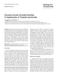
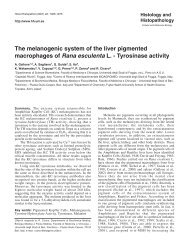
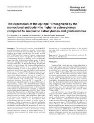
Fig. 1. Therapyrelated<br />
bone<br />
marrow (BM)<br />
lesions in IMF.<br />
a. Following HU<br />
treatment aplasia<br />
with a gelatinous<br />
edema may<br />
evolve. b. A<br />
strikingly<br />
expressed patchy<br />
appearance of<br />
residual<br />
hematopoiesis is<br />
detectable after<br />
IFN therapy<br />
revealing small<br />
clusters of<br />
(macrocytic)<br />
erythroblasts <strong>and</strong><br />
abnormal<br />
megakaryocytes<br />
with<br />
hypolobulated,<br />
dense nuclei. c.<br />
In some patients<br />
HU generates a<br />
dense sclerosing<br />
edema<br />
characterized by<br />
a fine network of<br />
reticulin, so-called<br />
scleredema (d).<br />
(a) Hematoxylin<br />
/Eosin, (b) CD61<br />
immunostaining,<br />
(c) PAS, (d) Silver<br />
impregnation; (a)<br />
x 80, (b-d) x 180
Sequential biopsies in IMF<br />
243<br />
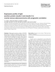
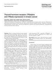
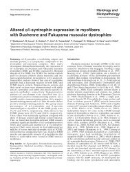
Fig. 2.<br />
Development of<br />
BM fibrosis in<br />
IMF. a. No<br />
increase in fibers<br />
(first biopsy on<br />
admission)<br />
consistent with a<br />
prefibrotic stage<br />
(IMF-0).<br />
b. Patchy<br />
distribution of<br />
hematopoiesis<br />
<strong>and</strong> a moderate<br />
increase in<br />
reticulin following<br />
IFN therapy for<br />
12 months (IMF-<br />
1) in the same<br />
patient. c.<br />
Advanced fibroosteosclerotic<br />
changes (IMF-3)<br />
arising from a<br />
prefibrotic lesion<br />
(first biopsy) in a<br />
patient with<br />
almost 10 years<br />
of symptomatic<br />
treatment (last<br />
examination).<br />
d. Prominent<br />
myelodysplastic<br />
features of<br />
megakaryopoiesis<br />
following HU<br />
therapy for about<br />
2.5 years include<br />
clustered smallto<br />
medium-sized<br />
megakaryocytes<br />
with<br />
hypolobulated<br />
(bulbous), dense<br />
nuclei<br />
surrounded by a<br />
relatively small<br />
portion of<br />
cytoplasm. (a-c)<br />
Silver<br />
impregnation, (d)<br />
PAS; (a-c) x 180,<br />
(d) x 380
244<br />
Sequential biopsies in IMF<br />
considering the first <strong>and</strong> the last BM biopsy in each<br />
patient <strong>and</strong> the stage of IMF at diagnosis a general<br />
progression of myelofibrosis was evident in 183 of the<br />
340 patients (54%) without or with only moderate<br />
fibrosis (grades 0 to 2) at onset (Fig. 3). This finding<br />
supports recently published data (Buhr et al., 2003;<br />
Thiele et al., 2003) that reveal a clear-cut tendency<br />
towards fiber increase in particular concerning patients<br />
with prefibrotic IMF (grade 0). Here a progression rate<br />
between 32% (Buhr et al., 2003) <strong>and</strong> 50 % (Thiele et al.,<br />
2003), depending on intervals between first <strong>and</strong> last BM<br />
biopsies (about 3 versus 4 years) was noted, thus<br />
implying a prodromal stage of overt (classical) IMF<br />
(Barosi, 1999; Tefferi, 2000). This feature was also<br />
explicitly expressed in the IFN-treated cohort of patients<br />
(Fig. 2a,b). A regression of fibrosis occurred in only 20<br />
patients, while in 66 patients (excluding the 71 patients<br />
with the last grade of myelofibrosis or IMF-3, which<br />
remained constant by definition) a stable state was<br />
apparent. In this con<strong>text</strong> it is noteworthy that the<br />
majority of patients with a regression of myelofibrosis<br />
which normally included only one grade, displayed<br />
severe hypoplasia to overt aplasia of the BM. This<br />
finding was consistent with a substantial myelo-ablative<br />
effect of treatment that was clinically associated with<br />
severe cytopenias <strong>and</strong> BM insufficiency (van den Berg<br />
et al., 1990; Michelson et al., 1993; Rousselet et al.,<br />
1996; Hurwitz, 1997). In six patients without<br />
cytoreductive therapy a grade-one regression of fiber<br />
density occurred during the course of the disease. In<br />
order to neutralize the shortcoming of variable biopsy<br />
intervals or endpoints of examination, cross-over of drug<br />
therapy <strong>and</strong> the significantly different numbers of<br />
trephines in each patient, we regarded the individual<br />
changes in the grades of fibrosis at a st<strong>and</strong>ardized<br />
median interval of 20 months (280 biopsy endpoints).<br />
When following this procedure calculation of possible<br />
changes in myelofibrosis revealed no significant<br />
differences in each therapy group implying a failing<br />
influence of any treatment modality on the development<br />
of myelofibrosis (Fig. 4).<br />
Although palliative therapy still remains the<br />
principal mode of treatment in IMF (Smith et al., 1988;<br />
Anger et al., 1990; Barosi, 1999; Tefferi, 2000), BM <strong>and</strong><br />
stem cell transplantation have recently gained more<br />
acceptance, especially in younger patients (Anderson et<br />
al., 1997; Guardiola et al., 1999). Because IFN<br />
preferentially inhibits the proliferation of the<br />
megakaryocytic cell lineage <strong>and</strong> consequently interferes<br />
with the cytokine-mediated generation of myelofibrosis<br />
(Le Bousse-Kerdiles <strong>and</strong> Martyre, 1999a,b) this agent<br />
seemed to be particularly suitable for treatment (Gilbert,<br />
1998). However, compared to HU as the drug of choice,<br />
IFN may not be well tolerated by many patients <strong>and</strong>,<br />
according to clinical data, was not shown to exert a<br />
clear-cut beneficial effect on the regression or inhibition<br />
of myelofibrosis (Parmeggiani et al., 1987; Barosi et al.,<br />
1990; Sacchi, 1995; Bachleitner-Hofmann <strong>and</strong><br />
Gisslinger, 1999). As has been demonstrated by this<br />
study in most patients a progressively developing BM<br />
fibrosis that was not influenced by any treatment<br />
modalities, especially not by IFN, could be observed in<br />
IMF (Thiele et al., 2003). This result is in keeping with<br />
data from a recently published study on IMF including<br />
109 patients of whom 48 received either BU or HU or<br />
combination therapies (Buhr et al., 2003).<br />
Maturation defects<br />
In addition to significant alterations concerning their<br />
quantity <strong>and</strong> distribution in the BM space<br />
(histotopography), hematopoietic cell lineages exhibited<br />
Fig. 3. Overview on the dynamics of myelofibrosis in our 340 patients<br />
with IMF regarding first <strong>and</strong> last trephine biopsies (median interval 32<br />
months).<br />
Fig. 4. Development of myelofibrosis in IMF according to 280 biopsy<br />
endpoints in the different therapy groups at st<strong>and</strong>ardized median<br />
intervals of 20 months (for abbreviations of therapy groups see Table 1).
245<br />
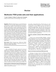
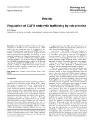
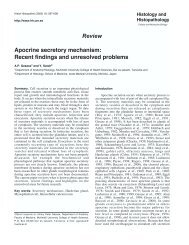
Fig. 5.<br />
Maturation<br />
defects<br />
(myelodysplastic<br />
features) of<br />
hematopoiesis<br />
<strong>and</strong> evolution of<br />
accelerated<br />
phase in IMF<br />
after HU therapy.<br />
a. Increase in<br />
megakaryopoiesis<br />
showing sheets<br />
of atypical<br />
micromegakaryocytes/-blasts.<br />
b. Large clusters<br />
of left-shifted<br />
erythropoiesis<br />
revealing an<br />
arrest in<br />
maturation <strong>and</strong> a<br />
macrocytic<br />
appearance (c).<br />
d. Slight increase<br />
in CD34 +<br />
progenitor cells<br />
exhibiting small<br />
clusters.<br />
e. Extensive<br />
proliferation of<br />
the left-shifted<br />
neutrophil<br />
granulopoiesis<br />
<strong>and</strong> cluster of<br />
atypical<br />
megakaryocytes<br />
abnormally<br />
located at the<br />
endosteal border<br />
(right lower<br />
corner). (a) CD61<br />
immunostaining,<br />
(b) <strong>and</strong> (e)<br />
chloroacetate<br />
esterase<br />
reaction, (c)<br />
antiglycophorin C<br />
immunostaining,<br />
(d) CD34<br />
immunostaining;<br />
(a,b,e) x 180,<br />
(d,c) x 380.
246<br />
Sequential biopsies in IMF<br />
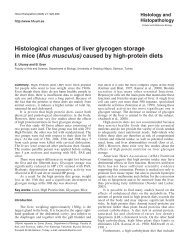
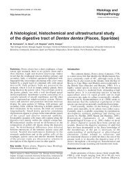
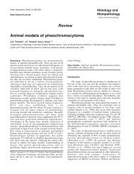
Fig. 6. Blastic<br />
crisis in IMF. a.<br />
Peripheral blood<br />
smear with<br />
pronounced<br />
leukoerythroblastic<br />
reaction. b.<br />
Bone marrow<br />
showing a diffuse<br />
(chloroacetate<br />
esterasenegative)<br />
blast<br />
infiltration.<br />
c. Packed growth<br />
of CD34 + blasts.<br />
d. Extensive<br />
staining of<br />
(myelomonocytic)<br />
blasts with<br />
lysozyme<br />
expression. e.<br />
Overall<br />
proliferation of<br />
atypical<br />
micromegakaryocytes/-blasts.<br />
(a)<br />
Pappenheim, (b)<br />
chloroacetate<br />
esterase, (c)<br />
CD34<br />
immunostaining,<br />
(d) lysozyme<br />
immunostaining,<br />
(e) CD61<br />
immunostaining;<br />
(a) x 1080, (b,c) x<br />
380, (d, e) x 180
Sequential biopsies in IMF<br />
247<br />
various degrees of maturation defects at the different<br />
endpoints of examination during follow-up. At extreme<br />
ranges these changes were comparable to so-called<br />
secondary or therapy-related myelodysplasia which<br />
predominantly involved the erythroid <strong>and</strong><br />
megakaryocytic cell lineages (Figs. 1b, 5a-c). In<br />
particular, megakaryocytes presented the diagnostic<br />
hallmark of these peculiar changes, since they showed a<br />
medium to small size <strong>and</strong> hypolobulated, dense nuclei<br />
surrounded by relatively small areas of cytoplasm <strong>and</strong><br />
frequent clustering (Fig. 2d). Relevant changes ranged<br />
from occurrence of grossly atypical micromegakaryocytes<br />
(Fig. 5a) to a maturation arrest <strong>and</strong><br />
macrocytic appearance of erythroid precursors (Figs. 3a,<br />
5b,c), i.e. features that clinically were consistent with an<br />
acceleration. According to the repeatedly performed BM<br />
biopsies during evolution of the disease process<br />
borderline to mild maturation defects developed in about<br />
23% of patients that received only symptomatic<br />
treatment, contrasting with the significantly expressed<br />
myelodysplastic changes in 3.3% patients of the HU<br />
group (Fig. 5a-e) that were not present after IFN<br />
treatment. It is reasonable to assume that minor to<br />
moderate maturation defects may develop during the<br />
normal course of disease even without interference by<br />
cytoreductive treatment. On the other h<strong>and</strong>, in 10<br />
patients blastic transformation of IMF indicating<br />
terminal stages was characterized by an increase in the<br />
peripheral blast count (Fig. 6a) accompanied by overt<br />
immaturity of hematopoiesis (Fig. 6b). Differentiation of<br />
the blast population may vary considerably <strong>and</strong> often<br />
includes CD34 + progenitors, lysozyme-expressing<br />
myelomonocytoid cells <strong>and</strong> CD61 + (micro-)<br />
megakaryoblasts (Fig. 6c-e).<br />
It should be emphasized that disturbances of<br />
maturation in CMPDs may present a diagnostic pitfall to<br />
hematopathologists, especially in those patients without<br />
any clinical information about previous therapy.<br />
Altogether conspicuous abnormalities of maturation may<br />
occur, which mimick myelodysplasia or so-called<br />
overlapping cases between a myeloproliferative <strong>and</strong><br />
myelodysplastic disorder (Bain, 1999; Gupta et al.,<br />
1999). These difficulties are also highlighted in our<br />
series, because at the beginning we had to exclude 34<br />
additional patients (14 females, 20 males) from further<br />
study. These peculiar cases showed IMF according to the<br />
first biopsy sample, but simultaneously exhibited<br />
strikingly expressed maturation defects of the erythroid<br />
<strong>and</strong> megakaryocytic lineage compatible with a<br />
myelodysplastic appearance. Although there was no<br />
indication of any preceding treatment on the report<br />
sheet, a more thoroughly performed investigation finally<br />
revealed a previous history of short-time cytostatic<br />
therapy. Since in this cohort no pretreatment biopsy<br />
specimens were available at diagnosis these patients<br />
were consequently excluded from this study. Therefore,<br />
much caution should be exerted, when diagnosing<br />
unclassifiable myelodysplastic/myelo-proliferative<br />
diseases in order not to miss any drug-related BM<br />
lesions.<br />
Maturation defects ranging from mild anomalies of<br />
differentiation to overt myelodysplastic features have<br />
repeatedly been described (Frisch et al., 1986; Rigolin et<br />
al., 1998) in association with cytotoxic therapy in<br />
CMPDs (Sterkers et al., 1998; R<strong>and</strong>i et al., 2000).<br />
Significantly expressed myelodysplastic changes may<br />
precede leukemic transformation especially after HU<br />
administration (Loefvenberg et al., 1990; Weinfeld et al.,<br />
1994; Higuchi et al., 1995; Furgerson et al., 1996;<br />
Liozon et al., 1997; Finazzi <strong>and</strong> Barbui, 1999;<br />
Mavrogianni et al.,2002; Nielsen <strong>and</strong> Hasselbalch,<br />
2003), thus contrasting with IFN therapy. In this con<strong>text</strong><br />
it is noteworthy that in our material myelodysplasia of<br />
megakaryocytes (so-called dysmegakaryopoiesis) was a<br />
most conspicuous feature following HU <strong>and</strong> BU<br />
administration <strong>and</strong> could be easily assessed (Figs. 2d, 5a,<br />
6e), whereas changes of the other lineages were more<br />
discrete. However, one should not overlook the fact that<br />
maturation defects, mostly of a mild degree, were also<br />
found in the control group of patients that received only<br />
supportive treatment for many years. Therefore, defects<br />
of hematopoietic cell differentiation eventually evolve in<br />
the lengthy course of disease signalling terminal stages,<br />
independently from therapeutic modalities. It may be<br />
speculated that such changes indicate a severe<br />
disturbance of lineage-specific maturation (Fig. 5a-e)<br />
<strong>and</strong> may herald blastic transformation of the disease<br />
process in a number of patients (Fig. 6a-e).<br />
Clinical features<br />
In keeping with the consideration of early to<br />
advanced stages of IMF at diagnosis of patients (first<br />
biopsy) clinical data showed a striking heterogeneity<br />
which, however, differed from the findings following<br />
therapy at the time of the last biopsy (Smith et al., 1988;<br />
Thiele et al., 1989, 1996, 2002; Cervantes et al., 1998;<br />
Georgii et al., 1998; Buhr et al., 2003). The 202 patients<br />
presenting with initial to early IMF without or with mild<br />
reticulin fibrosis were characterized by a more<br />
frequently occurring thrombocythemia exceeding<br />
1,000x10 9 /L (34%), a leukocytosis greater than<br />
10x10 9 /L (52 %) <strong>and</strong> a minimal to mild anemia (38 %)<br />
or splenomegaly (15%). In more than 20% of the<br />
patients of this cohort there was a relevant rise in the<br />
LDH value <strong>and</strong> the score of the leukocyte alkaline<br />
phosphatase (ALP) detectable. In a minority (4 %) of<br />
these patients a borderline to slightly expressed leukoerythroblastic<br />
blood picture with atypical precursors <strong>and</strong><br />
tear-drop poikilocytosis appeared on the blood films. At<br />
the endpoint of the last BM examination during the<br />
lengthy course of the disease differences concerning<br />
laboratory features of this group were not clearly<br />
distinguishable from the 138 patients that were initially<br />
diagnosed as overt (classical) IMF (Barosi, 1999;<br />
Tefferi, 2000). The latter cohort already presented with<br />
myelofibrosis, anemia, splenomegaly <strong>and</strong> tear-drop<br />
poikilocytosis at the beginning. The straightforward
248<br />
Sequential biopsies in IMF<br />
evolution of clinical data during follow-up underlines<br />
the dynamics of the disease process in IMF. However,<br />
when discriminating patients according to their therapy<br />
groups hematological variables failed to display different<br />
constellations of findings. In 10 patients a leukemic<br />
transformation was recognizable which occurred besides<br />
severe BM insufficiency as cause of death. In this<br />
con<strong>text</strong> the myelodysplastic <strong>and</strong> putative leukemogenic<br />
effects following chemotherapy with HU in CMPDs<br />
have to be emphasized (N<strong>and</strong> et al., 1996; Liozon et al.,<br />
1997; Rigolin et al., 1998; Sterkers et al., 1998; Finazzi<br />
<strong>and</strong> Barbui, 1999; Finazzi et al., 2000; Mavrogianni et<br />
al., 2002; Nielsen <strong>and</strong> Hasselbalch, 2003). This feature<br />
merits some attention because contrasting with IFN<br />
administration, prominent maturation defects were<br />
observed in a considerable number of our patients in<br />
particular following HU treatment.<br />
In conclusion, this review has systematically<br />
outlined drug-related lesions of the BM in IMF <strong>and</strong> in<br />
particular has focused on changes in cellularity,<br />
histotopography of residual hematopoiesis <strong>and</strong> the<br />
occurrence of maturation defects, including<br />
myelodysplastic features. Follow-up in a large series of<br />
patients (Buhr et al., 2003; Thiele et al., 2003) has not<br />
only validated the recently introduced diagnostic criteria<br />
of the WHO (Thiele et al., 2001), but also emphasized<br />
that evolution of myelofibrosis is not substantially<br />
modified by therapy.<br />
References<br />
Anderson J.E., Sale G., Appelbaum F.R., Chauncey T.R. <strong>and</strong> Storb R.<br />
(1997). Allogeneic marrow transplantation for primary myelofibrosis<br />
<strong>and</strong> myelofibrosis secondary to polycythaemia vera or essential<br />
thrombocytosis. Br. J. Haematol. 98, 1010-1016.<br />
Anger B., Seidler R., Haug U., Popp C. <strong>and</strong> Heimpel H. (1990).<br />
Idiopathic myelofibrosis: a retrospective study of 103 patients.<br />
Haematologica 75, 228-234.<br />
Bachleitner-Hofmann T. <strong>and</strong> Gisslinger H. (1999). The role of interferonalpha<br />
in the treatment of idiopathic myelofibrosis. Ann. Hematol. 78,<br />
533-538.<br />
Bain B.J. (1999). The relationship between the myelodysplastic<br />
syndromes <strong>and</strong> the myeloproliferative disorders. Leuk. Lymphoma.<br />
34, 443-449.<br />
Barosi G. (1999). Myelofibrosis with myeloid metaplasia: diagnostic<br />
definition <strong>and</strong> prognostic classification for clinical studies <strong>and</strong><br />
treatment guidelines. J. Clin. Oncol. 17, 2954-2970.<br />
Barosi G., Berzuini C., Liberato L.N., Costa A., Polino G. <strong>and</strong> Ascari E.<br />
(1988). A prognostic classification of myelofibrosis with myeloid<br />
metaplasia. Br. J. Haematol. 70, 397-401.<br />
Barosi G., Liberato L.N., Costa A., Buratti A., Di Dio F., Salvatore S. <strong>and</strong><br />
Ascari E. (1990). Induction <strong>and</strong> maintenance alpha-interferon<br />
therapy in myelofibrosis with myeloid metaplasia. Eur. J. Haematol.<br />
Suppl. 52, 12-14.<br />
Bartl R., Frisch B. <strong>and</strong> Wilmanns W. (1993). Potential of bone marrow<br />
biopsy in chronic myeloproliferative disorders (MPD). Eur. J.<br />
Haematol. 50, 41-52.<br />
Bauermeister D.E. (1971). Quantitation of bone marrow reticulin--a<br />
normal range. Am. J. Clin. Pathol. 56, 24-31.<br />
Bilgrami S. <strong>and</strong> Greenberg B.R. (1995). Polycythemia rubra vera.<br />
Semin. Oncol. 22, 307-326.<br />
Buesche G., Georgii A., Duensing A., Schmeil A., Schlue J. <strong>and</strong> Kreipe<br />
H.H. (2003). Evaluating the volume ratio of bone marrow affected by<br />
fibrosis: A parameter crucial for the prognostic significance of<br />
marrow fibrosis in chronic myeloid leukemia. Hum. Pathol. 34, 391-<br />
401.<br />
Buhr T., Georgii A. <strong>and</strong> Choritz H. (1993). Myelofibrosis in chronic<br />
myeloproliferative disorders. Incidence among subtypes according<br />
to the Hannover Classification. Pathol. Res. Pract. 189, 121-132.<br />
Buhr T., Buesche G., Choritz H., Langer F. <strong>and</strong> Kreipe H. (2003).<br />
Evolution of myelofibrosis in chronic idiopathic myelofibrosis as<br />
evidenced in sequential bone marrow biopsy specimens. Am. J.<br />
Clin. Pathol 119, 152-158.<br />
Cervantes F., Pereira A., Esteve J., Cobo F., Rozman C. <strong>and</strong><br />
Montserrat E. (1998). The changing profile of idiopathic<br />
myelofibrosis: a comparison of the presenting features of patients<br />
diagnosed in two different decades. Eur. J. Haematol. 60, 101-105.<br />
Cervantes F., Alvarez-Larran A., Talarn C., Gomez M. <strong>and</strong> Montserrat<br />
E. (2002). Myelofibrosis with myeloid metaplasia following essential<br />
thrombocythaemia: actuarial probability, presenting characteristics<br />
<strong>and</strong> evolution in a series of 195 patients. Br. J. Haematol. 118, 786-<br />
790.<br />
Ellis J.T., Peterson P., Geller S.A. <strong>and</strong> Rappaport H. (1986). Studies of<br />
the bone marrow in polycythemia vera <strong>and</strong> the evolution of<br />
myelofibrosis <strong>and</strong> second hematologic malignancies. Semin.<br />
Hematol. 23, 144-155.<br />
Finazzi G. <strong>and</strong> Barbui T. (1999). Treatment of essential<br />
thrombocythemia with special emphasis on leukemogenic risk. Ann.<br />
Hematol. 78, 389-392.<br />
Finazzi G., Ruggeri M., Rodeghiero F. <strong>and</strong> Barbui T. (2000). Second<br />
malignancies in patients with essential thrombocythaemia treated<br />
with busulphan <strong>and</strong> hydroxyurea: long-term follow-up of a<br />
r<strong>and</strong>omized clinical trial. Br. J. Haematol. 110, 577-583.<br />
Frisch B., Bartl R. <strong>and</strong> Chaichik S. (1986). Therapy-induced<br />
myelodysplasia <strong>and</strong> secondary leukaemia. Sc<strong>and</strong>. J. Haematol.<br />
Suppl. 45, 38-47.<br />
Furgerson J.L., Vukelja S.J., Baker W.J. <strong>and</strong> O'Rourke T.J. (1996).<br />
Acute myeloid leukemia evolving from essential thrombocythemia in<br />
two patients treated with hydroxyurea. Am. J. Hematol. 51, 137-140.<br />
Georgii A., Vykoupil K.F., Buhr T., Choritz H., Doehler U., Kaloutsi V.<br />
<strong>and</strong> Werner M. (1990). Chronic myeloproliferative disorders in bone<br />
marrow biopsies. Pathol. Res. Pract. 186, 3-27.<br />
Georgii A., Buesche G. <strong>and</strong> Kreft A. (1998). The histopathology of<br />
chronic myeloproliferative diseases. Baillieres Clin. Haematol. 11,<br />
721-749.<br />
Gilbert H.S. (1998). Long term treatment of myeloproliferative disease<br />
with interferon-alpha-2b: feasibility <strong>and</strong> efficacy. Cancer 83, 1205-<br />
1213.<br />
Guardiola P., Anderson J.E., B<strong>and</strong>ini G., Cervantes F., Runde V.,<br />
Arcese W., Bacigalupo A., Przepiorka D., O'Donnell M.R., Polchi P.,<br />
Buzyn A., Sutton L., Cazals-Hatem D., Sale G., de Witte T., Deeg<br />
H.J. <strong>and</strong> Gluckman E. (1999). Allogeneic stem cell transplantation<br />
for agnogenic myeloid metaplasia: a European Group for Blood <strong>and</strong><br />
Marrow Transplantation, Societe Francaise de Greffe de Moelle,<br />
Gruppo Italiano per il Trapianto del Midollo Osseo, <strong>and</strong> Fred<br />
Hutchinson Cancer Research Center Collaborative Study. Blood 93,<br />
2831-2838.<br />
Gupta R., Abdalla S.H. <strong>and</strong> Bain B.J. (1999). Thrombocytosis with
Sequential biopsies in IMF<br />
249<br />
sideroblastic erythropoiesis: a mixed myeloproliferative<br />
myelodysplastic syndrome. Leuk. Lymphoma 34, 615-619.<br />
Hasselbalch H.C. (1993). Idiopathic myelofibrosis--an update with<br />
particular reference to clinical aspects <strong>and</strong> prognosis. Int. J. Clin.<br />
Lab. Res. 23, 124-138.<br />
Hasselbalch H. <strong>and</strong> Lisse I. (1991). A sequential histological study of<br />
bone marrow fibrosis in idiopathic myelofibrosis. Eur. J Haematol.<br />
46, 285-289.<br />
Higuchi T., Okada S., Mori H., Niikura H., Omine M. <strong>and</strong> Terada H.<br />
(1995). Leukemic transformation of polycythemia vera <strong>and</strong> essential<br />
thrombocythemia possibly associated with an alkylating agent.<br />
Cancer 75, 471-477.<br />
Hurwitz N. (1997). Bone marrow trephine biopsy changes following<br />
chemotherapy <strong>and</strong>/or bone marrow transplantation. Curr. Diagn.<br />
Pathol. 4, 196-202.<br />
Krech R. <strong>and</strong> Thiele J. (1985). <strong>Histopathology</strong> of the bone marrow in<br />
toxic myelopathy. A study of drug induced lesions in 57 patients.<br />
Virchows Arch A. 405, 225-235.<br />
Kreft A., Reimann J. <strong>and</strong> Choritz H. (2000). Fibre content <strong>and</strong> cellularity<br />
of the bone marrow of the iliac crest, vertebral column <strong>and</strong> sternum<br />
in chronic myeloproliferative disorders. Leuk. Lymphoma 38, 165-<br />
173.<br />
Le Bousse-Kerdiles M.C. <strong>and</strong> Martyre M.C. (1999a). Myelofibrosis:<br />
pathogenesis of myelofibrosis with myeloid metaplasia. French<br />
INSERM Research Network on Myelofibrosis with Myeloid<br />
Metaplasia. Springer Semin. Immunopathol. 21, 491-508.<br />
Le Bousse-Kerdiles M.C. <strong>and</strong> Martyre M.C. (1999b). Dual implication of<br />
fibrogenic cytokines in the pathogenesis of fibrosis <strong>and</strong><br />
myeloproliferation in myeloid metaplasia with myelofibrosis. Ann.<br />
Hematol. 78, 437-444.<br />
Liozon E., Brigaudeau C., Trimoreau F., Desangles F., Fermeaux V.,<br />
Praloran V. <strong>and</strong> Bordessoule D. (1997). Is treatment with<br />
hydroxyurea leukemogenic in patients with essential<br />
thrombocythemia? An analysis of three new cases of leukaemic<br />
transformation <strong>and</strong> review of the literature. Hematol. Cell. Ther. 39,<br />
11-18.<br />
Loefvenberg E., Wahlin A., Roos G. <strong>and</strong> Ost A. (1990). Reversal of<br />
myelofibrosis by hydroxyurea. Eur. J .Haematol. 44, 33-38.<br />
Lohmann T.P. <strong>and</strong> Beckman E.N. (1983). Progressive myelofibrosis in<br />
agnogenic myeloid metaplasia. Arch. Pathol. Lab. Med. 107, 593-<br />
594.<br />
Mavrogianni D., Vianiou N., Michali E., Terpos E., Meletis J., Vaiopoulos<br />
G., Madzourani M., Pangalis G., Yataganas X. <strong>and</strong> Loukopoulos D.<br />
(2002). Leukemogenic risk of hydroxyurea therapy as a single agent<br />
in polycythemia vera <strong>and</strong> essential thrombocythemia: N- <strong>and</strong> K-ras<br />
mutations <strong>and</strong> microsatelite instability in chromosomes 5 <strong>and</strong> 7 in 69<br />
patients. Int. J. Hematol. 75, 394-400.<br />
Michelson J.D., Gornet M., Codd T., Torres J., Lanighan K. <strong>and</strong> Jones<br />
R. (1993). Bone morphology after bone marrow transplantation for<br />
Hodgkin's <strong>and</strong> non-Hodgkin's lymphoma. Exp. Hematol. 21, 475-<br />
482.<br />
N<strong>and</strong> S., Stock W., Godwin J. <strong>and</strong> Fisher S.G. (1996). Leukemogenic<br />
risk of hydroxyurea therapy in polycythemia vera, essential<br />
thrombocythemia, <strong>and</strong> myeloid metaplasia with myelofibrosis. Am. J.<br />
Hematol. 52, 42-46.<br />
Nielsen II. <strong>and</strong> Hasselbalch H.C. (2003) Acute leukemia <strong>and</strong><br />
myelodysplasia in patients with a Philadelphia chromosome<br />
negative chronic myeloproliferative disorder treated with<br />
hydroxyurea alone or with hydroxyurea after busulphan. Am. J.<br />
Hematol. 74, 26-31.<br />
Parmeggiani L., Ferrant A., Rodhain J., Michaux J.L. <strong>and</strong> Sokal G.<br />
(1987). Alpha interferon in the treatment of symptomatic<br />
myelofibrosis with myeloid metaplasia. Eur. J. Haematol. 39, 228-<br />
232.<br />
R<strong>and</strong>i M.L., Fabris F. <strong>and</strong> Girolami A. (2000). Leukemia <strong>and</strong><br />
myelodysplasia effect of multiple cytotoxic therapy in essential<br />
thrombocythemia. Leuk. Lymphoma 37, 379-385.<br />
Rigolin G.M., Cuneo A., Roberti M.G., Bardi A., Bigoni R., Piva N.,<br />
Minotto C., Agostini P., De Angeli C., Del Senno L., Spanedda R.<br />
<strong>and</strong> Castoldi G. (1998). Exposure to myelotoxic agents <strong>and</strong><br />
myelodysplasia: case-control study <strong>and</strong> correlation with<br />
clinicobiological findings. Br. J. Haematol. 103, 189-197.<br />
Rousselet M.C., Kerjean A., Guyetant S., Francois S., Saint-Andre J.P.<br />
<strong>and</strong> Ifrah N. (1996). <strong>Histopathology</strong> of bone marrow after allogeneic<br />
bone marrow transplantation for chronic myeloid leukaemia. Pathol.<br />
Res. Pract. 192, 790-795.<br />
Sacchi S. (1995). The role of alpha-interferon in essential<br />
thrombocythaemia, polycythaemia vera <strong>and</strong> myelofibrosis with<br />
myeloid metaplasia (MMM): a concise update. Leuk. Lymphoma 19,<br />
13-20.<br />
Smith R.E., Chelmowski M.K. <strong>and</strong> Szabo E.J. (1988). Myelofibrosis: a<br />
concise review of clinical <strong>and</strong> pathologic features <strong>and</strong> treatment.<br />
Am. J .Hematol. 29, 174-180.<br />
Spivak J.L. (2002). Polycythemia vera: myths, mechanisms, <strong>and</strong><br />
management. Blood 100, 4272-4290.<br />
Sterkers Y., Preudhomme C., Lai J.L., Demory J.L., Caulier M.T., Wattel<br />
E., Bordessoule D., Bauters F. <strong>and</strong> Fenaux P. (1998). Acute myeloid<br />
leukemia <strong>and</strong> myelodysplastic syndromes following essential<br />
thrombocythemia treated with hydroxyurea: high proportion of cases<br />
with 17p deletion. Blood 91, 616-622.<br />
Tefferi A. (2000). Myelofibrosis with myeloid metaplasia. N. Engl. J.<br />
Med. 342, 1255-1265.<br />
Tefferi A. (2001). Recent progress in the pathogenesis <strong>and</strong><br />
management of essential thrombocythemia. Leuk. Res. 25, 369-377.<br />
Thiele J., Laubert A., Vykoupil K.F. <strong>and</strong> Georgii A. (1985). Autopsy <strong>and</strong><br />
clinical findings in acute leukemia <strong>and</strong> chronic myeloproliferative<br />
diseases--an evaluation of 104 patients. Pathol. Res. Pract. 179,<br />
328-336.<br />
Thiele J., Simon K.G., Fischer R. <strong>and</strong> Zankovich R. (1988). Follow-up<br />
studies with sequential bone marrow biopsies in chronic myeloid<br />
leukaemia <strong>and</strong> so-called primary (idiopathic) osteo-myelofibrosis.<br />
Evolution of histopathological lesions <strong>and</strong> clinical course in 40<br />
patients. Pathol. Res. Pract. 183, 434-445.<br />
Thiele J., Zankovich R., Steinberg T., Fischer R. <strong>and</strong> Diehl V. (1989).<br />
Agnogenic myeloid metaplasia (AMM) - correlation of bone marrow<br />
lesions with laboratory data: a longitudinal clinicopathological study<br />
on 114 patients. Hematol. Oncol. 7, 327-343.<br />
Thiele J., Kvasnicka H.M., Werden C., Zankovich R., Diehl V. <strong>and</strong><br />
Fischer R. (1996). Idiopathic primary osteo-myelofibrosis: a clinicopathological<br />
study on 208 patients with special emphasis on<br />
evolution of disease features, differentiation from essential<br />
thrombocythemia <strong>and</strong> variables of prognostic impact. Leuk.<br />
Lymphoma 22, 303-317.<br />
Thiele J., Kvasnicka H.M., Boeltken B., Zankovich R., Diehl V. <strong>and</strong><br />
Fischer R. (1999a). Initial (prefibrotic) stages of idiopathic (primary)<br />
myelofibrosis (IMF) - a clinicopathological study. Leukemia 13, 1741-<br />
1748.<br />
Thiele J., Kvasnicka H.M. <strong>and</strong> Fischer R. (1999b). Histochemistry <strong>and</strong>
250<br />
Sequential biopsies in IMF<br />
morphometry on bone marrow biopsies in chronic myeloproliferative<br />
disorders - aids to diagnosis <strong>and</strong> classification. Ann. Hematol. 78,<br />
495-506.<br />
Thiele J., Kvasnicka H.M., Schmitt-Graeff A., Bundschuh S., Biermann<br />
T., Roessler G., Wasmus M., Diehl V., Zankovich R. <strong>and</strong> Schaefer<br />
H.E. (2000). Effects of chemotherapy (busulfan-hydroxyurea) <strong>and</strong><br />
interferon-alfa on bone marrow morphologic features in chronic<br />
myelogenous leukemia. Histochemical <strong>and</strong> morphometric study on<br />
sequential trephine biopsy specimens with special emphasis on<br />
dynamic features. Am. J. Clin. Pathol. 114, 57-65.<br />
Thiele J., Imbert M., Pierre R., Vardiman J.W., Brunning R.D. <strong>and</strong><br />
Fl<strong>and</strong>rin G. (2001). Chronic idiopathic myelofibrosis. In: WHO<br />
classification of tumours: tumours of haematopoietic <strong>and</strong> lymphoid<br />
tissues. Jaffe E.S., Harris N.L., Stein H. <strong>and</strong> Vardiman J.W. (eds)<br />
pp. 35-38. IARC Press: Lyon.<br />
Thiele J., Kvasnicka H.M., Zankovich R. <strong>and</strong> Diehl V. (2002). Early<br />
stage idiopathic (primary) myelofibrosis - current issues of diagnostic<br />
features. Leuk. Lymphoma 43, 1035-1041.<br />
Thiele J., Kvasnicka H.M., Schmitt-Gräff A. <strong>and</strong> Diehl V. (2003).<br />
Dynamics of fibrosis in chronic idiopathic (primary) myelofibrosis<br />
during therapy: a follow-up study on 309 patients. Leuk. Lymphoma<br />
44, 549-553.<br />
van den Berg H., Kluin P.M. <strong>and</strong> Vossen J.M. (1990). Early<br />
reconstitution of haematopoiesis after allogeneic bone marrow<br />
transplantation: a prospective histopathological study of bone<br />
marrow biopsy specimens. J. Clin. Pathol. 43, 365-369.<br />
Weinfeld A., Swolin B. <strong>and</strong> Westin J. (1994). Acute leukaemia after<br />
hydroxyurea therapy in polycythaemia vera <strong>and</strong> allied disorders:<br />
prospective study of efficacy <strong>and</strong> leukaemogenicity with therapeutic<br />
implications. Eur. J. Haematol 52, 134-139.<br />
Wilkins B.S., Bostanci A.G., Ryan M.F. <strong>and</strong> Jones D.B. (1993).<br />
Haemopoietic regrowth after chemotherapy for acute leukaemia: an<br />
immunohistochemical study of bone marrow trephine biopsy<br />
specimens. J. Clin. Pathol .46, 915-921.<br />
Wolf B.C. <strong>and</strong> Neiman R.S. (1985). Myelofibrosis with myeloid<br />
metaplasia: pathophysiologic implications of the correlation between<br />
bone marrow changes <strong>and</strong> progression of splenomegaly. Blood 65,<br />
803-809.<br />
Accepted August 7, 2003