Full text-PDF - Histology and Histopathology
Full text-PDF - Histology and Histopathology
Full text-PDF - Histology and Histopathology

Create successful ePaper yourself
Turn your PDF publications into a flip-book with our unique Google optimized e-Paper software.
Histol Histopathol (2005) 20: 785-789<br />
http://www.hh.um.es<br />
<strong>Histology</strong> <strong>and</strong><br />
<strong>Histopathology</strong><br />
Cellular <strong>and</strong> Molecular Biology<br />
Unusual circular annulate lamellae<br />
in hepatocytes of Torpedo marmorata<br />
A. Haggag 1 <strong>and</strong> J. Gilloteaux 1,2<br />
1 Department of Anatomy, American University of the Caribbean School of Medicine, M.E.I.O. Inc,<br />
St Maarten Campus, Netherl<strong>and</strong> Antilles, West Indies <strong>and</strong> 2 Laboratoire Arago, Observatoire Océanologique of the<br />
Université Pierre et Marie Curie (Paris VI) <strong>and</strong> of the Centre National de la Recherche Scientifique, Banyuls-Sur-Mer, France<br />
Summary. This report describes an unusual morphology<br />
of annulate lamellae (AL) in the hepatocytes of Torpedo<br />
marmorata Risso. These Als <strong>and</strong> fragments are detected<br />
amidst the main glycogen <strong>and</strong> lipid deposits. AL cisterns<br />
are circumscribed by parts of the smooth endoplasmic<br />
reticulum. Based on the finding of these unusual annular<br />
ALs, accompanied by other subcellular lesions such as a<br />
number of membranous whorls <strong>and</strong> altered<br />
mitochondria. These findings can concur <strong>and</strong> support<br />
other authors’ observations suggesting that these adult<br />
hepatocytes transient changes reflect that this species<br />
could be exposed to local, natural or likely human<br />
coastal seabed pollutants.<br />
Key words: Annulate lamellae, Liver, Microscopy,<br />
Hepatocyte, Torpedo marmorata, Cytotoxicity<br />
Introduction<br />
The term annulate lamellae (AL) can be used to<br />
describe an intracellular organelle composed of parallel<br />
arrays of cisterns bearing at regular intervals small<br />
annuli or fenestrrae. AL ressemble stacked pieces of<br />
nuclear envelope. In a few exceptional instances they<br />
have been detected in continuity with the nuclear<br />
envelope (Kessel, 1983) <strong>and</strong> have been located<br />
intranuclearly (Ghadially <strong>and</strong> Parry, 1974, surveys by<br />
Ghadially, 1985, 1997; Kessel 1983, 1992). AL contains<br />
ribonucleoproteins attached to their outer surface or to<br />
some of the endoplasmic-associated membranes.<br />
Originally observed by a few histologists in the oocytes<br />
of marine <strong>and</strong> terrestrial invertebrates, pancreatic acinar<br />
cells <strong>and</strong> rat spermatids, it is the electron microscope<br />
that allowed Swift (1956) to first coin the term. Several<br />
other terms have been used in describing this cell<br />
Offprint requests to: Professor J. Gilloteaux, Department of Anatomy,<br />
American University of the Caribbean School of Medicine, Campus St<br />
Maarten, Suite # 401, M.E.I.O.- M.E.A.S. Inc, 901 Ponce de Leon<br />
Boulevard, Coral Gables FL 33134, USA. e-mail: jagillot@hotmail.com<br />
infrastructure: “coarse fibrous component”, “periodic<br />
lamellae’ (Rebhun, 1956), “secondary membranes”<br />
(Merriam, 1959), “pitted membranes” <strong>and</strong> “porous<br />
cytomembranes” (Kessel, 1968a). If ALs were reviewed<br />
by Witschnitzer (1970), Ghadiallay (1985, 1997), these<br />
organelles were the topic of numerous publications by<br />
Kessel (1968a,b, 1983, 1992). It can be noted that these<br />
cell substructures are usually located in the cytoplasm of<br />
vertebrate <strong>and</strong> invertebrate germ cells, embryonic cells<br />
<strong>and</strong> several animal benign <strong>and</strong> malignant tumor cells<br />
(Wischnitzer, 1970; Kessel, 1983, 1992; Ghadially,<br />
1997). However this organelle is also found occasionally<br />
in some adult somatic cells. AL may also present<br />
complexes surrounded by adjacent membranous saccules<br />
of smooth or rough endoplasmic reticulum (ER). ER<br />
saccules are eventually continuous or contiguous with<br />
these cisternae. In addition, it is more common to find<br />
ALs in cells that have been exposed to cytotoxic<br />
compounds <strong>and</strong> viral infections.<br />
This report addresses the unusual morphology of AL<br />
structure found in the hepatocytes of an electric ray<br />
while surveying the structure of its biliary tract<br />
(Gilloteaux et al., 1996). From our observations, we<br />
hypothesize that the presence of AL in hepatocytes could<br />
be an intracellular signal of sea pollutant cytotoxicity.<br />
Materials <strong>and</strong> methods<br />
Samples of liver tissues of 5 specimens of Torpedo<br />
marmorata Risso, 17-32 cm in length (3 males <strong>and</strong> 2<br />
females) were captured by trawler fishing with the boat<br />
Nereis II, at 35-40 m depth from the s<strong>and</strong>y seabed (along<br />
a NNW-SSE line between Cape Béart to Cape de<br />
l’Abeille, 1 nautical mile before the edge of the National<br />
Marine Reserve) in the Bay of Banyuls (Gulf of the<br />
Lion, Mediterranean Sea, France). The fish were brought<br />
to the Arago Marine Biological Station of the University<br />
of Paris, <strong>and</strong> within 1/2 hour of capture they were kept in<br />
aerated sea water. The electric rays were decapitated to<br />
prevent any changes in the liver due to using dissolved<br />
anesthetic <strong>and</strong>, after removal of other organs of interest,
786<br />
Annulate lammellae in Torpedo hepatocytes<br />
liver specimens were cut in 1 mm- thick slices <strong>and</strong> fixed<br />
promptly at laboratory temperature (15°C) for 10 min in<br />
3% glutaraldehyde diluted by sea water, <strong>and</strong> buffered by<br />
0.1 M cacodylate buffer. Fixative was then changed <strong>and</strong><br />
specimens were immersed for another 2 hours at 4°C<br />
before they were washed by the buffer alone containing<br />
30 gm/L sodium chloride <strong>and</strong> 2 gm sucrose. Tissues<br />
were postfixed with a 2% aqueous osmium tetraoxide<br />
solution for 2h duration. Tissue samples were then<br />
processed <strong>and</strong> embedded in PolyBed 812 (Polysciences,<br />
Warrington, PA). Thin sections were observed in a Jeol<br />
101 transmission electron microscope.<br />
Results<br />
Torpedo display elongated, prismatic <strong>and</strong> polyhedral<br />
shaped hepatocytes from 15 to 25 µm in length <strong>and</strong> 5 to<br />
8 µm in width, <strong>and</strong> 6 to 7 µm in height; they are mostly<br />
uninucleate <strong>and</strong> contain very large amounts of storage<br />
glycogen as well as lipid droplets with dense bodies <strong>and</strong><br />
yolk deposits. The hepatocytes reveal irregular <strong>and</strong><br />
thick, microvilli extensions on their surfaces in the space<br />
of Disse (Fig. 1). A narrow 1.0 to 2.5 µm -thick<br />
bordering region of cytoplasm, rich in branching smooth<br />
endoplasmic reticulum, contains scattered mitochondrial<br />
profiles <strong>and</strong> bundles of actin-like filaments. These<br />
organelles appear to be confined to the encompassing<br />
cytoplasm among these filaments. Mitochondria, oblong<br />
to round shaped (1.5 µm long <strong>and</strong> 0.3-0.5 µm in<br />
diameter) display exaggerated electron densities, a<br />
poorly developed matrix, appears somewhat vacuolated<br />
<strong>and</strong> sometimes display minute whorls.<br />
The unique observation made throughout the 5 liver<br />
specimens of this selachian is the presence of unusual<br />
circular ALs surrounded by glycogen <strong>and</strong> lipid deposits.<br />
In addition, pieces (?) or linear segments of AL can be<br />
detected near the circular AL <strong>and</strong> at the edge of these<br />
storage components. The large glycogen storages<br />
typically segregate other organelles as expected in the<br />
hepatocytes. However, the glycogen aggregates appear<br />
spread as a dense network instead of rosettes as viewed<br />
in a more classic macromolecular storage. In addition,<br />
within the glycogen storage component, lysosomal (?)<br />
whorls of membrane appear as onion bodies. This type<br />
of AL has unique, ultrastructural features, again because<br />
it is circular <strong>and</strong> it comprises two concentric rings of<br />
nuclear-like pore complexes each s<strong>and</strong>wiched between<br />
two narrow smooth endoplasmic reticulum (SER)<br />
cisternae. The innermost <strong>and</strong> outermost SER cisterns are<br />
contiguous <strong>and</strong> continuous with some SER tubular<br />
cross-sections outside the ALs; these are also adjacent to<br />
the main glycogen deposits of the central storage area of<br />
the hepatocyte. The inner ring contains a central area of<br />
cytoplasm filled with glycogen, lipid deposits, <strong>and</strong><br />
smooth endoplasmic reticulum. A narrow cytoplasmic<br />
zone is s<strong>and</strong>wiched between the inner <strong>and</strong> the outer rings<br />
of AL-SER complexes <strong>and</strong> also contains sparse but<br />
delicate aggregates of glycogen-like storage.<br />
Discussion<br />
The intention of this report is not to describe the<br />
general architecture of the liver in this species that will<br />
be the object of another contribution but to describe a<br />
peculiar AL morphology present in this differentiated<br />
tissue. From these observations, the hepatocytes found in<br />
the Torpedo samples were of the same anatomical<br />
structure as that characterized by other works (Diaz <strong>and</strong><br />
Connes, 1988; Rocha et al., 2001).<br />
The name annulate lamellae (AL) varied early on,<br />
but the advances in electron microscopy allowed the<br />
study <strong>and</strong> description of AL occurrence, <strong>and</strong> their<br />
organization. However a full underst<strong>and</strong>ing of their tridimensional<br />
architecture as well as their function in<br />
cellular homeostasis needs further elucidation. In this<br />
report, we propose that the ALs observed originated<br />
from a spiral structure that could unwrap <strong>and</strong> dissect out<br />
small pieces, i.e. the segments found among the other<br />
organelles. The molecular organization of ALs has been<br />
related to the cell cycle <strong>and</strong> dissected by several groups<br />
(Dabauvalle <strong>and</strong> Scheer, 1991; Cordes et al., 1995, 1996;<br />
Meier et al., 1995; Dabauvalle et al., 1999; Beckheling<br />
et al., 2003). These recent publications help us to<br />
underst<strong>and</strong> the early studies reporting that AL usually<br />
belong to cells that have active cell cycles or are<br />
prepared to undergo mitosis, such as animal <strong>and</strong> human<br />
cell lines (Wischnitzer, 1970; Kessel, 1983, 1992),<br />
including gametes or early division after fertilization<br />
(Billard 1984; Romeo et al., 1992), embryonic cells<br />
(Benzo, 1972, 1974), <strong>and</strong> pathologic ones (Kato et al.,<br />
2001), including injured liver cells (Hruban et al.,<br />
1965a,b; Kohda et al., 1989), hepatomas <strong>and</strong> other<br />
tumors (Hoshino, 1963; Svoboda, 1964; Ma <strong>and</strong> Webber,<br />
1966; Locker et al., 1969; Kim et al., 1990; Eyden,<br />
2000; Biernat et al., 2001) <strong>and</strong> in virus-infected cells<br />
(Marshall et al., 1996). Additionally, recent data<br />
confirmed that ALs are associated with the complex<br />
control <strong>and</strong> regulation of the initial steps leading to the<br />
pre-mitotic phase of the cell cycle (Cordes et al., 1996;<br />
Beckheling et al., 2003), including the disassembly <strong>and</strong><br />
reassembly of the nuclear envelope (Erl<strong>and</strong>son <strong>and</strong> De<br />
Harven, 1971; Meier et al., 1995; Cordes et al., 1996).<br />
Furthermore, the presence of glycogen <strong>and</strong> lipids in<br />
these hepatocytes is not a surprise as this organ is the<br />
main reserve storage of glycogen <strong>and</strong> this storage favors<br />
segregation of organelles (Bruslé <strong>and</strong> González I<br />
Anadon (1996). Even though we managed to preserve<br />
the tissue as soon as the fish was captured, <strong>and</strong><br />
according to previous literature (Spielberg et al., 1993),<br />
one observed the glycogen clumped into loose<br />
aggregates, not forming the rosettes morphology as<br />
classically described <strong>and</strong> detected many droplets of fatty<br />
deposits found throughout hepatocytes were well<br />
preserved. As in other selachians, the lipid reserve is rich<br />
in squalene <strong>and</strong> dense bodies (Peyronel et al., 1984;<br />
Rocha et al., 2001; Sarasquete et al., 2002) that could<br />
make the liver cells more susceptible to lipophilic toxic
Annulate lammellae in Torpedo hepatocytes<br />
787<br />
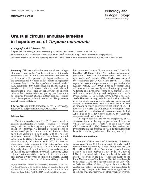
Fig. 1. Example of an hepatocyte content from Torpedo marmorata showing one annulate lamellae (AL) complex in a glycogen- <strong>and</strong> lipid-rich<br />
cytoplasmic area. A fragment of AL (curved arrow) is also shown. Bundles of actin-like filaments are indicated (straight arrows) among the smooth<br />
endoplasmic reticulum (sr). D: space of Disse; gly: glycogen deposits; l: lipid droplet; m: mitochondria. Bar scale: 1 µm.
788<br />
Annulate lammellae in Torpedo hepatocytes<br />
compounds that would induce membranous whorls or<br />
onion bodies. Five specimens captured were within the<br />
size expected (Filiz <strong>and</strong> Mater, 2002) <strong>and</strong> showed nor<br />
gross neighter histopathologic anomalies in their livers.<br />
It was only because the biliary tract was studied with the<br />
electron microscope that circular ALs were detected in<br />
the hepatocytes.<br />
The mere presence of peculiar ALs in differentiated<br />
hepatocytes co-localized with cytoplasmic whorls or<br />
onion bodies <strong>and</strong> the simultaneous finding of adjacent<br />
damaged mitochondria <strong>and</strong> dilated smooth endoplasmic<br />
reticulum with an altered pattern of glycogen aggregates<br />
led us to suspect that this could signal intrahepatic<br />
cytotoxic subcellular damage or sensitization to local<br />
shoreline or sea effluent effect reaching the sea floor <strong>and</strong><br />
affecting Torpedo, a bottom feeder <strong>and</strong> predator.<br />
These changes are not different to those observations<br />
made in the hepatocytes of several freshwater <strong>and</strong> sea<br />
water fish by various research groups showing lesions in<br />
ER, lysosomes, ALs <strong>and</strong> other organelles (Klaunig et al.,<br />
1979; Van der Heijden <strong>and</strong> Dormans, 1981; Hinton et al.,<br />
1984; Braunbeck <strong>and</strong> Völkl, 1991; Arnold et al., 1996;<br />
Zahn et al., 1996; Braunbeck <strong>and</strong> Applebaum, 1999).<br />
These changes were confirmed by the works of<br />
Biagianti-Risbourg (1990), Klaunig et al. (1979), Hinton<br />
et al. (1984) <strong>and</strong> reviewed by Bruslé <strong>and</strong> González-<br />
Anadon (1996) refer to the same cytological lesions<br />
detected in hepatocytes of other fish species. These<br />
changes were also observed as this was found in other<br />
fish <strong>and</strong> those found in a mammalian model treated by a<br />
toxic compound (Kohda et al., 1989).<br />
Even though these ALs were observed in a small<br />
number of specimens, as these fish were collected along<br />
with another series of bottom feeder species for a first<br />
description of the hepato-biliary system (Gilloteaux et<br />
al., 1996), we believe that the finding of this observation<br />
of ALs of peculiar shape in this selachian liver cells was<br />
of interest to be reported as an initial cytological marker<br />
for cytotoxicity <strong>and</strong> to maybe grant further<br />
investigations related to biomarine welfare.<br />
Acknowledgements. Sponsored by the Scholar <strong>and</strong> Academic<br />
Committee of A.U.C. School of Medicine, Campus St Maarten. Data<br />
was collected during a stay at the Observatoire Océanologique de<br />
l’Université Pierre et Marie Curie (ParisVI) <strong>and</strong> of the Centre National de<br />
la Recherche Scientifique that was supported by the Ministry of<br />
Education of Belgium. At the time A.H. is a medical student who<br />
committed to writing a scientific project.<br />
References<br />
Arnold H., Pluta H.J. <strong>and</strong> Braunbeck T. (1996). Sublethal effects of<br />
prolonged exposure to disulfoton in rainbow trout (Orcorhynchus<br />
mykiss): cytological alterations in the liver by a potent acetylcholine<br />
esterase inhibitor. Ecotoxicol. Environ. Saf. 34, 43-55.<br />
Beckleling C., Chang P., Chevalier S., Ford C. <strong>and</strong> Houliston E. (2003).<br />
Pre-M Phase – promoting factor associates with annulate lamellae<br />
in Xenopus oocytes <strong>and</strong> egg extracts. Mol. Biol. Cell 14, 1125-1137.<br />
Benzo C.A. (1972). The annulate lamella of chick embryo liver cells in<br />
organ culture. Anat. Rec. 174, 399-405.<br />
Benzo C.A. (1974). The annulate lamella in hepatic <strong>and</strong> pancreatic beta<br />
cells of the chick embryo. Am. J. Anat. 140, 139-143<br />
Biagianti-Risbourg S. (1990). Contribution à l’ étude du foie de juvéniles<br />
de muges (Téléosténs Mugilidés) contaminés expérimentalement<br />
par de l’atrazine (S -triazine herbicide): approche ultrastructurale et<br />
métabolique, intérêt en exotoxicologie. Thèse doctorale, Université<br />
de Perpignan, France.<br />
Biernat W., Liberski P., Kordek R., Zakrzewski K., Plois L. <strong>and</strong> Budka H.<br />
(2001). Dysembryoplastic neurectodermal tumor: an ultrastructural<br />
study of six cases. Ultrastruct. Pathol. 25, 455-467.<br />
Billard R. (1984). Ultrastructural changes in the spermatogonia <strong>and</strong><br />
spermatocyte of Poecilia reticulata during spermatogenesis. Cell<br />
Tissue Res. 237, 219-226.<br />
Braunbeck T. <strong>and</strong> Applebaum S. (1999). Ultrastructural alterations in the<br />
liver <strong>and</strong> intestine of carp Cyprinus carpio induced orally by ultra-low<br />
doses of endosulfan. Dis. Aquat. Organ 36, 183- 200.<br />
Braunbeck T. <strong>and</strong> Völkl A. (1991). Induction of biotransformation in the<br />
liver of eel Anguilla anguilla L.) by sublethal exposure to dinitro-ocresol:<br />
an ultrastrctural <strong>and</strong> biochemical study. Ecotoxicol. Environ.<br />
Saf.. 21, 109 - 127.<br />
Bruslé J. <strong>and</strong> González I Anadon G. (1996). The structure <strong>and</strong> function<br />
of fish liver. In: Fish Morphology - Horizon of new research. J.S.D.<br />
Munshi <strong>and</strong> Dutta H.M. (eds). Science Publ. Inc. Lebanon, N.H. pp<br />
77-93.<br />
Cordes V.C., Gajewski A., Stumpp S. <strong>and</strong> Krohne G. (1995).<br />
Immunocytochemistry of annulate lamellae: potential cell biological<br />
markers for studies of cell differentiation <strong>and</strong> pathology.<br />
Differentiation 58, 307-312.<br />
Cordes V.C., Reidenbach S. <strong>and</strong> Franke W.E. (1996). Cytoplasmic<br />
annulate lamellae in cultured cells: composition, distribution, <strong>and</strong><br />
mitotic behavior. Cell Tissue Res. 284, 177-191.<br />
Dabauvalle M.C. <strong>and</strong> Scheer U. (1991). Assembly of nuclear pore<br />
complexes in Xenopus egg extract. Biol. Cell. 72, 25-29 .<br />
Dabauvalle M.C., Muller E., Ewald A., Kress W., Krohne G. <strong>and</strong> Muller<br />
C.R. (1999). Distribution of emerin during the cell cycle. Eur. J. Cell<br />
Biol. 78, 749-756.<br />
Diaz J.P. <strong>and</strong> Connes R. (1998). Particularités de l’organisation<br />
ultrastructurale du foie du loup Dicentrarchus labrax L. (Poisson<br />
Téléostéen). Ann. Sci. Nat. Zool. 9, 123-141.<br />
Erl<strong>and</strong>son R.A. <strong>and</strong> De Harven E. (1971). The ultrastructure of<br />
synchronized HeLa cells. J. Cell Sci. 8, 353-397.<br />
Eyden B. (2000). Annulate lamellae in gastric carcinoma - Readers’<br />
Forum. Ultrastruct. Pathol. 24, 127-128.<br />
Filiz H. <strong>and</strong> Mater S. (2002). A preliminary study on length-weight<br />
relationships for seven Elasmobranch species from North Aegean<br />
Sea, Turkey. E.U. J. Fish. 19, 401- 409.<br />
Ghadially F.N. (1985). Diagnostic electron microscopy of tumours. 2nd<br />
edition. Butterworths. London.<br />
Ghadially F.N. (1997). Ultrastructural pathology of the cell <strong>and</strong> matrix.<br />
4th edition, vol I, pp. 603-617, Butterworth - Heinemann, Boston.<br />
Ghadially F.N. <strong>and</strong> Parry E.W. (1974). Intranuclear annulate lamella in<br />
Ehrlich ascites tumor cells. Virchows Arch. (B) 15, 131-137.<br />
Gilloteaux J., Oldham C.K. <strong>and</strong> Biagianti-Risbourg S. (1996). The biliary<br />
tract <strong>and</strong> the gallbladder morphologic diversity in fish. In: Fish<br />
Morphology - Horizon of new research. J.S.D. Munshi <strong>and</strong> Dutta<br />
H.M. (eds). Science Publ. Inc. Lebanon, N.H. pp 95-110.<br />
Hinton D.E., Walker E.R., Pinkstaff C.A. <strong>and</strong> Zuchelkowski E.M. (1984).
Annulate lammellae in Torpedo hepatocytes<br />
789<br />
Morphological survey of teleost organs important in carcinogenesis<br />
with attention to fixation. J. Natl. Cancer Inst. Monogr. 65, 291-320.<br />
Hoshino M. (1963). Submicroscopic characteristics of four strains of<br />
Yoshida ascites hepatoma of rats: A comparative study. Cancer<br />
Res. 23, 209-216.<br />
Hruban Z., Swift H. <strong>and</strong> Slesers A. (1965a). Effect of azaserine on the<br />
fine structure of Yoshida ascites hepatoma of rats: A comparative<br />
study. Cancer Res. 25, 708-723.<br />
Hruban Z., Swift H., Dunn F.W. <strong>and</strong> Lewis D.E. (1965b). Effects of ß-3-<br />
furylalanine on the ultrastructure of the hepatocytes <strong>and</strong> pancreatic<br />
acinar cells. Lab. Invest. 14, 70-80.<br />
Kato N., Fukase M., Ono I., Matsumoto K. <strong>and</strong> Motoyama T. (2001).<br />
Sertoli-stromal cell tumor of the ovary: immunohistochemical,<br />
ultrastructural, <strong>and</strong> genetic studies. Hum. Pathol. 32, 796-802.<br />
Kessel R.G. (1968a). Annulate lamellae. J. Ultrastruct. Res. 10, 1-82.<br />
Kessel R.G. (1968b). Fine structure of annulate lamellae. J. Cell Biol.<br />
36, 658-663.<br />
Kessel R.G. (1983). The structure <strong>and</strong> function of annulate lamellae:<br />
Porous cytoplasmic <strong>and</strong> intranuclear membranes. Int. Rev. Cytol.<br />
82, 181 - 303.<br />
Kessel R.G. (1992). Annulate lamellae: a last frontier in cellular<br />
organelles. Int. Rev. Cytol. 133, 43-120.<br />
Kim Y.I., Kim S.T., Lee G.T. <strong>and</strong> Choi B.I. (1990). Papillary cystic tumor<br />
of the liver. A case report with ultrastructural observation. Cancer 65,<br />
2740-2746.<br />
Klaunig J.E., Lipsky M.M., Trump B.F. <strong>and</strong> Hinton D.E. (1979).<br />
Biochemical <strong>and</strong> ultrastructural changes in teleost liver following<br />
subacute exposure to PCB. J. Environm. Pathol. Toxicol. 2, 953 -<br />
963.<br />
Kohda S., Nishikawa H., Sumino M., Sano M., Kuroda J., Miura K.,<br />
Tsuchitani M., Narama I., Yada H. <strong>and</strong> Yamashita K. (1989). One -<br />
year chronic oral toxicity study of propiverine hydrochloride in dogs<br />
followed by one -month recovery. J. Toxicol. Sci. 14 (suppl 2), 111-<br />
160.<br />
Locker J., Goldblatt P.J. <strong>and</strong> Leighton J. (1969). Hematogenous<br />
metastasis of Yoshida ascites hepatoma in the chick embryo liver:<br />
Ultrastructural changes in tumor cells. Cancer Res. 29, 1245-1253.<br />
Ma M.H. <strong>and</strong> Webber A. J. (1966). Fine structure of liver tumors induced<br />
in the rat by 3’-methyl-4-dimethyl-aminobenzene. Cancer Res. 26,<br />
935-946.<br />
Marshall J.A., Borg J., Coulepis A.G. <strong>and</strong> Anderson D.A. (1996).<br />
Annulate lamellae <strong>and</strong> lytic HAV infection in vitro. Tissue Cell 28,<br />
205-214.<br />
Meier E., Miller B.R. <strong>and</strong> Forbes D.J. (1995). Nuclear pore complex<br />
assembly studied with a biochemical assay for annulate lamellae<br />
formation. J. Cell Biol. 129, 1459-1472.<br />
Merriam R.W. (1959). The origin <strong>and</strong> fate of annulate lamellae in<br />
maturing <strong>and</strong> dollar eggs. J. Biophys. Biochem. Cytol. 5, 117-<br />
122.<br />
Peyronel D., Artaud J., Iatrides M.C., Rancurel P. <strong>and</strong> Chevalier J.L.<br />
(1984). Fatty acid <strong>and</strong> squalene compositions of Mediterranean<br />
Centrophorus sp. egg <strong>and</strong> liver oils in relation to age. Lipids 19, 643-<br />
648.<br />
Rebhun L.I. (1956). Electron microscopy of basophilic structures of<br />
some invertebrate oocytes. I. Periodic lamellae <strong>and</strong> the nuclear<br />
envelope. J. Biophys. Biochem. Cytol. 2, 93-104.<br />
Rocha E., Monteiro R.A., Oliveira M.H. <strong>and</strong> Silva M.W. (2001). The<br />
hepatocytes of the brown trout (Salmo trutta f. fario): a quantitative<br />
study using design-based stereology. Histol. Histopathol. 16, 423-<br />
437.<br />
Romeo R., Castorina S. <strong>and</strong> Marcello M.F. (1992). Morpho-functional<br />
considerations on the annulate lamellae of the human oocyte. Boll.<br />
Soc. Ital. Biol. Sper. 68, 513-520.<br />
Sarasquete C., Cárdenas S., González de Canales M.L. <strong>and</strong> Pascual E.<br />
(2002). Oogenesis in the bluefin tuna, Thunnus thynnus L : A<br />
histological <strong>and</strong> histochemical study. Histol. Histopathol. 17, 775-<br />
788.<br />
Spielberg L., Evensen O. Btratberg B. <strong>and</strong> Skjerve E. (1993). Evaluation<br />
of five immersion fixatives for light microscopic studies of liver tissue<br />
in Atlantic salmon Salmo salar. Dis. Aquat. Org. 17, 47-55.<br />
Svoboda D.J. (1964). Fine structure of hepatomas induced in rats with<br />
p-dimethylaminobenzene. J. Natl. Cancer Inst. 33, 315 -338.<br />
Swift H. (1956). The fine structure of annulate lamellae. J. Biophys.<br />
Biochem. Cytol. 2 (Suppl. 4), 415.- 418.<br />
Van der Heijden C.A. <strong>and</strong> Dormans J.A. (1981). Short- term induction of<br />
neoplastic nodules in the rat liver. II. Study of their development <strong>and</strong><br />
the effects of withdrawal of 2-acetylaminofluorene. Carcinogenesis<br />
2, 47 - 156.<br />
Wischnitzer S. (1970). The annulate lamellae. Int. Rev. Cytol. 27, 65-<br />
100.<br />
Zahn T., Arnold H. <strong>and</strong> Braunbeck T. (1996). Cytological <strong>and</strong><br />
biochemical response of R1 cells <strong>and</strong> isolated hepatocytes from<br />
rainbow trout (Oncorhynchus mykiss) to subacute in vitro exposure<br />
to disulfoton. Exp. Toxicol. Pathol. 48, 47-64.<br />
Accepted March 7, 2005