Full text-PDF - Histology and Histopathology
Full text-PDF - Histology and Histopathology
Full text-PDF - Histology and Histopathology

Create successful ePaper yourself
Turn your PDF publications into a flip-book with our unique Google optimized e-Paper software.
Histol Histopathol (2006) 21: 23-34<br />
http://www.hh.um.es<br />
<strong>Histology</strong> <strong>and</strong><br />
<strong>Histopathology</strong><br />
Cellular <strong>and</strong> Molecular Biology<br />
Altered α1-syntrophin expression in myofibers<br />
with Duchenne <strong>and</strong> Fukuyama muscular dystrophies<br />
Y. Wakayama 1 , M. Inoue 1 , H. Kojima 1 , T. Jimi 1 , S. Yamashita 2 , T. Kumagai 3 , S. Shibuya 1 , H. Hara 1 <strong>and</strong> H. Oniki 4<br />
1 Department of Neurology <strong>and</strong> 4 Electron Microscope Laboratory, Showa University Fujigaoka Hospital, Yokohama, Japan,<br />
2 Department of Neurology, Kanagawa Children’s Medical Center, Yokohama, Japan <strong>and</strong><br />
3 Department of Pediatric Neurology, Aichi Prefectural Colony, Kasugai, Japan<br />
Summary. α1-Syntrophin, a scaffolding adapter <strong>and</strong><br />
modular protein, is a cytoplasmic component of the<br />
dystrophin glycoprotein complex. This study<br />
investigated immunohistochemically the expression of<br />
α1-syntrophin in Duchenne <strong>and</strong> Fukuyama muscular<br />
dystrophies (DMD <strong>and</strong> FCMD, respectively). Biopsied<br />
muscles of five DMD, five FCMD, five normal controls<br />
<strong>and</strong> five disease controls (three myotonic <strong>and</strong> two<br />
facioscapulohumeral dystrophies) were analyzed.<br />
Immunoblot analysis showed that anti-α1-syntrophin<br />
antibody had a decreased reaction in both DMD <strong>and</strong><br />
FCMD muscle extracts. Biopsied muscle sections <strong>and</strong><br />
their serial sections were immunostained with rabbit<br />
anti-α1-syntrophin <strong>and</strong> rabbit anti-muscle-specific ß-<br />
spectrin antibodies, respectively. Immunoreactive<br />
patterns of sarcolemma were classified into (i) a<br />
continuously positive immunostaining pattern, (ii) a<br />
partially positive immunostaining pattern, (iii) a negative<br />
immunostaining pattern <strong>and</strong> (iv) a faint but entire surface<br />
positive immunostaining pattern. The group mean<br />
percentages of α1-syntrophin <strong>and</strong> ß-spectrin<br />
immunonegative myofibers in the DMD group were<br />
39.3% <strong>and</strong> 10.8%, respectively, while those in the<br />
FCMD group were 45.5% <strong>and</strong> 10.4%, respectively.<br />
These values were statistically significant compared with<br />
those of disease control <strong>and</strong> normal control muscles.<br />
Thus we found that dystrophin-deficient DMD muscles<br />
contained significant numbers of α1-syntrophin-positive<br />
fibers <strong>and</strong> significant numbers of α1-syntrophinnegative<br />
fibers were present in dystrophin-positive<br />
muscles of severe muscular dystrophy such as FCMD.<br />
α-Dystrobrevin immunoreactivity was tested in DMD<br />
muscles <strong>and</strong> appreciable amounts of α-dystrobrevin that<br />
binds to syntrophin were found in DMD muscle<br />
membranes.<br />
Key words: α1-Syntrophin, Immunohistochemistry,<br />
Immunoblot, Skeletal myofiber, Human muscular<br />
dystrophies<br />
Offprint requests to: Yoshihiro Wakayama, MD, PhD, Department of<br />
Neurology, Showa University Fujigaoka Hospital, 1-30 Fujigaoka, Aobaku,<br />
Yokohama, 227-8501 Japan. e-mail: wakayama@showa-universityfujigaoka.gr.jp<br />
Introduction<br />
Duchenne muscular dystrophy (DMD) is the most<br />
common form of human muscular dystrophy <strong>and</strong> is<br />
caused by mutations in the megagene encoding a large<br />
sarcolemmal protein dystrophin (Hoffman et al., 1987;<br />
Koenig et al., 1988). Syntrophins are a family of<br />
scaffolding proteins of the dystrophin glycoprotein<br />
complex that contains extracellular α-dystroglycan, the<br />
transmembrane ß-dystroglycan, α-, ß‚-, γ-, δ-sarcoglycan<br />
<strong>and</strong> sarcospan, <strong>and</strong> cytoplasmic syntrophins <strong>and</strong><br />
dystrobrevins (Ozawa, 2004). Syntrophins α, ß1, ß2, γ1<br />
<strong>and</strong> γ2 have been characterized so far (Ahn et al., 1996;<br />
Piluso et al., 2000). Each syntrophin isoform shares a<br />
similar domain organization <strong>and</strong> contains two t<strong>and</strong>em<br />
pleckstrin homology domains at the N terminus (Tyers et<br />
al., 1988; Chockalingam et al., 1999): a single postsynaptic<br />
density-95, Drosophila disc large protein <strong>and</strong><br />
Zona occludens protein 1 (PDZ) protein-interaction<br />
domain (Fanning <strong>and</strong> Anderson, 1999) <strong>and</strong> a highly<br />
conserved C terminal syntrophin-unique domain (Ahn et<br />
al., 1996; Piluso et al., 2000; Ou et al., 2003).<br />
Syntrophins bind to two sites in the COOH-terminal<br />
region of dystrophin so that two syntrophins can attach<br />
to one dystrophin (Newey et al., 2000; Adams et al.,<br />
2001). α1-Syntrophin binds at its PDZ domain to<br />
various plasma membrane proteins, such as neuronal<br />
nitric oxide synthase (nNOS) (Brenman et al., 1996;<br />
Adams et al., 2000), SCN4A <strong>and</strong> SCN5A, Na + -channel,<br />
(Gee et al., 1998; Schultz et al., 1998; Hogan et al.,<br />
2001) <strong>and</strong> kinases (Hasegawa et al., 1999; Lumeng et<br />
al., 1999). PDZ domain-disrupted α1-syntrophin<br />
transgenic mice lack membrane localization of<br />
aquaporin (AQP) 4 (Adams et al., 2001), which is absent<br />
at the sarcolemma <strong>and</strong> perivascular astrocyte endfeet in<br />
α1-syntrophin knockout mice (Yokota et al., 2000). PDZ<br />
domains of syntrophins ß1, ß2 interact with SCN4A, a<br />
skeletal muscle Na + -channel, <strong>and</strong> SCN5A, a cardiac<br />
muscle <strong>and</strong> intestinal smooth muscle Na + -channel, by<br />
the intermediary of the C terminal sequence motif (Gee<br />
et al., 1998; Schultz et al., 1998; Hogan et al., 2001);<br />
however, it is yet unknown if ß1 <strong>and</strong> ß2 syntrophins<br />
interact with the AQP4 molecule. α1-Syntrophin has<br />
binding sites for actin cytoskeleton <strong>and</strong> calmodulin
24<br />
α1-syntrophin in muscular dystrophies<br />
(Iwata et al., 1998). Syntrophins have been hypothesized<br />
to couple SCN5A with the actin cytoskeleton to provide<br />
a mechanism for mechanical regulation of voltagedependent<br />
ion channel gating (Ou et al., 2003).<br />
Dystrophin also binds to dystrobrevin, which is a<br />
protein having a sequence homology with dystrophin<br />
(Sadoulet-Puccio et al., 1996; Peters et al., 1998; Adams<br />
et al., 2001) <strong>and</strong> which can also bind two syntrophins<br />
(Newey et al., 2000). Dystrobrevins consist of different<br />
isoforms <strong>and</strong> are encoded by different genes (Blake et<br />
al., 1996). Mouse <strong>and</strong> human α-dystrobrevin (Blake et<br />
al., 1996; Sadoulet-Puccio et al., 1996) <strong>and</strong> ß-<br />
dystrobrevin (Peters et al., 1997b; Blake et al., 1998;<br />
Puca et al., 1998) have been cloned, <strong>and</strong> ß-dystrobrevin<br />
is expressed in a broader range of tissues than α-<br />
dystrobrevin (Enigk <strong>and</strong> Maimone, 1999). α-<br />
Dystrobrevin is the predominant isoform in skeletal<br />
muscle <strong>and</strong> alternative splicing of the dystrobrevin gene<br />
in skeletal <strong>and</strong> cardiac muscle generates α-dystrobrevin<br />
1, 2 <strong>and</strong> 3 (Blake, 2002). α-Dystrobrevin 1 <strong>and</strong> 2 have<br />
two syntrophins <strong>and</strong> one dystrophin binding site<br />
(Sadoulet-Puccio et al., 1997; Newey et al., 2000). The<br />
dystrophin glycoprotein complex can bind up to four<br />
syntrophins, which provides a scaffold for multiple<br />
functionally interdependent signaling proteins (Adams et<br />
al., 2001). Expression of syntrophins <strong>and</strong> dystrobrevins<br />
in human dystrophic muscles has so far been<br />
underinvestigated.<br />
Fukuyama type congenital muscular dystrophy<br />
(FCMD) is a severe muscular dystrophy widespread in<br />
Japan <strong>and</strong> is characterized by generalized <strong>and</strong> severe<br />
muscle atrophy having an extremely early onset, joint<br />
contracture at an early stage <strong>and</strong> mental retardation<br />
(Fukuyama et al., 1981). FCMD is caused by mutation<br />
of the fukutin gene (Kobayashi et al., 1998), but the<br />
molecular architecture of the skeletal muscle plasma<br />
membrane observed by using freeze fracture electron<br />
microscopy is similar to that of DMD (Schotl<strong>and</strong> et al.,<br />
1981; Wakayama et al., 1984, 1985). The AQP4<br />
expression in the muscle plasma membrane is markedly<br />
reduced in DMD <strong>and</strong> FCMD (Wakayama et al., 2002,<br />
2003). The AQP4 molecule is associated with α1-<br />
syntrophin at its PDZ protein-interacting domain<br />
(Fanning <strong>and</strong> Anderson, 1999; Adams et al., 2001). Thus<br />
we analyzed α1-syntrophin expression in muscle plasma<br />
membranes of both DMD <strong>and</strong> FCMD muscles <strong>and</strong><br />
compared this expression in these two muscular<br />
dystrophies. We also surveyed α-dystrobrevin<br />
expression in DMD myofibers.<br />
Materials <strong>and</strong> methods<br />
Muscle samples<br />
All muscle samples were biopsied from quadriceps<br />
femoris muscles under local anesthesia. Muscle samples<br />
contained five DMD muscles, five FCMD muscles, three<br />
muscles with myotonic dystrophy <strong>and</strong> two muscles with<br />
facioscapulohumeral (FSH) muscular dystrophy. Boys<br />
aged 5 to 8 years affected with DMD had proximal<br />
muscle weakness, atrophy in various degrees, calf<br />
pseudohypertrophy <strong>and</strong> markedly high serum creatine<br />
kinase levels. The diagnosis of DMD was confirmed by<br />
examination of leukocyte genomic DNA <strong>and</strong> negative<br />
dystrophin immunostaining of biopsied muscle. Children<br />
(3 boys <strong>and</strong> 2 girls aged 8 months to 2 years) affected<br />
with FCMD had muscle weakness <strong>and</strong> hypotonia from<br />
infancy, delayed motor milestone, myopathic faces,<br />
mental retardation, high serum creatine kinase values<br />
<strong>and</strong> myopathic changes shown by electromyograms<br />
(Fukuyama et al., 1981). Myotonic dystrophy <strong>and</strong> FSH<br />
dystrophy were diagnosed by their characteristic clinical<br />
manifestations. Muscle samples of myotonic dystrophy<br />
<strong>and</strong> FSH dystrophy patients (4 males <strong>and</strong> 1 female aged<br />
23 to 45 years) were the disease control. For normal<br />
control specimens, five histochemically normal biopsy<br />
specimens of quadriceps femoris muscles were collected<br />
from patients (1 male <strong>and</strong> 4 females aged 10 to 48 years)<br />
who were thought to have myopathy, but were free of<br />
neuromuscular disorders after histochemical <strong>and</strong><br />
immunologic examinations. All muscle samples were<br />
taken under informed consent. The procedure of muscle<br />
biopsy was approved by the ethics committee of Showa<br />
University.<br />
Immunoblot analysis<br />
Immunoblot analyses of α1-syntrophin, ß-spectrin<br />
<strong>and</strong> myosin heavy chain in the histochemically normal<br />
quadriceps femoris muscles <strong>and</strong> those of children with<br />
DMD <strong>and</strong> FCMD were done by using previously<br />
described methods (Wakayama et al., 2003) with minor<br />
modifications. Sodium dodecyl sulfate polyacrylamide<br />
gel electrophoresis was done for α1-syntrophin<br />
(Wakayama et al., 1997) <strong>and</strong> myosin heavy chain<br />
(Upstate Inc. NY, USA) on a 12.5% homogeneous gel<br />
<strong>and</strong> for ß-spectrin (Wakayama et al., 1995) on a 3-10%<br />
gradient gel. The protein was transferred from the gel to<br />
a clear blot P membrane sheet (ATTO, Tokyo, Japan) by<br />
using horizontal electrophoresis at 108mA for 90 min at<br />
room temperature. Immunostainings were done with<br />
rabbit anti-α1-syntrophin antibody diluted to 3µg<br />
IgG/ml, rabbit anti-ß-spectrin antiserum diluted to 1:200<br />
<strong>and</strong> mouse monoclonal anti-myosin heavy chain<br />
antibody diluted to 1:3000.<br />
Immunohistochemistry<br />
Antibodies against α1-syntrophin <strong>and</strong> a skeletal<br />
muscle specific isoform of ß-spectrin were generated in<br />
our laboratory by commercial base (Wakayama et al.,<br />
1995, 1997). The muscle biopsy specimens were<br />
immediately frozen in isopentane cooled with liquid<br />
nitrogen. Frozen 6 µ m-thick cross sections of the<br />
muscles were placed on cover slips <strong>and</strong> were incubated<br />
with primary rabbit anti-α1-syntrophin antibody diluted<br />
to 5 µg IgG/ml. Serial sections of muscles were<br />
incubated with primary rabbit anti-ß-spectrin antiserum
α1-syntrophin in muscular dystrophies<br />
25<br />
diluted to 1:200 <strong>and</strong> with primary monoclonal Dys 3<br />
antibody (Novocastra Laboratories Ltd. Newcastle upon<br />
Tyne, U.K.) diluted to 1:30. Other serial muscle section<br />
was further histochemically stained with myosin ATPase<br />
at pH 4.6. Muscle sections from the five DMD boys<br />
were immunostained with mouse monoclonal anti-αdystrobrevin<br />
antibody (BD Bioscience CA, USA)<br />
diluted to 1:50. Muscle sections from the five FCMD<br />
children were immunostained with mouse monoclonal<br />
anti-neonatal myosin antibody (Vector Laboratory Inc.<br />
CA, USA) diluted to 1:50. Indirect immunofluorescent<br />
staining was done as previously described (Wakayama et<br />
al., 1995).<br />
Analysis of immunohistochemically stained muscles<br />
Muscle specimens with immunofluorescent staining<br />
by anti-ß-spectrin <strong>and</strong> anti-α1-syntrophin antibodies<br />
were examined, <strong>and</strong> photographs were taken <strong>and</strong> printed<br />
at a final magnification of x 250. The immunostaining<br />
patterns of the surface membrane of myofibers were<br />
classified from these printed photographs into the<br />
following four patterns: group 1, continuously positive<br />
immunostaining pattern with more than 90% myofiber<br />
surface immunolabeling <strong>and</strong> normal immunoreactivity;<br />
group 2, partially positive immunostaining pattern with<br />
10 to 90% myofiber surface immunolabeling <strong>and</strong> normal<br />
immunoreactivity; group 3, negative immunostaining<br />
pattern with less than 10% myofiber surface<br />
immunolabeling; group 4, faint but entire surface<br />
positive immunostaining pattern. The immunostaining<br />
patterns in the muscle samples stained both with anti-ßspectrin<br />
<strong>and</strong> anti-α1-syntrophin antibodies were<br />
analyzed in each muscle by using about 200 myofibers<br />
in each case. The group mean percentage of each<br />
immunostaining pattern was calculated for DMD,<br />
FCMD, disease control <strong>and</strong> normal control groups. The<br />
difference in the group mean percentage in each<br />
immunostaining pattern in each muscle group was<br />
statistically compared by using the two-tailed t test.<br />
DMD muscle sections immunostained with anti-αdystrobrevin<br />
antibody <strong>and</strong> FCMD muscle sections<br />
immunostained with anti-neonatal myosin antibody were<br />
evaluated qualitatively.<br />
Results<br />
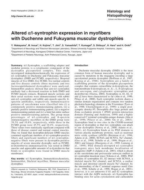
Immunoblot analysis<br />
Immunoblot analysis showed that anti-ß-spectrin<br />
antibody reacted with 270kDa protein extracts of normal<br />
muscles. The reactions of anti-ß-spectrin antibody both<br />
in DMD <strong>and</strong> FCMD muscle extracts slightly decreased<br />
compared with those of extracts in normal control<br />
muscles (Fig. 1A). Anti-α1-syntrophin antibody reacted<br />
with 59kDa protein extracts of normal muscles, but the<br />
reactions of anti-α1-syntrophin antibody decreased both<br />
in DMD <strong>and</strong> FCMD muscle extracts (Fig. 1B). The<br />
reactions for anti-myosin heavy chain antibody showed a<br />
fairly broad b<strong>and</strong> <strong>and</strong> its center was about 220kDa in<br />
normal, DMD <strong>and</strong> FCMD muscles. The reactions for<br />
anti-myosin heavy chain antibody in DMD <strong>and</strong> FCMD<br />
Fig. 1. Immunoblots of ß-spectrin (A), α1-syntrophin (B) <strong>and</strong> myosin heavy chain (C) expressions in normal human quadriceps femoris muscles (lane 1<br />
of A, B <strong>and</strong> C, respectively), in muscles of boys with Duchenne muscular dystrophy (DMD) (lane 2 of A, B <strong>and</strong> C, respectively) <strong>and</strong> in muscles of<br />
children with Fukuyama-type congenital muscular dystrophy (FCMD) (lane 3 of A, B <strong>and</strong> C, respectively). Electrophoresis <strong>and</strong> blotting were done as<br />
described in the Materials <strong>and</strong> Methods. In normal human muscle extracts, b<strong>and</strong>s of ß-spectrin <strong>and</strong> α1-syntrophin were at 270kDa <strong>and</strong> 59kDa,<br />
respectively (A <strong>and</strong> B, respectively); the b<strong>and</strong> of myosin heavy chain was rather broad <strong>and</strong> its center was about 220kDa (C). The reaction for ß-spectrin<br />
in DMD <strong>and</strong> FCMD muscle extracts showed a slight decrease in comparison with normal control muscle extracts (A); the reaction for α1-syntrophin<br />
both in DMD <strong>and</strong> FCMD muscle extracts moderately decreased compared with normal muscle extracts (B). Reactions for myosin heavy chain in<br />
extracts of normal, DMD <strong>and</strong> FCMD muscles had almost the same intensity (C). Numbers to the left of each figure are the molecular masses of<br />
st<strong>and</strong>ards.
26<br />
α1-syntrophin in muscular dystrophies<br />
muscle extracts were almost as intense as the reaction of<br />
normal muscle extract (Fig. 1C).<br />
Immunohistochemistry <strong>and</strong> its analysis<br />
The immunoreaction of normal skeletal myofibers<br />
stained with anti-ß-spectrin <strong>and</strong> anti-ß1-syntrophin<br />
antibodies was at the myofiber surface (Fig. 2A, B). All<br />
myofibers including both slow twitch type 1 fibers <strong>and</strong><br />
fast twitch type 2A <strong>and</strong> 2B fibers (Fig. 2C), were<br />
similarly immunostained.<br />
In DMD muscles, scattered myofibers were present<br />
with variously surface-immunostained myofibers stained<br />
with anti-ß-spectrin antibody. These myofibers included<br />
continuous surface immunolabeling myofibers,<br />
discontinuous surface immunolabeling myofibers <strong>and</strong><br />
negative surface immunolabeling myofibers (Fig. 3A1,<br />
2); but dystrophin immunostaining of these myofibers<br />
showed a negative immunoreaction (Fig. 3B1, 2).<br />
Immunostaining with anti-α1-syntrophin antibody in<br />
DMD myofibers showed similar immunostaining<br />
patterns to those with anti-ß-spectrin antibody, <strong>and</strong> the<br />
number of immunopositive myofibers with anti-α1-<br />
syntrophin antibody was large, irrespective of the<br />
absence of dystrophin (Fig. 3B1, B2, C1, C2). However<br />
the percentage of immunopositive surface labeling<br />
Fig. 2. Immunohistochemical staining of serial normal muscle sections with anti-ß-spectrin (A), anti-α1-syntrophin (B) antibodies <strong>and</strong> myosin ATPase<br />
(pH 4.6) (C). The cell surface of individual myofibers in normal muscle shows a thin layer of immunofluorescense with these antibodies. The muscle<br />
section with myosin ATPase staining (C) contains type 1 fiber (1), type 2A fiber (A) <strong>and</strong> type 2B fiber (B). The immunostaining pattern has no fiber type<br />
selectivity. Scale bars: 50 µm. x 250
α1-syntrophin in muscular dystrophies<br />
27<br />
Fig. 3A1-C1. Immunohistochemical staining of serial sections of Duchenne muscular dystrophy (DMD) muscle with anti-ß-spectrin (A1), anti-dystrophin<br />
(B1) <strong>and</strong> anti-α1-syntrophin (C1) antibodies. Immunoreactions for both ß-spectrin (A1) <strong>and</strong> α1-syntrophin (C1) at the surface of DMD myofibers show a<br />
positive immunoreaction at many DMD myofibers in various degrees <strong>and</strong> negative immunoreaction in scattered remaining myofibers; the<br />
immunoreactivity for dystrophin was negative (B1). Generally, the number of myofibers with negative immunoreactivity for α1-syntrophin was more than<br />
for ß-spectrin. Positively immunostained myofibers with anti-ß-spectrin antibody (A1, arrows) showed negative immunoreaction for anti-α1-syntrophin<br />
antibody in the serial muscle section (C1, arrows). Scale bars: 50 µm. x 250
28<br />
α1-syntrophin in muscular dystrophies<br />
Fig. 3A2-C2. Immunohistochemical staining of serial muscle sections with anti-ß-spectrin (A2), anti-dystrophin (B2) <strong>and</strong> anti-α1-syntrophin (C2)<br />
antibodies from another boy with Duchenne muscular dystrophy (DMD). Anti-α1-syntrophin immunopositive myofibers (C2) are less numerous than<br />
those seen in Fig. 3C1, although the immunostaining patterns at the myofiber surface for both ß-spectrin (A2) <strong>and</strong> dystrophin (B2) in this case reveal<br />
similar patterns to those observed in Fig. 3A1 <strong>and</strong> Fig. 3B1, respectively. Again the number of myofibers with negative immunoreactivity for α1-<br />
syntrophin was more than for ß-spectrin. Positively immunostained myofibers with anti-ß-spectrin antibody (A2, arrows) disclosed negative<br />
immunoreaction for anti-α1-syntrophin antibody in the serial DMD muscle section (C2, arrows). Scale bars: 50 µm. x 330
α1-syntrophin in muscular dystrophies<br />
29<br />
Fig. 3D1-F. Representative myofibers immunostained with anti-ß-spectrin <strong>and</strong> anti-α1-syntrophin antibodies. Fig. 3D1, 3D2, 3D3, 3D4 are examples of<br />
myofibers in group 1, 2, 3, 4, respectively. 3E, F. Immunohistochemical staining of serial DMD muscle sections with anti-α1-syntrophin (E) <strong>and</strong> anti-αdystrobrevin<br />
(F) antibodies. Appreciable amounts of α1-syntrophin (E) <strong>and</strong> α-dystrobrevin (F) are in DMD muscle membranes. α1-Syntrophin-negative<br />
myofibers (E, asterisks) are α-dystrobrevin immunopositive myofibers (F, asterisks). Scale bars: 50 µm. x 330
30<br />
Fig. 4. Immunohistochemical staining of serial sections of Fukuyama-type congenital muscular dystrophy (FCMD) muscle with anti-ß-spectrin (A) <strong>and</strong><br />
anti-α1-syntrophin (B) antibodies. Immunoreactivities for both anti-ß-spectrin <strong>and</strong> anti-α1-syntrophin antibodies in FCMD myofibers show positive<br />
immunoreaction in various degrees in many FCMD myofibers <strong>and</strong> negative immunostaining in the remaining myofibers. The number of FCMD<br />
myofibers with negative immunostaining by anti-α1-syntrophin antibody was more than by anti-ß-spectrin antibody. Positively immunostained myofiber<br />
with anti-ß-spectrin antibody (A, arrows) shows negative immunoreaction for anti-α1-syntrophin antibody in the serial muscle section (B, arrows).<br />
Immunohistochemical staining of serial FCMD muscle sections with anti-α1-syntrophin (C) <strong>and</strong> anti-neonatal myosin (D) antibodies. Partially α1-<br />
syntrophin-expressed FCMD myofibers at their surface membranes (C, asterisks) show negative immunostaining with anti-neonatal myosin antibody<br />
(D, asterisks). α1-Syntrophin immunonegative FCMD myofibers at their surface membranes (C, arrows) show positive immunostaining with antineonatal<br />
myosin antibody (D, arrows). Scale bars: 50 µm. x 250
α1-syntrophin in muscular dystrophies<br />
31<br />
myofibers with anti-α1-syntrophin antibody was less<br />
than these myofibers stained with anti-ß-spectrin<br />
antibody. From the classification of immunostaining<br />
patterns into group 1 to group 4 (Fig. 3D1-3D4), the<br />
group mean percentages of groups 1, 2, 3 <strong>and</strong> 4 DMD<br />
myofibers stained with anti-α1-syntrophin antibody<br />
were 15.4%, 39.6%, 39.3% <strong>and</strong> 5.7%, respectively, <strong>and</strong><br />
those of groups 1, 2, 3 <strong>and</strong> 4 DMD myofibers stained<br />
with anti-ß-spectrin antibody were 50.2%, 39.0%, 10.8%<br />
<strong>and</strong> 0%, respectively (Table 1). DMD myofibers<br />
immunostained with anti-dystrobrevin antibody showed<br />
numerous dystrobrevin positive myofibers. Appreciable<br />
amounts of α-dystrobrevin, a molecule of syntrophindystrobrevin<br />
subcomplex, were found in the DMD<br />
muscle membranes. Dystrobrevin-immunopositive DMD<br />
myofibers appeared to be more numerous than α1-<br />
syntrophin-immunopositive DMD myofibers in all five<br />
DMD muscles used to survey the two antibody-stained<br />
serial muscle sections (Fig. 3E, F).<br />
FCMD muscles immunostained with anti-ß-spectrin<br />
<strong>and</strong> anti-α1-syntrophin antibodies showed<br />
immunostaining patterns similar to those of DMD<br />
muscles (Fig. 4A, B). The number of immunonegative<br />
myofibers stained with anti-α1-syntrophin antibody was<br />
large (Fig. 4B) irrespective of the presence of<br />
dystrophin. The percentage of immunonegative<br />
myofibers stained with anti-α1-syntrophin antibody was<br />
more than these myofibers stained with anti-ß-spectrin<br />
antibody. From the classification into group 1 to group 4,<br />
the group mean percentages of groups 1, 2, 3 <strong>and</strong> 4<br />
FCMD myofibers stained with anti-α1-syntrophin<br />
antibody were 15.3%, 31.9%, 45.5% <strong>and</strong> 7.3%,<br />
respectively, <strong>and</strong> those of groups 1, 2, 3 <strong>and</strong> 4 FCMD<br />
myofibers stained with anti-ß-spectrin antibody were<br />
52.7%, 36.4%, 10.4% <strong>and</strong> 0.5%, respectively (Table 1).<br />
Immunonegative FCMD myofibers stained with α1-<br />
syntrophin at the myofiber surface were consistent with<br />
neonatal myosin immunopositive FCMD myofibers in<br />
serial muscle sections (Fig. 4C, D).<br />
The disease control muscle samples immunostained<br />
with anti-ß-spectrin <strong>and</strong> anti-α1-syntrophin antibodies<br />
contained fewer myofibers with group 2 <strong>and</strong> 3 staining<br />
patterns (Table 1). The group mean percentages of α1-<br />
syntrophin- <strong>and</strong> ß-spectrin-immunonegative myofibers<br />
were 39.3% <strong>and</strong> 10.8%, respectively, in the DMD group,<br />
<strong>and</strong> were 45.5% <strong>and</strong> 10.4%, respectively, in the FCMD<br />
group. The group mean percentages of α1-syntrophinimmunonegative<br />
myofibers in DMD <strong>and</strong> FCMD groups<br />
were statistically significant compared with those of ß-<br />
spectrin-immunonegative myofibers (Table 2).
32<br />
α1-syntrophin in muscular dystrophies<br />
Table 1. Immunoreaction for ß-spectrin <strong>and</strong> α1-syntrophin in dystrophic muscles.<br />
CASES (1) (2) (3) (4)<br />
Anti-ß-spectrin immunoreactiviy*<br />
5 DMD 50.22±8.24 39.01±6.33 10.78±2.51 0<br />
5 FCMD 52.73±8.51 36.43±5.40 10.38±3.08 0.47±0.30<br />
5 DC 84.93±3.15 13.34±2.38 1.73±1.65 0<br />
5 NC 96.40±0.76 3.60±0.76 0 0<br />
Anti-α1-syntrophin immunoreactivity*<br />
5 DMD 15.41±7.29 39.60±1.56 39.31±7.71 5.67±1.60<br />
5 FCMD 15.31±6.37 31.89±4.21 45.51±10.25 7.30±1.22<br />
5 DC 78.73±1.80 17.38±1.32 3.89±0.98 0<br />
5 NC 95.03±1.43 4.97±1.43 0 0<br />
(1)=group 1: Continuously positive immunostaining pattern with more than 90% myofiber surface immunolabeling <strong>and</strong> normal immunoreactivity.<br />
(2)=group 2: Partially positive immunostaining pattern with 10 to 90% myofiber surface immunolabeling <strong>and</strong> normal immunoreactivity. (3)=group 3:<br />
Negative immunostaining pattern with less than 10% myofiber surface immunolabeling. (4)=group 4: Faint but entire surface positive immunostaining<br />
pattern. *: Group mean percentage of myofibers ± st<strong>and</strong>ard error of the mean. DMD: Duchenne muscular dystrophy. FCMD: Fukuyama type congenital<br />
muscular dystrophy. DC: Disease control (myotonic dystrophy + facioscapulohumeral dystrophy). NC: Normal control.<br />
Table 2. Immunonegative fiber ratio (group 3).<br />
Discussion<br />
ANTI-ß-SPECTRIN ANTI-α1-SYNTROPHIN P<br />
ANTIBODY<br />
ANTIBODY<br />
IMMUNOREACTIVITY* IMMUNOREACTIVITY*<br />
5 DMD 10.78±2.51% 39.31±7.71% P
α1-syntrophin in muscular dystrophies<br />
33<br />
In DMD muscles, morphometric analysis showed that<br />
motor nerve terminals are normal, but at nearly 50<br />
percent of the end plates, focal degeneration of the<br />
junctional folds is seen <strong>and</strong> some postsynaptic regions<br />
are simpler than normal (Jerusalem et al., 1974).<br />
Desmuslin is at the Z-line <strong>and</strong> binds to desmin (Mizuno<br />
et al., 2001). Desmuslin expression in DMD myofibers<br />
has not been reported so far, but desmuslin expression in<br />
DMD muscles is plausible. The appreciable amounts of<br />
α-dystrobrevin expressed in DMD muscles in this study<br />
may bind desmuslin despite the absence of dystrophin.<br />
FCMD muscles have dystrophin molecules, but<br />
dystrophin-associated protein α1-syntrophin was absent<br />
in about 45% FCMD myofibers in this study. This<br />
finding was also consistent with our previous finding of<br />
the expression of the α1-syntrophin-binding molecule<br />
AQP4 being markedly reduced in FCMD muscles<br />
(Wakayama et al., 2003). The mechanism of the<br />
downregulated expression of AQP4 in FCMD muscle is<br />
unknown, but the immaturity of the FCMD myofibers is<br />
one cause. Immunostaining of FCMD myofibers with<br />
anti-neonatal myosin antibody shows that α1-syntrophin<br />
negative FCMD myofibers are immature, because this<br />
antibody immunostained the human fatal myosin (Ecob-<br />
Prince et al., 1989). Present immunohistochemical<br />
studies of α1-syntrophin generally agreed with the<br />
results of our immunoblot analysis. Further studies of<br />
other isoforms of syntrophin, such as ß1-, ß2- <strong>and</strong> γ2-<br />
syntrophins <strong>and</strong> a detailed study of dystrobrevins will<br />
throw light on the precise pathogenesis of human<br />
muscular dystrophies.<br />
Acknowledgements. We thank Mrs. Y. Hirayama <strong>and</strong> Ms. C. Tochimono<br />
for their technical assistance. This study was supported by the<br />
Research grant (14B-4) for Nervous <strong>and</strong> Mental Disorders from the<br />
Ministry of Health, Labour <strong>and</strong> Welfare, Japan<br />
References<br />
Adams M.E., Kramarcy N., Krall S.P., Rossi S.G., Rotundo R.L.,<br />
Sealock R. <strong>and</strong> Froehner S.C. (2000). Absence of α-syntrophin<br />
leads to structurally aberrant neuromuscular synapses deficient in<br />
utrophin. J. Cell Biol. 150, 1385-1398.<br />
Adams M.E., Mueller H.A. <strong>and</strong> Froehner S.C. (2001). In vivo<br />
requirement of the α-syntrophin PDZ domain for the sarcolemmal<br />
localization of nNOS <strong>and</strong> aquaporin-4. J. Cell Biol. 155, 113-122.<br />
Ahn A.H., Freener C.A., Gussoni E., Yoshida M. <strong>and</strong> Ozawa E. (1996).<br />
The three human syntrophin genes are expressed in diverse tissues,<br />
have distinct chromosomal locations, <strong>and</strong> each bind to dystrophin<br />
<strong>and</strong> its relatives. J. Biol. Chem. 271, 2724-2730.<br />
Benson M.A., Newey S.E., Martin-Rendon E., Hawkes R. <strong>and</strong> Blake<br />
D.J. (2001). Dysbindin, a novel coiled-coil-containing protein that<br />
interacts with the dystrobrevins in muscle <strong>and</strong> brain. J. Biol. Chem.<br />
276, 24232-24241.<br />
Blake D.J., Nawrotzki R., Peters M.F., Froehner S.C. <strong>and</strong> Davies K.E.<br />
(1996). Isoform diversity of dystrobrevin, the murine 87-kDa<br />
postsynaptic protein. J. Biol. Chem. 271, 7802-7810.<br />
Blake D.J. (2002). Dystrobrevin dynamics in muscle-cell signalling: a<br />
possible target for therapeutic intervention in Duchenne muscular<br />
dystrophy? Neuromuscul. Disord. 12 Suppl 1, S110-S117.<br />
Blake D.J., Nawrotzki R., Loh N.Y., Gorecki D.C. <strong>and</strong> Davies K.E.<br />
(1998). ß-Dystrobrevin, a member of the dystrophin-related protein<br />
family. Proc. Natl. Acad. Sci. USA 95, 241-246.<br />
Brenman J.E., Chao D.S., Gee S.H., McGee A.W., Craven S.E.,<br />
Santillano D.R., Wu Z., Huang F., Xia H., Peters M.F., Froehner S.C.<br />
<strong>and</strong> Bredt D.S. (1996). Interaction of nitric oxide synthase with the<br />
postsynaptic density protein PSD-95 <strong>and</strong> α1-syntrophin mediated by<br />
PDZ domains. Cell 84, 757-767.<br />
Chockalingam P.S., Gee S.H. <strong>and</strong> Jarrett H.W. (1999). Pleckstrin<br />
homology domain 1 of mouse α1-syntrophin binds<br />
phosphatidylinositol 4,5-bisphosphate. Biochemistry 38, 5596-5602.<br />
Di Blasi C., Mor<strong>and</strong>i L., Barresi R., Blasevich F., Cornelio F. <strong>and</strong> Mora<br />
M. (1996). Dystrophin-associated protein abnormalities in<br />
dystrophin-deficient muscle fibers from symptomatic <strong>and</strong><br />
asymptomatic Duchenne/Becker muscular dystrophy carriers. Acta<br />
Neuropathol. (Berl) 92, 369-377.<br />
Ecob-Prince M., Hill M. <strong>and</strong> Brown W. (1989). Immunocytochemical<br />
demonstration of myosin heavy chain expression in human muscle.<br />
J. Neurol. Sci. 91, 71-78.<br />
Enigk R.E. <strong>and</strong> Maimone M.M. (1999). Differential expression <strong>and</strong><br />
developmental regulation of a novel α-dystrobrevin isoform in<br />
muscle. Gene 238, 479-488.<br />
Fanning A.S. <strong>and</strong> Anderson J.M. (1999). PDZ domains: fundamental<br />
building blocks in the organization of protein complexes at the<br />
plasma membrane. J. Clin. Invest. 103, 767-772.<br />
Fukuyama Y., Osawa M. <strong>and</strong> Suzuki H. (1981). Congenital progressive<br />
muscular dystrophy of the Fukuyama type: clinical, genetic <strong>and</strong><br />
pathologic considerations. Brain Dev. 3, 1-29.<br />
Gee S.H., Madhavan R., Levinson S.R., Caldwell J.H., Sealock R. <strong>and</strong><br />
Froehner S.C. (1998). Interaction of muscle <strong>and</strong> brain sodium<br />
channels with multiple members of the syntrophin family of<br />
dystrophin-associated proteins. J. Neurosci. 18, 128-137.<br />
Grady R.M., Grange R.W., Lau K.S., Maimone M.M., Nichol M.C., Stull<br />
J.T. <strong>and</strong> Sanes J.R. (1999). Role for α-dystrobrevin in the<br />
pathogenesis of dystrophin-dependent muscular dystrophies. Nat.<br />
Cell Biol. 1, 215-220.<br />
Hasegawa M., Cuenda A., Spillantini M.G., Thomas G.M., Buee-<br />
Scherrer V., Cohen P. <strong>and</strong> Goedert M. (1999). Stress-activated<br />
protein kinase-3 interacts with the PDZ domain of α1-syntrophin. A<br />
mechanism for specific substrate recognition. J. Biol. Chem. 274,<br />
12626-12631.<br />
Hoffman E.P., Brown Jr R.H. <strong>and</strong> Kunkel L.M. (1987). Dystrophin: the<br />
protein product of the Duchenne muscular dystrophy locus. Cell 51,<br />
919-928.<br />
Hogan A., Shepherd L., Chabot J., Quenneville S., Prescott S.M.,<br />
Topham M.K. <strong>and</strong> Gee S.H. (2001). Interaction of γ1-syntrophin with<br />
diacylglycerol kinase-ζ: regulation of nuclear localization by PDZ<br />
interactions. J. Biol. Chem. 276, 26526-26533.<br />
Iwata Y., Pan Y., Yoshida T., Hanada H. <strong>and</strong> Shigekawa M. (1998). α1-<br />
Syntrophin has distinct binding sites for actin <strong>and</strong> calmodulin. FEBS<br />
Lett. 423, 173-177.<br />
Jerusalem F., Engel A.G. <strong>and</strong> Gomez M.R. (1974). Duchenne<br />
dystrophy. II. Morphometric study of motor end-plate fine structure.<br />
Brain 97, 123-130.<br />
Jones K.J., Compton A.G., Yang N., Mills M.A., Peters M.F., Mowat D.,<br />
Kunkel L.M., Froehner S.C. <strong>and</strong> North K.N. (2003). Deficiency of the<br />
syntrophins <strong>and</strong> α-dystrobrevin in patients with inherited myopathy.<br />
Neuromuscul. Disord. 13, 456-467.<br />
Kobayashi K., Nakahori Y., Miyake M., Matsumura K., Kondo-Iida E.,
34<br />
α1-syntrophin in muscular dystrophies<br />
Nomura Y., Segawa M., Yoshioka M., Saito K., Osawa M., Hamano<br />
K., Sakakihara Y., Nonaka I., Nakagome Y., Kanazawa I.,<br />
Nakamura Y., Tokunaga K. <strong>and</strong> Toda T. (1998). An ancient<br />
retrotransposal insertion causes Fukuyama-type congenital<br />
muscular dystrophy. Nature 394, 388-392.<br />
Koenig M., Monaco A.P. <strong>and</strong> Kunkel L.M. (1988). The complete<br />
sequence of dystrophin predicts a rod-shaped cytoskeletal protein.<br />
Cell 53, 219-226.<br />
Kramarcy N.R., Vidal A., Froehner S.C. <strong>and</strong> Sealock R. (1994).<br />
Association of utrophin <strong>and</strong> multiple dystrophin short forms with the<br />
mammalian Mr 58,000 dystrophin-associated protein (syntrophin). J.<br />
Biol. Chem. 269, 2870-2876.<br />
Lumeng C., Phelps S., Crawford G.E., Walden P.D., Barald K. <strong>and</strong><br />
Chamberlain J.S. (1999). Interactions between ß2-syntrophin <strong>and</strong> a<br />
family of microtubule-associated serine/threonine kinases. Nat.<br />
Neurosci. 2, 611-617.<br />
Metzinger L., Blake D.J., Squier M.V., Anderson L.V., Deconinck A.E.,<br />
Nawrotzki R., Hilton-Jones D. <strong>and</strong> Davies K.E. (1997). Dystrobrevin<br />
deficiency at the sarcolemma of patients with muscular dystrophy.<br />
Hum. Mol. Genet. 6, 1185-1191.<br />
Mizuno Y., Yoshida M., Nonaka I., Hirai S. <strong>and</strong> Ozawa E. (1994).<br />
Expression of utrophin (dystrophin-related protein) <strong>and</strong> dystrophinassociated<br />
glycoproteins in muscles from patients with Duchenne<br />
muscular dystrophy. Muscle Nerve 17, 206-216.<br />
Mizuno Y., Thompson T.G., Guyon J.R., Lidov H.G., Brosius M.,<br />
Imamura M., Ozawa E., Watkins S.C. <strong>and</strong> Kunkel L.M. (2001).<br />
Desmuslin, an intermediate filament protein that interacts with α-<br />
dystrobrevin <strong>and</strong> desmin. Proc. Natl. Acad. Sci. USA 98, 6156-6161.<br />
Newey S.E., Benson M.A., Ponting C.P., Davies K.E. <strong>and</strong> Blake D.J.<br />
(2000). Alternative splicing of dystrobrevin regulates the<br />
stoichiometry of syntrophin binding to the dystrophin protein<br />
complex. Curr. Biol. 10, 1295-1298.<br />
Newey S.E., Howman E.V., Ponting C.P., Benson M.A., Nawrotzki R.,<br />
Loh N.Y., Davies K.E. <strong>and</strong> Blake D.J. (2001). Syncoilin, a novel<br />
member of the intermediate filament superfamily that interacts with<br />
α-dystrobrevin in skeletal muscle. J. Biol. Chem. 276, 6645-6655.<br />
Ou Y., Strege P., Miller S.M., Makielski J., Ackerman M., Gibbons S.J.<br />
<strong>and</strong> Farrugia G. (2003). Syntrophin γ2 regulates SCN5A gating by a<br />
PDZ domain-mediated interaction. J. Biol. Chem. 278, 1915-1923.<br />
Ozawa E. (2004). The muscle fiber cytoskeleton: the dystrophin system.<br />
In: Myology. 3rd ed. Engel A.G. <strong>and</strong> Franzini-Armstrong C. (eds).<br />
The McGraw-Hill Companies. New York. pp 455-470.<br />
Peters M.F., Kramarcy N.R., Sealock R. <strong>and</strong> Froehner S.C. (1994). ß2-<br />
Syntrophin: localization at the neuromuscular junction in skeletal<br />
muscle. Neuroreport 5, 1577-1580.<br />
Peters M.F., Adams M.E. <strong>and</strong> Froehner S.C. (1997a). Differential<br />
association of syntrophin pairs with the dystrophin complex. J. Cell<br />
Biol. 138, 81-93.<br />
Peters M.F., O'Brien K.F., Sadoulet-Puccio H.M., Kunkel L.M., Adams<br />
M.E. <strong>and</strong> Froehner S.C. (1997b). ß-Dystrobrevin, a new member of<br />
the dystrophin family. Identification, cloning, <strong>and</strong> protein<br />
associations. J. Biol. Chem. 272, 31561-31569.<br />
Peters M.F., Sadoulet-Puccio H.M., Grady M.R., Kramarcy N.R., Kunkel<br />
L.M., Sanes J.R., Sealock R. <strong>and</strong> Froehner S.C. (1998). Differential<br />
membrane localization <strong>and</strong> intermolecular associations of α-<br />
dystrobrevin isoforms in skeletal muscle. J. Cell Biol. 142, 1269-<br />
1278.<br />
Piluso G., Mirabella M., Ricci E., Belsito A., Abbondanza C., Servidei S.,<br />
Puca A.A., Tonali P., Puca G.A. <strong>and</strong> Nigro V. (2000). γ1- <strong>and</strong> γ2-<br />
Syntrophins, two novel dystrophin-binding proteins localized in<br />
neuronal cells. J. Biol. Chem. 275, 15851-15860.<br />
Puca A.A., Nigro V., Piluso G., Belsito A., Sampaolo S., Quaderi N.,<br />
Rossi E., Di Iorio G., Ballabio A. <strong>and</strong> Franco B. (1998). Identification<br />
<strong>and</strong> characterization of a novel member of the dystrobrevin gene<br />
family. FEBS Lett. 425, 7-13.<br />
Sadoulet-Puccio H.M., Khurana T.S., Cohen J.B. <strong>and</strong> Kunkel L.M.<br />
(1996). Cloning <strong>and</strong> characterization of the human homologue of a<br />
dystrophin related phosphoprotein found at the Torpedo electric<br />
organ post-synaptic membrane. Hum. Mol. Genet. 5, 489-496.<br />
Sadoulet-Puccio H.M., Rajala M. <strong>and</strong> Kunkel L.M. (1997). Dystrobrevin<br />
<strong>and</strong> dystrophin: An interaction through coiled-coil motifs. Proc. Natl.<br />
Acad. Sci. USA 94, 12413-12418.<br />
Schotl<strong>and</strong> D.L., Bonilla E. <strong>and</strong> Wakayama Y. (1981). Freeze fracture<br />
studies of muscle plasma membrane in human muscular dystrophy.<br />
Acta Neuropathol. (Berl) 54, 189-197.<br />
Schultz J., Hoffmuller U., Krause G., Ashurst J., Macias M.J., Schmieder<br />
P., Schneider-Mergener J. <strong>and</strong> Oschkinat H. (1998). Specific<br />
interactions between the syntrophin PDZ domain <strong>and</strong> voltage-gated<br />
sodium channels. Nat. Struct. Biol. 5, 19-24.<br />
Sewry C.A., Matsumura K., Campbell K.P. <strong>and</strong> Dubowitz V. (1994).<br />
Expression of dystrophin-associated glycoproteins <strong>and</strong> utrophin in<br />
carriers of Duchenne muscular dystrophy. Neuromuscul. Disord. 4,<br />
401-409.<br />
Tyers M., Rachubinski R.A., Stewart M.I., Varrichio A.M., Shorr R.G.L.,<br />
Haslam R.J. <strong>and</strong> Harley C.B. (1988). Molecular cloning <strong>and</strong><br />
expression of the major protein kinase C substrate of platelets.<br />
Nature 333, 470-473.<br />
Wakayama Y., Okayasu H., Shibuya S. <strong>and</strong> Kumagai T. (1984).<br />
Duchenne dystrophy: Reduced density of orthogonal array subunit<br />
particles in the muscle plasma membrane. Neurology 34, 1313-<br />
1317.<br />
Wakayama Y., Kumagai T. <strong>and</strong> Shibuya S. (1985). Freeze-fracture<br />
studies of muscle plasma membrane in Fukuyama-type congenital<br />
muscular dystrophy. Neurology 35, 1587-1593.<br />
Wakayama Y., Shibuya S., Takeda A., Jimi T., Nakamura Y. <strong>and</strong> Oniki<br />
H. (1995). Ultrastructural localization of the C-terminus of the 43-kd<br />
dystrophin-associated glycoprotein <strong>and</strong> its relation to dystrophin in<br />
normal murine skeletal myofiber. Am. J. Pathol. 146, 189-196.<br />
Wakayama Y., Inoue M., Murahashi M., Shibuya S., Jimi T., Kojima H.<br />
<strong>and</strong> Oniki H. (1997). Ultrastructural localization of α1-syntrophin <strong>and</strong><br />
neuronal nitric oxide synthase in normal skeletal myofiber, <strong>and</strong> their<br />
relation to each other <strong>and</strong> to dystrophin. Acta Neuropathol. (Berl) 94,<br />
455-464.<br />
Wakayama Y., Jimi T., Inoue M., Kojima H., Murahashi M., Kumagai T.,<br />
Yamashita S., Hara H. <strong>and</strong> Shibuya S. (2002). Reduced aquaporin 4<br />
exression in the muscle plasma membrane of patients with<br />
Duchenne muscular dystrophy. Arch. Neurol. 59, 431-437.<br />
Wakayama Y., Jimi T., Inoue M., Kojima H., Yamashita S., Kumagai T.,<br />
Murahashi M., Hara H. <strong>and</strong> Shibuya S. (2003). Altered aquaporin 4<br />
expression in muscles of Fukuyama-type congenital muscular<br />
dystrophy. Virchows Arch. 443, 761-767.<br />
Yang B., Jung D., Rafael J.A., Chamberlain J.S. <strong>and</strong> Campbell K.P.<br />
(1995). Identification of α-syntrophin binding to syntrophin triplet,<br />
dystrophin, <strong>and</strong> utrophin. J. Biol. Chem. 270, 4975-4978.<br />
Yokota T., Miyagoe Y., Hosaka Y., Tsukita K., Kameya S., Shibuya S.,<br />
Matsuda R., Wakayama Y., <strong>and</strong> Takeda S. (2000). Aquaporin-4 is<br />
absent at the sarcolemma <strong>and</strong> at perivascular astrocyte endfeet in<br />
α1-syntrophin knockout mice. Proc. Jpn. Acad. 76, 22-27.<br />
Accepted August 9, 2005