Real-time Virtual Sonography の
Real-time Virtual Sonography の
Real-time Virtual Sonography の

Create successful ePaper yourself
Turn your PDF publications into a flip-book with our unique Google optimized e-Paper software.
CT<br />
<strong>Real</strong>-<strong>time</strong> <strong>Virtual</strong> <strong>Sonography</strong>XCT<br />
<br />
<br />
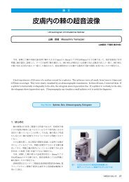
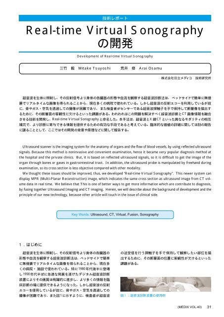
Ultrasound scanner is the imaging system for the anatomy of organs and the flow of blood vessels, by using reflected ultrasound<br />
signals. Because this method is noninvasive and convenient examination, hence it became very popular diagnosis method at<br />
the hospital and the private clinics. But, it is based on reflected ultrasound signals, so it is difficult to get the image of the<br />
organ through bones or gases in gastrointestinal tract. In addition, the ultrasound probe is manipulated by freehand during<br />
examination, so its cross section is less objective compared with other modality.<br />
We thought these issues should be improved, thus, we developed "<strong>Real</strong>-<strong>time</strong> <strong>Virtual</strong> <strong>Sonography</strong>". This newer system can<br />
display MPR (Multi-Planar Reconstruction) image, which indicates the same cross section as ultrasound image from CT volume<br />
data in real <strong>time</strong>. We believe that This is one of better ways to get more information which are contribute to diagnosis,<br />
by fusing together Ultrasound Imaging and CT imaging. Herein, we will describe about the background of development and the<br />
principle of our new technology, because other article will touch in the issue of clinical side.<br />
Key Words: Ultrasound, CT, <strong>Virtual</strong>, Fusion, <strong>Sonography</strong><br />
<br />
<br />
<br />
<br />
1980<br />
1990<br />
<br />
<br />
<br />
1<br />
<br />
<br />
<br />
<br />
MEDIX VOL.40 31
CT<br />
<br />
<br />
CT<br />
<br />
CT<br />
<br />
(2)<br />
CT<br />
<strong>Real</strong>-<strong>time</strong> <strong>Virtual</strong> <strong>Sonography</strong>XCT<br />
<br />
<br />
<br />
<br />
<br />
<br />
LANPCPC<br />
<br />
<br />
<br />
<br />
1<br />
<br />
<br />
PC<strong>Virtual</strong> <strong>Sonography</strong><br />
CTMPR<br />
<br />
<br />
<br />
<br />
<br />
<br />
<br />
<br />
<br />
<br />
<br />
<br />
1996<br />
1) <br />
CT<br />
CT<br />
<br />
15cm30<br />
CT5mm<br />
CTMPR(Multi-<br />
Planar Reconstruction)<br />
<br />
MDCT(Multi-Detector CT)<br />
1mm<br />
<br />
(PC)<br />
MPRPC<br />
<br />
<br />
3<br />
PC<br />
<br />
<br />
CTCDROM<br />
<br />
<br />
<br />
<br />
<br />
<br />
<br />
<br />
<br />
<br />
<br />
<br />
<br />
<br />
<br />
<br />
<br />
<br />
CTPC<br />
DICOM(Digital Imaging and Communication in<br />
Medicine)<br />
CTCT<br />
<br />
MDCT<br />
4<br />
161416<br />
CT<br />
201mm<br />
<br />
MPR<br />
<br />
32 MEDIX VOL.40
DICOM<br />
()<br />
150MPR<br />
<br />
<br />
<br />
RS232C<br />
PCPC<br />
<br />
(xyz)(xyz<br />
)<br />
<br />
MPRCT<br />
<br />
<br />
<br />
<br />
<br />
<br />
<br />
2) xyzx'y'z'<br />
r11r33<br />
DxDyDz<br />
i ) <br />
UR<br />
ii) RT<br />
iii) CT<br />
TC<br />
PusCT<br />
Pvol<br />
PvolPus<br />
<br />
UR RT TC (1)<br />
<br />
UR<br />
3) <br />
()UR<br />
<br />
RT<br />
<br />
CT<br />
TC<br />
<br />
<br />
TC<br />
(5)<br />
<br />
<br />
x' y' z' 1 x y z 1<br />
<br />
<br />
r 11 r 12 r 13 0<br />
r 21 r 22 r 23 0<br />
r 31 r 32 r 33 0<br />
Dx Dy Dz 1<br />
4<br />
CT(CT)<br />
<br />
()<br />
4<strong>Virtual</strong> <strong>Sonography</strong><br />
<br />
<br />
<br />
<br />
<br />
<br />
<br />
<br />
<br />
<br />
CT<br />
UC<br />
UC UR RT TC<br />
(2)<br />
<br />
<br />
<br />
<br />
<br />
<br />
<br />
<br />
(2)TC<br />
TCRT 1 UR 1 UC<br />
(3)<br />
(3)RTUR<br />
<br />
MEDIX VOL.40 33
TC<br />
<br />
<br />
UCTC<br />
<br />
<br />
<br />
CT<br />
CT<br />
CT<br />
SxSySzUC<br />
<br />
UC<br />
Sx 0 0 0<br />
0 Sy 0 0<br />
0 0 Sz 0<br />
Dx Dy Dz 1<br />
(4)<br />
<strong>Virtual</strong> <strong>Sonography</strong><br />
<br />
<br />
70cm5mm<br />
(a) <br />
(b) <br />
(c) <br />
<br />
<br />
<br />
<br />
<br />
<br />
CT<br />
PvolCal<br />
PvolCal XvolC YvolC ZvolC<br />
(5)<br />
<br />
<br />
<br />
<br />
<br />
()<br />
<br />
RTcal<br />
<br />
<br />
<br />
<br />
<br />
<br />
<br />
<br />
<br />
<br />
PusCal<br />
XusC YusC ZusC<br />
(6)<br />
CT<br />
<br />
PvolCalPusCal<br />
<br />
UCCal<br />
(7)<br />
(7)(4)DxDyDzUCCal<br />
(3)TC<br />
TCRT 1 UR 1<br />
<br />
UCCal<br />
(8)<br />
<br />
(1)CT<br />
<strong>Virtual</strong> <strong>Sonography</strong><br />
<br />
<br />
6<br />
CT<br />
7 CT<br />
<br />
<br />
8<br />
<br />
CTCT<br />
<br />
<br />
34 MEDIX VOL.40
<strong>Virtual</strong> <strong>Sonography</strong><br />
<br />
3D<br />
<br />
<br />
<br />
<br />
<br />
<br />
<br />
<br />
<br />
<br />
<br />
RFA(Radio Frequency Ablation)<br />
RFA<br />
CT<br />
RFA<br />
<br />
<br />
CT<br />
<br />
<br />
<br />
<br />
CT<br />
<br />
<br />
<br />
9<strong>Virtual</strong><br />
<strong>Sonography</strong><br />
8<br />
<br />
<strong>Virtual</strong> <strong>Sonography</strong><br />
<br />
<br />
<br />
<br />
<br />
<br />
<br />
<br />
<br />
<br />
<br />
<br />
<br />
<br />
<br />
1) James D. Fouley, and Andries Van Dam : "Fundamentals<br />
of Interactive Computer Graphics", Addison-<br />
Welsey Publishing Company, Inc. 253-274 , 1982.<br />
2) K. Oshio, and H. Shinmoto : "Simulation of US Imaging<br />
by Using a CT Data Set", 1996 Scientific Program of<br />
Radiological Society of North America, 517, 1996.<br />
3) N. Pagoulatos, D. R. Haynor, and Y. Kim : "Calibration<br />
and validation of free-hand 3D ultrasound systems<br />
based on DC magnetic tracking", in Proceedings of the<br />
SPIE, Vol. 3335, 59-71, 1998.<br />
2GHz4<br />
PC256 256<strong>Virtual</strong> <strong>Sonography</strong><br />
11/<br />
<br />
<br />
<strong>Virtual</strong> <strong>Sonography</strong><br />
CT<br />
<br />
MEDIX VOL.40 35