DVT - Hong Kong College of Emergency Medicine
DVT - Hong Kong College of Emergency Medicine
DVT - Hong Kong College of Emergency Medicine

Create successful ePaper yourself
Turn your PDF publications into a flip-book with our unique Google optimized e-Paper software.
2009/7/22<br />
Compression USG<br />
• Compression USG<br />
Advocated for >20 years<br />
Now recognized as the primary diagnostic modality +<br />
clinical probability + D-dimer for lower limb <strong>DVT</strong><br />
Noninvasive<br />
Cheap, repeatable<br />
Portable at bedside [esp unstable patient]<br />
Lack <strong>of</strong> Complications [no contrast is used]<br />
Compression USG<br />
• However, formal ultrasound study usually not<br />
24-hourly a/v in Department <strong>of</strong> Diagnostic<br />
Radiology<br />
• OR even not accessible to ED patients<br />
• <strong>Emergency</strong> Physicians/Intensivists have to<br />
perform bedside USG to rule out/rule in <strong>DVT</strong>.<br />
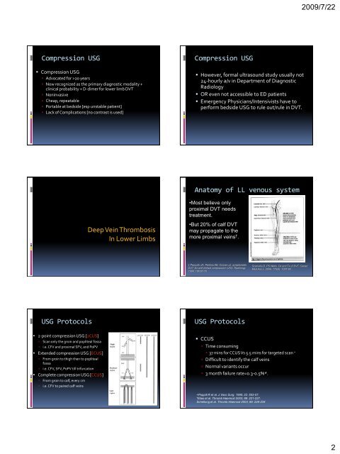
Anatomy <strong>of</strong> LL venous system<br />
•Most believe only<br />
proximal <strong>DVT</strong> needs<br />
treatment.<br />
Deep Vein Thrombosis<br />
In Lower Limbs<br />
•But 20% <strong>of</strong> calf <strong>DVT</strong><br />
may propagate to the<br />
more proximal veins † .<br />
† Pezzullo JA, Perkins AB, Cronan JJ: symptomatic<br />
<strong>DVT</strong>: Dx with limited compression USG. Radiology<br />
1996; 196:67-70<br />
Scarvelis D, PS Wells. Dx and Tx <strong>of</strong> <strong>DVT</strong>. Canad<br />
Med Ass L. 2006; 175(9): 1087-92.<br />
USG Protocols<br />
USG Protocols<br />
• 2-point compression USG [2CUS]<br />
Scan only the groin and popliteal fossa<br />
i.e. CFV and proximal SFV, and PoPV<br />
• Extended compression USG [ECUS]<br />
From groin to thigh then to popliteal<br />
fossa<br />
i.e. CFV, SFV, PoPV till trifurcation<br />
• Complete compression USG [CCUS]<br />
From groin to calf, every cm<br />
i.e. CFV to paired calf veins<br />
• CCUS<br />
Time consuming<br />
• 37 mins for CCUS Vs 5.5 mins for targeted scan +<br />
Difficult to identify the calf veins<br />
Normal variants occur<br />
3 month failure rate=0.3-0.5%*.<br />
+Poppiti R et al. J Vasc Surg. 1995; 22: 553-57.<br />
*Elias et al. Thromb Haemost 2003; 89: 221-227.<br />
Schellong et al. Thromb Haemost 2003; 89: 228-234<br />
2