DVT - Hong Kong College of Emergency Medicine
DVT - Hong Kong College of Emergency Medicine
DVT - Hong Kong College of Emergency Medicine

Create successful ePaper yourself
Turn your PDF publications into a flip-book with our unique Google optimized e-Paper software.
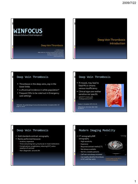
2009/7/22<br />
WINFOCUS<br />
Ultrasound Enhanced Critical Management<br />
Deep Vein Thrombosis<br />
Deep Vein Thrombosis<br />
Introduction<br />
Dr. Mok Ka Leung<br />
Associate Consultant<br />
MBBS, MRCS Ed., FHKCEM, FHKAM (<strong>Emergency</strong> <strong>Medicine</strong>)<br />
Accident and <strong>Emergency</strong> Department<br />
Ruttonjee and Tang Shiu Kin Hospital<br />
<strong>Hong</strong> <strong>Kong</strong> SAR, China<br />
Deep Vein Thrombosis<br />
• Thrombosis in the deep veins, esp in the<br />
lower limbs<br />
• 0.1% annual incidence in white population*<br />
• Frequent DDx to be ruled out in <strong>Emergency</strong><br />
care settings<br />
Deep Vein Thrombosis<br />
• If missed, may lead to<br />
fatal PE or chronic<br />
venous insufficiency<br />
• Clinical signs are neither<br />
sensitive nor specific<br />
• [50% <strong>DVT</strong> missed, 30%<br />
misdiagnosed as <strong>DVT</strong>]<br />
*White RH. The epidemiology <strong>of</strong> venous thromboembolism. Ciruclation 2003; 107<br />
(supp 1): I-4-I-8.<br />
Kakkar V. Circulation 1975; 51:8-19.<br />
Hillner B et al. Arch Intern Med 1992;<br />
52:165-75<br />
Deep Vein Thrombosis<br />
• Gold standard=contrast venography<br />
• Rarely performed because:<br />
Invasive, expensive<br />
Time consuming not 24 hourly a/v in most institutions<br />
Complications: anaphylaxis, inducing <strong>DVT</strong>, extravasation<br />
<strong>of</strong> contrast<br />
Non-diagnostic: around 1%<br />
Modern Imaging Modality<br />
• CT venography/MR<br />
venography<br />
High sensitivity<br />
Expensive<br />
Required contrast media [CT]<br />
Not always available<br />
Not for unstable patients<br />
Calf vein not always included<br />
Can readily identify thrombosis<br />
in IVC and iliac veins<br />
A clot is seen in the IVC in<br />
Contrast venography<br />
1
2009/7/22<br />
Compression USG<br />
• Compression USG<br />
Advocated for >20 years<br />
Now recognized as the primary diagnostic modality +<br />
clinical probability + D-dimer for lower limb <strong>DVT</strong><br />
Noninvasive<br />
Cheap, repeatable<br />
Portable at bedside [esp unstable patient]<br />
Lack <strong>of</strong> Complications [no contrast is used]<br />
Compression USG<br />
• However, formal ultrasound study usually not<br />
24-hourly a/v in Department <strong>of</strong> Diagnostic<br />
Radiology<br />
• OR even not accessible to ED patients<br />
• <strong>Emergency</strong> Physicians/Intensivists have to<br />
perform bedside USG to rule out/rule in <strong>DVT</strong>.<br />
Anatomy <strong>of</strong> LL venous system<br />
•Most believe only<br />
proximal <strong>DVT</strong> needs<br />
treatment.<br />
Deep Vein Thrombosis<br />
In Lower Limbs<br />
•But 20% <strong>of</strong> calf <strong>DVT</strong><br />
may propagate to the<br />
more proximal veins † .<br />
† Pezzullo JA, Perkins AB, Cronan JJ: symptomatic<br />
<strong>DVT</strong>: Dx with limited compression USG. Radiology<br />
1996; 196:67-70<br />
Scarvelis D, PS Wells. Dx and Tx <strong>of</strong> <strong>DVT</strong>. Canad<br />
Med Ass L. 2006; 175(9): 1087-92.<br />
USG Protocols<br />
USG Protocols<br />
• 2-point compression USG [2CUS]<br />
Scan only the groin and popliteal fossa<br />
i.e. CFV and proximal SFV, and PoPV<br />
• Extended compression USG [ECUS]<br />
From groin to thigh then to popliteal<br />
fossa<br />
i.e. CFV, SFV, PoPV till trifurcation<br />
• Complete compression USG [CCUS]<br />
From groin to calf, every cm<br />
i.e. CFV to paired calf veins<br />
• CCUS<br />
Time consuming<br />
• 37 mins for CCUS Vs 5.5 mins for targeted scan +<br />
Difficult to identify the calf veins<br />
Normal variants occur<br />
3 month failure rate=0.3-0.5%*.<br />
+Poppiti R et al. J Vasc Surg. 1995; 22: 553-57.<br />
*Elias et al. Thromb Haemost 2003; 89: 221-227.<br />
Schellong et al. Thromb Haemost 2003; 89: 228-234<br />
2
2009/7/22<br />
USG Protocols<br />
Hardware preparation<br />
• 2CUS/ECUS<br />
Easy & fast to perform- ili<strong>of</strong>emoral & popliteal<br />
regions are superficial<br />
Repeat if 1st USG –ve in high risk case<br />
EP are competent in performing 2-CUS +<br />
• Kappa=0.9, 98% agreement with vascular<br />
sonorgraphers<br />
• Median time=3 min 28 sec<br />
3-month failure rate was 0.7 to 2%*<br />
+Blaivas M et al. Acad Emerg Med 2000 7(2): 120-126<br />
*Ginsberg JS. N Eng J Med 1996; 335:1816-1828<br />
*Cogo A et al. Thromb Hemost 1995; 73:1098<br />
• High frequency linear transducer<br />
• 5-10MHz<br />
• Mainly B mode imaging<br />
• Colour flow / Pulse wave Doppler application<br />
optional<br />
• Adequate transonic Gel<br />
• Adjust TGC and Depth when scanning midthigh<br />
level [veins go deeper]<br />
Image Acquisition<br />
Image Acquisition<br />
• Patient in supine position with slightly<br />
externally rotation and flexion <strong>of</strong> hip<br />
• Transverse scan<br />
• Start just below the inguinal ligament<br />
• Identify the Common Femoral<br />
Vein [CFV] in cross section<br />
• It comes close with the<br />
Common Femoral Artery [CFA].<br />
• Apply firm and direct pressure<br />
to assess the compressibility <strong>of</strong><br />
the CFV<br />
• Then go distal 1cm by 1cm to<br />
assess the sapheno-femoral<br />
junction<br />
CFV<br />
CFA<br />
CFA<br />
Image Acquisition<br />
• Further distal movement <strong>of</strong><br />
probe to notice splitting <strong>of</strong> CFV<br />
into Superficial Femoral Vein<br />
[SFV] and Deep Femoral Vein<br />
[DFV].<br />
Image Acquisition<br />
• Put the transducer to the<br />
Popliteal fossa behind the knee to<br />
assess the Popliteal vein [PopV]<br />
• SFV is actually part <strong>of</strong> the deep<br />
vein system!<br />
• PoPV is above the popliteal artery<br />
at this point.<br />
• SFV goes medial and deep into<br />
muscle layer and turns<br />
posteriorly into the popliteal<br />
fossa in the adductor canal.<br />
• Need to use bimannual<br />
technique to compress the SFV<br />
at this point.<br />
• Assess the compressibility at<br />
least on the proximal 2cm <strong>of</strong> the<br />
vein and just distal to the<br />
trifurcation<br />
POP V<br />
POP A<br />
POP A<br />
3
2009/7/22<br />
Compressibility<br />
Compressibility<br />
• Apply firm and direct<br />
pressure in transverse scan<br />
• Probe must be<br />
perpendicular to the vein.<br />
• Normal compressibility<br />
Complete collapse <strong>of</strong> the vein<br />
as shown in the ultrasound<br />
monitor.<br />
Adequate pressure=Pressure<br />
just enough to deform the<br />
correspondent arteries<br />
• Pitfalls<br />
Pressure at wrong angles and vectors<br />
Difficult areas: adductor canal<br />
• need both hands<br />
Longitudinal scans<br />
• slip away from the vein!<br />
• better avoid it<br />
<strong>DVT</strong> features<br />
<strong>DVT</strong> features<br />
• Acute <strong>DVT</strong>:<br />
Fresh clots may not be visible,<br />
depending on the echogenicity.<br />
Only complete compressibility<br />
rule out <strong>DVT</strong><br />
Avoid excessive compression to<br />
prevent dislodgement <strong>of</strong> the<br />
clot<br />
risk <strong>of</strong> PE!<br />
An echogenic clot at the CFV<br />
Left SFV and Pop V failed to compress ie. <strong>DVT</strong> +ve. Also absence<br />
<strong>of</strong> colour flow doppler in these 2 viens<br />
<strong>DVT</strong> features<br />
<strong>DVT</strong> features<br />
Dilated left CFV and Pop V with echogenic intramural clots<br />
• Chronic <strong>DVT</strong>:<br />
Clots begin to organize within 5-<br />
10 days.<br />
Aging thrombi recannulize<br />
centrally and blood flow can be<br />
possible<br />
Complete compression is still<br />
not possible but near-complete<br />
collapse occur!<br />
Appears as thickened venous<br />
wall<br />
Longitudinal scan with colour<br />
flow doppler can help.<br />
4
2009/7/22<br />
Doppler Assessment<br />
Colour Flow Doppler<br />
• Not an essential examination for identifying<br />
<strong>DVT</strong><br />
• But provide more info about the blood flow<br />
within the vessels ie. Venous obstruction<br />
• Help to differentiate artery from vein<br />
• Colour flow doppler shows flow defect in<br />
thrombosed veins.<br />
• Pulse wave doppler shows absence <strong>of</strong> normal<br />
phasic changes and loss <strong>of</strong> augmentation.<br />
Normal and complete fill-up <strong>of</strong> the colour map in the<br />
Common Femoral Vein No <strong>DVT</strong> was found<br />
Colour Flow Doppler<br />
Pulse-wave Doppler<br />
FA<br />
CFV<br />
SFV<br />
Absence <strong>of</strong> flow in the CFV and SFV with echogenic clots<br />
Normal phasic changes and augmentation in the Common Femoral Vein<br />
Sensitivity and Specificity<br />
• However, compression<br />
only USG not 100%<br />
sensitive and specific.<br />
• Sensitivity falls when<br />
scanning calf veins: 30-<br />
70% †<br />
†Birdwell, Raskob GE, WhitsettTL, et al. The clinical validity <strong>of</strong> normal compression USG in outpatients<br />
suspected <strong>of</strong> having <strong>DVT</strong>. Ann Intern Med 1998; 128:1-7.<br />
Lensing AW, Prandoni P, Brandjes D et al. Detection <strong>of</strong> <strong>DVT</strong> by real time B mode USG. N Engl J med 1989; 320:<br />
342-5.<br />
Kearon C, Ginsberg JS, Hirsh J. The role <strong>of</strong> venous UGS in the Dx <strong>of</strong> suspected <strong>DVT</strong> and PE. Ann Intern Med 1998;<br />
129:1044-9.<br />
Sensitivity and Specificity<br />
• A recent Meta-analysis by Goodacre et al<br />
100 studies comparing all USG techniques Vs<br />
venography<br />
Sensitivity: 94.2% for proximal <strong>DVT</strong> and 63.5% for<br />
distal <strong>DVT</strong><br />
Specificity: 93.8% overall<br />
With duplex:<br />
• sensitivity 96% for proximal <strong>DVT</strong> and 71% for calf<br />
<strong>DVT</strong><br />
• Specificity 94% overall<br />
Goodacre S et al Health Technol Assess 2006; 10:168, iii-iv.<br />
5
2009/7/22<br />
EP performed Venous Scan<br />
Pitfalls<br />
• Burnside et al. did a systemic review <strong>of</strong><br />
emergency physician-performed USG for LL<br />
<strong>DVT</strong><br />
6 studies identified<br />
Criterion standard: Vascular<br />
sonorgrapher/radiologist performed USG.<br />
Pooled data analysis yielded overall sensitivitiy <strong>of</strong><br />
95% [CI=0.87-0.99] and specificity <strong>of</strong> 96%<br />
[CI=0.87-0.99].<br />
• Anatomical variation e.g. Double pop veins.<br />
32.5% have multiple SFV †<br />
42% have more than 1 POP veins in popliteal fossa;<br />
5% true duplications †<br />
• Technical aspect [Doppler may help]<br />
Groin Lymph Nodes: mistaken as a thrombosed<br />
vein<br />
Mistaken an artery as a vein with incompressibility<br />
Burnside et al Acad Emerg Med 2008; 15:6, 493-498.<br />
† Quinlan DJ, AAlikhan R, Gishen P Sidhu PS. Variations in Lower limb venous<br />
anatomy: implications for USG diagnosis <strong>of</strong> <strong>DVT</strong>. Rdaiology 2003; 228:443-448.<br />
Limitation <strong>of</strong> USG<br />
Common DDx<br />
• Acute on chronic <strong>DVT</strong><br />
Not accurate to differentiate old/new clots<br />
Need venography<br />
• Physical obstacles<br />
POP/Cast insitu<br />
Surgical emphysema/open laceration wound<br />
Iliac viens/IVC: relies on doppler flow assessment<br />
because compression is impossible<br />
• Cellulitis/subcutaneous abscess<br />
• Superficial thrombophlebitis<br />
• Calf muscle tear: medical head <strong>of</strong><br />
gastronemicus<br />
• Ruptured Baker’s cyst<br />
Cellulitis<br />
Superficial thrombophlebitis<br />
Non-compressibility and echogenic intramural<br />
clots <strong>of</strong> the superficial veins<br />
6
2009/7/22<br />
Gastrocnemius tear<br />
Gastrocnemius tear<br />
Transverse scan <strong>of</strong> the right medial<br />
gastrocnemius muscle tear with<br />
formation <strong>of</strong> haematoma<br />
Tear <strong>of</strong> right medial gastrocnemius<br />
muscle with formation <strong>of</strong> hypechoic<br />
collection <strong>of</strong> fluid between<br />
gastrocnemius and soleus muscles<br />
Normal gastrocnemius for comparison<br />
Ruptured Baker’s cyst<br />
Deep Vein Thrombosis<br />
In Upper Limbs<br />
Appearance <strong>of</strong> Baker’s cyst in MRI<br />
USG appearance <strong>of</strong> Baker’s cyst with sign <strong>of</strong><br />
rupture and dissection into the calf<br />
Upper Limb <strong>DVT</strong><br />
• Less common than lower limb <strong>DVT</strong><br />
• More common in patients with iv lines and<br />
malignancy<br />
Presence <strong>of</strong> a central venous catheter (72%)<br />
Infection (28%)<br />
Extra-thoracic malignancy (22%)<br />
Thoracic malignancy (21%)<br />
Renal failure (21%)<br />
Prior LL <strong>DVT</strong> (18%)<br />
Clinical Features<br />
• Predisposing factors:<br />
iv lines, Malignancy<br />
• Arm swelling, pain, heaviness<br />
• Dilated subcutaneous veins<br />
• Upper limb cyanosis<br />
• Can be asymptomatic<br />
Again clinical features are not specific!<br />
Marinella MA, Kathula SK, Markert RJ. Heart Lung. 2000 Mar-<br />
Apr;29(2):113-7. Spectrum <strong>of</strong> upper-extremity deep venous<br />
thrombosis in a community teaching hospital.<br />
7
2009/7/22<br />
Upper limb venous system<br />
Common Site <strong>of</strong> UL <strong>DVT</strong><br />
• Mainly Subclavian and/or Axillary Veins<br />
• Usually multiple site involvement<br />
Subclavian vein (18-69%)<br />
Axillary vein (5-42%)<br />
Internal jugular vein (8-29%)<br />
Brachial vein (4-13%)<br />
Scanning Protocol<br />
Diagnostic Criteria<br />
• Patient is placed supine and place the arm on<br />
bed.<br />
• Linear Transducer 5-7MHz<br />
• Start proximally from IJV, SV, AV, to BVs.<br />
• Compression Scan [same as LL scan] from<br />
Basilic and brachial veins to axillary veins.<br />
• Colour Doppler to assess the Subcalvian and<br />
Internal Jugular Veins<br />
• Same as LL Scan<br />
• Incomplete compressibility<br />
• Presence <strong>of</strong> clots<br />
• Loss <strong>of</strong> normal pulsatile flow pattern in the IJV<br />
and SV<br />
• Valsalva manoeuvre: widening <strong>of</strong> veins and<br />
reduction in flow<br />
• Sniff test: narrowing <strong>of</strong> vein and increase in flow<br />
• But flow abnormalities are only suggestive <strong>of</strong><br />
thrombosis need venography<br />
Normal Scan<br />
Abnormal Scan<br />
Echogenic clot in Internal Jugular Vein<br />
with loss <strong>of</strong> compressibility<br />
Normal Compressibility <strong>of</strong> Axillary Vein<br />
Normal Brachial Veins<br />
Echogenic clot in Brachial Vein with loss<br />
<strong>of</strong> compressibility<br />
8
2009/7/22<br />
Sensitivity and Specificity<br />
• Limited number <strong>of</strong> published reports on Sn<br />
and Sp <strong>of</strong> USG Vs Contrast Venography<br />
• Reported Sensitivity=78-100%<br />
• Reported Specificity=82-100%<br />
Diagnostic Pitfalls<br />
• USG not sensitive for <strong>DVT</strong> involving proximal<br />
Subcalvian viens and Innominate viens.<br />
need other modality <strong>of</strong> imaging<br />
Baarslag HJ et al. European Radiology 2004 14:1263-74.<br />
• Presence <strong>of</strong> collaterals mistaken as a normal vein,<br />
causing false –ve result.<br />
• Other DDx: Superficial vein thrombosis<br />
Any Questions?<br />
THANK YOU<br />
9