Emergency Ultrasound - Hong Kong College of Emergency Medicine
Emergency Ultrasound - Hong Kong College of Emergency Medicine
Emergency Ultrasound - Hong Kong College of Emergency Medicine

You also want an ePaper? Increase the reach of your titles
YUMPU automatically turns print PDFs into web optimized ePapers that Google loves.
<strong>Emergency</strong><br />
<strong>Ultrasound</strong><br />
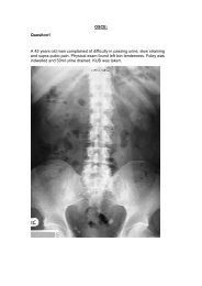
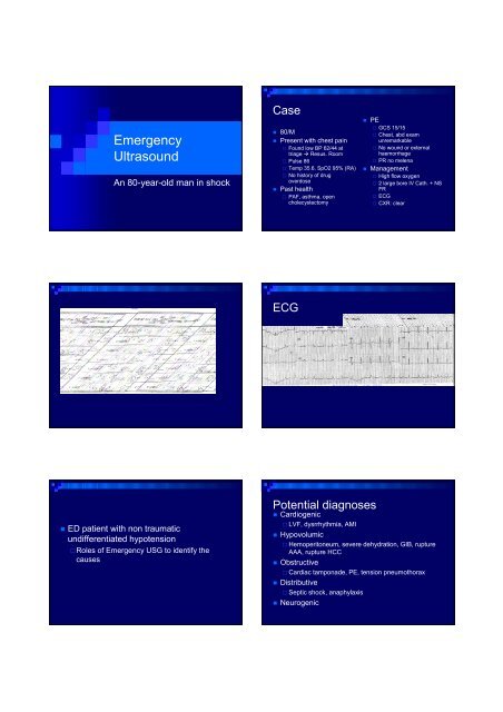
An 80-year-old man in shock<br />
Case<br />
80/M<br />
Present with chest pain<br />
Found low BP 62/44 at<br />
triage Resus. Room<br />
Pulse 86<br />
Temp 35.6, SpO2 95% (RA)<br />
No history <strong>of</strong> drug<br />
overdose<br />
Past health<br />
PAF, asthma, open<br />
cholecystectomy<br />
<br />
<br />
PE<br />
GCS 15/15<br />
Chest, abd exam<br />
unremarkable<br />
No wound or external<br />
haemorrhage<br />
PR no melena<br />
Management<br />
High flow oxygen<br />
2 large bore IV Cath. + NS<br />
FR<br />
ECG<br />
CXR: clear<br />
ECG<br />
ED patient with non traumatic<br />
undifferentiated hypotension<br />
Roles <strong>of</strong> <strong>Emergency</strong> USG to identify the<br />
causes<br />
Potential diagnoses<br />
Cardiogenic<br />
LVF, dysrrhythmia, AMI<br />
Hypovolumic<br />
Hemoperitoneum, severe dehydration, GIB, rupture<br />
AAA, rupture HCC<br />
Obstructive<br />
Cardiac tamponade, PE, tension pneumothorax<br />
Distributive<br />
Septic shock, anaphylaxis<br />
Neurogenic
Focused, goal-directed USG in non-traumatic,<br />
undifferentiated shock<br />
Aim: to find diagnoses that warrant specific treatments<br />
beyond fluid resuscitation<br />
Subcostal view:<br />
subcostal region <strong>of</strong> abdomen, pericardium, right ventricle diastolic collapse<br />
IVC view<br />
IVC diameter, collapsibility with inspiration (intravascular volume status)<br />
Parasternal long cardiac view<br />
LV function, pericardial effusion (qualitative estimate <strong>of</strong> LV function)<br />
Apical 4 chamber view<br />
Ventricular size estimate, (qualitative estimate <strong>of</strong> LV function)<br />
Abdominal aorta<br />
Aneurysm<br />
Hepatorenal recess view<br />
Free intraperitoneal fluid<br />
Pelvis and retrovesical area<br />
Fluid collections<br />
Right subcostal + subxiphoid view<br />
Free pericardial fluid or diastolic right ventricular<br />
collapse (tamponade/pericardial effusion)<br />
global cardiac function and chamber size<br />
R pleural effusion, free fluid in Morison's pouch,<br />
and free fluid in the R paracolic gutter.<br />
Liver mass<br />
IVC<br />
Subxiphoid view<br />
Size and resp. variations <strong>of</strong> proximal IVC<br />
CVP and fluid status<br />
Initial size and resp. variation not as helpful as<br />
the changes <strong>of</strong> these parameters in response<br />
to a fluid challenge serial monitoring<br />
Extreme: lack <strong>of</strong> respiratory variation in the<br />
IVC and hepatic veins (tamponade)<br />
Parasternal long axis<br />
Pericardial<br />
effusion/tamponade<br />
Dilated aortic root<br />
(
Suprasternal window<br />
For the arch <strong>of</strong> aorta<br />
Abdominal aorta<br />
Transverse view >3cm<br />
Intraluminal thrombus<br />
Splenorenal recess<br />
left pleural effusion, free fluid in the<br />
subphrenic space and splenorenal recess,<br />
and free fluid in the left paracolic gutter<br />
Pelvic/retro vesicle<br />
Free fluid in the anterior pelvis or cul-desac<br />
(pouch <strong>of</strong> Douglas).<br />
What is next?<br />
Recheck IVC after fluid challenge<br />
respiratory variation still present after IV<br />
fluid resuscitation<br />
No rash or petechiae over the body<br />
Erythema noted over left lower ankle<br />
Recent minor trauma to the left ankle at<br />
home a few days ago – kick to wooden<br />
furniture<br />
appea<br />
Dusky red<br />
appearance
scan over the lesions<br />
Skin and s<strong>of</strong>t tissue infection<br />
Focused question<br />
Can NF be differentiated from cellulitis by USG<br />
findings?<br />
Anatomical considerations<br />
Awareness <strong>of</strong> the proximity <strong>of</strong> adjacent structures<br />
Technique<br />
5–10 MHz linear array transducer<br />
Transducer pressure should be kept to a minimum<br />
systematically scanned in two perpendicular planes (short &<br />
long axis)<br />
compared with the contralateral position on the<br />
corresponding normal limb<br />
Limitation<br />
limitation being when the anatomic sites are difficult<br />
to ascertain (too deep) by US<br />
Normal Scan: short-axis sonogram <strong>of</strong> a normal arm.<br />
<br />
Images <strong>of</strong> cellulitis<br />
Non specific<br />
Diffuse infection,<br />
accumulation <strong>of</strong><br />
edema fluid<br />
Images <strong>of</strong> NF<br />
Studies on the use <strong>of</strong> bedside<br />
USG in the diagnosis <strong>of</strong> NF are<br />
limited.<br />
It should NOT be used to exclude<br />
the presence <strong>of</strong> NF.<br />
Characteristics<br />
diffuse thickening fascia<br />
a layer <strong>of</strong> fluid accumulation<br />
(anechoic fluid layer) >4mm in<br />
depth along the deep fascial layer<br />
Loculated abscesses can coexist
Progress<br />
Working diagnoses<br />
Septic shock, Necrotizing fasciitis<br />
Septic work up + IV Rocephin, ampicllin, cloxacillin<br />
Dopamine and adrenaline drips<br />
Consult O&T<br />
Needle aspirate over skin near lat maleollus <strong>of</strong> left ankle <br />
turbid fluid<br />
Book E OT from AED<br />
Consult ICU<br />
Echo: good LV/RV function, no dilated aorta, no pericardial<br />
effusion<br />
Intraoperative findings<br />
Left leg redness, bluish color involved medial<br />
and lateral malleolus as well as calf<br />
Fascia underneath lateral side is necrotic<br />
Turbid fluid++<br />
10% full thickness<br />
Left leg above knee amputation<br />
Excisional debridement <strong>of</strong> bil UL (Day 4)<br />
Spreading <strong>of</strong> NF to both arms after LL<br />
amputation<br />
Revision amputation and multiple<br />
debridement done<br />
Died day 21<br />
Microbiology results<br />
subcutaneous aspirate<br />
Vibrio vulnificus<br />
<br />
<br />
<br />
<br />
References<br />
<strong>Emergency</strong> ultrasound Ma, O. John.<br />
New York : McGraw-Hill Medical ; London : McGraw-Hill,<br />
2007.<br />
Jones AE. Tayal VS. Sullivan DM. Kline JA. Randomized,<br />
controlled trial <strong>of</strong> immediate versus delayed goal-directed<br />
ultrasound to identify the cause <strong>of</strong> nontraumatic hypotension<br />
in emergency department patients Critical Care <strong>Medicine</strong>.<br />
32(8):1703-8, 2004 Aug.<br />
Yen ZS. Wang HP. Ma HM. Chen SC. Chen WJ.<br />
Ultrasonographic screening <strong>of</strong> clinically-suspected necrotizing<br />
fasciitis. Academic <strong>Emergency</strong> <strong>Medicine</strong>. 9(12):1448-51,<br />
2002 Dec.<br />
Chao HC, <strong>Kong</strong> MS, Lin TY: Diagnosis <strong>of</strong> necrotizing fasciitis<br />
in children. J <strong>Ultrasound</strong> Med 18(4):277–281, 1999.