Large Intestine (Colon) - Axon
Large Intestine (Colon) - Axon
Large Intestine (Colon) - Axon

Create successful ePaper yourself
Turn your PDF publications into a flip-book with our unique Google optimized e-Paper software.
<strong>Large</strong> <strong>Intestine</strong> n <br />
Only polyps containing invasive adenocarcinoma require a<br />
decision for additional treatment on the part of the clinician.<br />
Adenoma, adenocarcinoma in situ, and even intramucosal<br />
adenocarcinoma lack metastatic capability and are<br />
considered adequately treated by polypectomy alone. 1,11,13,14,16<br />
As a result, some pathologists advocate modification of the<br />
nomenclature to account for clinical behavior and promulgate<br />
use of the term high-grade glandular dysplasia to<br />
encompass high-grade dysplasia, adenocarcinoma in situ,<br />
and even intramucosal adenocarcinoma. 10,14 Although<br />
the 1989 WHO guidelines accepted and defined two<br />
(low-grade, high-grade) or three (mild, moderate, severe)<br />
grades of dysplasia, adenocarcinoma in situ, and intramucosal<br />
adenocarcinoma, the authors of those guidelines<br />
recommended a similar behavior-based modification for<br />
intramucosal carcinoma and stated that “… intramucosal<br />
adenocarcinoma of the colon has not been shown to<br />
metastasize, and for this reason ‘carcinoma in situ’ is more<br />
appropriate.” 11<br />
The 2000 version of the WHO classification added little<br />
clarification and introduced new and even more confusing<br />
terms. 14 The authors stated that the defining feature of<br />
colorectal adenocarcinoma is invasion through the muscularis<br />
mucosae into the submucosa. However, once defined,<br />
worrisome lesions not fulfilling this criterion become difficult<br />
to describe. For example, the 2000 WHO classification<br />
defines adenocarcinoma in situ and intramucosal<br />
adenocarcinoma as lesions with morphologic characteristics<br />
of “adenocarcinoma” confined to the epithelium or that<br />
“invade” the lamina propria alone and lack invasion through<br />
the muscularis mucosae. The WHO goes on to state that<br />
these lesions have virtually no risk of metastasis. According<br />
to the WHO, the term “… high-grade intraepithelial neoplasia<br />
is more appropriate than adenocarcinoma in situ<br />
and … intramucosal neoplasia is more appropriate than<br />
intramucosal adenocarcinoma.” In the 2000 version, the<br />
WHO believes that use of these terms will help avoid<br />
overtreatment. 14<br />
The problems with this classification are many. The inaccurate<br />
use of the term invasion to describe lesions that are<br />
not by definition invasive carcinoma is confusing. The lesser<br />
lesion of high-grade intraepithelial neoplasia sounds worse<br />
than the term used to describe intramucosal adenocarcinoma<br />
(intramucosal neoplasia). Furthermore, all adenomas,<br />
strictly speaking, are intraepithelial neoplasia. An effort to<br />
achieve consensus (largely between Eastern [Japanese] and<br />
Western pathologists) 17-20 resulted in the Vienna classification<br />
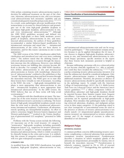
of gastrointestinal (GI) neoplasia, 20 presented in Table<br />
23-1.<br />
Problems with the Vienna system include the following:<br />
(1) inaccurate use of the word invasion; (2) category 4<br />
“noninvasive” high-grade neoplasia including potentially<br />
dangerous lesions (e.g., suspicious for invasive adenocarcinoma);<br />
and (3) category 5 “invasive neoplasms” including<br />
intramucosal adenocarcinoma, which is widely accepted to<br />
be clinically benign in the colon and rectum. It is unlikely<br />
that this system of categories without clinical correlation<br />
will ever gain widespread acceptance.<br />
a pragmatic view<br />
As modified from the 1989 WHO classification, low-grade<br />
dysplasia, high-grade dysplasia, adenocarcinoma in situ,<br />
TABLE 23-1<br />
Vienna Classification of Gastrointestinal Neoplasia<br />
Category<br />
Definition<br />
1 Negative for neoplasia/dysplasia<br />
2 Indefinite for neoplasia/dysplasia<br />
3 Noninvasive low-grade neoplasia (low-grade adenoma/<br />
dysplasia)<br />
4 Noninvasive high-grade neoplasia<br />
• High-grade adenoma/dysplasia<br />
• Noninvasive carcinoma (carcinoma in situ)<br />
• Suspicious for invasive carcinoma<br />
5 Invasive neoplasia<br />
• Intramucosal carcinoma<br />
• Submucosal carcinoma or beyond<br />
and intramucosal adenocarcinoma exist and can be recognized<br />
by pathologists. 1,11 This nomenclature remains attractive<br />
because it can be applied throughout the GI tract. If<br />
one chooses to diagnose high-grade dysplasia, adenocarcinoma<br />
in situ, and intramucosal adenocarcinoma in colorectal<br />
biopsy specimens, specific mention in the report<br />
that these lesions lack metastatic potential is helpful to<br />
clinicians.<br />
Because infiltrating carcinoma cells in a colorectal polyp<br />
do not become clinically significant (i.e., able to metastasize)<br />
until they have invaded the submucosa, 1,14-16,21-48 only<br />
a polyp containing invasive adenocarcinoma (invasion of at<br />
least the submucosa) should be considered malignant. Only<br />
invasive adenocarcinoma requires a decision regarding<br />
additional treatment. Therefore, the presence or absence of<br />
invasive adenocarcinoma should be specifically mentioned<br />
in the pathology report. To comply with the American<br />
College of Gastroenterology (ACG), the U.S. Multi-Society<br />
Task Force on Colorectal Cancer, and the American Cancer<br />
Society guidelines, 13,28,49 a villous component (villous or<br />
tubulovillous adenoma) and high-grade dysplasia should<br />
be reported because these features require more frequent<br />
surveillance. Carcinoma in situ and intramucosal adenocarcinoma<br />
can be reported parenthetically as high-grade<br />
dysplasia. Most mistakes that pathologists make in reporting<br />
colorectal adenomas, dysplasia, and malignant polyps<br />
occur in three major categories: (1) the pathology report is<br />
not clear (nonspecific or noncommittal terms are used or<br />
the presence or absence of invasive adenocarcinoma is not<br />
clearly stated); (2) mispositioned glands (pseudocarcinomatous<br />
invasion) are misinterpreted as invasive adenocarcinoma;<br />
and (3) the margin of excision is either not<br />
identified or not discussed.<br />
Malignant Polyps<br />
differential diagnosis<br />
A common problem concerns differentiating invasive carcinoma<br />
complicating a colorectal adenoma from pseudocarcinomatous<br />
invasion (pseudoinvasion). Pseudoinvasion<br />
describes a situation in which neoplastic glands of the<br />
adenoma are mispositioned, presumably by trauma, into or<br />
beneath the muscularis mucosae. 50-58 Pseudoinvasion is<br />
relatively common, reported in 3% to 10% of resected<br />
colorectal polyps. 50,52,53 Distinguishing this epithelial mis-