Bohra nullarbora sp. nov., a second tree-kangaroo - Murdoch ...
Bohra nullarbora sp. nov., a second tree-kangaroo - Murdoch ...
Bohra nullarbora sp. nov., a second tree-kangaroo - Murdoch ...
You also want an ePaper? Increase the reach of your titles
YUMPU automatically turns print PDFs into web optimized ePapers that Google loves.
174 G. Prideaux, N. Warburton<br />
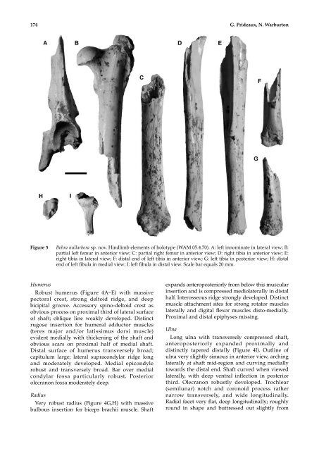
Figure 5 <strong>Bohra</strong> <strong>nullarbora</strong> <strong>sp</strong>. <strong>nov</strong>. Hindlimb elements of holotype (WAM 05.4.70). A: left innominate in lateral view; B:<br />
partial left femur in anterior view; C: partial right femur in anterior view; D: right tibia in anterior view; E:<br />
right tibia in lateral view; F: distal end of left tibia in anterior view; G: left tibia in posterior view; H: distal<br />
end of left fibula in medial view; I: left fibula in distal view. Scale bar equals 20 mm.<br />
Humerus<br />
Robust humerus (Figure 4A–E) with massive<br />
pectoral crest, strong deltoid ridge, and deep<br />
bicipital groove. Accessory <strong>sp</strong>ino-deltoid crest as<br />
obvious process on proximal third of lateral surface<br />
of shaft; oblique line weakly developed. Distinct<br />
rugose insertion for humeral adductor muscles<br />
(teres major and/or latissimus dorsi muscle)<br />
evident medially with thickening of the shaft and<br />
obvious scars on proximal half of medial shaft.<br />
Distal surface of humerus transversely broad;<br />
capitulum large; lateral supracondylar ridge long<br />
and moderately developed. Medial epicondyle<br />
robust and transversely broad. Bar over medial<br />
condylar fossa particularly robust. Posterior<br />
olecranon fossa moderately deep.<br />
Radius<br />
Very robust radius (Figure 4G,H) with massive<br />
bulbous insertion for biceps brachii muscle. Shaft<br />
expands anteroposteriorly from below this muscular<br />
insertion and is compressed mediolaterally in distal<br />
half. Interosseous ridge strongly developed. Distinct<br />
muscle attachment sites for strong rotator muscles<br />
laterally and digital flexor muscles disto-medially.<br />
Proximal and distal epiphyses missing.<br />
Ulna<br />
Long ulna with transversely compressed shaft,<br />
anteroposteriorly expanded proximally and<br />
distinctly tapered distally (Figure 4I). Outline of<br />
ulna very slightly sinuous in anterior view, arching<br />
laterally at shaft mid-region and curving medially<br />
towards the distal end. Shaft curved when viewed<br />
laterally, with deep ventral inflection in posterior<br />
third. Olecranon robustly developed. Trochlear<br />
(semilunar) notch and coronoid process rather<br />
narrow transversely, and wide longitudinally.<br />
Radial facet very flat, deep longitudinally; roughly<br />
round in shape and buttressed out slightly from


