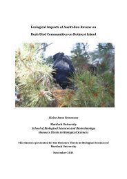
Bohra nullarbora sp. nov., a second tree-kangaroo - Murdoch ...
Bohra nullarbora sp. nov., a second tree-kangaroo - Murdoch ...
Bohra nullarbora sp. nov., a second tree-kangaroo - Murdoch ...

Create successful ePaper yourself
Turn your PDF publications into a flip-book with our unique Google optimized e-Paper software.
Records of the Western Australian Museum 25: 165–179 (2009).<br />
<strong>Bohra</strong> <strong>nullarbora</strong> <strong>sp</strong>. <strong>nov</strong>., a <strong>second</strong> <strong>tree</strong>-<strong>kangaroo</strong> (Marsupialia:<br />
Macropodidae) from the Pleistocene of the Nullarbor Plain,<br />
Western Australia<br />
Gavin J. Prideaux 1,3 and Natalie Warburton 2,3<br />
1<br />
School of Biological Sciences, Flinders University, Bedford Park, South Australia 5042, Australia.<br />
E-mail: gavin.prideaux@flinders.edu.au<br />
2<br />
School of Veterinary & Biomedical Sciences, <strong>Murdoch</strong> University, <strong>Murdoch</strong>, Western Australia 6150, Australia.<br />
E-mail: n.warburton@murdoch.edu.au<br />
3<br />
Department of Earth and Planetary Sciences, Western Australian Museum, Welshpool,<br />
Western Australia 6106, Australia.<br />
Abstract – <strong>Bohra</strong> <strong>nullarbora</strong> <strong>sp</strong>. <strong>nov</strong>. is described from a partial skeleton<br />
collected from a diverse Pleistocene vertebrate assemblage preserved in<br />
Leaena’s Breath Cave, Nullarbor Plain, Western Australia. It is distinguished<br />
from its Nullarbor contemporary, B. illuminata, by having different cranial<br />
proportions, smaller cheek teeth and a relatively narrower upper premolar.<br />
It also differs in a number of postcranial attributes, which may reflect slight<br />
variation in locomotory capabilities. The unexpected discovery that, in the<br />
relatively recent geological past, two large arboreal <strong>kangaroo</strong>s inhabited<br />
the now ‘Treeless’ Plain effectively highlights how little we still know about<br />
the Pleistocene history of Western Australia, and of the drier regions of the<br />
continent in general.<br />
INTRODUCTION<br />
Extant <strong>tree</strong>-<strong>kangaroo</strong>s (Dendrolagus) inhabit the<br />
forests of far northeastern Queensland and New<br />
Guinea (Flannery 1990; Strahan 1995). Their ancestors<br />
appear to have evolved from terrestrial <strong>kangaroo</strong>s<br />
some time in the latter part of the Miocene (Flannery<br />
1989). Tree-<strong>kangaroo</strong>s are rarely preserved, or at least<br />
have been rarely recognised, as fossils. Flannery<br />
and Szalay (1982) described a large new genus and<br />
<strong>sp</strong>ecies of <strong>tree</strong>-<strong>kangaroo</strong> (<strong>Bohra</strong> paulae) from the<br />
Wellington Caves in central eastern New South<br />
Wales (Figure 1) on the basis of some hind limb<br />
elements. Craniodental material from the Pliocene of<br />
Chinchilla in Queensland was referred to the same<br />
genus by Dawson (2004), a decision later supported<br />
when the first of the new <strong>sp</strong>ecies, B. illuminata,<br />
was described by Prideaux and Warburton (2008)<br />
from Pleistocene deposits in the Thylacoleo Caves,<br />
Nullarbor Plain, Western Australia (Prideaux et al.<br />
2007; Figure 1). The holotype of B. illuminata consists<br />
of much of one skeleton, including the complete<br />
cranium. Hocknull (2005a,b) recorded remains<br />
referable to the genera <strong>Bohra</strong> and Dendrolagus<br />
from the Mt Etna cave-fills (near Rockhampton in<br />
Queensland) of middle Pleistocene age (Hocknull<br />
et al. 2007). Undescribed Pleistocene <strong>tree</strong>-<strong>kangaroo</strong><br />
elements are also known from Curramulka Quarry,<br />
Yorke Peninsula, South Australia, and the eastern<br />
Darling Downs (pers. obs.; Figure 1).<br />
This paper describes the holotype and only<br />
known <strong>sp</strong>ecimen of <strong>Bohra</strong> <strong>nullarbora</strong> <strong>sp</strong>. <strong>nov</strong>., a<br />
partial skeleton from Leaena’s Breath Cave, one<br />
of the three caves near the centre of the Nullarbor<br />
Plain known collectively as the Thylacoleo Caves<br />
(Figure 1). The caves contain well-preserved<br />
skeletal remains of a diverse middle Pleistocene<br />
vertebrate fauna (Prideaux et al. 2007), including<br />
eight <strong>kangaroo</strong> <strong>sp</strong>ecies entirely new to science. An<br />
overview of the significance of the sites and likely<br />
palaeoenvironment was provided by Prideaux et<br />
al. (2007). Analyses of the functional morphology<br />
of the forelimb and hind limb, which confirm the<br />
arboreal aptitude of the Nullarbor <strong>sp</strong>ecies of <strong>Bohra</strong>,<br />
are presented in Harvey (2006) and Warburton<br />
and Prideaux (in press), re<strong>sp</strong>ectively. Refer to<br />
Prideaux and Warburton (2008) for a more detailed<br />
comparison of <strong>Bohra</strong> and Dendrolagus.<br />
The holotype of B. <strong>nullarbora</strong> is registered<br />
in the vertebrate palaeontological collections<br />
of the Western Australian Museum, Perth<br />
(abbreviation WAM). Description style, terminology<br />
and mensuration follow Prideaux (2004) for the<br />
craniodental system, and Owen (1876), Murray<br />
(1995), Wells and Tedford (1995), the Nomina
166 G. Prideaux, N. Warburton<br />
Anatomica Veterinaria (2005), Weisbecker<br />
and Sánchez-Villagra (2006) and Prideaux and<br />
Warburton (2008) for the postcranial skeleton.<br />
Serial designation of the cheek dentition follows<br />
Flower (1867) and Luckett (1993). Upper teeth are<br />
designated by upper case abbreviations (e.g. P3,<br />
M2); lower teeth are designated by lower case<br />
abbreviations (e.g. i1, m3).<br />
SYSTEMATIC PALAEONTOLOGY<br />
Diprotodontia Owen, 1866<br />
Superfamily Macropodoidea (Gray, 1821)<br />
Family Macropodidae Gray, 1821<br />
Subfamily Macropodinae Gray, 1821<br />
Tribe Dendrolagini Flannery, 1989<br />
Genus <strong>Bohra</strong> Flannery & Szalay, 1982<br />
Type <strong>sp</strong>ecies<br />
<strong>Bohra</strong> paulae Flannery and Szalay, 1982.<br />
Material examined<br />
<strong>Bohra</strong> <strong>nullarbora</strong> <strong>sp</strong>. <strong>nov</strong>.<br />
Figures 2–6, Tables 1–3<br />
Figure 1<br />
Map showing Australian localities yielding<br />
Pleistocene <strong>tree</strong>-<strong>kangaroo</strong>s. 1, Thylacoleo Caves,<br />
middle Pleistocene, <strong>Bohra</strong> illuminata and B.<br />
<strong>nullarbora</strong>; 2, Curramulka Quarry, ?Pleistocene,<br />
<strong>Bohra</strong> <strong>sp</strong>. indet. (pers. obs.); 3, Wellington Caves,<br />
?Pleistocene, <strong>Bohra</strong> paulae (Flannery and Szalay,<br />
1982); 4, eastern Darling Downs, <strong>Bohra</strong> <strong>sp</strong>. indet.<br />
(pers. obs.); 5, Mt Etna cave-fills, <strong>Bohra</strong> <strong>sp</strong>.<br />
indet., Dendrolagus <strong>sp</strong>. indet., middle Pleistocene<br />
(Hocknull 2005a,b; Hocknull et al. 2007).<br />
Holotype<br />
Australia: Western Australia: WAM 05.4.70,<br />
partial adult cranium, partial left and right<br />
dentaries; vertebrae (atlas, axis, cervical 3–5,<br />
thoracic 1, ?5–6, ?10, ?12, dorsal lumbar fragment,<br />
two mid-caudal and two distal-caudal vertebrae,<br />
1 mid-caudal chevron), left and right clavicles, six<br />
ribs, humerus (left proximal and distal fragments,<br />
right partial diaphysis), left ulna, left radius<br />
(diaphysial fragment), carpals (right hamatum, left<br />
scaphoid), metacarpals (left and right III, left IV–V),<br />
manual phalanges (digits I–IV proximal; digits<br />
III–V medial; digits III–IV distal), innominates<br />
(partial left; fragments of right), left epipubic,<br />
femur (proximal portion of left, abraded distal<br />
fragments of right), tibia (right diaphysis, left distal<br />
diaphysis fragments), fibula (distal portion), left<br />
calcaneus, talus and navicular, right entocuneiform,<br />
metatarsals IV–V, pedal phalanges (digit II, IV–V<br />
proximal, digit II–V medial; digits IV–V distal)<br />
(Figure 1; Tables 1–2).<br />
Type locality and age<br />
The holotype was collected by P. D. Devine<br />
and G. J. Prideaux in July 2002 from the floor of<br />
Leaena’s Breath Cave, Nullarbor Plain, southeastern<br />
Western Australia (Figure 1). Precise location and<br />
site details are registered with the Department of<br />
Earth and Planetary Sciences, Western Australian<br />
Museum, Perth. Fossils from the cave-floor and<br />
upper sediment unit of Leaena’s Breath Cave are<br />
probably early middle Pleistocene in age (Prideaux<br />
et al. 2007).<br />
Diagnosis<br />
Distinguished from <strong>Bohra</strong> paulae by having<br />
smaller calcaneus with tuber calcanei less barrelshaped,<br />
partial separation of posterior calcanealtalar<br />
facets, longer sustentaculum, shorter plantar<br />
sulcus, calcaneal-cuboid facets flat rather than<br />
V-shaped in transverse plane, smaller, relatively<br />
longer talus, narrower trochlea, higher lateral<br />
trochlear crest of talus.<br />
Distinguished from <strong>Bohra</strong> wilkinsonorum by<br />
having distinctly smaller cheek teeth and by<br />
lacking posterobuccal accessory cu<strong>sp</strong>, thin low<br />
buccal cingulum and thick lingual cingulum on<br />
P3, and distinct lingual ‘hook’ of postparacrista on<br />
upper molars.<br />
Distinguished from <strong>Bohra</strong> illuminata on the basis<br />
of the following craniodental attributes. Incisorbearing<br />
portion of premaxilla deep; buccinator<br />
fossa shallow; narial aperture near-circular;
Pleistocene <strong>tree</strong>-<strong>kangaroo</strong> from Nullarbor Plain 167<br />
Table 1<br />
Dimensions (in mm) of the cranium and dentary of the holotype of <strong>Bohra</strong><br />
<strong>nullarbora</strong> <strong>sp</strong>. <strong>nov</strong>. (WAM 05.4.70) compared with those of B. illuminata<br />
(WAM 03.5.10).<br />
Dimension B. <strong>nullarbora</strong> B. illuminata<br />
Width across zygomatic arches 79.5 75.2<br />
Width across frontals ca 29.7 23.8<br />
Distance between masseteric processes ca 49.4 48.1<br />
Palatal width at M1 protoloph 20.9 18.6<br />
Palatal width at M4 protoloph 23.4 17.8<br />
Dentary depth at m2–3 18.3 14.4<br />
Dentary width at m2–3 8.23 7.83<br />
masseteric process tiny; supraorbital crest distinct,<br />
positioned above posterior portion of orbit, with<br />
distinct sulcus beneath; zygomatic arch moderately<br />
deep; ectoglenoid process rather wide; postglenoid<br />
process ventrally projected. Cheek teeth smaller<br />
relative to size of cranium; P3 wider anteriorly than<br />
posteriorly, bearing small, low posterolingual cu<strong>sp</strong><br />
and narrow lingual cingulum; buccal enamel on<br />
i1 forms quite thick dorsal flange, posterior end<br />
of ventral enamel flange very thin; lower molars<br />
bear low paracristid and premetacristid, with<br />
latter extending to centre of trigonid basin, small<br />
precingulid, and very low cristid obliqua.<br />
Distinguished from <strong>Bohra</strong> illuminata on the<br />
basis of the following postcranial attributes.<br />
Clavicle more robust and less flattened, with<br />
expanded articular facets and more laterally<br />
positioned anterior inflection. Humerus more<br />
robust with stronger pectoral crest, deltoid ridge,<br />
bicipital groove and <strong>sp</strong>ino-deltoid insertion, but<br />
Table 2<br />
Cheek tooth dimensions (in mm) of the holotype of <strong>Bohra</strong> <strong>nullarbora</strong> <strong>sp</strong>.<br />
<strong>nov</strong>. (WAM 05.4.70) compared with those of B. illuminata (WAM 03.5.10).<br />
Abbreviations: L = length, AW = anterior width, PW = posterior width,<br />
AH = anterior height, PH = posterior height.<br />
Species Tooth L AW PW AH PH<br />
B. <strong>nullarbora</strong> P3 8.93 4.42 4.14 4.23 3.61<br />
M1 7.39 6.44 6.92 - -<br />
M2 8.38 7.39 7.07 - -<br />
M3 8.64 7.94 6.62 3.74 3.68<br />
M4 8.57 7.14 5.67 3.86 4.17<br />
B. illuminata P3 9.40 4.02 5.24 5.69 5.27<br />
M1 7.17 6.99 6.69 - -<br />
M2 8.82 7.50 6.93 4.05 4.65<br />
M3 9.41 7.81 7.04 4.40 4.74<br />
M4 10.00 7.65 6.49 4.68 5.25<br />
B. <strong>nullarbora</strong> p3 - - - - -<br />
m1 6.94 4.57 4.58 - -<br />
m2 7.90 5.44 5.47 - -<br />
m3 9.12 6.03 6.08 3.65 4.32<br />
m4 8.51 6.04 5.47 3.76 -<br />
B. illuminata p3 - - - - -<br />
m1 - 4.61 5.14 - -<br />
m2 8.34 5.58 5.71 3.67 4.61<br />
m3 10.65 6.56 6.81 5.30 5.87
168 G. Prideaux, N. Warburton<br />
Table 3<br />
Dimensions (in mm) of postcranial elements of the holotype of <strong>Bohra</strong> <strong>nullarbora</strong><br />
<strong>sp</strong>. <strong>nov</strong>. (WAM 05.4.70) compared with those of B. illuminata (WAM 03.5.10) and<br />
B. paulae (AM F62099–F62101). Abbreviations: C, cervical; est., estimated; H,<br />
height; L, length; W, width; T, thoracic.<br />
Element<br />
<strong>Bohra</strong><br />
<strong>nullarbora</strong><br />
<strong>Bohra</strong><br />
illuminata<br />
<strong>Bohra</strong> paulae<br />
VERTEBRAE<br />
C1 (width across postzygapophyses) 50.6 25.8<br />
C2 (centrum dimensions)<br />
9.2x6.8<br />
C2 (height including <strong>sp</strong>ine) 29<br />
C2 (Ventral length including odontoid process) 23.4<br />
C3,4 (posterior centrum width) 8.9, 9.3<br />
C3,4 (centra length) 13.8, 12.5<br />
T1 (anterior centra dimensions) 9.6x6.3 8.1x5.7<br />
T1 (centrum length) 10.7 10.6<br />
T?5,6 (centra length) 12.7, 12.8<br />
T?10 (centrum length) 13.8<br />
Mid-caudals (centra length) 45.8, 47.2<br />
Distal caudal (centrum length) 34.8<br />
CLAVICLE<br />
Length 49.5 42.4<br />
HUMERUS<br />
Maximum length (est.) 110 105<br />
Maximum distal width 39 37.4<br />
Width distal articular surface 21.8 25.5<br />
RADIUS<br />
Dimensions mid-shaft (HxW)<br />
9.2x7.2<br />
ULNA<br />
Maximum length (est.) 150 140<br />
Anteroposterior length at coronoid fossa 13.5 13.4<br />
Transverse width at coronoid fossa 8.7 9.0<br />
METACARPALS<br />
Metacarpal III length (left, right) 28.4, 28.1 22.9<br />
Metacarpal IV length 25.3 18.8<br />
Metacarpal V length 20.3 15.7<br />
CARPALS<br />
Hamatum maximum length 10.5 12.4<br />
Hamatum maximum width 14.5 7.8<br />
Hamatum maximum height 11.3 8.6<br />
Scaphoid maximum length 9.1<br />
Scaphoid maximum width 18<br />
Scaphoid maximum height 6.3<br />
INNOMINATE<br />
Sacroiliac articular surface width 26.3<br />
Acetabular fossa (LxW) 23.5x23.4 21.8x24.2<br />
EPIPUBIC<br />
Maximum length 78.2 62<br />
Articular surface (LxW) 11.7x6.3 – x16.6<br />
FEMUR<br />
Maximum diaphysis length 150
Pleistocene <strong>tree</strong>-<strong>kangaroo</strong> from Nullarbor Plain 169<br />
Table 3 (continued)<br />
Element<br />
<strong>Bohra</strong><br />
<strong>nullarbora</strong><br />
<strong>Bohra</strong><br />
illuminata<br />
<strong>Bohra</strong> paulae<br />
Articular head (LxW)<br />
23.0x21.5<br />
Mid-shaft dimensions (HxW)<br />
17.4x18.8<br />
TIBIA<br />
Maximum diaphysis length (est.) 175 183<br />
FIBULA<br />
Proximal epiphysis<br />
CALCANEUS<br />
Maximum length 43.4 36.1<br />
Maximum width 27.7 23.7<br />
Depth tuber calcanei 16.5 13.7<br />
Talar articulation width 22 19.9<br />
Dorsomedial facet width 10.9 10.0<br />
Dorsolateral facet width 6.8 6.2<br />
Width ventromedian facet 8.4 8.2<br />
TALUS<br />
Maximum length 24.2 21.0<br />
Maximum width 28.7 26.1<br />
Transverse distance between trochleas 15.2 14.3<br />
Navicular facet (head) 11.0x9.7 10.6x9.1<br />
NAVICULAR<br />
Maximum height 13.3<br />
Length of dorsal surface 16.1<br />
Dorsal transverse width 9.6<br />
METATARSAL IV<br />
Diaphysis length 68.6<br />
Proximal transverse width 16.2<br />
Distal transverse width (epiphysis) 14.9<br />
Metatarsal V facet<br />
10.4x5.1<br />
Sesamoid facet<br />
7.0x5.0<br />
METATARSAL V<br />
Maximum shaft length 60.1<br />
Proximal transverse width 13.9<br />
Distal transverse width 11.6<br />
PHALANGES<br />
Proximal phalanx IV length 33.6<br />
Proximal phalanx IV mid-width 10.7<br />
Medial phalanx IV length 23.6<br />
Medial phalanx IV mid-width 9.5<br />
Distal phalanx IV length 29.4<br />
Distal phalanx IV mid-width 10.5<br />
Proximal phalanx V length 24.1<br />
Proximal phalanx V mid-width 7.1<br />
Medial phalanx V length 15.4<br />
Medial phalanx V mid-width 6.7<br />
Distal phalanx V length 21.4<br />
Distal phalanx V width 8.2
170 G. Prideaux, N. Warburton<br />
Figure 2<br />
<strong>Bohra</strong> <strong>nullarbora</strong> <strong>sp</strong>. <strong>nov</strong>. Cranium and dentaries of holotype (WAM 05.4.70). A: neurocranium in dorsal view;<br />
B: left squamosal and ali<strong>sp</strong>henoid in ventral view; C: neurocranium in lateral view; D: left premaxilla in lateral<br />
view; E: left maxilla in lateral view; F: Stereopair of palate in occlusal view; G: partial right dentary in<br />
lateral view; H: partial right dentary in mesial view; I: right i1 in lingual view; J: right i1 in buccal view; K:<br />
stereopair of partial right dentary in occlusal view; L: stereopair of left dentary in occlusal view; M: left dentary<br />
in mesial view; N: left dentary in lateral view. Scale bar equals 20 mm.
Pleistocene <strong>tree</strong>-<strong>kangaroo</strong> from Nullarbor Plain 171<br />
Figure 3<br />
Craniodental features of <strong>Bohra</strong> illuminata that<br />
differentiate it from B. <strong>nullarbora</strong> <strong>sp</strong>. <strong>nov</strong>.<br />
with lower teres major insertion; distal articular<br />
surface (e<strong>sp</strong>ecially capitulum) relatively narrow.<br />
Ilium relatively broader anteroposteriorly, with<br />
medial border more sinuous; marked oval fossa<br />
contiguous with rugose muscle scar of M. rectus<br />
femoris dorsal to acetabulum. Epipubic very<br />
large. Calcaneus larger with partial separation of<br />
posterior calcaneal-talar facets, shorter anterior<br />
plantar sulcus, better developed plantar tuberosity,<br />
wider sustentaculum tali, and lateral talar facet<br />
of calcaneus relatively narrow and less tapered<br />
mesially. Talus relatively longer with higher lateral<br />
trochlear ridge. Metatarsal IV more robust with<br />
larger cuboid facet, less medially constricted facet<br />
for metatarsal V and less rounded, less distally<br />
oriented sesamoid facet. Metatarsal V more robust.<br />
Etymology<br />
The <strong>sp</strong>ecies refers to the region of origin, the<br />
Nullarbor Plain.<br />
Description<br />
Cranium<br />
Incisor-bearing portion of premaxilla short and<br />
deep; upright portion essentially vertical. Anterior<br />
edge of premaxilla gently curved in lateral view<br />
(Figure 2D). Diastema short, straight and deflected<br />
anteroventrally relative to cheek tooth row; maxilla<br />
contributes to most of diastema length. Central<br />
region of diastema not preserved. Most of incisive<br />
foramina not preserved, but anterior remnants<br />
suggest they were broad, and extended anteriorly to<br />
adjacent to midpoint on I3 alveolus. Most of rostrum<br />
not preserved, but lower portion of lateral surface<br />
of left maxilla reveals shallow, poorly demarcated<br />
posterior half of buccinator fossa (Figure 2E–F).<br />
Mesial portions of premaxillae not preserved, but<br />
narial aperture apparently near-circular. Masseteric<br />
process not preserved on either side of cranium, but<br />
enough of maxilla preserved beneath right orbit to<br />
indicate process was tiny and positioned adjacent<br />
to M2–3 abutment (Figure 2E–F). Infraorbital<br />
foramen opens anteriorly; positioned directly above<br />
posterior root of P3. Lacrimals, jugals, nasals and<br />
anterior half of frontals not preserved.<br />
Posterior part of frontal slightly concave on<br />
dorsal surface and bears distinct, but incomplete,<br />
supraorbital crest above posterior portion of<br />
orbit (Figure 2A). Distinct sulcus formed beneath<br />
supraorbital crest. Palatine bones well developed;<br />
palate non-fenestrate except for two small foramina<br />
on maxilla-palatine suture, adjacent to M3<br />
protoloph (Figure 2F). Palate terminates posteriorly<br />
adjacent to M4 metaloph. Temporal (parietal)<br />
crests weakly developed, confluent anteriorly<br />
with supraorbital crests (Figure 2A). Interparietal,<br />
occiput and basicranium not preserved, except<br />
for left ali<strong>sp</strong>henoid, which is markedly extended<br />
posteriorly, evidently to paroccipital process (Figure<br />
2B). Dorsal surface of neurocranium domed (Figure<br />
2C). Zygomatic arch moderately deep; jugal not<br />
preserved, but shallow jugal fossa on squamosal<br />
reflects rather wide ectoglenoid process (Figure 2B).<br />
Zygomatic process of squamosal arises well anterior<br />
of occiput. Large, ventrally projected postglenoid<br />
process forms posterior border of glenoid fossa<br />
(Figure 2C). Ectotympanic missing. Subsquamosal<br />
foramen large; positioned adjacent to posterior<br />
extremity of zygomatic process of squamosal.<br />
Upper Dentition<br />
P3 crown oriented anterodorsally at 20° to<br />
palatal plane (Figure 2E). P3 longer than all molars.<br />
Crown is wider anteriorly than posteriorly due<br />
to rounded eminence on buccal side, absence of<br />
posterobuccal accessory cu<strong>sp</strong>, and small, low nature<br />
of posterolingual cu<strong>sp</strong> (Figure 2F). Main crest<br />
straight and oriented along longitudinal axis of<br />
tooth. Posterior basin very poorly demarcated. Very<br />
low, rounded lingual cingulum extends smoothly<br />
from immediately anterior to posterolingual cu<strong>sp</strong><br />
to anterior end of crown. Buccal cingulum absent.<br />
Main crest composed of four connected cu<strong>sp</strong>ules,<br />
with anteriormost and posteriormost more distinct<br />
(Figure 2E). Two very weak vertical ridgelets lie<br />
between slightly larger eminences corre<strong>sp</strong>onding to<br />
anteriormost and posteriormost cu<strong>sp</strong>ules on buccal<br />
surface of tooth.<br />
Upper molars low crowned. M1 very worn,<br />
dentine of protoloph, metaloph and postprotocrista
172 G. Prideaux, N. Warburton<br />
breached and continuous; M2 worn, dentine of<br />
protoloph and metaloph crests breached; M3<br />
crests moderately worn, dentine breached only at<br />
protocone apex; M4 crests slightly worn. Protoloph<br />
narrow than metaloph on M1, but wider on M2–4,<br />
becoming progressively more so from M2 to M4<br />
(Figure 2F). M1–2 shorter relative to width than M3–<br />
4. Loph faces smooth with no enamel crenulations<br />
or <strong>second</strong>ary cristae, except for extremely slight<br />
eminence buccal to posterior end of postprotocrista<br />
on M1, which represents cu<strong>sp</strong> C portion of stylar<br />
crest. Preparacrista low, but distinct; maintains<br />
connection with paracone apex on all molars.<br />
Postprotocrista barely evident; manifested as<br />
very low, rounded eminence on posterior face of<br />
protoloph, and only marginally more distinct in<br />
interloph valley (Figure 2F). Postparacrista low<br />
and oriented anteroposteriorly. Premetacrista<br />
very weakly developed. Postmetaconulecrista<br />
very low near metaconule apex, and thickens only<br />
marginally posterodorsally as it extends across<br />
posterior face of metaloph. Postmetacrista very low<br />
and weakly developed.<br />
Dentary<br />
Ramus stout; deep relative to width, particularly<br />
beneath posterior molars. Depth below m3<br />
interlophid valley 19.8 mm; width 9.3 mm. Dentary<br />
depth gradually increases posteriorly from<br />
symphyseal region to beneath m4, where digastric<br />
eminence is deepest (Figure 2M,N). Diastema<br />
region broken, but clearly short and quite deep.<br />
Symphyseal plate rugose; restricted to ventral<br />
half of anteromesial a<strong>sp</strong>ect of dentary. Genial pit<br />
well developed. Symphysis extends posteriorly to<br />
beneath anterior root of p3 (Figure 2M). Anterior<br />
mental foramen large; positioned below anterior<br />
extremity of p3. Buccinator sulcus straight and<br />
moderately deep along entire length, extending<br />
from immediately behind anterior mental foramen<br />
to beneath m2 hypolophid. Digastric sulcus<br />
distinct, extending from beneath anterior end of<br />
medial pterygoid fossa to anteriorly to beneath m3<br />
protolophid (Figure 2H,M). Anterior root of vertical<br />
ascending ramus adjacent to portion of postalveolar<br />
shelf immediately posterior to m4 hypolophid<br />
(Figure 2G,H). Postalveolar process distinct and<br />
posteromesially projected. Angular process (medial<br />
pterygoid fossa) slightly inflated posteriorly; tip<br />
of angular process distinct and posteromesially<br />
projected. Masseteric fossa quite deep, largely due<br />
to laterally expanded posteroventral border. Ventral<br />
border of masseteric fossa at level of posterior end<br />
of buccinator sulcus (Figure 2N). Anterior insertion<br />
area for <strong>second</strong> layer of masseter muscle distinct,<br />
but not e<strong>sp</strong>ecially large. Masseteric foramen<br />
moderately large (Figure 2L), anteroventrally<br />
oriented and leads into masseteric canal, which<br />
extends to beneath m3. Inferior mandibular<br />
foramen egg shaped, opening largely posteriorly.<br />
Articular and coronoid processes not preserved.<br />
Lower Dentition<br />
Stout i1 upturned 20° relative to longitudinal<br />
axis of ramus (Figure 2N). Straight occlusal surface<br />
lies in same plane as longitudinal axis of ramus,<br />
i.e., worn at 20° relative to axis of i1 (Figure 2I,J).<br />
Lingual surface of i1 devoid of enamel. Extension<br />
of buccal enamel forms quite thick dorsal flange.<br />
Posterior end of ventral enamel flange very thin<br />
(Figure 2I).<br />
Lower molars low crowned; m1–2 very worn,<br />
dentine of protoloph and metaloph crests breached;<br />
m3 crests moderately worn, dentine breached<br />
only at protoconid apex; m4 crests slightly worn.<br />
Protolophid and hypolophid crests only slightly<br />
curved posteriorly and close to parallel; oriented<br />
perpendicular to molar midline (Figure 2K,L).<br />
Lophid faces smooth; anterior faces gently sloping<br />
due to marked anteroposterior thickness of<br />
lophids, particularly toward base. Posterior portion<br />
of paracristid low and rounded; point at which<br />
paracristid inflects lies in midline of tooth at<br />
anterior extremity of crown. Anterior portion<br />
of paracristid thickened; terminates short of<br />
anterolingual corner creating distinct anterolingual<br />
notch (Figure 2K,L). Precingulid small, low and<br />
oriented at 45° to tooth midline. Low broad<br />
parametacristid most distinct on m4; extends into<br />
center of trigonid basin, terminating against middle<br />
of paracristid (Figure 2K). Cristid obliqua very low,<br />
barely more than a low eminence. Preentocristid<br />
not evident.<br />
Vertebrae<br />
Atlas (C1) large and robust laterally, marked<br />
posteriorly by semicircular depressions on either<br />
side of small, anterior mid-dorsal crest. Halves of<br />
atlas unfused mid-ventrally. Lateral masses of atlas<br />
possess articular facets anteriorly and posteriorly.<br />
Anterior facets medially directed and deeply<br />
concave with marked dorsal lip corre<strong>sp</strong>onding to<br />
occipital condyles. Posterior concave facets very<br />
shallow. Transverse processes are broad and extend<br />
slightly ventrally. Processes constricted at base,<br />
then expand distally with a semicircular margin.<br />
Epineural canal positioned at base of transverse<br />
process, posterior to margin of occipital fossa.<br />
Axis (C2) with neural arch relatively square<br />
in lateral view, slightly convex anteriorly and<br />
thickened along dorsal border. Neural canal<br />
dorso-ventrally compressed resulting in oval<br />
section. Broad, convex prezygapophyses cover<br />
anterior a<strong>sp</strong>ect of centrum on either side of<br />
odontoid process. Process short (though abraded)<br />
and circular in section. Slender pleurapophyses<br />
ventrally directed. Postzygapophyses very short,
Pleistocene <strong>tree</strong>-<strong>kangaroo</strong> from Nullarbor Plain 173<br />
Figure 4<br />
<strong>Bohra</strong> <strong>nullarbora</strong> <strong>sp</strong>. <strong>nov</strong>. Forelimb elements of holotype (WAM 05.4.70). A: incomplete right humerus in anterior<br />
view; B: right humerus in medial view; C: distal end of left humerus in anterior view; D: proximal end of<br />
left humerus in medial view; E: left humerus in anterior view; F: left clavicle in anterior view; G: left radius in<br />
ventral view; H: left radius in medial view; I: left ulna in dorsal view. Scale bar equals 20 mm.<br />
facets posteroventrally directed. Centra with<br />
small mid-ventral keel, and two small protuberant<br />
ventrolateral muscle/ligament insertion scars.<br />
Centra of fragmentary cervical vertebrae 3–5<br />
rectangular in section. Low, broad neural canal;<br />
stout neural arches anteriorly inclined. Anterior<br />
zygapophyses large rounded articular surfaces;<br />
posterior zygapophyses very short and broad.<br />
Pleurapophyses long, tapered and posteriorly<br />
inflected. Small parapophyses arise from anterior<br />
base of pleurapophyses.<br />
Anterior thoracic vertebrae (T) with small centra,<br />
semicircular in cross-section, becoming deeper<br />
and longer along thoracic series. Neural <strong>sp</strong>ine of<br />
T1 robust and slightly posteriorly directed. Neural<br />
<strong>sp</strong>ines of T5–6 at approximately 50° to horizontal;<br />
neural <strong>sp</strong>ines of T10–12 relatively low, and almost<br />
vertical. Neural canal sub-triangular in T1;<br />
becomes circular through mid-post thoracic region.<br />
Transverse processes moderately long and rounded<br />
with lateral costal demifacets.<br />
Single fragment of lumbar vertebrae badly<br />
degraded and uninformative.<br />
Mid-caudal vertebrae large and robust. Long<br />
centra bear modestly developed zygapophyses.<br />
Neural canal present as narrow, mid-dorsal<br />
groove. Ventral haemal canal shallow; relatively<br />
broader than neural canal. Transverse diapophysial<br />
projections moderately wide and robust.<br />
Clavicle<br />
Robust, flattened anteroposteriorly. The sternal<br />
end is expanded and globular, with an oval articular<br />
surface and non-articular depression (Figure 4F).<br />
The shaft constricts, becomes compressed, then<br />
broadens at its acromial end. The bone is bent,<br />
arcing forwards in its lateral third. Obvious line<br />
of muscle attachment for pectoral muscles along<br />
anterior edge. Acromial end flattened, the concave<br />
ventral articular facet elongate and bounded<br />
by rugose ligament scars. Distinct dorso-lateral<br />
projection from acromial end.
174 G. Prideaux, N. Warburton<br />
Figure 5 <strong>Bohra</strong> <strong>nullarbora</strong> <strong>sp</strong>. <strong>nov</strong>. Hindlimb elements of holotype (WAM 05.4.70). A: left innominate in lateral view; B:<br />
partial left femur in anterior view; C: partial right femur in anterior view; D: right tibia in anterior view; E:<br />
right tibia in lateral view; F: distal end of left tibia in anterior view; G: left tibia in posterior view; H: distal<br />
end of left fibula in medial view; I: left fibula in distal view. Scale bar equals 20 mm.<br />
Humerus<br />
Robust humerus (Figure 4A–E) with massive<br />
pectoral crest, strong deltoid ridge, and deep<br />
bicipital groove. Accessory <strong>sp</strong>ino-deltoid crest as<br />
obvious process on proximal third of lateral surface<br />
of shaft; oblique line weakly developed. Distinct<br />
rugose insertion for humeral adductor muscles<br />
(teres major and/or latissimus dorsi muscle)<br />
evident medially with thickening of the shaft and<br />
obvious scars on proximal half of medial shaft.<br />
Distal surface of humerus transversely broad;<br />
capitulum large; lateral supracondylar ridge long<br />
and moderately developed. Medial epicondyle<br />
robust and transversely broad. Bar over medial<br />
condylar fossa particularly robust. Posterior<br />
olecranon fossa moderately deep.<br />
Radius<br />
Very robust radius (Figure 4G,H) with massive<br />
bulbous insertion for biceps brachii muscle. Shaft<br />
expands anteroposteriorly from below this muscular<br />
insertion and is compressed mediolaterally in distal<br />
half. Interosseous ridge strongly developed. Distinct<br />
muscle attachment sites for strong rotator muscles<br />
laterally and digital flexor muscles disto-medially.<br />
Proximal and distal epiphyses missing.<br />
Ulna<br />
Long ulna with transversely compressed shaft,<br />
anteroposteriorly expanded proximally and<br />
distinctly tapered distally (Figure 4I). Outline of<br />
ulna very slightly sinuous in anterior view, arching<br />
laterally at shaft mid-region and curving medially<br />
towards the distal end. Shaft curved when viewed<br />
laterally, with deep ventral inflection in posterior<br />
third. Olecranon robustly developed. Trochlear<br />
(semilunar) notch and coronoid process rather<br />
narrow transversely, and wide longitudinally.<br />
Radial facet very flat, deep longitudinally; roughly<br />
round in shape and buttressed out slightly from
Pleistocene <strong>tree</strong>-<strong>kangaroo</strong> from Nullarbor Plain 175<br />
Figure 6<br />
<strong>Bohra</strong> <strong>nullarbora</strong> <strong>sp</strong>. <strong>nov</strong>. Pedal elements of holotype (WAM 05.4.70). A: left calcaneus in dorsal view; B: calcaneus<br />
in plantar view; C: calcaneus in medial view; D: calcaneus in anterior view; E: left talus in anterior view;<br />
F: talus in dorsal view; G: talus in ventral view; H: talus in medial view; I: left metatarsal IV in lateral view;<br />
J: metatarsal IV in plantar view; K: metatarsal IV in dorsal view; L: metatarsal IV in posterior view; M: left<br />
metatarsal V in lateral view; N: metatarsal V in medial view; O: metatarsal V in plantar view; P: metatarsal V<br />
in dorsal view; Q: phalanges of digit IV. Scale bar equals 20 mm.<br />
shaft. Obvious muscular attachment for digital<br />
flexor with deep proximo-medial sulcus and rugose<br />
scar from M. brachialis anterior to the trochlear<br />
notch. Interosseous border bear strong ridge in<br />
distal half. Styloid process well-formed with<br />
hemi<strong>sp</strong>herical articular surface.<br />
Carpals<br />
Hamatum large; elongate in anterior view,<br />
with mesiodistal expansion for metacarpal IV<br />
articulation. Facet for metacarpal V concave.<br />
Posterior surface of hamatum comprises bulbous<br />
medial projection and elongate posterolateral<br />
process separated by oblique fossa. Posterolateral<br />
projection large, rounded and at its tip ventrally<br />
expanded. Medial projection dorsally convex, oval<br />
in outline, and obliquely oriented. Laterally, a short,<br />
irregular shelf present upon which triquetrum<br />
sits. Scaphoid large, transversely elongate, and<br />
proximodistally compressed. Proximal surface<br />
convex for articulation with distal radius. Medially<br />
expanded with round ended projection. Mesial<br />
surface with large, shallow concave facets for<br />
hamatum and capitulum; small distal facets for<br />
trapezoid and trapezium.<br />
Metacarpals and Phalanges<br />
Five metacarpals short and robust. Metacarpal<br />
I is particularly stout; remainder increase in<br />
relative shaft thickness from metacarpal II<br />
through V. Metacarpal III is longest. Asymmetrical<br />
proximal ends bear distinct lateral expansions<br />
and shallow, concave articular facets. Palmar<br />
surface of metacarpal shafts concave in profile;<br />
dorsal surface straight. Proximal phalanges long<br />
and thin compared to middle series. Distal carpal<br />
phalanges long and curved, proportionately more<br />
transversely compressed and gracile than distal<br />
tarsal phalanges.<br />
Innominate<br />
Partial left innominate consists of acetabulum,<br />
and fused bases of ilium, and pubis; separate<br />
fragments of both right and left ischial bases<br />
(Figure 5A). Ilium subtriangular in section;<br />
anterior ridge swollen at sacroiliac facet but
176 G. Prideaux, N. Warburton<br />
diminishes in height rapidly as it continues<br />
forwards. The rugose muscle scar (M. rectus<br />
femoris) sit uated i m mediately dorsal to<br />
acetabulum is irregular with both an elongate<br />
raised portion and a (an unusual) dorsal deep<br />
oval fossa. Acetabulum large and deep with deep<br />
acetabular fossa; dorsal ischiocotylar portion<br />
of articular facet large; pubocotylar articular<br />
(ventral) portion reduced. Base of pubis deep<br />
and flattened; iliopectineal tuberosity apparently<br />
moderately well-developed. Base of ischium<br />
transversely flattened.<br />
Epipubic<br />
Large, long, dorsoventrally flattened epipubic<br />
bones. Proximally, epipubic is ventrally thickened<br />
for articular surface. Ascending body of epipubic<br />
bone slightly laterally convex, constricting<br />
towards distal tip.<br />
Femur<br />
Head not quite hemi<strong>sp</strong>herical in shape with<br />
fovea located slightly posteriorly. Greater<br />
tuberosity large (Figure 5B). Lesser tuberosity<br />
obvious with a distally extended ridge (Figure<br />
5B). Diaphysis slightly antero-posteriorly<br />
compressed. Mid-posterior surface of shaft<br />
marked by obvious muscle scar (Figure 5C).<br />
Distal epiphyseal condyles badly abraded.<br />
Tibia<br />
Tibia (Figure 5D–G) with longitudinal axis<br />
of diaphysis straight anteroposteriorly and<br />
sinuous laterally in proximal third. Strongly<br />
developed anterior tibial crest bears laterally<br />
inflected anterior ridge, which ends abruptly<br />
approximately one-quarter along length of<br />
diaphysis. Fibular articulation marked on distal<br />
half of lateral surface of shaft. Proximal end of<br />
diaphysis expanded for triangular proximal<br />
epiphysis.<br />
Fibula<br />
Proximal epiphysis (Figure 5H,I) with large<br />
eminence from lateral-posterior region of<br />
proximal surface for articulation with femur<br />
epicondyle. Medially, a proximally directed<br />
articular shelf and elongate medial facet for<br />
tibial articulation. Anterior knob for origin of<br />
M. peroneus longus and posterior pit for tendon<br />
of M. popliteus are distinct. Proximal diaphysis<br />
transversely compressed (Figure 5I).<br />
Calcaneus<br />
Tuber calcanei very broad and medially expanded<br />
(Figure 6A,B); rounded trapezium shape in posterior<br />
view. Deep sulcus crosses posterior a<strong>sp</strong>ect of<br />
tuberosity ventrolaterally from medial border. Stout<br />
shaft subtriangular in cross-section. Dorsomedial<br />
crest flattened and expands posteriorly. Broad<br />
plantar surface roughened and slightly concave;<br />
expanded posteriorly onto epiphysis (Figure 6B).<br />
Plantar surface interrupted anteriorly by broad<br />
sulcus that crosses obliquely from flexor sulcus on<br />
sustentaculum tali (Figure 6B). Anterior margin<br />
of plantar surface marked by distinct (but slightly<br />
abraded) plantar tuberosity on boundary of<br />
ventromedial facet of calcaneal-cuboid articulation.<br />
Sustentaculum tali broad transversely (Figure 6B);<br />
flexor sulcus relatively shallow and almost level<br />
with plantar surface anteriorly (Figure 6C). From<br />
medial a<strong>sp</strong>ect, sustentaculum tali is quite deep<br />
dorsoventrally, almost horizontal, and only slightly<br />
convex ventrally.<br />
Articular facets for talus expansive (Figure 6A).<br />
Ovoid medial facet is contiguous with sustentacular<br />
facet mesially. Lateral talar facet high, rounded and<br />
convex; expands laterally. Lateral and medial facets<br />
separated by distinct groove, although posteriorly<br />
lateral facet extends slightly medially (Figure 6A).<br />
Anteromedial facet for articulation with talar<br />
head indistinct. Subtriangular posterolateral<br />
a<strong>sp</strong>ect of lateral calcaneal-talar facet expanded<br />
for articulation with fibula (Figure 6D). Irregular<br />
concave area immediately anterior to fibular contact<br />
represents attachment of fibular ligaments. Large,<br />
broad subarticular ridge extends anteroventrally<br />
from fibular contact. Dorsomedial facet for cuboid<br />
transversely broad, ovoid and slightly convex<br />
(Figure 6D). Dorsolateral facet smaller and squarer<br />
in outline. Step between two facets smoothed and<br />
obliquely oriented when viewed dorsally. Narrow<br />
ventromedian facet contiguous with dorsolateral<br />
facet, and separated mesially from lateral half of<br />
dorsomedial facet by steep notch (Figure 6D).<br />
Talus (= astragalus)<br />
Transversely very broad, with marked medial<br />
expansion of medial malleolus and talar head<br />
(Figure 6E–H). Sub-parallel trochlear crests high,<br />
rounded and widely separated; medial crest<br />
positioned slightly more posteriorly than lateral<br />
crest (Figure 6F). Medial crest steeper and higher<br />
than lateral crest. Broad, concave trochlear sulcus<br />
slightly more steeply inclined medially (Figure<br />
6E). Greatly expanded medial malleolus separated<br />
from body of talus by large, circular malleolar fossa<br />
(Figure 6F). Medially di<strong>sp</strong>laced talar head obliquely<br />
oriented and expanded ventromedially (Figure<br />
6G,H). Distolateral process broad dorsoventrally.<br />
Two deeply concave facets for articulation with<br />
calcaneus dominate ventral surface of talus<br />
(Figure 6G). Facets separated by distinct groove.<br />
Posteromedially, medial facet overhung by large<br />
posteroventral process. Anteromedially, medial<br />
facet separated from articular surface of talar
Pleistocene <strong>tree</strong>-<strong>kangaroo</strong> from Nullarbor Plain 177<br />
head by wide groove across the talar neck (Figure<br />
6G).<br />
Navicular<br />
Robust bone, transversely compressed. Proximally<br />
concave for head of talus. Lateral surface concave<br />
for articulation with cuboid; distinct depression for<br />
cuboid-navicular ligaments. Distal surface marked<br />
by teardrop shaped dorsal facet for entocuneiform<br />
and elongate, irregularly shaped medial facet for<br />
ectocuneiform separated by distinct but irregularly<br />
shaped groove. Plantar surface swollen and<br />
medially inflected.<br />
Entocuneiform<br />
Proximally swollen and distally transversely<br />
compressed. Proximal facet shallowly concave<br />
and roughly teardrop shaped for corre<strong>sp</strong>onding<br />
surface of navicular. Medial surface smoothly<br />
concave. Lateral surface irregular in form with<br />
small dorsal facets, one proximal and one distal,<br />
for ectocuneiform and mesocuneiform re<strong>sp</strong>ectively.<br />
Distal surface with shallow ovoid depression for<br />
medial a<strong>sp</strong>ect of head of metatarsal II.<br />
Metatarsal IV<br />
Large, dorsoventrally flattened and slightly<br />
expanded distally. Shaft slightly concave<br />
ventrally; expands gently to meet distal epiphysis<br />
when viewed dorsally (Figure 6K). Proximal<br />
epiphysis subtriangular in section and expanded<br />
dorsolaterally (Figure 6L). Two facets for metatarsal<br />
V continuous and greatly expanded transversely,<br />
forming elongate, ovoid, slightly concave articular<br />
surface (Figure 6I,J). Very slight medial constriction<br />
separates facets. Plantar tuberosity bears large flat,<br />
ovoid facet for plantar sesamoid (Figure 6J). Shallow,<br />
concave sulcus for articulation of metatarsals II–III<br />
present proximomedially. Proximal third of plantar<br />
surface of metatarsal IV marked by two roughened<br />
ridges extending distally from either side of plantar<br />
tuberosity (Figure 6J). Steep groove for interosseous<br />
connections present laterally. Distal epiphysis broad<br />
and convex. Medial and lateral faces marked by<br />
deep, circular fossae (Figure 6I,J). Medial and lateral<br />
plantar crests roughly equal in height. Narrow,<br />
high, central crest slightly di<strong>sp</strong>laced toward lateral<br />
side (Figure 6J).<br />
Metatarsal V<br />
Narrow shaft strongly curved laterally (Figure 6P)<br />
and markedly dorsally (Figure 6M). Small lateral<br />
tuberosity present on dorsal a<strong>sp</strong>ect of shaft below<br />
articular facets. Proximally, subtriangular facet<br />
for lateral tuberosity of cuboid slightly concave<br />
along dorsoventral axis. Facet extends posteriorly<br />
onto enlarged proximolateral tuberosity (Figure<br />
6P). Tuberosity elongate and rugose on swollen<br />
plantar surface (Figure 6O). Groove demarcating<br />
border of cuboid facet extends along proximal<br />
boundary. Facets for articulation with metatarsal IV<br />
combined to make single, enlarged articular area<br />
corre<strong>sp</strong>onding combined facets of metatarsal IV<br />
(Figure 6N). Small ventromedial process positioned<br />
just posterior to medial boundary of facets for<br />
metatarsal IV. Asymmetrical distal epiphysis<br />
greatly reduced on lateral side (Figure 6O,P).<br />
Together, enlarged central and medial crests form<br />
trochlear articulation for proximal phalanx.<br />
Phalanges of Digits II–III<br />
Proximal and medial phalanges of digits II or III<br />
have been identified. Proximal phalanx elongate and<br />
very gracile. Proximal articular surface expanded<br />
from shaft and near-circular. Distal articular surface<br />
transversely compressed forming deep trochlea.<br />
Medial phalanx very small, approximately half the<br />
length of the proximal phalanx. Articular surfaces<br />
transversely compressed; proximal facet with small<br />
medial keel; distal facet with deep trochlear shape.<br />
Phalanges of Digits IV<br />
Proximal and, in particular, distal articular<br />
surfaces compressed dorsoventrally. Proximal<br />
articular surface elliptical and shallowly concave.<br />
Well-developed ventral tuberosities for plantar<br />
sesamoid bones separated by wide V-shaped<br />
groove. Distal articular surface hourglass shaped.<br />
Medial phalanx approximately two-thirds length<br />
of proximal phalanx. Stout diaphysis constricted<br />
between expanded articular surfaces. Proximal<br />
articular surface dorsoventrally compressed,<br />
saddle shaped and deeply concave. Expansive<br />
distal articular surface bears crests ventrolaterally<br />
oriented on ventral surface. Distal phalanx long,<br />
laterally compressed and hooked.<br />
Phalanges of Digits V<br />
Proximal phalanx of digit V approximately half<br />
size of digit IV equivalent. Proximal articular<br />
surface slightly ovoid and obliquely aligned,<br />
so that articulation with head of metatarsal V<br />
is asymmetrical. Sesamoid tuberosities large,<br />
rounded, and separated by small ‘V’ shaped notch.<br />
Medial phalanx stout. Proximal articular surface<br />
deeply concave.<br />
DISCUSSION<br />
The description of <strong>Bohra</strong> <strong>nullarbora</strong> brings the<br />
total number of described <strong>sp</strong>ecies in this genus to<br />
four. Features of the limbs, and in particular the<br />
hind foot, clearly indicate that the three <strong>sp</strong>ecies<br />
represented by postcranial remains (B. <strong>nullarbora</strong>,<br />
B. illuminata, B. paulae) were well adapted to living<br />
in <strong>tree</strong>s. Differences between the <strong>sp</strong>ecies probably<br />
reflect slight variations in climbing styles and
178 G. Prideaux, N. Warburton<br />
abilities, and precise ecological niches occupied, as<br />
they do in modern <strong>sp</strong>ecies of Dendrolagus (Flannery<br />
et al. 1996; Harvey 2006; Warburton and Prideaux<br />
in press). On balance, the realisation that two<br />
<strong>tree</strong>-<strong>kangaroo</strong>s were inhabitants, around half-amillion<br />
years ago, of the region we now call the<br />
‘Treeless’ Plain is about as good an indication as<br />
one can get of how incompletely we understand<br />
even the relatively recent historical zoogeography<br />
of the Australian biota. In particular, it says two<br />
very interesting things. First, de<strong>sp</strong>ite having a<br />
remarkably similar climate to today, the Nullarbor<br />
Plain was once much better vegetated, a shift that<br />
may have been wrought by increasing frequency<br />
and/or intensity of bushfires (Prideaux et al. 2007;<br />
Warburton and Prideaux in press). Second, <strong>tree</strong><strong>kangaroo</strong>s<br />
might now be denizens of the wetter<br />
forests of northeastern Australia and New Guinea,<br />
but this was not always so. Indeed, one of the key<br />
advances facilitated by the relatively complete<br />
preservation of the fossil skeletons retrieved from<br />
the Thylacoleo Caves is the ongoing identification<br />
of incomplete <strong>tree</strong>-<strong>kangaroo</strong> <strong>sp</strong>ecimens (e.g. partial<br />
jaws, isolated teeth and foot bones) from other late<br />
Cenozoic localities in Australia and New Guinea.<br />
A picture is just beginning to emerge of a diverse<br />
group of arboreal folivores adapted to a wide range<br />
of wooded habitats from rainforest (e.g. Hocknull<br />
2005a,b) to open woodland (e.g. Prideaux et al.<br />
2007), occupying niches perhaps most comparable<br />
with larger lemurs or monkeys elsewhere.<br />
ACKNOWLEDGEMENTS<br />
We thank the following individuals and institutions<br />
for the loan of or access to comparative <strong>sp</strong>ecimens:<br />
Norah Cooper, Western Australian Museum; Robert<br />
Jones, Australian Museum; Scott Hocknull, Steve<br />
Van Dyck, Heather Janetski and Kristen Spring,<br />
Queensland Museum; Robert Palmer, Australian<br />
National Wildlife Collection; David Stemmer, South<br />
Australian Museum; Robert Voss, American Museum<br />
of Natural History. Clay Bryce, Paul Devine, Lindsay<br />
Hatcher, John Long, Mark Norton and Eve Taylor<br />
provided field assistance. We are grateful to Grant<br />
Gully for taking the photographs in figures 3–4. This<br />
study was supported by the Rio Tinto WA Future<br />
Fund and an anonymous donation to the Western<br />
Australian Museum Foundation. Bernard Cooke, Tim<br />
Flannery and an anonymous reviewer are thanked for<br />
their comments.<br />
REFERENCES<br />
Dawson, L. (2004). A new Pliocene <strong>tree</strong> <strong>kangaroo</strong> <strong>sp</strong>ecies<br />
(Marsupialia, Macropodinae) from the Chinchilla Local<br />
Fauna, southeastern Queensland. Alcheringa 28: 267–273.<br />
Flannery, T.F. (1989). Phylogeny of the Macropodoidea; a<br />
study in convergence. pp1–46 In: Grigg, G.C., Jarman,<br />
P.J. and Hume, I.D. (eds), Kangaroos, Wallabies and<br />
Rat-<strong>kangaroo</strong>s: Surrey Beatty & Sons: Chipping Norton,<br />
NSW.<br />
Flannery, T.F. (1990). Mammals of New Guinea. Robert Brown<br />
and Associates: Brisbane, QLD.<br />
Flannery, T.F., Martin, R. and Szalay, A. (1996). Tree-<strong>kangaroo</strong>s:<br />
a Curious Natural History. Reed Books: Melbourne, VIC.<br />
Flannery, T.F. and Szalay, F.S. (1982). <strong>Bohra</strong> paulae: a new<br />
giant fossil <strong>tree</strong> <strong>kangaroo</strong> (Marsupialia: Macropodidae)<br />
from New South Wales, Australia. Australian Mammalogy<br />
5: 83–94.<br />
Flower, W.H. (1867). On the development and succession of<br />
teeth in the Marsupiala. Philosophical Transactions of the<br />
Royal Society of London B 157: 631–641.<br />
Gray, J.E. (1821). On the natural arrangement of vertebrose<br />
animals. London Medical Repository 15: 296–310.<br />
Harvey, K. (2006). A Morpho-functional Assessment of the<br />
Forelimb in the Macropodinae, with Implications for the<br />
Paleobiology of the Pleistocene Tree Kangaroo, <strong>Bohra</strong><br />
illuminata: 1–128, unpublished B.Sc.(Hons) thesis, School<br />
of Animal Biology, University of Western Australia:<br />
Perth, WA.<br />
Hocknull, S.A. (2005a). Additional <strong>sp</strong>ecimens of <strong>Bohra</strong><br />
(Marsupialia: Macropodidae) from the Pliocene of<br />
Queensland. Memoirs of the Queensland Museum 51: 26.<br />
Hocknull, S.A. (2005b). Ecological succession during the late<br />
Cainozoic of central eastern Queensland: extinction of a<br />
diverse rainforest community. Memoirs of the Queensland<br />
Museum 51: 39–122.<br />
Hocknull, S.J., Zhao, J.-x., Feng, Y.-x. and Webb, G.E. (2007).<br />
Re<strong>sp</strong>onses of Quaternary rainforest vertebrates to<br />
climate change in Australia. Earth and Planetary Science<br />
Letters 264: 317–331.<br />
Luckett, W.P. (1993). An ontogenetic assessment of dental<br />
homologies in therian mammals pp182–204. In:<br />
Szalay, F.S., Novacek, M.J. and McKenna, M.C. (eds),<br />
Mammal phylogeny, volume 1, Mesozoic differentiation,<br />
multituberculates, monotremes, early therians, and marsupials:<br />
Springer-Verlag: New York, NY, USA.<br />
Owen, R. (1866). On the Anatomy of Vertebrates. Volume II.<br />
Longmans, Green: London, UK.<br />
Owen, R. (1876). On the osteology of the Marsupialia. Part<br />
V. Fam. Poephaga, Genus Macropus. Transactions of the<br />
Zoological Society of London 9: 417–446.<br />
Murray, P.F. (1995). The postcranial skeleton of the<br />
Miocene <strong>kangaroo</strong>, Hadronomas puckridgi Woodburne<br />
(Marsupialia, Macropodidae). Alcheringa 19: 119–170.<br />
Nomina Anatomica Veterinaria. (2005). International<br />
Committee on Veterinary Gross Anatomical<br />
Nomenclature, 5th ed. http://www.wava-amav.org/<br />
Downloads/nav_2005.pdf.<br />
Prideaux, G.J., Long, J.A., Ayliffe, L.K., Hellstrom, J.C.,<br />
Pillans, B., Boles, W.E., Hutchinson, M.N., Roberts, R.G.,<br />
Cupper, M.L., Arnold, L.J., Devine, P.D. and Warburton,<br />
N.M. (2007). An arid-adapted middle Pleistocene<br />
vertebrate fauna from south-central Australia. Nature<br />
445: 422–425.<br />
Prideaux, G.J. and Warburton, N.M. (2008). A new<br />
Pleistocene <strong>tree</strong>-<strong>kangaroo</strong> (Diprotodontia: Macropodidae)<br />
from the Nullarbor Plain of south-central Australia.<br />
Journal of Vertebrate Paleontology 28: 463–478.<br />
Prideaux, G.J. (2004). Systematics and evolution of the<br />
sthenurine <strong>kangaroo</strong>s. University of California Publications
Pleistocene <strong>tree</strong>-<strong>kangaroo</strong> from Nullarbor Plain 179<br />
in Geological Sciences 146: i–xvii, 1–623.<br />
Strahan, R. (ed.) (1995). Mammals of Australia. Reed Books:<br />
Sydney, NSW.<br />
Warburton, N.M. and Prideaux, G.J. (in press). Functional<br />
pedal morphology of the extinct <strong>tree</strong>-<strong>kangaroo</strong> <strong>Bohra</strong><br />
(Diprotodontia: Macropodidae). In: Coulson, G.C. and<br />
Eldridge, M.D.B. (eds), Macropods: the biology of <strong>kangaroo</strong>s,<br />
wallabies and rat-<strong>kangaroo</strong>s, Surrey Beatty & Sons: Sydney,<br />
NSW.<br />
Weisbecker, V. and Sánchez-Villagra, M.R. (2006). Carpal<br />
evolution in diprotodontian marsupials. Zoological<br />
Journal of the Linnean Society 146: 369–384.<br />
Wells, R.T. and Tedford. R.H. (1995). Sthenurus<br />
(Macropodidae: Marsupialia) from the Pleistocene of<br />
Lake Callabonna, South Australia. Bulletin of the American<br />
Museum of Natural History 225: 1–111.<br />
Manuscript received 16 May 2008; accepted 9 September 2008.