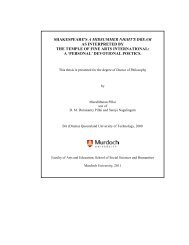
Rickettsiales and rickettsial diseases in Australia - Murdoch ...
Rickettsiales and rickettsial diseases in Australia - Murdoch ...
Rickettsiales and rickettsial diseases in Australia - Murdoch ...

You also want an ePaper? Increase the reach of your titles
YUMPU automatically turns print PDFs into web optimized ePapers that Google loves.
<strong>Rickettsiales</strong> <strong>and</strong> <strong>rickettsial</strong><br />
<strong>diseases</strong> <strong>in</strong> <strong>Australia</strong><br />
Leonard He<strong>in</strong>z Izzard<br />
Bachelor of Applied Science (Honours)<br />
This thesis is presented for the degree of Doctor of<br />
Philosophy of <strong>Murdoch</strong> University<br />
2010
Declaration<br />
I declare that this thesis is my own account of my<br />
research <strong>and</strong> conta<strong>in</strong>s as its ma<strong>in</strong> content work which<br />
has not previously been submitted for a degree at any<br />
tertiary <strong>in</strong>stitution.<br />
………………………….<br />
Leonard He<strong>in</strong>z Izzard<br />
i
Abstract<br />
Currently, there are 12 known <strong>Rickettsiales</strong> species <strong>in</strong> <strong>Australia</strong>. However<br />
research <strong>in</strong>to the diversity <strong>and</strong> range of these agents <strong>in</strong> <strong>Australia</strong> is still far from<br />
complete.<br />
A sero-epidemiological study was undertaken around the city of Launceston <strong>in</strong><br />
Tasmania, <strong>Australia</strong> to determ<strong>in</strong>e the level of exposure to spotted fever group<br />
(SFG) rickettsia among the local cat <strong>and</strong> dog population. The study showed that<br />
over 50% of the dogs <strong>and</strong> cats tested were positive for SFG rickettsiae<br />
antibodies. However, no correlation was observed between the animals’ health<br />
<strong>and</strong> seropositivity at the time of test<strong>in</strong>g.<br />
Ixodes tasmani ticks collected from Tasmanian devils <strong>in</strong> Tasmania were tested<br />
for the presence of SFG <strong>and</strong> typhus group (TG) rickettsiae us<strong>in</strong>g a specific real<br />
time PCR (qPCR), <strong>and</strong> 55% were found to be positive. The gltA, rompA, rompB<br />
<strong>and</strong> sca4 genes were then sequenced. Us<strong>in</strong>g the current criteria this new<br />
rickettsia qualified as a C<strong>and</strong>idatus species, <strong>and</strong> was named C<strong>and</strong>idatus<br />
Rickettsia tasmanensis, after the location from which it was first detected.<br />
Soft ticks of the species Argas dewae were collected from bat roost<strong>in</strong>g boxes<br />
north of Melbourne. Of the ten ticks collected, seven (70%) were positive for<br />
SFG rickettsiae us<strong>in</strong>g the qPCR mentioned above. An isolate was obta<strong>in</strong>ed<br />
us<strong>in</strong>g cell culture isolation methods <strong>and</strong> the rrs, gltA, rompA, rompB <strong>and</strong> sca4<br />
genes were sequenced. Us<strong>in</strong>g the current criteria this new rickettsia qualified as<br />
ii
a novel species, <strong>and</strong> was tentatively named Rickettsia argasii sp. nov. after the<br />
tick genus from which it was isolated.<br />
Four family members <strong>and</strong> their neighbour liv<strong>in</strong>g <strong>in</strong> metropolitan Victoria became<br />
ill after exposure to a flea-<strong>in</strong>fested kitten. Initial serological analysis <strong>in</strong>dicated a<br />
typhus group (TG) <strong>rickettsial</strong> <strong>in</strong>fection. However, test<strong>in</strong>g of fleas from the group<br />
of cats <strong>in</strong> Lara, Victoria, where the kitten orig<strong>in</strong>ated, revealed the presence of R.<br />
felis, the agent of cat flea typhus. This was the first case of human <strong>in</strong>fection with<br />
R. felis <strong>in</strong> <strong>Australia</strong> <strong>and</strong> the first detection of R. felis <strong>in</strong> fleas <strong>in</strong> Victoria.<br />
A tourist return<strong>in</strong>g to <strong>Australia</strong> from the United Arab Emirates was diagnosed<br />
with a scrub typhus group (STG) <strong>rickettsial</strong> <strong>in</strong>fection <strong>and</strong> the agent was isolated<br />
from their blood. Analysis of the rrs <strong>and</strong> 47kDa genes showed significant<br />
divergence compared to all available stra<strong>in</strong>s of Orientia tsutsugamushi. Due to<br />
the degree of genetic divergence <strong>and</strong> the geographically unique orig<strong>in</strong> of this<br />
isolate it was considered to be a new species, which has been tentatively<br />
named Orientia chuto, with ‘chuto’ be<strong>in</strong>g Japanese for ‘Middle East’.<br />
Dogs <strong>in</strong> central <strong>and</strong> northern <strong>Australia</strong> were tested for Anaplasma platys us<strong>in</strong>g a<br />
specifically designed real-time PCR (qPCR) assay. Of the 68 dogs tested, 27<br />
(40%) were positive for A. platys DNA, <strong>in</strong>clud<strong>in</strong>g six dogs from Western<br />
<strong>Australia</strong>. This was the first report of A. platys <strong>in</strong> Western <strong>Australia</strong>.<br />
These studies offer an <strong>in</strong>sight <strong>in</strong>to the range <strong>and</strong> diversity of <strong>Rickettsiales</strong> <strong>and</strong><br />
<strong>rickettsial</strong> <strong>diseases</strong> previously unrecognised <strong>in</strong> <strong>Australia</strong>.<br />
iii
Table of Contents<br />
<strong>Rickettsiales</strong> <strong>and</strong> <strong>rickettsial</strong> <strong>diseases</strong> <strong>in</strong> <strong>Australia</strong> ...................................... i<br />
Declaration ...................................................................................................... i<br />
Abstract .......................................................................................................... ii<br />
Table of Contents .......................................................................................... iv<br />
List of Figures ................................................................................................ x<br />
List of Tables ............................................................................................... xiii<br />
Acknowledgements .................................................................................... xiv<br />
Preface ......................................................................................................... xvi<br />
Abbreviations ............................................................................................... xx<br />
Chapter 1. Literature Review .................................................................... 1<br />
1.1. Introduction ........................................................................................ 1<br />
1.2. Taxonomy <strong>and</strong> Pathogenicity of the order <strong>Rickettsiales</strong> .................... 4<br />
1.2.1. Anaplasmataceae ......................................................................... 4<br />
1.2.1.1. Anaplasma ............................................................................. 4<br />
1.2.1.2. Ehrlichia ................................................................................. 5<br />
1.2.2. Rickettsiaceae .............................................................................. 6<br />
1.2.2.1. Rickettsia ............................................................................... 6<br />
1.2.2.2. Orientia .................................................................................. 7<br />
1.3. Genomics of the order <strong>Rickettsiales</strong> .................................................. 9<br />
1.4. Methods of identification <strong>and</strong> characterisation of <strong>Rickettsiales</strong> ........ 13<br />
1.4.1. Sta<strong>in</strong><strong>in</strong>g ...................................................................................... 13<br />
1.4.2. Serology ..................................................................................... 13<br />
iv
1.4.3. Isolation ...................................................................................... 14<br />
1.4.4. Polymerase Cha<strong>in</strong> Reaction (PCR) ............................................ 14<br />
1.4.4.1. Conventional PCR ............................................................... 15<br />
1.4.4.2. Real-time PCR (qPCR) ........................................................ 15<br />
1.4.4.3. Molecular speciation ............................................................ 16<br />
1.5. Geographic distribution of the <strong>Rickettsiales</strong> ..................................... 18<br />
1.6. <strong>Rickettsiales</strong> species <strong>in</strong> <strong>Australia</strong> ..................................................... 18<br />
1.6.1. Anaplasma ................................................................................. 18<br />
1.6.1.1. <strong>Australia</strong>n (cattle) tick fever (Anaplasma marg<strong>in</strong>ale) .............. 18<br />
1.6.1.2. ... Can<strong>in</strong>e <strong>in</strong>fectious cyclic thrombocytopenia (Anaplasma platys)<br />
............................................................................................................ 19<br />
1.6.2. Ehrlichia ..................................................................................... 20<br />
1.6.3. Rickettsia .................................................................................... 20<br />
1.6.3.1. Mur<strong>in</strong>e typhus (Rickettsia typhi) ........................................... 22<br />
1.6.3.2. Queensl<strong>and</strong> tick typhus (Rickettsia australis) ...................... 25<br />
1.6.3.3. Fl<strong>in</strong>ders isl<strong>and</strong> spotted fever (R. honei) ............................... 27<br />
1.6.3.4. <strong>Australia</strong>n spotted fever (R. honei stra<strong>in</strong> marmionii) ............ 28<br />
1.6.4. Orientia ....................................................................................... 30<br />
1.6.4.1. Scrub typhus (Orientia tsutsugamushi) ................................ 30<br />
1.6.5. This Study .................................................................................. 32<br />
Chapter 2. Materials <strong>and</strong> Methods ......................................................... 34<br />
2.1. Rickettsial Isolation .......................................................................... 34<br />
2.1.1. Blood sample process<strong>in</strong>g ........................................................... 34<br />
2.1.2. Tick sample process<strong>in</strong>g .............................................................. 34<br />
2.1.3. Flea sample process<strong>in</strong>g ............................................................. 35<br />
v
2.2. Cell Culture ...................................................................................... 35<br />
2.3. Freez<strong>in</strong>g Samples ............................................................................ 36<br />
2.4. Molecular Methods .......................................................................... 37<br />
2.4.1. DNA sample preparation ............................................................ 37<br />
2.4.2. Primer/probe design <strong>and</strong> validation ............................................ 37<br />
2.4.2.1. Primer/probe set design ....................................................... 37<br />
2.4.2.2. Test<strong>in</strong>g Sensitivity ................................................................ 38<br />
2.4.2.3. Test<strong>in</strong>g Specificity ................................................................ 39<br />
2.4.3. qPCR detection .......................................................................... 39<br />
2.4.4. Conventional PCR ...................................................................... 41<br />
2.4.5. Sequenc<strong>in</strong>g ................................................................................ 45<br />
2.4.6. Bio<strong>in</strong>formatics ............................................................................. 45<br />
2.5. Serology .......................................................................................... 46<br />
2.5.1. Microimmunofluorescence ......................................................... 46<br />
Chapter 3. A serological prevalence study for <strong>rickettsial</strong> exposure of<br />
cats <strong>and</strong> dogs <strong>in</strong> Launceston, Tasmania, <strong>Australia</strong> .................................. 48<br />
3.1. Abstract ........................................................................................... 48<br />
3.2. Introduction ...................................................................................... 49<br />
3.3. Materials <strong>and</strong> methods .................................................................... 50<br />
3.3.1. Sample <strong>and</strong> data collection ........................................................ 50<br />
3.3.2. Detection of antibodies to Spotted Fever Group Rickettsia ........ 51<br />
3.3.3. Statistical analysis of serological results .................................... 51<br />
3.4. Results ............................................................................................. 51<br />
3.4.1. Serology Results ........................................................................ 51<br />
3.4.2. Statistical analysis ...................................................................... 52<br />
vi
3.5. Discussion ....................................................................................... 53<br />
Chapter 4. Novel Rickettsia (C<strong>and</strong>idatus Rickettsia tasmanensis) <strong>in</strong><br />
Tasmania, <strong>Australia</strong> ..................................................................................... 57<br />
4.1. Abstract ........................................................................................... 57<br />
4.2. Introduction ...................................................................................... 58<br />
4.3. Methods ........................................................................................... 58<br />
4.4. Results ............................................................................................. 59<br />
4.5. Discussion ....................................................................................... 63<br />
Chapter 5 Isolation of Rickettsia argas sp. nov. from the bat tick Argas<br />
dewae. ........................................................................................................... 65<br />
5.1. Abstract ........................................................................................... 65<br />
5.2. Introduction ...................................................................................... 65<br />
5.3. Materials <strong>and</strong> methods .................................................................... 66<br />
5.4. Results ............................................................................................. 66<br />
5.5. Discussion ....................................................................................... 73<br />
Chapter 6. First reported human cases of Rickettsia felis (cat flea<br />
typhus) <strong>in</strong> <strong>Australia</strong> ..................................................................................... 75<br />
6.1. Abstract ........................................................................................... 75<br />
6.2. Introduction ...................................................................................... 75<br />
6.3. Case Study ...................................................................................... 77<br />
6.4. Methods ........................................................................................... 80<br />
6.4.1. Serology ..................................................................................... 80<br />
6.4.2. PCR ............................................................................................ 80<br />
6.4.3. Molecular Characterisation ......................................................... 81<br />
6.4.4. Attempted Isolation <strong>and</strong> Culture ................................................. 81<br />
vii
6.5. Results ............................................................................................. 82<br />
6.5.1. Serology ..................................................................................... 82<br />
6.5.2. Molecular Analysis ..................................................................... 83<br />
6.5.3. Attempted Isolation <strong>and</strong> Culture ................................................. 84<br />
6.6. Discussion ....................................................................................... 85<br />
Chapter 7. Isolation of a highly variant Orientia species (O. chuto sp.<br />
nov.) from a patient return<strong>in</strong>g from Dubai ................................................. 87<br />
7.1. Abstract ........................................................................................... 87<br />
7.2. Introduction ...................................................................................... 88<br />
7.3. Cl<strong>in</strong>ical case history ......................................................................... 90<br />
7.4. Materials <strong>and</strong> methods .................................................................... 93<br />
7.4.1. Microimmunofluorescence assay (IFA) ...................................... 93<br />
7.4.2. Culture ........................................................................................ 93<br />
7.4.3. DNA extraction <strong>and</strong> PCR assays................................................ 93<br />
7.4.3.1. 16S rRNA gene .................................................................... 93<br />
7.4.3.2. 47kDa gene ......................................................................... 93<br />
7.4.4. Sequenc<strong>in</strong>g ................................................................................ 94<br />
7.4.5. qPCR design .............................................................................. 95<br />
7.5. Results ............................................................................................. 95<br />
7.5.1. Serology ..................................................................................... 95<br />
7.5.2. Culture ........................................................................................ 96<br />
7.5.3. Molecular analysis ...................................................................... 96<br />
7.5.4. qPCR .......................................................................................... 99<br />
7.6. Discussion ..................................................................................... 100<br />
viii
Chapter 8. Anaplasma platys <strong>in</strong> <strong>Australia</strong>n dogs detected by a novel<br />
real-time PCR assay. ................................................................................. 104<br />
8.1. Abstract ......................................................................................... 104<br />
8.2. Introduction .................................................................................... 104<br />
8.3. Methods ......................................................................................... 106<br />
8.3.1. Probe Design ........................................................................... 106<br />
8.3.2. Assay Optimisation ................................................................... 106<br />
8.3.3. Sample Preparation .................................................................. 106<br />
8.3.4. PCR reaction ............................................................................ 107<br />
8.4. Results ........................................................................................... 107<br />
8.4.1. Assay Optimisation ................................................................... 107<br />
8.4.2. PCR Results ............................................................................. 108<br />
8.5. Discussion ..................................................................................... 109<br />
Chapter 9. Conclud<strong>in</strong>g Remarks .......................................................... 112<br />
Appendices ................................................................................................. 118<br />
Appendix 1: Mathematical determ<strong>in</strong>ation of copy numbers ...................... 118<br />
References................................................................................................... 119<br />
ix
List of Figures<br />
Figure 1. Phylogenetic relationship of members of the order <strong>Rickettsiales</strong><br />
adapted from the Taxonomy browser with<strong>in</strong> the NCBI website<br />
(http://www.ncbi.nlm.nih.gov) show<strong>in</strong>g the order (red), family (purple), genus<br />
(blue) <strong>and</strong> species group (orange). .................................................................. 3<br />
Figure 2. Flow diagram of phylogenetic classification of Rickettsia adapted from<br />
Fournier et al. 81 .............................................................................................. 17<br />
Figure 3. Map of Tasmania, <strong>Australia</strong>, show<strong>in</strong>g number of positive (black) <strong>and</strong><br />
negative (white) ticks <strong>and</strong> their locations. The question mark <strong>in</strong>dicates unknown<br />
locations. A total of 55% of the ticks were positive for a spotted fever group<br />
rickettsia. ........................................................................................................ 60<br />
Figure 4. Phylogenetic tree show<strong>in</strong>g the relationship of a 4,834-bp fragment of<br />
the outer membrane prote<strong>in</strong> B gene of C<strong>and</strong>idatus Rickettsia tasmanensis (<strong>in</strong><br />
boldface) compared to all validated rickettsia species. The tree was prepared<br />
us<strong>in</strong>g the neighbor-jo<strong>in</strong><strong>in</strong>g algorithm with<strong>in</strong> the MEGA 4 software 245 . Bootstrap<br />
values are <strong>in</strong>dicated at each node. Scale bar <strong>in</strong>dicates 2% nucleotide<br />
divergence. .................................................................................................... 62<br />
Figure 5. Phylogenetic tree show<strong>in</strong>g the relationship of a 1,098bp fragment of<br />
the gltA gene of Rickettsia argasii sp. nov. to all validated <strong>rickettsial</strong> species.<br />
The tree was prepared us<strong>in</strong>g the neighbor-jo<strong>in</strong><strong>in</strong>g algorithm, with<strong>in</strong> the Mega 4<br />
software. Bootstrap values are <strong>in</strong>dicated at each node. The scale bar<br />
represents a 2% nucleotide divergence. ........................................................ 68<br />
x
Figure 6. Phylogenetic tree show<strong>in</strong>g the relationship of a 4,881bp fragment of<br />
the rOmpB gene of Rickettsia argasii sp. nov. to all validated <strong>rickettsial</strong> species.<br />
The tree was prepared us<strong>in</strong>g the neighbor-jo<strong>in</strong><strong>in</strong>g algorithm, with<strong>in</strong> the Mega 4<br />
software. Bootstrap values are <strong>in</strong>dicated at each node. The scale bar<br />
represents a 5% nucleotide divergence. ........................................................ 69<br />
Figure 7. Phylogenetic tree show<strong>in</strong>g the relationship of a 530bp fragment of the<br />
rOmpA gene of Rickettsia argasii sp. nov. to all validated <strong>rickettsial</strong> species.<br />
The tree was prepared us<strong>in</strong>g the neighbor-jo<strong>in</strong><strong>in</strong>g algorithm, with<strong>in</strong> the Mega 4<br />
software. Bootstrap values are <strong>in</strong>dicated at each node. The scale bar<br />
represents a 10% nucleotide divergence. ...................................................... 70<br />
Figure 8. Phylogenetic tree show<strong>in</strong>g the relationship of a 1413bp fragment of<br />
the rrs gene of Rickettsia argasii sp. nov. to all validated <strong>rickettsial</strong> species. The<br />
tree was prepared us<strong>in</strong>g the neighbor-jo<strong>in</strong><strong>in</strong>g algorithm, with<strong>in</strong> the Mega 4<br />
software. Bootstrap values are <strong>in</strong>dicated at each node. The scale bar<br />
represents a 0.2% nucleotide divergence. ..................................................... 71<br />
Figure 9. Phylogenetic tree show<strong>in</strong>g the relationship of a 2,901bp fragment of<br />
the sca4 gene of Rickettsia argasii sp. nov. to all validated <strong>rickettsial</strong> species.<br />
The tree was prepared us<strong>in</strong>g the neighbor-jo<strong>in</strong><strong>in</strong>g algorithm, with<strong>in</strong> the Mega 4<br />
software. Bootstrap values are <strong>in</strong>dicated at each node. The scale bar<br />
represents a 2% nucleotide divergence. ........................................................ 72<br />
Figure 10. A condensed phylogenetic tree show<strong>in</strong>g the relationship of a 1077bp<br />
fragment of the gltA gene of Rickettsia felis (Lara) among other validated<br />
<strong>rickettsial</strong> species, with the core spotted fever group rickettsiae truncated. The<br />
tree was prepared us<strong>in</strong>g the neighbor-jo<strong>in</strong><strong>in</strong>g algorithm, with<strong>in</strong> the Mega 4<br />
xi
software. Bootstrap values are <strong>in</strong>dicated at each node. The scale bar<br />
represents a 2% nucleotide divergence. ........................................................ 84<br />
Figure 11. A regional map show<strong>in</strong>g the distribution of scrub typhus <strong>and</strong> the<br />
location of Dubai with<strong>in</strong> the United Arab Emirates. ........................................ 89<br />
Figure 12. Eschar on the abdomen of the patient. ......................................... 91<br />
Figure 13. Scrub Typhus serology, show<strong>in</strong>g a marked change <strong>in</strong> antibody titres<br />
to three stra<strong>in</strong>s of O. tsutsugamushi over a 57 day period. ............................ 92<br />
Figure 14. Phylogenetic trees show<strong>in</strong>g the relationship between the 16S rRNA<br />
<strong>and</strong> 47kDa genes of Orientia chuto stra<strong>in</strong> Churchill to various O. tsutsugamushi<br />
stra<strong>in</strong>s. The tree was prepared us<strong>in</strong>g the neighbor-jo<strong>in</strong><strong>in</strong>g algorithm, with<strong>in</strong> the<br />
Mega 4 software. Bootstrap values are <strong>in</strong>dicated at each node. The scale bars<br />
represents a 0.2% <strong>and</strong> 2.0% nucleotide divergence for the 16S rRNA <strong>and</strong><br />
47kDa gene respectively. ............................................................................... 98<br />
Figure 15. A st<strong>and</strong>ard curve show<strong>in</strong>g the Ct versus the number of copies of the<br />
template conta<strong>in</strong><strong>in</strong>g plasmid. ......................................................................... 99<br />
Figure 16. St<strong>and</strong>ard curve show<strong>in</strong>g the relative Ct versus the number of copies<br />
of the template conta<strong>in</strong><strong>in</strong>g plasmid. .............................................................. 108<br />
Figure 17: The geographic distribution <strong>and</strong> number of positive <strong>and</strong> total A.<br />
platys samples collected <strong>in</strong> <strong>Australia</strong>. .......................................................... 109<br />
xii
List of Tables<br />
Table 1. Current list of validated <strong>rickettsial</strong> species. ...................................... 11<br />
Table 2. Current list of validated Anaplasma <strong>and</strong> Ehrlichia species. .............. 12<br />
Table 3. Name, conditions <strong>and</strong> literature references for oligonucleotides used<br />
for conventional PCR. .................................................................................... 43<br />
Table 4. Seropositivity for Spotted Fever Group rickettsiae <strong>in</strong> dog <strong>and</strong> cat serum<br />
tested at 1/50, 1/100 <strong>and</strong> 1/200 dilutions show<strong>in</strong>g a lack of statistical<br />
relationship (p>0.05) between cl<strong>in</strong>ically sick animals <strong>and</strong> seropositive animals.<br />
....................................................................................................................... 53<br />
Table 5. GenBank accession numbers of additional sequences used <strong>in</strong> this<br />
study. ............................................................................................................. 59<br />
Table 6. Serology results from five patients <strong>and</strong> a cat post-exposure to a<br />
<strong>rickettsial</strong> agent. ............................................................................................. 83<br />
Table 7. Primer sequences used to amplify the 47 kDa genes (Richards et al.).<br />
....................................................................................................................... 94<br />
Table 8. qPCR primer <strong>and</strong> probe set sequences target<strong>in</strong>g the 16S rRNA gene.<br />
....................................................................................................................... 95<br />
Table 9. Percentage pairwise divergence plot of O. chuto stra<strong>in</strong> Churchill with<br />
various stra<strong>in</strong>s of O. tsutsugamushi show<strong>in</strong>g the significant level of divergence<br />
of O. chuto stra<strong>in</strong> Churchill. ............................................................................ 97<br />
xiii
Acknowledgements<br />
I would first like to thank my supervisors A/Prof. John Stenos <strong>and</strong> A/Prof.<br />
Stephen Graves from the <strong>Australia</strong>n Rickettsial Reference Laboratory, <strong>and</strong> Prof.<br />
Stan Fenwick from the School of Veter<strong>in</strong>ary <strong>and</strong> Biomedical Science, <strong>Murdoch</strong><br />
University, for their patience <strong>and</strong> support over the previous few years. I would<br />
especially like to thank John <strong>and</strong> Stephen for offer<strong>in</strong>g their experience <strong>and</strong><br />
ideas when I was problem solv<strong>in</strong>g <strong>and</strong> for review<strong>in</strong>g my drafts over the years. I<br />
would also like to thank the <strong>Australia</strong>n Rickettsial Reference Laboratory <strong>and</strong><br />
<strong>Murdoch</strong> University for their f<strong>in</strong>ancial support throughout my c<strong>and</strong>idature.<br />
With<strong>in</strong> the <strong>Australia</strong>n Rickettsial Reference Laboratory I would first like to thank<br />
Chelsea Nguyen for teach<strong>in</strong>g me serological <strong>and</strong> culture methods <strong>and</strong> Michelle<br />
Lockhart for her support <strong>in</strong> assist<strong>in</strong>g me to bra<strong>in</strong>storm. Furthermore, I would like<br />
to thank all of the staff at the <strong>Australia</strong>n Rickettsial Reference Laboratory for<br />
their support <strong>and</strong> friendship.<br />
I would also like to thank collaborators <strong>and</strong> colleagues who have assisted me<br />
with most of my chapters. These <strong>in</strong>clude for chapter three, Helen Owen for<br />
provid<strong>in</strong>g the cat control sera. For chapter 4, I would like to thank Ian Norton,<br />
his colleagues, <strong>and</strong> Dydee Mann for collect<strong>in</strong>g ticks from sites <strong>in</strong> Tasmania. I<br />
would also like to thank Ian Beveridge for his assistance with tick speciation.<br />
For chapter five, I would like to thank Lisa Evans for supply<strong>in</strong>g me with the <strong>in</strong>itial<br />
tick samples <strong>and</strong> contacts, <strong>and</strong> Natasha Schedv<strong>in</strong>, Robert Bender, Marissa<br />
Izzard <strong>and</strong> Stephen Weste for assist<strong>in</strong>g me with collect<strong>in</strong>g ticks. For chapter six,<br />
xiv
I would like to thank Molly Williams <strong>and</strong> Julian Kelly for br<strong>in</strong>g<strong>in</strong>g this <strong>in</strong>terest<strong>in</strong>g<br />
case to my attention <strong>and</strong> for supply<strong>in</strong>g me with the case study. I would also like<br />
to thank Am<strong>in</strong>ul Islam for his assistance with attempted isolation. For Chapter<br />
seven, I would like to thank Andrew Fuller for br<strong>in</strong>g<strong>in</strong>g this case to my attention<br />
<strong>and</strong> supply<strong>in</strong>g the case study. As well, I would like to thank Stuart Blacksell,<br />
Daniel Paris, Al Richards, Nuntipa Aukkanit, <strong>and</strong> Ju Jiang for their assistance<br />
with sequenc<strong>in</strong>g various genes <strong>and</strong> Chelsea Nguyen for her assistance <strong>in</strong><br />
cultur<strong>in</strong>g this isolate. F<strong>in</strong>ally, for chapter eight, I would like to thank Graeme<br />
Brown for supply<strong>in</strong>g me with dog blood samples used <strong>in</strong> this study.<br />
On a personal note I would like to thank all of my family <strong>in</strong>clud<strong>in</strong>g, Mum <strong>and</strong><br />
Dad, Debbie, Steve, David, Sue <strong>and</strong> my sisters Jo <strong>and</strong> Bianca. They may not<br />
have always known what my projects entailed, nevertheless they always<br />
supported me <strong>and</strong> had faith that I would succeed.<br />
Most of all I would like to thank my new wife Marissa Izzard (née McCaw) for<br />
her support throughout not only my PhD but my entire University education. She<br />
has been my rock when I was lost for direction <strong>and</strong> was always there to show<br />
me the positives when I was feel<strong>in</strong>g negative. I would also like to thank her for<br />
help<strong>in</strong>g to support me f<strong>in</strong>ancially over my school<strong>in</strong>g life <strong>and</strong> for look<strong>in</strong>g at more<br />
drafts than I can remember.<br />
xv
Preface<br />
The work <strong>in</strong> this thesis is my own research. Collaborat<strong>in</strong>g authors mentioned <strong>in</strong><br />
chapters 3 were ma<strong>in</strong>ly responsible for sample collection <strong>and</strong> manuscript<br />
revision prior to submission, while <strong>in</strong> chapter 4, collaborat<strong>in</strong>g authors were<br />
primarily responsible for manuscript revision prior to submission. The<br />
contributions of all other parties are mentioned <strong>in</strong> the acknowledgments section<br />
of this thesis.<br />
Orig<strong>in</strong>al manuscripts<br />
Izzard L, Graves S, Cox E, Fenwick S, Unsworth N, Stenos J. Novel rickettsia <strong>in</strong><br />
ticks, Tasmania, <strong>Australia</strong>. Emerg Infect Dis 2009; 15:1654 - 56.<br />
Izzard L, Cox E, Stenos J, Waterston M, Fenwick S, Graves S. A serological<br />
prevalence study of <strong>rickettsial</strong> exposure of cats <strong>and</strong> dogs <strong>in</strong> Launceston,<br />
Tasmania, <strong>Australia</strong>. Aust Vet J 2010; 88:29 - 31.<br />
Izzard L, Fuller A, Blacksell S, Daniel Paris D, Al Richards A, Aukkanit N,<br />
Nguyen C, Jiang J, Fenwick S, Day N, Graves S, Stenos J. Isolation of a highly<br />
variant Orientia species (O. chuto sp. nov.) from a patient return<strong>in</strong>g from Dubai.<br />
Submitted to the J Cl<strong>in</strong> Microbiol.<br />
xvi
Izzard L, Graves S, Fenwick S, Stenos J. Isolation of Rickettsia argasii sp. nov.<br />
from Argas dewae ticks <strong>in</strong> Victoria, <strong>Australia</strong>. In Preparation for submission<br />
to Int J Syst Evol Microbiol.<br />
Izzard L, Stenos J, Fenwick S, Graves S. <strong>Rickettsiales</strong> <strong>and</strong> <strong>rickettsial</strong> <strong>diseases</strong><br />
<strong>in</strong> <strong>Australia</strong>. In Preparation for submission to Annals of the ACTM.<br />
Orig<strong>in</strong>al published abstracts<br />
Izzard L, Stenos J, Fenwick S, Graves S. Two Novel Rickettsiae Identified <strong>in</strong><br />
<strong>Australia</strong> – Oral presentation at the 21st Meet<strong>in</strong>g of the American Society<br />
for Rickettsiology, Colorado, USA (2007)<br />
Izzard L, Stenos J, Fenwick S, Graves S. Two Novel Rickettsiae Identified <strong>in</strong><br />
<strong>Australia</strong> – Poster presentation at <strong>Murdoch</strong> University WA (2007)<br />
xvii
Izzard L, Fuller A, Blacksell S, Paris D, Aukkanit N, Graves S, Fenwick S,<br />
Nguyen C, Day N, Stenos J. Isolation of a highly variant Orientia sp. from a<br />
patient return<strong>in</strong>g from Dubai – Poster presentation at the 5th International<br />
Conference on Rickettsiae <strong>and</strong> Rickettsial Diseases, Marseille, France<br />
(2008)<br />
Izzard L, Williams M, Kelly J, Graves S, Fenwick S, Stenos J. First cases of<br />
Rickettsia felis (cat flea typhus) <strong>in</strong> <strong>Australia</strong> - Poster presentation at <strong>Murdoch</strong><br />
University WA (2009)<br />
Co-authored manuscripts<br />
Stenos J, Graves S, Izzard L: Chapter 25: Rickettsia. Schuller M, Sloots T,<br />
James G, Halliday I, Carter I, eds. PCR for Cl<strong>in</strong>ical Microbiology: An<br />
<strong>Australia</strong>n <strong>and</strong> International Perspective. New York: Spr<strong>in</strong>ger 2010, 197-99.<br />
Williams M, Izzard L, Islam A, Graves S, Stenos J, Kelly J. First cases of<br />
Rickettsia felis (cat flea typhus) <strong>in</strong> <strong>Australia</strong>. Submissed to the Med J Aust.<br />
xviii
Co-authored published abstracts<br />
Lockhart M, Izzard L, Nguyen C, Fenwick S, Stenos J, Graves S. Asymptomatic<br />
chronic Coxiella burnetii bacteraemia without seroconversion – Oral<br />
presentation at the 5th International Conference on Rickettsiae <strong>and</strong><br />
Rickettsial Diseases, Marseille, France (2008)<br />
Islam A, Izzard L, Unsworth N, Givney R, Ferguson J, Stenos J, Graves S. A<br />
note on rickettsiae <strong>in</strong> <strong>Australia</strong> – Poster presentation at the 23rd Meet<strong>in</strong>g of<br />
the American Society for Rickettsiology, South Carol<strong>in</strong>a, USA (2009)<br />
Ja<strong>in</strong>g J, Blacksell S, Paris D, Aukkanit N, Newton P, Phetsouvanh R, Izzard L,<br />
Stenos J, Graves S, Day N, Richards A. Diversity of the 47kDa/HtrA translated<br />
am<strong>in</strong>o acid sequences among human isolates of Orientia tsutsugamushi –<br />
Poster presentation at the 23rd Meet<strong>in</strong>g of the American Society for<br />
Rickettsiology, South Carol<strong>in</strong>a, USA (2009)<br />
xix
Abbreviations<br />
AG – Ancestral group<br />
AGRF – The <strong>Australia</strong>n Genomic Research Facility<br />
ASF – <strong>Australia</strong>n spotted fever<br />
ATF – <strong>Australia</strong> tick fever<br />
BHQ-1 – Black hole quencher-1<br />
Bp – Base pairs<br />
CCFM – Cell culture freez<strong>in</strong>g media<br />
CF – Complement Fixation<br />
Ct – Threshold cycle<br />
DNA – Deoxyribonucleic acid<br />
dNTP – Deoxyribonucleic triphosphate<br />
EDTA – Ethylenediam<strong>in</strong>etetraacetic Acid<br />
ELISA – Enzyme-l<strong>in</strong>ked immuno sorbent assay<br />
FISF – Fl<strong>in</strong>ders isl<strong>and</strong> spotted fever<br />
FITC – Fluoresce<strong>in</strong> isothiocyanate<br />
gltA – Citrate synthase gene<br />
HEPES – 4-(2-hydroxyethyl)-1-piperaz<strong>in</strong>eethanesulfonic acid<br />
xx
IFA – Immunofluorescent assay<br />
kDa – Kilodalton<br />
NCBI – National Centre for Biotechnology Information<br />
NSW – New South Wales<br />
NTC – No template control<br />
OPNP – Organ Pipes National Park<br />
PBMC – Peripheral Blood Mononuclear Cells<br />
PBS – Phosphate buffered sal<strong>in</strong>e<br />
PCR – Polymerase cha<strong>in</strong> reaction<br />
QLD – Queensl<strong>and</strong><br />
qPCR – Real-time PCR<br />
QTT – Queensl<strong>and</strong> tick typhus<br />
RBC – Red blood cell<br />
RNA – Ribonucleic acid<br />
rompA – Rickettsial outer membrane prote<strong>in</strong> A gene<br />
rompB – Rickettsial outer membrane prote<strong>in</strong> B gene<br />
RPM – Revolutions per m<strong>in</strong>ute<br />
rRNA – Ribosomal RNA.<br />
rrs – 16S ribosomal RNA gene<br />
xxi
sca4 – The <strong>rickettsial</strong> gene D / PS–120 gene<br />
SDS – Sodium dodecyl sulphate<br />
SFG – Spotted fever group<br />
TAE – Tris-acetate-EDTA<br />
TG – Typhus group<br />
Tm – Melt<strong>in</strong>g temperature<br />
TRG – Transitional group<br />
xxii
Chapter 1. Literature Review<br />
1.1. Introduction<br />
In 1909 Howard T Ricketts first described small organisms that appeared to be<br />
associated with the disease Rocky Mounta<strong>in</strong> spotted fever 209 . This was the first<br />
report of the organism Rickettsia. The <strong>diseases</strong> associated with these<br />
organisms however have been described <strong>in</strong> literature dat<strong>in</strong>g back possibly as<br />
far as 1083, with typhus extensively described by Fracastoro <strong>in</strong> 1546 204 .<br />
In 1910 Theiler proposed the new genus <strong>and</strong> species Anaplasma marg<strong>in</strong>ale as<br />
the aetiological agent of anaplasmosis, found <strong>in</strong> the erythrocytes of cattle 249 <strong>and</strong><br />
<strong>in</strong> 1916 Rocha Lima proposed the genus name Rickettsia. At this stage the only<br />
species <strong>in</strong> the genus was the agent of epidemic typhus Rickettsia prowazekii 187 .<br />
By 1953, the family Rickettsiaceae conta<strong>in</strong>ed 4 genera, namely Rickettsia,<br />
Ehrlichia, Cowdria, <strong>and</strong> Coxiella. The genus Rickettsia conta<strong>in</strong>ed seven<br />
species: R. prowazekii, R. typhi, R. tsutsugamushi, R. rickettsii, R. conorii, R.<br />
australis <strong>and</strong> R. akari. The genus Ehrlichia conta<strong>in</strong>ed E. bovis, E. canis, E.<br />
ov<strong>in</strong>a <strong>and</strong> E. kurlovi. Cowdria <strong>and</strong> Coxiella each only conta<strong>in</strong>ed one species,<br />
Cowdria rum<strong>in</strong>antium <strong>and</strong> Coxiella burnetii respectively 187 .<br />
In 1974 the eighth edition of Bergey’s manual of determ<strong>in</strong>ative bacteriology was<br />
published. By this stage the order <strong>Rickettsiales</strong> conta<strong>in</strong>ed three families;<br />
Rickettsiaceae, Bartonellaceae <strong>and</strong> Anaplasmataceae. The family<br />
Rickettsiaceae conta<strong>in</strong>ed among others the genus Rickettsia (which <strong>in</strong>cluded<br />
the species R. tsutsugamushi), Ehrlichia <strong>and</strong> Coxiella, while the genus<br />
Anaplasma was with<strong>in</strong> the Anaplasmataceae family 158 .<br />
1
In 1989, Coxiella burnetii was removed from the Rickettsiaceae family after 16S<br />
gene sequenc<strong>in</strong>g revealed that it was more closely related to the genus<br />
Legionella than Rickettsia 277 . This was followed by the removal of Bartonella<br />
spp. <strong>in</strong> 1993 29 .<br />
The species R. tsutsugamushi was moved <strong>in</strong>to the novel genus Orientia <strong>in</strong> 1995<br />
<strong>and</strong> renamed Orientia tsutsugamushi 244 .<br />
In 2001 the genera with<strong>in</strong> the Rickettsiaceae <strong>and</strong> Anaplasmataceae underwent<br />
extensive reorganisation with the help of molecular analysis. The tribes<br />
Rickettsiae, Ehrlichieae <strong>and</strong> Wolbachieae were removed. The genus Wolbachia<br />
was transferred from the Rickettsiaceae to the Anaplasmataceae family.<br />
Several species of Ehrlichia <strong>and</strong> Anaplasma were regrouped <strong>in</strong>to various<br />
genera 66 .<br />
The order <strong>Rickettsiales</strong> currently conta<strong>in</strong>s six dist<strong>in</strong>ct families <strong>in</strong>clud<strong>in</strong>g the<br />
families Anaplasmataceae <strong>and</strong> Rickettsiaceae (Figure 1). The family<br />
Anaplasmataceae is broken <strong>in</strong>to five genera; Aegyptianella, Anaplasma,<br />
Ehrlichia, Neorickettsia <strong>and</strong> Wolbachia while the family Rickettsiaceae is<br />
separated <strong>in</strong>to two genera; Rickettsia <strong>and</strong> Orientia. Rickettsia was traditionally<br />
broken up <strong>in</strong>to spotted fever group (SFG), typhus group (TG) <strong>and</strong> ancestral<br />
group (AG), although recent articles have suggested the formation of a 4th<br />
group called the transitional group (TRG) (Figure 1) 91 .<br />
Although there are several genera with<strong>in</strong> the order <strong>Rickettsiales</strong> this thesis<br />
focused on Rickettsia, Orientia <strong>and</strong> Anaplasma as these genera are the cause<br />
of most <strong>rickettsial</strong> <strong>in</strong>fections of humans <strong>and</strong> animals <strong>in</strong> <strong>Australia</strong>.<br />
2
Rickettsiaales<br />
Figure 11.<br />
Phylogeenetic<br />
relaationship<br />
of o membeers<br />
of the<br />
adapted<br />
Anaplassmataceae<br />
Rickettssiaceae<br />
Holospooraceae<br />
SAR11 CCluster<br />
AAnaplasma<br />
Rickettssiales<br />
genera<br />
<strong>in</strong>certae sedis<br />
Unclassiified<br />
Ricketttsiales<br />
from tthe<br />
Taxoonomy<br />
browser<br />
wwith<strong>in</strong><br />
thhe<br />
NCBI<br />
(blue) <strong>and</strong>d<br />
species group (oraange).<br />
AAegyptianella<br />
Ehrlichia<br />
Neorickettssia<br />
Wolbachia a<br />
Rickettsia<br />
OOrientia<br />
Scrubb<br />
typhus grooup<br />
HHolospora<br />
Spott ted fever grooup<br />
Typhhus<br />
group<br />
Ancenntral<br />
group<br />
Transitional<br />
grouup<br />
Oligobacte er<br />
Caedibacte er<br />
S<strong>in</strong>oricke ettsia<br />
order Ric ckettsialess<br />
websitee<br />
(http://wwww.ncbi.nlmm.nih.gov)<br />
show<strong>in</strong>g the order (red), fammily<br />
(purpl le), genuss<br />
3
1.2. Taxonomy <strong>and</strong> Pathogenicity of the order<br />
<strong>Rickettsiales</strong><br />
1.2.1. Anaplasmataceae<br />
1.2.1.1. Anaplasma<br />
Anaplasma are small Gram-negative-like coccoidal cells 0.2 – 0.4 µm <strong>in</strong><br />
diameter. They survive with<strong>in</strong> vacuoles <strong>in</strong> the cytoplasm of eukaryotic cells <strong>and</strong><br />
divide by b<strong>in</strong>ary fission. Each vacuole can conta<strong>in</strong> numerous organisms 211 .<br />
Currently there are six validated species of Anaplasma (Table 2), all of which<br />
<strong>in</strong>fect animal <strong>and</strong>/or human haematopoietic cells 31, 211 . The three species; A.<br />
marg<strong>in</strong>ale, A. centrale <strong>and</strong> A. ovis, <strong>in</strong>fect erythrocytes 31, 134, 211 ; A.<br />
phagocytophilum primarily <strong>in</strong>fects neutrophils 31, 158, 211 ; A. bovis <strong>in</strong>fects<br />
monocytes 31 ; <strong>and</strong> A. platys <strong>in</strong>fects megakaryocytes <strong>and</strong> platelets 31, 232 .<br />
Symptoms caused by Anaplasma spp. vary from species to species, <strong>and</strong><br />
<strong>in</strong>clude anaemia, leukopaenia, thrombocytopenia <strong>and</strong> anorexia 31, 172, 211 .<br />
Anaplasma spp. are not transovarially transmitted <strong>in</strong> ticks, mean<strong>in</strong>g they act as<br />
vectors of Anaplasma but not reservoirs, with vertebrate animals be<strong>in</strong>g the<br />
primary reservoirs 31 . For example white tailed deer are considered to be a<br />
natural reservoir for Anaplasma phagocytophilum 65, 246 .<br />
4
Ticks <strong>in</strong>volved with transmission of Anaplasma vary from species to species<br />
<strong>and</strong> from country to country with Ixodes, Rhipicephalus, Dermacentor 31 <strong>and</strong><br />
Amblyomma 74 ticks be<strong>in</strong>g some of the species recognised as vectors.<br />
1.2.1.2. Ehrlichia<br />
The size <strong>and</strong> shape of Ehrlichia species are similar to Anaplasma, <strong>and</strong> like<br />
Anaplasma, the target cells for Ehrlichia are primarily haematopoietic cells. E.<br />
chaffeensis, E. canis <strong>and</strong> E. muris <strong>in</strong>fect monocytes 31, 211 <strong>and</strong> E. ew<strong>in</strong>gii <strong>in</strong>fects<br />
neutrophils 232 . The exception is E. rum<strong>in</strong>antium (previously known as Cowdria<br />
rum<strong>in</strong>antium) which <strong>in</strong>fects both neutrophils <strong>and</strong> endothelial cells 31, 211 . They<br />
survive with<strong>in</strong> membrane-surrounded vacuoles derived from an <strong>in</strong>itial endosome<br />
<strong>in</strong> the cytoplasm of eukaryotic cells. The vacuoles differ to Anaplasma however,<br />
<strong>in</strong> that they are filled with fibrillar material 194 .<br />
As with Anaplasma, illness due to Ehrlichia is usually a result of damage to the<br />
cells that the organisms are <strong>in</strong>fect<strong>in</strong>g, result<strong>in</strong>g <strong>in</strong> leukopaenia,<br />
thrombocytopenia <strong>and</strong>, depend<strong>in</strong>g on the species, anaemia 179 . Other<br />
generalised symptoms <strong>in</strong>clude fever, headache, myalgia, <strong>and</strong> arthralgia 170 .<br />
To date, the only known vectors for Ehrlichia are ticks, <strong>and</strong> like Anaplasma they<br />
cannot be transovarially transmitted <strong>and</strong> therefore a number of mammals are<br />
the presumed reservoirs for Ehrlichia <strong>in</strong>clud<strong>in</strong>g white tailed deer, domestic dogs,<br />
wolves <strong>and</strong> goats 31, 87, 179 .<br />
The species of ticks <strong>in</strong>volved with transmission vary, although as most ehrlichial<br />
<strong>in</strong>fections of animals are asymptomatic, there is limited cl<strong>in</strong>ical data available 31,<br />
211 . Some of the known tick genera that harbour this agent <strong>in</strong>clude<br />
5
Amblyomma 13, 31, 232 , Rhipicephalus 161, 232 , Dermacentor 232 , Haemaphysalis 126<br />
<strong>and</strong> Ixodes 72 .<br />
Some Ehrlichia species however appear to be quite specific for their tick<br />
vectors, for example E. canis appears to be primarily transmitted by<br />
Rhipicephalus sangu<strong>in</strong>eus ticks 161, 232 , while the only known vectors of E.<br />
rum<strong>in</strong>antium are Amblyomma sp. ticks 31 .<br />
1.2.2. Rickettsiaceae<br />
1.2.2.1. Rickettsia<br />
Rickettsiae are Gram-negative-like coccobacilli approximately 0.8 to 2µm <strong>in</strong><br />
length <strong>and</strong> 0.3 to 0.5µm <strong>in</strong> diameter 172 . Like Anaplasma <strong>and</strong> Ehrlichia,<br />
Rickettsia are obligate <strong>in</strong>tracellular organisms that survive <strong>in</strong> the cytoplasm, <strong>and</strong><br />
<strong>in</strong> the case of SFG rickettsiae, the cytoplasm <strong>and</strong> nucleus of eukaryotic cells 190 .<br />
However, unlike Anaplasma <strong>and</strong> Ehrlichia, Rickettsia do not form vacuoles 158 .<br />
To date, there are 18 SFG, 2 TG 2 Ancestral group (AG), <strong>and</strong> 3 Transitional<br />
group (TRG) species of Rickettsia that have been validated (Table 1), many of<br />
which cause disease <strong>in</strong> humans <strong>and</strong> animals 179 . Rickettsiae are able to <strong>in</strong>fect<br />
any nucleated cell <strong>in</strong> the host, therefore their primary targets are the cells with<br />
which they first come <strong>in</strong>to contact 268 . As the organism spreads through the<br />
blood, endothelial cells tend to be the primary target, especially with<strong>in</strong> vessels <strong>in</strong><br />
the bra<strong>in</strong> <strong>and</strong> lungs 262 . Illness from <strong>rickettsial</strong> <strong>in</strong>fection is a result of damage to<br />
the endothelial cells caused by an <strong>in</strong>creased cell membrane permeability <strong>and</strong>/or<br />
attack by the host’s immune system <strong>and</strong>/or apoptosis. This <strong>in</strong> turn <strong>in</strong>creases the<br />
permeability of the blood vessels result<strong>in</strong>g <strong>in</strong> leakage of <strong>in</strong>travascular fluid <strong>in</strong>to<br />
6
the surround<strong>in</strong>g extra-vascular space 269 . Although <strong>rickettsial</strong> <strong>in</strong>fection can affect<br />
every organ, the organism does not commonly spread beyond the vascular <strong>and</strong><br />
lymphatic systems 268 .<br />
As with Anaplasma <strong>and</strong> Ehrlichia, Rickettsia are transmitted by an arthropod<br />
vector, primarily ticks, although unlike Anaplasma <strong>and</strong> Ehrlichia, ticks typically<br />
act as both vector <strong>and</strong> reservoir for rickettsiae. Tick species from the genus<br />
Amblyomma 137 , Bothriocroton (formally Aponomma) 237 , Haemaphysalis 137 ,<br />
Dermacentor 157, 164 , Rhipicephalus 254 , Ixodes 41 <strong>and</strong> Argas 188 have all been<br />
associated with <strong>rickettsial</strong> species. Chigger mites (Leptotrombidium<br />
deliense) 254 , fleas (Ctenocephalides spp.) 195, 221 <strong>and</strong> lice (primarily Pediculus<br />
humanus) 158 have been recorded as vectors of various <strong>rickettsial</strong> species.<br />
1.2.2.2. Orientia<br />
Like all <strong>Rickettsiales</strong>, Orientia are obligate <strong>in</strong>tracellular organisms. They are rod<br />
shaped cells approximately 1.2 to 3.0µm <strong>in</strong> length <strong>and</strong> 0.5µm <strong>in</strong> width 271 <strong>and</strong><br />
until recently were considered to be a species of Rickettsia 244 .<br />
The genus currently conta<strong>in</strong>s only the s<strong>in</strong>gle species Orientia tsutsugamushi,<br />
although the species is broken <strong>in</strong>to several serotypes <strong>in</strong>clud<strong>in</strong>g the three<br />
orig<strong>in</strong>al serotypes; Kato, Karp <strong>and</strong> Gilliam 271 .<br />
As with Rickettsia, the primary site of <strong>in</strong>fection is the vascular endothelial<br />
cells 159 , but, Orientia can also <strong>in</strong>fect leukocytes 210 <strong>in</strong>clud<strong>in</strong>g monocytes <strong>and</strong><br />
macrophages 43 . As with <strong>rickettsial</strong> disease, Orientia <strong>in</strong>fection causes an<br />
<strong>in</strong>crease <strong>in</strong> vascular permeability due to damage to the vascular endothelial<br />
cells, result<strong>in</strong>g <strong>in</strong> an <strong>in</strong>crease <strong>in</strong> extra-vascular fluid <strong>and</strong> subsequently<br />
7
<strong>in</strong>flammation around the bra<strong>in</strong> <strong>and</strong> fluid <strong>in</strong> the lungs, <strong>and</strong>, <strong>in</strong> severe cases,<br />
multi-organ failure 266 .<br />
The vector of Orientia tsutsugamushi is the larval trombiculid mite<br />
(Leptotrombidium spp.). The vector species vary from region to region, however<br />
the primary species are L. deliense, L. fletcheri <strong>and</strong> L. arenicola 225 .<br />
8
1.3. Interactions of <strong>Rickettsiales</strong> <strong>and</strong> arthropods<br />
The route of transmission of all species of Anaplasma, Ehrlichia <strong>and</strong> Rickettsia<br />
is via an arthropod vector 248 . The hard ticks (Ixodidae) are the primary vectors<br />
for Anaplasma, Ehrlichia <strong>and</strong> the spotted fever <strong>and</strong> transitional group rickettsia,<br />
with the exception of R. akeri <strong>and</strong> R. felis, which are transmitted by mites <strong>and</strong><br />
fleas respectively 2, 113, 248 . Typhus group rickettsiae are transmitted by fleas<br />
while scrub typhus group rickettsiae are transmitted by trombiculid mites 220, 225 .<br />
Infection <strong>in</strong> vertebrates is typically via a bite from an <strong>in</strong>fected arthropod 248 .<br />
Only selected animals produce rickettsemias of sufficient extent <strong>and</strong> duration to<br />
allow un<strong>in</strong>fected ticks to become <strong>in</strong>fected, for example, Anaplasma<br />
phagocytophilum <strong>in</strong> the white tailed deer 65, 246 . For the majority of <strong>rickettsial</strong><br />
species, the tick vector is also the reservoir for the organism <strong>and</strong> these are<br />
ma<strong>in</strong>ta<strong>in</strong>ed by <strong>in</strong>heritance of the agent, specifically the tick’s progeny are born<br />
<strong>in</strong>fected due to transovarial transmission 248 . Un<strong>in</strong>fected ticks however are<br />
believed to primarily acquire <strong>rickettsial</strong>es by co‐feed<strong>in</strong>g, when a number of ticks<br />
feed with<strong>in</strong> close proximity result<strong>in</strong>g <strong>in</strong> direct spread 248 .<br />
1.4. Genomics of the order <strong>Rickettsiales</strong><br />
Genetically, the species Anaplasma, Ehrlichia, Rickettsia <strong>and</strong> Orientia have one<br />
major trait <strong>in</strong> common, <strong>and</strong> that is the small size of their genomes (between 1.1<br />
to 1.5Mb) 167 with G+C content of approximately 30% 14 . Even with such a small<br />
genome, as much as 24% of the DNA of some <strong>Rickettsiales</strong> species is non-<br />
cod<strong>in</strong>g DNA or pseudogenes 5 . This is believed to be a result of genome<br />
degradation <strong>and</strong> reduction accompany<strong>in</strong>g adaptation to a parasitic <strong>in</strong>tracellular<br />
lifestyle<br />
9<br />
22 . Many biosynthetic pathways present <strong>in</strong> free-liv<strong>in</strong>g bacteria are
eplaced by transport systems <strong>in</strong> <strong>Rickettsiales</strong>, result<strong>in</strong>g <strong>in</strong> a complete<br />
dependence upon the host cell for survival 207 .<br />
Horizontal gene transfer is a common practice between prokaryotic organisms.<br />
As <strong>Rickettsiales</strong> are obligate <strong>in</strong>tracellular organisms, they rarely come <strong>in</strong>to<br />
contact with microorganisms of other species <strong>and</strong> therefore have little<br />
opportunity to exchange DNA 167 .<br />
In 2005 Ogata reported on the presence of two plasmids with<strong>in</strong> the species R.<br />
felis. This was the first report of a plasmid with<strong>in</strong> a <strong>Rickettsiales</strong> 168 . Plasmids<br />
have s<strong>in</strong>ce been detected <strong>in</strong> R. helvetica, R. peacockii <strong>and</strong> R. massiliae along<br />
with a number of non-validated species 14 .<br />
Members of the genus Rickettsia are phylogenetically the closest microbial<br />
relative to the eukaryotic mitochondria, with speculation that they evolved from<br />
a common ancestor 6 .<br />
10
Table 1. Current list of validated <strong>rickettsial</strong> species.<br />
Species Group Common Vector Diseases Reference<br />
16<br />
R. aeschlimannii SFG Hyalomma marg<strong>in</strong>atum Unnamed Disease<br />
131<br />
R. africae SFG Amblyomma spp. African tick bite fever<br />
85<br />
R. asiatica SFG Ixodes ovatus Unknown<br />
36<br />
R. conorii SFG Rhipicephalus sangu<strong>in</strong>eus Mediterranean spotted fever<br />
284<br />
R. heilongjiangensis SFG Dermacentor silvarum Unnamed Disease<br />
17<br />
R. helvetica SFG I. ric<strong>in</strong>us Unnamed Disease<br />
240<br />
R. honei SFG Bothriocroton hydrosauri Fl<strong>in</strong>ders Isl<strong>and</strong> spotted fever<br />
Dermacentor, Ixodes <strong>and</strong><br />
256<br />
R. japonica SFG<br />
Oriental spotted fever<br />
Haemaphysalis spp.<br />
18<br />
R. massiliae SFG Rhipicephalus spp. Unnamed Disease<br />
19<br />
R. montanensis SFG Dermacentor spp. Unknown<br />
138, 268<br />
R. parkeri SFG Amblyomma spp. Maculatum disease<br />
164<br />
R. peacockii SFG D. <strong>and</strong>ersoni Unknown<br />
157<br />
R. raoultii SFG Dermacentor spp. Unknown<br />
38<br />
R. rhipicephali SFG R. sangu<strong>in</strong>eus Unknown<br />
281<br />
R. rickettsii SFG D. <strong>and</strong>ersoni Rocky Mounta<strong>in</strong> spotted fever<br />
283<br />
R. sibirica SFG D. silvarum Siberian tick typhus<br />
224<br />
R. slovaca SFG D. marg<strong>in</strong>atus Tick-borne lymphadenopathy<br />
83<br />
R. tamurae SFG A. testud<strong>in</strong>arium Unknown<br />
55<br />
R. prowazekii TG Pediculus humanus humanus Epidemic Typhus<br />
187<br />
R. typhi TG Xenopsylla cheopis Mur<strong>in</strong>e/Endemic Typhus<br />
188<br />
R. bellii AG Dermacentor spp. Unknown<br />
156<br />
R. canadensis AG H. leporispalustris Unknown<br />
113<br />
R. akari TRG Allodermanyssus sangu<strong>in</strong>eus Rickettsialpox<br />
9<br />
R. australis TRG Ixodes spp. Queensl<strong>and</strong> tick typhus<br />
107<br />
R. felis TRG Ctenocephalides felis Cat flea rickettsiosis<br />
11
Table 2. Current list of validated Anaplasma <strong>and</strong> Ehrlichia species.<br />
Species Common Vector Common<br />
Disease Reference<br />
Pathogenic Host<br />
Haemaphysalis sp.,<br />
62<br />
A. bovis<br />
Rhipicephalus sp. <strong>and</strong><br />
Bov<strong>in</strong>e Tick Fever<br />
Amblyomma sp.<br />
Ixodes sp. <strong>and</strong><br />
250<br />
A. central<br />
Bov<strong>in</strong>e Tick Fever<br />
Haemaphysalis sp.<br />
Ixodes sp., Dermacentor sp.<br />
249<br />
A. marg<strong>in</strong>ale<br />
Rum<strong>in</strong>ant Tick Fever<br />
<strong>and</strong> Boophilus sp.<br />
143<br />
A. ovis Dermacentor sp. Rum<strong>in</strong>ant Unnamed Disease<br />
Can<strong>in</strong>e <strong>in</strong>fectious cyclic<br />
102<br />
A. platys R. sangu<strong>in</strong>eus Can<strong>in</strong>e<br />
thrombocytopenia<br />
Ixodes sp. <strong>and</strong> Dermacentor Can<strong>in</strong>e, Rum<strong>in</strong>ant, Human granulocytic anaplasmosis<br />
79<br />
A. phagocytophilum<br />
sp.<br />
Human, Others Tick-borne fever<br />
61<br />
E. canis R. sangu<strong>in</strong>eus Can<strong>in</strong>e Can<strong>in</strong>e monocytic ehrlichiosis<br />
Amblyomma americanum<br />
3<br />
E. chaffeensis<br />
Can<strong>in</strong>e, Human Human monocytic ehrlichiosis<br />
<strong>and</strong> Dermacentor sp.<br />
Can<strong>in</strong>e granulocytic ehrlichiosis<br />
4<br />
E. ew<strong>in</strong>gii A. americanum Can<strong>in</strong>e, Human<br />
E. ew<strong>in</strong>gii granulocytic ehrlichiosis<br />
278<br />
E. muris H. flava Mice, Voles Unknown<br />
144<br />
E. ov<strong>in</strong>a Unknown Ov<strong>in</strong>e Unnamed Disease<br />
47<br />
E. rum<strong>in</strong>antium Amblyomma spp. Rum<strong>in</strong>ant Heartwater fever<br />
12
1.5. Methods of identification <strong>and</strong> characterisation of<br />
<strong>Rickettsiales</strong><br />
1.5.1. Sta<strong>in</strong><strong>in</strong>g<br />
Organisms <strong>in</strong> the order <strong>Rickettsiales</strong> are considered Gram-negative-like,<br />
however they sta<strong>in</strong> very poorly with the Gram sta<strong>in</strong><strong>in</strong>g method 136 <strong>and</strong> are<br />
typically sta<strong>in</strong>ed us<strong>in</strong>g Giemsa or Gimenez sta<strong>in</strong><strong>in</strong>g methods 92 . This technique<br />
works particularly well with members of the Anaplasmataceae family, as they<br />
form clusters with<strong>in</strong> vacuoles <strong>in</strong> the host cell <strong>and</strong> are therefore quite clearly<br />
visible when sta<strong>in</strong>ed 31, 76 . The exception to this is Anaplasma platys, which is<br />
often difficult to detect due to low or absent numbers dur<strong>in</strong>g certa<strong>in</strong> stages of its<br />
cyclic parasitaemia 28, 101, 148 . Immunohistochemical sta<strong>in</strong><strong>in</strong>g of biopsy samples<br />
can also be an effective method of <strong>rickettsial</strong> detection; however this method<br />
has limited specificity <strong>in</strong> detect<strong>in</strong>g different species 176 .<br />
1.5.2. Serology<br />
The first serological method for detection of <strong>rickettsial</strong> disease (the Weil-Felix<br />
test) was designed <strong>in</strong> 1916 276 . This method relied on the cross-reaction of<br />
<strong>rickettsial</strong> antibodies with Proteus vulgaris stra<strong>in</strong>s OX2 <strong>and</strong> OX19 for SFG <strong>and</strong><br />
TG <strong>and</strong> P. mirabilis stra<strong>in</strong> OXK for STG rickettsia 201 . This test however has both<br />
low sensitivity <strong>and</strong> specificity 97 . Rickettsial antibodies <strong>in</strong> human <strong>and</strong> animal<br />
specimens have s<strong>in</strong>ce been detected us<strong>in</strong>g various methods <strong>in</strong>clud<strong>in</strong>g<br />
complement fixation (CF) 229 , enzyme-l<strong>in</strong>ked immunosorbent assay (ELISA) 100<br />
<strong>and</strong> immunofluorescence assay (IFA) 97 . Of these, IFA is considered the<br />
reference method for the detection of antibodies to both Rickettsiaceae <strong>and</strong><br />
13
Anaplasmataceae 136, 179, 181 . The ma<strong>in</strong> limitations with this method are the<br />
degree of cross-reactivity between species of the same genus 181 , <strong>and</strong> that it is a<br />
retrospective analysis, as it can take several weeks to detect antibodies, usually<br />
well after the patient has recovered.<br />
1.5.3. Isolation<br />
Isolation of <strong>Rickettsiales</strong> from a cl<strong>in</strong>ical sample is the most def<strong>in</strong>itive diagnostic<br />
method for detection of a <strong>rickettsial</strong> <strong>in</strong>fection 181 . It can be performed us<strong>in</strong>g<br />
various samples <strong>in</strong>clud<strong>in</strong>g buffy coat from hepar<strong>in</strong>ised or EDTA-treated whole<br />
blood, triturated blood clot, sk<strong>in</strong> biopsy <strong>and</strong> necropsy tissue. Alternatively, if a<br />
suspect arthropod is available, cultures can be performed directly on it 136, 181 . In<br />
the past animal <strong>in</strong>oculation <strong>and</strong> yolk sac culture were the commonly used<br />
isolation techniques 48, 98 . Cell culture methods are now the ma<strong>in</strong> techniques<br />
used for isolation of Rickettsiaceae <strong>and</strong> Anaplasmataceae 106, 136 , although<br />
animal <strong>in</strong>oculation is still useful for the removal of contam<strong>in</strong>ants, as cell cultures<br />
are typically antibiotic-free 71 . Cell l<strong>in</strong>es used <strong>in</strong>clude Vero, L929, HEL, XTC-2,<br />
MRC5 <strong>and</strong> DH-82 54, 136, 181 . Aga<strong>in</strong>, this method is retrospective, as it can take<br />
weeks to obta<strong>in</strong> an isolate, usually well after the patient has recovered.<br />
1.5.4. Polymerase Cha<strong>in</strong> Reaction (PCR)<br />
Cl<strong>in</strong>ical manifestations of <strong>rickettsial</strong> disease are typically non-specific; therefore<br />
laboratory test<strong>in</strong>g of samples is crucial for accurate diagnosis. Typically this is<br />
performed by serology, although this method has limitations, with antibodies not<br />
present at the early stages of <strong>in</strong>fection <strong>and</strong> a degree of cross-reactivity among<br />
species 77, 78 . Advancements <strong>in</strong> molecular methods have allowed the<br />
14
development of highly specific, sensitive <strong>and</strong> rapid assays for detection of<br />
<strong>Rickettsiales</strong> <strong>in</strong> many different samples <strong>in</strong>clud<strong>in</strong>g blood, tissue <strong>and</strong> arthropods 77,<br />
78 . The key advantage to this assay is its speed, as it can be performed <strong>in</strong> one<br />
day.<br />
1.5.4.1. Conventional PCR<br />
The first PCR to detect Rickettsia was published <strong>in</strong> 1989 <strong>and</strong> targeted the<br />
17kDa gene (orf17) 255 . S<strong>in</strong>ce then, assays have been developed that target a<br />
number of <strong>rickettsial</strong> genes. These <strong>in</strong>clude the 16S rRNA gene (rrs) 216 , citrate<br />
synthase gene (gltA) 218 , OmpA gene (rompA) 82 , OmpB gene (rompB) 217 <strong>and</strong><br />
gene D (sca4) 223 . Assays target<strong>in</strong>g the 16S rRNA gene 169 , 22kD, 47kD, 110kD,<br />
groESL <strong>and</strong> 56kD genes for Orientia tsutsugamushi have also been<br />
designed 128 . Assays have also been developed that amplify various genes<br />
<strong>in</strong>clud<strong>in</strong>g the 16S rRNA, groESL, surface prote<strong>in</strong> 66 , citrate synthase 115 , msp2 64<br />
<strong>and</strong> “dsb” genes 63 of the family Anaplasmataceae.<br />
1.5.4.2. Real-time PCR (qPCR)<br />
Real time PCR (qPCR) <strong>in</strong>volves the use of a fluorescent probe to detect the<br />
amplification of target DNA as it occurs. One common method used for<br />
<strong>rickettsial</strong> identification is the TaqMan probe method 105 . This method has been<br />
successfully used to detect members of the Anaplasmataceae 196 <strong>and</strong><br />
Rickettsiaceae 122 families. This is considered one of the most sensitive <strong>and</strong><br />
specific assays <strong>and</strong> is the fastest way to diagnose an <strong>in</strong>fection. Therefore it is<br />
often the molecular method of choice.<br />
15
1.5.4.3. Molecular speciation<br />
Initially, 16S gene analysis was used for speciation of <strong>Rickettsiales</strong> 116, 216 . With<br />
the <strong>in</strong>creas<strong>in</strong>g number of gene targets, molecular methods <strong>in</strong>volv<strong>in</strong>g<br />
comparison of multiple genes were developed 206 , but it was not until 2003 that a<br />
widely recognised molecular criteria for the speciation of Rickettsiae was<br />
published 81 . It specified m<strong>in</strong>imum <strong>and</strong> maximum percentage homologies of 5<br />
genes required to classify an isolate <strong>in</strong>to a genus <strong>and</strong> species (Figure 2). No<br />
such guidel<strong>in</strong>e has been published to speciate members of the<br />
Anaplasmataceae family, although a prelim<strong>in</strong>ary guidel<strong>in</strong>e was used <strong>in</strong> 2001 to<br />
reorganise the family us<strong>in</strong>g 16S rRNA, groESL <strong>and</strong> surface prote<strong>in</strong> genes 66 .<br />
Furthermore, all members of the <strong>Rickettsiales</strong> require that an isolate be<br />
deposited <strong>in</strong>to a culture collection to be def<strong>in</strong>ed as a novel species. Until this<br />
has been achieved the new agent is referred to as a “C<strong>and</strong>idatus” species 160 .<br />
16
Figure 2. Flow diagram of phylogenetic classification of Rickettsia adapted from<br />
Fournier et al. 81<br />
17
1.6. Geographic distribution of the <strong>Rickettsiales</strong><br />
Members of the genus Rickettsia are found on every cont<strong>in</strong>ent <strong>in</strong> the world that<br />
has been exam<strong>in</strong>ed 203 . Orientia tsutsugamushi is endemic to a 13 million<br />
square kilometre triangle-shaped area of the Asia-Pacific rim from Afghanistan<br />
<strong>in</strong> the north west, across to Kamchatka pen<strong>in</strong>sula, Russia <strong>in</strong> the north east,<br />
down to northern <strong>Australia</strong> <strong>in</strong> the south 129, 271 . Like Rickettsia, species of the<br />
family Anaplasmataceae are also found on every cont<strong>in</strong>ent exam<strong>in</strong>ed 33, 87 .<br />
1.7. <strong>Rickettsiales</strong> species <strong>in</strong> <strong>Australia</strong><br />
1.7.1. Anaplasma<br />
Three species of Anaplasma are known to occur <strong>in</strong> <strong>Australia</strong>. They are<br />
Anaplasma platys 33 , Anaplasma marg<strong>in</strong>ale <strong>and</strong> Anaplasma centrale 215 . Both A.<br />
platys <strong>and</strong> A. marg<strong>in</strong>ale are naturally occurr<strong>in</strong>g <strong>in</strong> northern <strong>Australia</strong>, while a low<br />
virulence stra<strong>in</strong> of A. centrale was imported from South Africa <strong>in</strong> 1934 for use as<br />
a live vacc<strong>in</strong>e aga<strong>in</strong>st A. marg<strong>in</strong>ale <strong>in</strong>fection <strong>in</strong> cattle 215 .<br />
1.7.1.1. <strong>Australia</strong>n (cattle) tick fever (Anaplasma<br />
marg<strong>in</strong>ale)<br />
<strong>Australia</strong>n tick fever (ATF) is a disease of cattle <strong>in</strong> northern Queensl<strong>and</strong> caused<br />
primarily by the agents Babesia bovis, Babesia bigem<strong>in</strong>a <strong>and</strong> Anaplasma<br />
marg<strong>in</strong>ale 124 , with <strong>in</strong>fection percentages of 81.5, 11.0 <strong>and</strong> 7.5 respectively 24 ,<br />
although other estimates put the percentage of cases caused by A. marg<strong>in</strong>ale at<br />
more than 13% 23 .<br />
18
Believed to have been imported <strong>in</strong>to <strong>Australia</strong> with Boophilus microplus ticks on<br />
cattle <strong>in</strong> the 19 th century 26 , the economic cost to the country has been great. In<br />
1998 it was estimated that the annual cost <strong>in</strong> lost farm<strong>in</strong>g profits due to tick<br />
fever was around $22 million 26 .<br />
The effectiveness of the A. centrale vacc<strong>in</strong>e is quite variable <strong>in</strong> <strong>Australia</strong> <strong>and</strong><br />
worldwide 26 . Immunity to A. marg<strong>in</strong>ale by <strong>in</strong>oculation with A. centrale is believed<br />
to be due to the antigenic similarities between them result<strong>in</strong>g <strong>in</strong> cross protection<br />
from the animal’s immune system 1 . A new vacc<strong>in</strong>e us<strong>in</strong>g a low virulent<br />
<strong>Australia</strong>n isolate of A. marg<strong>in</strong>ale has been trialled 23 . The study <strong>in</strong>dicated that<br />
this may serve as a useful vacc<strong>in</strong>e <strong>in</strong> <strong>Australia</strong>, although more research <strong>in</strong>to the<br />
safety <strong>and</strong> potency of the vacc<strong>in</strong>e is required before it can be used<br />
commercially 23 .<br />
Even with the estimated f<strong>in</strong>ancial impact of this organism <strong>and</strong> the availability of<br />
commercial vacc<strong>in</strong>es, only around 33% of beef farmers <strong>in</strong> high risk areas of<br />
<strong>Australia</strong> vacc<strong>in</strong>ate aga<strong>in</strong>st tick fever 25 .<br />
1.7.1.2. Can<strong>in</strong>e <strong>in</strong>fectious cyclic thrombocytopenia<br />
(Anaplasma platys)<br />
In 2001, the agent of can<strong>in</strong>e <strong>in</strong>fectious cyclic thrombocytopenia (A. platys) was<br />
first detected <strong>in</strong> <strong>Australia</strong> <strong>in</strong> free-roam<strong>in</strong>g dogs from the Northern Territory 33 .<br />
This was the first report of a member of the Anaplasmataceae <strong>in</strong>fect<strong>in</strong>g dogs <strong>in</strong><br />
<strong>Australia</strong> <strong>and</strong> was reported as be<strong>in</strong>g “among the most excit<strong>in</strong>g discoveries<br />
with<strong>in</strong> the area of can<strong>in</strong>e <strong>in</strong>fectious <strong>diseases</strong> <strong>in</strong> <strong>Australia</strong> <strong>in</strong> the past decade” 117 .<br />
19
The brown dog tick (Rhipicephalus sangu<strong>in</strong>eus) was always believed to be the<br />
only vector for the transmission of A. platys, <strong>and</strong> the rate of <strong>in</strong>fection among<br />
dogs altered with the seasonal effects on tick populations <strong>and</strong> life cycles 34 . In<br />
<strong>Australia</strong> however, the rate of <strong>in</strong>fection among dogs did not correlate with the<br />
seasonal levels of R. sangu<strong>in</strong>eus ticks. This suggested the presence of a<br />
second vector 34 . In 2005 Brown et al. discovered that a species of chew<strong>in</strong>g lice<br />
(Heterodoxus sp<strong>in</strong>iger) collected from dogs conta<strong>in</strong>ed A. platys DNA. He<br />
described these lice as the second vector for A. platys <strong>in</strong> <strong>Australia</strong> 34 .<br />
1.7.2. Ehrlichia<br />
As yet, no evidence of an established Ehrlichia species has been documented<br />
<strong>in</strong> <strong>Australia</strong>, although a few reports of the high potential of ehrlichial <strong>in</strong>fection<br />
spread <strong>in</strong> <strong>Australia</strong>n quarant<strong>in</strong>e facilities have been published. These reports<br />
describe the potential risks of serologically positive horses be<strong>in</strong>g imported <strong>in</strong>to<br />
<strong>Australia</strong> 236 <strong>and</strong> the potential spread of <strong>in</strong>fection among dogs <strong>and</strong> native wildlife<br />
by Ehrlichia <strong>in</strong>fected ticks 235 .<br />
Symptoms consistent with Anaplasmataceae <strong>in</strong>fection have been noted by<br />
veter<strong>in</strong>arians <strong>in</strong> the Northern Territory for many years 33 . Ehrlichia canis was the<br />
suspected agent <strong>and</strong> several serological surveys to <strong>in</strong>vestigate Ehrlichia canis<br />
<strong>in</strong> dogs were undertaken, with no success 88, 149 . It was later determ<strong>in</strong>ed that<br />
serological cross-reactions <strong>in</strong> these studies were caused by A. platys.<br />
1.7.3. Rickettsia<br />
The first <strong>rickettsial</strong> species identified <strong>in</strong> <strong>Australia</strong> was R. prowazekii, which was<br />
brought to <strong>Australia</strong> by convict <strong>and</strong> immigrant ships from as early as the first<br />
20
fleet (1788) to 1868 49 . The disease was never able to establish a foothold <strong>in</strong><br />
<strong>Australia</strong>, probably due to the different climate <strong>and</strong> cleaner liv<strong>in</strong>g conditions 95 ,<br />
although several outbreaks were reported <strong>in</strong> new settlements <strong>in</strong> the early days<br />
<strong>in</strong> every state except Western <strong>Australia</strong>, with the last outbreak occurr<strong>in</strong>g <strong>in</strong> the<br />
central Victorian goldfields <strong>in</strong> 1853 49 . Cases of Brill-Z<strong>in</strong>sser disease<br />
(recrudescence of latent R. prowazekii disease) have occurred <strong>in</strong> <strong>Australia</strong><br />
s<strong>in</strong>ce then, although <strong>in</strong> each case R. prowazekii was orig<strong>in</strong>ally contracted<br />
overseas 183, 251 . More cases of Brill-Z<strong>in</strong>sser disease can be anticipated as<br />
people still migrate to <strong>Australia</strong> from regions that are endemic for R.<br />
prowazekii 93 .<br />
Three <strong>rickettsial</strong> species known to cause human disease are endemic <strong>in</strong><br />
<strong>Australia</strong>, Rickettsia typhi 86 , R. australis 9 <strong>and</strong> R. honei 98 . The first diagnosed<br />
case of R. typhi was <strong>in</strong> South <strong>Australia</strong> <strong>in</strong> 1913. S<strong>in</strong>ce this time it has been<br />
detected <strong>in</strong> Queensl<strong>and</strong> 279 , New South Wales 247 , Western <strong>Australia</strong> 220 , <strong>and</strong>,<br />
more recently, Victoria 123 . It is likely that this organism is present <strong>Australia</strong>-wide.<br />
Historically, R. australis was believed to only occur <strong>in</strong> northern Queensl<strong>and</strong> <strong>and</strong><br />
consequently was named “North Queensl<strong>and</strong> Tick Typhus” 133 . It is now known<br />
to occur as far north as the Torres Strait isl<strong>and</strong>s 258 , <strong>and</strong> down the east coast of<br />
<strong>Australia</strong> as far south as Gippsl<strong>and</strong>, Victoria 67 .<br />
The agent of Fl<strong>in</strong>ders Isl<strong>and</strong> spotted fever (R. honei) was believed to be present<br />
only on a small isl<strong>and</strong> off the coast of Tasmania <strong>in</strong> the south east of <strong>Australia</strong>. It<br />
has now been shown to be more widespread, with cases occurr<strong>in</strong>g <strong>in</strong> South<br />
<strong>Australia</strong> <strong>and</strong> Tasmania 260 . More recently a new stra<strong>in</strong> (R. honei stra<strong>in</strong><br />
21
marmionii) was detected <strong>in</strong> Victoria 257 , Queensl<strong>and</strong>, South <strong>Australia</strong>,<br />
Tasmania 259 <strong>and</strong> the Torres Strait Isl<strong>and</strong>s 258 .<br />
Cases of <strong>in</strong>fection with other <strong>rickettsial</strong> agents have been reported <strong>in</strong>clud<strong>in</strong>g R.<br />
africae 270 , <strong>and</strong> R. conorii 99 , although the <strong>in</strong>fections were all <strong>in</strong> patients return<strong>in</strong>g<br />
from overseas<br />
R. felis has been detected <strong>in</strong> cat fleas <strong>in</strong> Western <strong>Australia</strong> 221 <strong>and</strong> more recently<br />
Victoria, with suspected human cases be<strong>in</strong>g reported (Chapter 6).<br />
Several SFG rickettsiae of unknown pathogenicity have been detected <strong>in</strong><br />
<strong>Australia</strong>n ticks. These <strong>in</strong>clude ‘C<strong>and</strong>idatus R. gravesii’ isolated from<br />
Amblyomma triguttatum ticks <strong>in</strong> Western <strong>Australia</strong> 175 , ‘C<strong>and</strong>idatus R. antech<strong>in</strong>i’,<br />
which has been detected <strong>in</strong> I. antech<strong>in</strong>i ticks 174 , “C<strong>and</strong>idatus R. tasmanensis”<br />
detected <strong>in</strong> I. tasmani ticks 120 , “C<strong>and</strong>idatus R. argasii” from Argas dewae ticks<br />
(Chapter 5), a Rickettsia detected <strong>in</strong> I. tasmani ticks collected from koalas <strong>in</strong><br />
Port Macquarie 265 <strong>and</strong> another SFG Rickettsia detected <strong>in</strong> I. tasmani collected<br />
from Tasmanian devils 264 .<br />
1.7.3.1. Mur<strong>in</strong>e typhus (Rickettsia typhi)<br />
In 1922 the doctor Frank Hone <strong>in</strong> Adelaide reported several cases of what he<br />
described as a disease ‘closely resembl<strong>in</strong>g typhus fever’ also known as ‘wheat<br />
disease’ by the local wheat lumpers (dock workers). Sixteen cases were<br />
reported with a mortality rate of around 20%. All cases had a rapid onset of<br />
symptoms <strong>in</strong>clud<strong>in</strong>g fever, malaise <strong>and</strong> a macular rash. He was quick to dismiss<br />
the assumption that the patients were suffer<strong>in</strong>g typhoid fever, due to the lack of<br />
abnormalities <strong>in</strong> the bowel when perform<strong>in</strong>g post mortem autopsies, <strong>and</strong> a rash<br />
22
that did not look consistent with typhoid. Weil-Felix test results showed<br />
agglut<strong>in</strong>ation with the OX19 stra<strong>in</strong>, which was consistent with a <strong>rickettsial</strong><br />
<strong>in</strong>fection (not scrub typhus). This was the first report of R. typhi (the agent of<br />
mur<strong>in</strong>e typhus) <strong>in</strong>fection <strong>in</strong> <strong>Australia</strong> 108 . A year later, Hone reported 21 more<br />
cases of disease ‘closely resembl<strong>in</strong>g typhus fever’. He noted that unlike the<br />
previous cases that had occurred around Port Adelaide, a number of these new<br />
cases occurred throughout Adelaide <strong>and</strong> its suburbs. He also noted that the<br />
cases were distributed evenly throughout the year with a slightly higher<br />
prevalence <strong>in</strong> Autumn 109 .<br />
In 1926, Wheatl<strong>and</strong> described thirty eight cases as ‘(resembl<strong>in</strong>g) very closely’<br />
the symptoms reported by Hone <strong>in</strong> Adelaide, with fever, headache <strong>and</strong> a<br />
macular rash of the body <strong>and</strong> limbs, although with only a 3% mortality rate 279 .<br />
Locals referred to the disease as ‘mouse fever’ as it usually occurred dur<strong>in</strong>g<br />
mouse plagues, where thous<strong>and</strong>s of mice would swarm on farms <strong>in</strong> the area 279 .<br />
Wheatl<strong>and</strong> concluded that the disease was likely the same as Hone had<br />
described <strong>in</strong> Adelaide, that it was not contagious <strong>and</strong> that it was most likely<br />
transmitted by an ‘ecto-parasite associated with (mice)’ 279 .<br />
In 1927, Hone published an update on mur<strong>in</strong>e typhus <strong>in</strong> Adelaide, report<strong>in</strong>g an<br />
additional 85 cases with ‘scarcely a month (pass<strong>in</strong>g) without one or more of<br />
these cases be<strong>in</strong>g under observation somewhere <strong>in</strong> or about Adelaide’ 110 . He<br />
also confirmed Wheatl<strong>and</strong>’s assumptions that the disease was associated with<br />
rats or mice or their arthropod vectors (primarily rat fleas), although he<br />
suspected <strong>in</strong>fection by <strong>in</strong>halation rather than a bite, not<strong>in</strong>g that the ‘disturbance<br />
23
of material <strong>and</strong> of rodent population also plays an important part <strong>in</strong> production of<br />
fresh cases’ 110 .<br />
In 1930 the first two cases of mur<strong>in</strong>e typhus <strong>in</strong> New South Wales were<br />
reported 163 , followed by a third case <strong>in</strong> 1936 247 . A retrospective study published<br />
<strong>in</strong> 1936 reported an additional 29 cases <strong>in</strong> New South Wales from 1927 to 1935<br />
<strong>and</strong> <strong>in</strong>cluded the first reported fatal case of mur<strong>in</strong>e typhus <strong>in</strong> the state 114 . Over<br />
the next 18 years several more cases from Queensl<strong>and</strong> 152, 153 , South <strong>Australia</strong> 68<br />
<strong>and</strong> a questionable case from Melbourne, (possibly contracted <strong>in</strong> Queensl<strong>and</strong>)<br />
were reported 141 .<br />
In 1954 the first report of mur<strong>in</strong>e typhus from Western <strong>Australia</strong> was published.<br />
This study reported 1332 cases of mur<strong>in</strong>e typhus with a mortality rate of 3.7%<br />
occurr<strong>in</strong>g between 1927 <strong>and</strong> 1952, primarily <strong>in</strong> the city of Perth <strong>and</strong> surround<strong>in</strong>g<br />
suburbs 220 . Sa<strong>in</strong>t demonstrated seasonal fluctuations <strong>in</strong> the number of cases<br />
<strong>and</strong> noted that the common denom<strong>in</strong>ator with all of the cases was exposure to,<br />
<strong>and</strong> <strong>in</strong>halation of dust from rat-<strong>in</strong>fested areas rather than contact with rats <strong>and</strong><br />
mice or their ectoparasites 220 .<br />
After a report of mur<strong>in</strong>e typhus follow<strong>in</strong>g a mouse plague <strong>in</strong> 1960 58 , there was a<br />
lapse <strong>in</strong> reported cases until 1991 when Forbes published a case study of a 41<br />
year old male from Queensl<strong>and</strong>, who was only diagnosed with a <strong>rickettsial</strong><br />
<strong>in</strong>fection as an afterthought when other possibilities were exhausted. The author<br />
questioned the number of cases of <strong>rickettsial</strong> <strong>in</strong>fection that go undiagnosed due<br />
to a lack of awareness <strong>and</strong> test<strong>in</strong>g of the disease <strong>in</strong> <strong>Australia</strong> 80 . Over the next<br />
few years several other cases were reported <strong>in</strong> Queensl<strong>and</strong> 96 <strong>and</strong> Western<br />
24
<strong>Australia</strong> 15, 130, 165 <strong>and</strong> <strong>in</strong> 2004 the first confirmed Victorian case of mur<strong>in</strong>e<br />
typhus was reported 123 .<br />
1.7.3.2. Queensl<strong>and</strong> tick typhus (Rickettsia australis)<br />
In 1946 Brody reported the case of a 50 year old female who developed fever,<br />
headache <strong>and</strong> a rash several days after collect<strong>in</strong>g wood from scrub l<strong>and</strong> near<br />
Gordonvale <strong>in</strong> North Queensl<strong>and</strong>. It was noted that her symptoms were milder<br />
than was usual for scrub typhus <strong>in</strong>fection <strong>in</strong> the area 30 . Serology results us<strong>in</strong>g<br />
the Weil-Felix test returned positive agglut<strong>in</strong>ation for both the OX2 <strong>and</strong> OX19<br />
stra<strong>in</strong>s, which was consistent with a SFG <strong>rickettsial</strong> <strong>in</strong>fection. However up to this<br />
po<strong>in</strong>t only TG (R. typhi) <strong>and</strong> scrub typhus group (STG) (O. tsutsugamushi)<br />
Rickettsia were known to be present <strong>in</strong> Queensl<strong>and</strong> 30 .<br />
Later the same year a second report of 12 cases of suspected tick typhus were<br />
reported from around the Atherton Tablel<strong>and</strong> <strong>in</strong> North Queensl<strong>and</strong>. As with a<br />
number of the scrub typhus cases reported at the time, the subjects were<br />
soldiers tra<strong>in</strong><strong>in</strong>g <strong>in</strong> the jungles of northern Queensl<strong>and</strong> 9 . All 12 patients were<br />
tested us<strong>in</strong>g the Weil-Felix test <strong>and</strong> returned positive agglut<strong>in</strong>ation for the both<br />
the OX2 <strong>and</strong> OX19 stra<strong>in</strong>s, confirm<strong>in</strong>g a SFG <strong>in</strong>fection, which Andrew<br />
tentatively named ‘North Queensl<strong>and</strong> tick typhus’ 9 . This was later amended to<br />
‘Queensl<strong>and</strong> tick typhus’ (QTT) after it was noted that the disease was not<br />
conf<strong>in</strong>ed to northern Queensl<strong>and</strong> 8 . Blood from the 12 patients was <strong>in</strong>oculated<br />
<strong>in</strong>to gu<strong>in</strong>ea pigs <strong>and</strong> from this both the PHS <strong>and</strong> FIK stra<strong>in</strong>s were isolated 9 . The<br />
same year a further study compar<strong>in</strong>g both the PHS <strong>and</strong> FIK stra<strong>in</strong>s with the<br />
agent of mur<strong>in</strong>e typhus (R. typhi) found that QTT was not caused by R. typhi. It<br />
also mentioned Ixodes holocyclus as a possible vector 86 . Later the same year a<br />
25
third article was published that confirmed the PHS stra<strong>in</strong> as a novel <strong>rickettsial</strong><br />
species, most likely of the SFG 192 .<br />
In 1948 three cases of QTT were reported from southern Queensl<strong>and</strong>. All three<br />
cases had similar symptoms to those mentioned before with fever, headache,<br />
macular rash <strong>and</strong> malaise. The Weil-Felix tests confirmed SFG <strong>in</strong>fection, <strong>and</strong><br />
Ixodes holocyclus was aga<strong>in</strong> the suspected vector 243 . In 1950, the name<br />
Rickettsia australis was proposed for the aetiological agent of QTT 186, 187 .<br />
In 1954, a sixteen year old youth from Mount Tambor<strong>in</strong>e <strong>in</strong> southern<br />
Queensl<strong>and</strong> was admitted to Brisbane hospital suffer<strong>in</strong>g fever, headache <strong>and</strong> a<br />
rash several days after discover<strong>in</strong>g a tick attached to his scalp. Weil-Felix tests<br />
confirmed a SFG <strong>rickettsial</strong> <strong>in</strong>fection 162 . A SFG rickettsia was isolated from his<br />
blood by a series of passages <strong>in</strong>to mice <strong>and</strong> this isolate was named the J.C.<br />
stra<strong>in</strong> 193 . Serological test<strong>in</strong>g showed both the J.C. <strong>and</strong> PHS stra<strong>in</strong>s to be<br />
serologically identical 146 .<br />
Ixodes holocyclus was often considered to be the vector for QTT 60 but it was not<br />
until 1974 that R. australis was successfully isolated from both I. holocyclus <strong>and</strong><br />
I. tasmani 41 .<br />
In 1979, the first non-Queensl<strong>and</strong> case of QTT was reported from Sydney 40 ,<br />
followed by several other reports from, Sydney 111, 112, 270 10, 191, 213,<br />
, Queensl<strong>and</strong><br />
228 , <strong>and</strong> Victoria 67 . There have, however, been only two confirmed deaths due to<br />
QTT 191, 228 <strong>and</strong> it is typically considered to be a mild <strong>rickettsial</strong> illness 154 . In<br />
contrast, three cases from northern Queensl<strong>and</strong> were shown to be quite severe<br />
with symptoms <strong>in</strong>clud<strong>in</strong>g renal failure, severe pneumonia <strong>and</strong> digital<br />
gangrene 154 .<br />
26
1.7.3.3. Fl<strong>in</strong>ders isl<strong>and</strong> spotted fever (R. honei)<br />
In 1991 Fl<strong>in</strong>ders Isl<strong>and</strong>, located <strong>in</strong> the Bass Strait between Tasmania <strong>and</strong><br />
Victoria, was reported as a new endemic focus for SFG rickettsia <strong>in</strong> <strong>Australia</strong>.<br />
With a population of around 1000 people the isl<strong>and</strong> had only one medical<br />
practitioner 242 . Over the 17 year period that he worked on Fl<strong>in</strong>ders Isl<strong>and</strong> he<br />
noticed many patients present<strong>in</strong>g with fever <strong>and</strong> a rash, with 26 cases <strong>in</strong><br />
particular that all had very similar cl<strong>in</strong>ical manifestations, such as fever,<br />
headache, myalgia, arthralgia <strong>and</strong> a maculopapular rash 242 .<br />
IFA was performed on sera from all 26 patients <strong>and</strong> 335 healthy <strong>in</strong>dividuals <strong>and</strong><br />
the Weil-Felix test was performed on 11 of the 26 patients. Of the 26 samples<br />
tested with IFA, 20 (77%) were positive for SFG rickettsia, with only 1% of the<br />
healthy population test<strong>in</strong>g positive for SFG antibodies. With the Weil-Felix test<br />
4 of the 11 sera (36%) reacted with the OX19 <strong>and</strong> OX2 antigens confirm<strong>in</strong>g<br />
SFG <strong>in</strong>fection, which was named Fl<strong>in</strong>ders Isl<strong>and</strong> spotted fever (FISF) 97 .<br />
Between 1987 <strong>and</strong> 1990, twenty four cases of suspected FISF from Victoria,<br />
Fl<strong>in</strong>ders Isl<strong>and</strong> <strong>and</strong> ma<strong>in</strong>l<strong>and</strong> Tasmania were tested for the presence of SFG<br />
antibodies. Four of the patients were sero-positive <strong>and</strong> the other 20<br />
subsequently sero-converted. The blood of 14 of the patients was <strong>in</strong>oculated<br />
<strong>in</strong>to gu<strong>in</strong>ea pigs, rats <strong>and</strong> neonatal mice. From the 14 patient samples, two<br />
isolates of the <strong>rickettsial</strong> agent were obta<strong>in</strong>ed 98 . This rickettsia was thus<br />
confirmed as the causative agent for these two cases.<br />
At the same time numerous vertebrate <strong>and</strong> <strong>in</strong>vertebrate specimens (ma<strong>in</strong>ly<br />
ticks) were tested from the same regions. Twenty two percent of the<br />
vertebrates but none of the <strong>in</strong>vertebrate specimens from Fl<strong>in</strong>ders Isl<strong>and</strong> were<br />
27
positive for SFG rickettsia, whereas 84% of vertebrate samples <strong>and</strong> two pools<br />
of <strong>in</strong>vertebrate specimens from Gippsl<strong>and</strong> were positive 98 . The two FISF<br />
isolates obta<strong>in</strong>ed were compared to R. australis by sequenc<strong>in</strong>g <strong>and</strong> southern<br />
blott<strong>in</strong>g techniques <strong>and</strong> it was found that the two differed sufficiently to suggest<br />
that the FISF agent may be a novel Rickettsia species 11 . Further genetic 12 <strong>and</strong><br />
monoclonal antibody 239 comparisons confirmed that it was a new species of<br />
SFG rickettsia <strong>and</strong> <strong>in</strong> 1998 it was officially named R. honei 240 . In 2003 the<br />
vector for FISF was proven to be Bothriocroton (previously Aponomma)<br />
hydrosauri, a reptile tick, commonly affect<strong>in</strong>g blue-tongue lizards (Tiliqua<br />
spp.) 280 .<br />
1.7.3.4. <strong>Australia</strong>n spotted fever (R. honei stra<strong>in</strong><br />
marmionii)<br />
Between 2001 <strong>and</strong> 2003 four cases of <strong>rickettsial</strong> <strong>in</strong>fection were detected near<br />
Adelaide <strong>in</strong> South <strong>Australia</strong>. All four had symptoms consistent with a SFG<br />
<strong>in</strong>fection <strong>in</strong>clud<strong>in</strong>g fevers, rigor, malaise <strong>and</strong> a macular rash, although one<br />
patient’s symptoms were milder. In two of the four patients, there was a clear<br />
rise <strong>in</strong> antibody levels to SFG rickettsia. Rickettsial DNA was detected <strong>in</strong> two of<br />
the patients, <strong>in</strong>clud<strong>in</strong>g the patient who had milder symptoms <strong>and</strong> no antibodies<br />
present dur<strong>in</strong>g the acute phase. Sequenc<strong>in</strong>g of the <strong>rickettsial</strong> DNA identified the<br />
agent as hav<strong>in</strong>g the closest phylogenetic similarity to R. honei (Fl<strong>in</strong>ders Isl<strong>and</strong><br />
spotted fever) 69 . Up to this time, FISF had not been detected elsewhere, other<br />
than Fl<strong>in</strong>ders Isl<strong>and</strong> 242 .<br />
The same year as the first case was published a Haemaphysalis novaegu<strong>in</strong>eae<br />
tick was removed from a male subject <strong>in</strong> Queensl<strong>and</strong>. A week later he<br />
28
developed symptoms consistent with a SFG <strong>in</strong>fection <strong>in</strong>clud<strong>in</strong>g fever, muscle<br />
aches, malaise <strong>and</strong> a spotted rash. Rickettsial DNA was detected <strong>in</strong> the tick <strong>and</strong><br />
sequenc<strong>in</strong>g identified it as hav<strong>in</strong>g the closest phylogenetic similarity to R.<br />
honei 139 .<br />
While the pr<strong>in</strong>cipal reservoir for FISF on Fl<strong>in</strong>ders Isl<strong>and</strong> is the reptile tick<br />
Bothriocroton hydrosauri 237 this R. honei-like Rickettsia was detected <strong>in</strong> H.<br />
novaegu<strong>in</strong>eae ticks, which are typically associated with mammals <strong>in</strong><br />
<strong>Australia</strong> 212 .<br />
A third article from 2005 reported three cases of FISF, with two from South<br />
<strong>Australia</strong> <strong>and</strong> the third from Schouten Isl<strong>and</strong> off the east coast of Tasmania. All<br />
three had symptoms consistent with FISF <strong>and</strong> <strong>rickettsial</strong> DNA was detected <strong>in</strong><br />
all three samples, with the closest homology to R. honei 260 . The causative agent<br />
for the previous mentioned cases was identified <strong>in</strong> 2007 as be<strong>in</strong>g Rickettsia<br />
honei stra<strong>in</strong> marmionii, <strong>and</strong> this was def<strong>in</strong>ed as the agent of <strong>Australia</strong>n spotted<br />
fever 259 . This article also identified seven additional cases of <strong>Australia</strong>n spotted<br />
fever (ASF), with five <strong>in</strong> Queensl<strong>and</strong>, one <strong>in</strong> South <strong>Australia</strong> <strong>and</strong> one <strong>in</strong><br />
Tasmania.<br />
In 2008 ASF was shown to be associated with 14 cases of chronic fatigue<br />
syndrome from Victoria, with the detection of <strong>rickettsial</strong> DNA <strong>in</strong> peripheral blood<br />
mononuclear cells (PBMC). The agent was isolated from the blood of two<br />
patients, but was not successfully ma<strong>in</strong>ta<strong>in</strong>ed <strong>in</strong> culture 257 .<br />
29
1.7.4. Orientia<br />
1.7.4.1. Scrub typhus (Orientia tsutsugamushi)<br />
A report of various <strong>diseases</strong> with fever was published <strong>in</strong> <strong>Australia</strong> <strong>in</strong> 1934 151 .<br />
This review <strong>in</strong>cluded ‘Mossman fever’, ‘coastal fever’ <strong>and</strong> ‘scrub fever’,<br />
mention<strong>in</strong>g cases dat<strong>in</strong>g back to 1910, although no conclusions were drawn on<br />
the cause of these <strong>diseases</strong>. In 1935 a report on the reclassification of<br />
‘Mossman’ <strong>and</strong> ‘coastal fever’ was also the first confirmed report of scrub<br />
typhus <strong>in</strong> <strong>Australia</strong> 140 . It had been noted that the first settlers of the region <strong>in</strong><br />
1877 suffered outbreaks of fever, <strong>and</strong> that the fevers were known to the local<br />
aborig<strong>in</strong>als well before the arrival of European settlers 104 . Symptoms varied with<br />
these <strong>diseases</strong> but <strong>in</strong> a number of cases the patients had fever, muscle aches<br />
<strong>and</strong> a rash. Symptoms lasted for around 14 days <strong>and</strong> results from the Weil-Felix<br />
test <strong>in</strong>dicated Orientia (then Rickettsia) tsutsugamushi as the causative<br />
agent 140 . It is now known that these <strong>diseases</strong> were probably a mixture of<br />
<strong>diseases</strong> <strong>in</strong>clud<strong>in</strong>g malaria, leptospirosis, dengue fever <strong>and</strong> scrub typhus 93 .<br />
Later the same year a second report on ‘coastal fever’ was published, <strong>in</strong>clud<strong>in</strong>g<br />
eight more cases from northern Queensl<strong>and</strong>, with results consistent with scrub<br />
typhus, <strong>and</strong> suggestions that a mite may be <strong>in</strong>volved <strong>in</strong> the transmission of the<br />
disease 261 .<br />
In 1938 Mathew noted that while some of the cases of endemic typhus <strong>in</strong><br />
Northern Queensl<strong>and</strong> reacted with the OX-19 stra<strong>in</strong> <strong>in</strong> the Weil-Felix test, <strong>and</strong><br />
had symptoms consistent with mur<strong>in</strong>e typhus, there were cases that reacted to<br />
only the OXK stra<strong>in</strong>, with eschars on the patients which resembled those of<br />
30
‘mite borne Japanese river fever’. He also noted that the symptoms were the<br />
same as those <strong>in</strong> patients with scrub typhus 153 .<br />
A article by Heaslip published <strong>in</strong> 1940 reported a case of ‘coastal fever’<br />
contracted south of Cairns that was <strong>in</strong> fact scrub typhus <strong>and</strong> noted that ‘coastal<br />
fever’ was caused by many different <strong>diseases</strong> 103 . Heaslip later reported the<br />
similarities between the earlier reports of coastal <strong>and</strong> Mossman fever <strong>and</strong><br />
Japanese river fever <strong>and</strong> reported a number of cases of scrub typhus <strong>in</strong><br />
Queensl<strong>and</strong>. He also suggested that transmission of the disease was via a mite<br />
vector 104 .<br />
With the beg<strong>in</strong>n<strong>in</strong>g of the 2 nd world war, areas of bushl<strong>and</strong> <strong>and</strong> jungle <strong>in</strong><br />
northern Queensl<strong>and</strong> were used for military tra<strong>in</strong><strong>in</strong>g exercises 234 . This resulted<br />
<strong>in</strong> a marked <strong>in</strong>crease <strong>in</strong> scrub typhus cases, typically <strong>in</strong>volv<strong>in</strong>g soldiers 7, 46, 234 .<br />
This <strong>in</strong>flux of cases, comb<strong>in</strong>ed with the accurately recorded locations <strong>and</strong> times<br />
of exposure, allowed researchers to learn a great deal about the characteristics<br />
of <strong>Australia</strong>n scrub typhus.<br />
Over the next 40 years numerous cases of scrub typhus <strong>in</strong> Queensl<strong>and</strong> were<br />
reported 41, 57, 59, 155, 230 , <strong>and</strong> it was believed this was the only area <strong>in</strong> <strong>Australia</strong><br />
where scrub typhus occurred, although <strong>in</strong> 1961 Derrick speculated that the<br />
range of scrub typhus may be broader 57 . In 1991 a article was published<br />
report<strong>in</strong>g two cases of scrub typhus from the Northern Territory 52 . The two near<br />
fatal cases were contracted <strong>in</strong> Litchfield National Park, a recently opened park<br />
allow<strong>in</strong>g access to ra<strong>in</strong>forest areas 52 . Up to this po<strong>in</strong>t, Northern Territory was<br />
not considered to be a focus for scrub typhus or Leptotrombidium deliense, the<br />
vector for the disease, although a study of Litchfield National Park <strong>in</strong> 1993<br />
31
eported the presence of L. deliense on rats 20 . In the same year five additional<br />
cases of scrub typhus were reported from the same region 51 . The first, <strong>and</strong> so<br />
far only fatal, <strong>Australia</strong>n case of scrub typhus from Litchfield National Park<br />
occurred <strong>in</strong> 1996 50 . The Litchfield stra<strong>in</strong> of O. tsutsugamushi was isolated from<br />
a patient <strong>in</strong> 1999, <strong>and</strong> it was found to be serologically divergent from other<br />
known stra<strong>in</strong>s. It was named O. tsutsugamushi stra<strong>in</strong> Litchfield 166 . Up to this<br />
po<strong>in</strong>t, all reported cases of scrub typhus <strong>in</strong> the Northern Territory had been<br />
contracted <strong>in</strong> Litchfield National Park. In 2004 however, a 40 year old male<br />
contracted scrub typhus while visit<strong>in</strong>g a patch of ra<strong>in</strong>forest 20km east of<br />
Litchfield National Park 200 , so the geographical boundaries may extend well<br />
beyond this park.<br />
The range of scrub typhus was broadened even further when, after visit<strong>in</strong>g the<br />
Kimberley region <strong>in</strong> Western <strong>Australia</strong>, a 19 year old eng<strong>in</strong>eer<strong>in</strong>g student<br />
developed rigors, sweats, myalgia <strong>and</strong> a fever. The presence of a scrub typhus<br />
<strong>in</strong>fection was serologically confirmed <strong>and</strong> the symptoms subsided when he was<br />
treated with appropriate antibiotics 197 . A serological survey of 920 patients for<br />
the presence of SFG, TG <strong>and</strong> STG Rickettsia <strong>in</strong> northern Western <strong>Australia</strong><br />
confirmed the presence of scrub typhus <strong>in</strong> the state with 2.6% of the sera<br />
hav<strong>in</strong>g antibodies to O. tsutsugamushi 94 .<br />
1.7.5. This Study<br />
Numerous studies over the years have given an <strong>in</strong>sight <strong>in</strong>to the species <strong>and</strong><br />
epidemiology of <strong>Rickettsiales</strong> <strong>and</strong> <strong>rickettsial</strong> <strong>diseases</strong> <strong>in</strong> <strong>Australia</strong>. However,<br />
significantly more research is still required to elucidate the full picture. This<br />
32
thesis conta<strong>in</strong>s several studies that add to the knowledge of epidemiology <strong>and</strong><br />
diversity of <strong>Rickettsiales</strong> <strong>in</strong> <strong>Australia</strong>.<br />
33
Chapter 2. Materials <strong>and</strong> Methods<br />
2.1. Rickettsial Isolation<br />
2.1.1. Blood sample process<strong>in</strong>g<br />
Blood was collected <strong>in</strong> EDTA tubes to prevent clott<strong>in</strong>g. The tubes were<br />
centrifuged at 6000xg us<strong>in</strong>g a 3K15 centrifuge (Sigma Laboratory Centrifuges,<br />
Germany) to separate packed red blood cells (RBC), the buffy coat (white blood<br />
cells) <strong>and</strong> plasma. The buffy coat was collected us<strong>in</strong>g a plastic transfer pipette<br />
(Samco, USA) <strong>and</strong> placed <strong>in</strong> a 10ml centrifuge tube (Techno-plas, <strong>Australia</strong>)<br />
conta<strong>in</strong><strong>in</strong>g Puregene RBC lysis solution (Gentra Systems, USA). This mix was<br />
<strong>in</strong>cubated at 37°C for 10 m<strong>in</strong>utes for red blood cell lysis, after which it was<br />
centrifuged aga<strong>in</strong> at 6000xg for 10 m<strong>in</strong>utes. The buffy coat pellet was r<strong>in</strong>sed<br />
twice with phosphate buffered sal<strong>in</strong>e (PBS) <strong>and</strong> resuspended <strong>in</strong> approximately<br />
300μl of PBS. The buffy coat mix was then divided, with half be<strong>in</strong>g placed <strong>in</strong> cell<br />
culture for growth <strong>and</strong> isolation of the <strong>rickettsial</strong> agent, <strong>and</strong> the rest subjected to<br />
DNA extraction for real-time polymerase cha<strong>in</strong> reaction (qPCR) detection <strong>and</strong><br />
subsequent sequenc<strong>in</strong>g.<br />
2.1.2. Tick sample process<strong>in</strong>g<br />
Ticks were collected by remov<strong>in</strong>g them from the host with forceps, <strong>and</strong> plac<strong>in</strong>g<br />
them <strong>in</strong> a humid conta<strong>in</strong>er. Once at the laboratory, they were washed first with<br />
70% ethanol for 30 seconds to kill any surface bacteria. As the Rickettsia are an<br />
<strong>in</strong>ternal organism they were unharmed.<br />
34
The ticks were then r<strong>in</strong>sed with sterile PBS for a further 30 seconds to remove<br />
any residual ethanol that may affect the Rickettsia once they were exposed.<br />
After wash<strong>in</strong>g the ticks, they were placed <strong>in</strong> a 1.5ml centrifuge tube (SSI,<br />
<strong>Australia</strong>) <strong>in</strong> 400μl of PBS. They were then homogenised us<strong>in</strong>g disposable<br />
centrifuge pestles (Edwards Instruments Co., <strong>Australia</strong>).<br />
The crushed tick mix was then filtered us<strong>in</strong>g a 0.45μm sterile syr<strong>in</strong>ge filter<br />
(Millex, USA), to remove the tick debris <strong>and</strong> any other larger bacteria. Rickettsia<br />
are small enough to pass through this filter. As with the blood sample<br />
preparation, the filtrate was divided, with half be<strong>in</strong>g put <strong>in</strong>to culture <strong>and</strong> the<br />
other half subjected to DNA extraction for qPCR <strong>and</strong> sequenc<strong>in</strong>g.<br />
2.1.3. Flea sample process<strong>in</strong>g<br />
Fleas were collected us<strong>in</strong>g a commercial flea comb. Animals were brushed with<br />
the flea comb <strong>and</strong> any fleas removed were placed <strong>in</strong> a humid conta<strong>in</strong>er. Once at<br />
the laboratory, the fleas were separated <strong>in</strong>to batches of 5 to 10 fleas <strong>and</strong> then<br />
processed us<strong>in</strong>g the same protocol used for the tick samples <strong>in</strong> section 2.1.2.<br />
2.2. Cell Culture<br />
Cell cultures were derived from a number of sources, four of which formed a<br />
monolayer <strong>and</strong> three which did not. The four that formed a monolayer were<br />
derived from Green Monkey kidney cells (Vero), South African Clawed Toad<br />
epithelial cells (XTC-2), Mouse Fibroblasts (L929) <strong>and</strong> dog macrophage cells<br />
(DH-82). The three which rema<strong>in</strong>ed <strong>in</strong> suspension were derived from Human<br />
Promyelocytic Leukaemia cells (HL-60) <strong>and</strong> Human Megakaryocytic Leukaemia<br />
cells (DAMI <strong>and</strong> HI-MEG).<br />
35
All cell l<strong>in</strong>es except XTC-2 were grown at 35°C <strong>in</strong> 25cm 2 cell culture flasks<br />
(Iwaki, Canada). The cell culture media for grow<strong>in</strong>g the cells was RPMI 1640<br />
medium (Gibco, <strong>Australia</strong>), supplemented with 25mM HEPES (Gibco,<br />
<strong>Australia</strong>), 200mM L-Glutam<strong>in</strong>e (Gibco, <strong>Australia</strong>) <strong>and</strong> 5% newborn calf serum<br />
(Gibco, <strong>Australia</strong>). The XTC-2 cells were grown at 28°C <strong>in</strong> 25cm 2 cell culture<br />
flasks. The media used was Leibrovitz L-15 (Gibco, <strong>Australia</strong>) supplemented<br />
with 200mM L-Glutam<strong>in</strong>e (Gibco, <strong>Australia</strong>), 0.4% tryptose phosphate broth<br />
(Oxoid, <strong>Australia</strong>) <strong>and</strong> 10% newborn calf serum (Gibco, <strong>Australia</strong>).<br />
The cell l<strong>in</strong>es were exam<strong>in</strong>ed daily under a light microscope to determ<strong>in</strong>e their<br />
confluence. Once confluent, the cell l<strong>in</strong>es were <strong>in</strong>oculated with material<br />
suspected of conta<strong>in</strong><strong>in</strong>g rickettsiae.<br />
2.3. Freez<strong>in</strong>g Samples<br />
Rickettsial cultures were frozen as viable stock for later use. This procedure<br />
<strong>in</strong>volved first us<strong>in</strong>g a cell scraper (TPP, Switzerl<strong>and</strong>) to lift the cell monolayer.<br />
The cell-medium mix was transferred <strong>in</strong>to a 10ml centrifuge tube <strong>and</strong><br />
centrifuged at 6000xg for 10 m<strong>in</strong>utes us<strong>in</strong>g a 3K15 centrifuge. The supernatant<br />
was removed <strong>and</strong> the pellet was resuspended <strong>in</strong> cell culture freez<strong>in</strong>g media<br />
(CCFM) (Gibco, <strong>Australia</strong>). This mix was then aliquoted <strong>in</strong>to 2ml plastic<br />
cryogenic vials (Iwaki, Canada) <strong>and</strong> stored <strong>in</strong> liquid nitrogen.<br />
36
2.4. Molecular Methods<br />
2.4.1. DNA sample preparation<br />
DNA was extracted us<strong>in</strong>g either the QIAmp DNA Blood M<strong>in</strong>i Kit (Qiagen,<br />
Germany) or the Real Genomics Gene DNA extraction kit (Real<br />
Biotechnology Corporation, Taiwan). A 200µl sample was used for the Qiagen<br />
kit while 100µl was used for the Real Genomics kit. Extractions were performed<br />
as per the manufacturer’s <strong>in</strong>structions. The extracted DNA was eluted <strong>in</strong>to 50-<br />
200µl of the elution buffer supplied <strong>in</strong> the kit.<br />
2.4.2. Primer/probe design <strong>and</strong> validation<br />
2.4.2.1. Primer/probe set design<br />
The first step of primer or probe design <strong>in</strong>volved select<strong>in</strong>g the target gene <strong>and</strong><br />
organism(s). The rrs, gltA, rOmp-A, rOmp-B, sca4 <strong>and</strong> orf17 genes were<br />
targeted for Rickettsia spp. While the rrs <strong>and</strong> gltA genes were targeted for<br />
Anaplasma spp. <strong>and</strong> Ehrlichia spp. For Orientia spp., the 110kDa, 56kDa <strong>and</strong><br />
47kDa genes were the targets for primer/probe design. Three representative<br />
sequences of the target gene for each organism were obta<strong>in</strong>ed us<strong>in</strong>g the<br />
BLAST program (National Center for Biotechnology Information, USA). The<br />
sequences were aligned us<strong>in</strong>g “MegAlign”, which is part of the DNA Star<br />
package (DNASTAR Inc., USA), to obta<strong>in</strong> a consensus sequence.<br />
The consensus sequence was entered <strong>in</strong>to the AlleleID software package<br />
(Premier Biosoft, USA). This software produced a list of primers <strong>and</strong> probes that<br />
targeted the consensus sequence entered. They were sorted us<strong>in</strong>g a scor<strong>in</strong>g<br />
37
system that took <strong>in</strong>to consideration such parameters as melt<strong>in</strong>g temperature,<br />
primer dimers, <strong>and</strong> GC group<strong>in</strong>g with<strong>in</strong> the primer <strong>and</strong> probe sequence.<br />
The primer/probe set with the highest score was selected <strong>and</strong> aligned aga<strong>in</strong>st<br />
the set of sequences with<strong>in</strong> “MegAlign” to check that it aligned with all targets. If<br />
this set was unsuccessful the next one on the list was selected <strong>and</strong> tested. This<br />
cont<strong>in</strong>ued until a successful set was obta<strong>in</strong>ed.<br />
The primers were purchased from Invitrogen (<strong>Australia</strong>), <strong>and</strong> the probes from<br />
Biosearch Technologies (USA). The assay was optimised by runn<strong>in</strong>g multiple<br />
reactions with vary<strong>in</strong>g MgCl2 <strong>and</strong> primer/probe concentrations. The comb<strong>in</strong>ation<br />
of concentrations that obta<strong>in</strong>ed the lowest Ct us<strong>in</strong>g the least amount of reagents<br />
was chosen for future assays.<br />
2.4.2.2. Test<strong>in</strong>g Sensitivity<br />
The primer set’s target region was amplified us<strong>in</strong>g the optimised PCR m<strong>in</strong>us the<br />
probe. This PCR product was <strong>in</strong>serted <strong>in</strong>to a pCR ® 2.1 Plasmid (Invitrogen,<br />
<strong>Australia</strong>), which was subsequently transformed <strong>in</strong>to One Shot ® Top10<br />
chemically competent Escherichia coli cells (Invitrogen, <strong>Australia</strong>) us<strong>in</strong>g the TA<br />
Clon<strong>in</strong>g Kit (Invitrogen, <strong>Australia</strong>) manufacturer’s <strong>in</strong>structions. After subsequent<br />
selection <strong>and</strong> growth of a positive clone, it was pelleted <strong>and</strong> the plasmid was<br />
extracted us<strong>in</strong>g a QuickLyse M<strong>in</strong>i Prep kit (Qiagen, Germany). The plasmid<br />
concentration was determ<strong>in</strong>ed us<strong>in</strong>g a ND-1000 spectrophotometer (NanoDrop<br />
Technologies, USA), <strong>and</strong> from this the theoretical number of copies was<br />
mathematically obta<strong>in</strong>ed (see section 10.1).<br />
38
Next, a titration was prepared us<strong>in</strong>g a series of 10-fold dilutions <strong>in</strong> duplicate to<br />
determ<strong>in</strong>e a copy number endpo<strong>in</strong>t to the assay <strong>and</strong> to determ<strong>in</strong>e its sensitivity.<br />
2.4.2.1. Test<strong>in</strong>g Specificity<br />
To test the specificity of the assay, DNA was extracted from the target<br />
organisms as well as other representative members of the <strong>Rickettsiales</strong><br />
(Rickettsia spp., Anaplasma spp. Ehrlichia spp., <strong>and</strong> Orientia spp.). DNA of<br />
other medically important organisms was also extracted (Bartonella v<strong>in</strong>sonii,<br />
Coxiella burnetii, Escherichia coli, Enterococcus faecalis, Haemophilus<br />
<strong>in</strong>fluenzae, Klebsiella oxytoca, Klebsiella pneumoniae, Legionella pneumophilia,<br />
Pseudomonas aerug<strong>in</strong>osa, Proteus mirabilis, Proteus vulgaris, Staphylococcus<br />
aureus, Staphylococcus epidermidis <strong>and</strong> Streptococcus pneumoniae).<br />
The qPCR was run us<strong>in</strong>g the extracted DNA along with a positive control to test<br />
if it amplified any organisms other than those <strong>in</strong>tended. Each DNA sample was<br />
also tested us<strong>in</strong>g a universal 16S conventional PCR described <strong>in</strong> section 2.4.4.<br />
to confirm the presence of bacterial DNA.<br />
2.4.3. qPCR detection<br />
When perform<strong>in</strong>g a qPCR reaction two different mastermixes were prepared<br />
depend<strong>in</strong>g on the reaction to be performed. Each mastermix conta<strong>in</strong>ed all of the<br />
reagents except the DNA sample. The first mix was used if concentrations of<br />
greater than 3mM MgCl2 were needed. This mix conta<strong>in</strong>ed 2XPlat<strong>in</strong>um ® qPCR<br />
SuperMix-UDG Mastermix (Invitrogen, <strong>Australia</strong>), MgCl2, forward <strong>and</strong> reverse<br />
primers, <strong>and</strong> the probe. Primer, probe <strong>and</strong> MgCl2 concentrations varied<br />
39
depend<strong>in</strong>g on the specific conditions of each primer/probe set determ<strong>in</strong>ed <strong>in</strong><br />
section 2.4.2.1.<br />
The second mastermix was used when concentrations of less than 3mM MgCl2<br />
were required <strong>and</strong> conta<strong>in</strong>ed 10X PCR reaction buffer (Invitrogen, <strong>Australia</strong>),<br />
MgCl2 (Invitrogen, <strong>Australia</strong>), forward <strong>and</strong> reverse primers, probe, dNTPs<br />
(Invitrogen, <strong>Australia</strong>) <strong>and</strong> Taq DNA polymerase (Invitrogen, <strong>Australia</strong>). As with<br />
the first mix; primer, probe <strong>and</strong> MgCl2 concentrations varied depend<strong>in</strong>g on the<br />
specific conditions determ<strong>in</strong>ed <strong>in</strong> section 2.4.2.1.<br />
After the mastermix was prepared it was aliquoted <strong>in</strong>to each reaction tube along<br />
with the specific DNA samples. A known positive sample was added to one tube<br />
as a positive control <strong>and</strong> water was added to a second tube as a ‘no template<br />
control’ (NTC).<br />
The qPCR reaction was performed <strong>in</strong> a Rotor-Gene 3000 (Corbett Research,<br />
<strong>Australia</strong>), with an <strong>in</strong>itial 3 m<strong>in</strong>ute 50ºC <strong>in</strong>cubation to allow the uracil DNA<br />
glycosylase (UDG) to digest any PCR template contam<strong>in</strong>ation, followed by a<br />
95ºC <strong>in</strong>cubation to activate the Taq DNA polymerase. After this the temperature<br />
was cycled 40-60 times; first with a 95ºC denaturation step, followed by a low<br />
anneal<strong>in</strong>g temperature of between 40-60ºC depend<strong>in</strong>g on the specific<br />
primer/probe set used. A few primer/probe sets also required a further 72ºC<br />
extension step <strong>in</strong> the cycle. The level of fluorescence for each tube was<br />
detected <strong>and</strong> recorded after every cycle with the results be<strong>in</strong>g displayed<br />
graphically.<br />
40
2.4.4. Conventional PCR<br />
Conventional PCR served two purposes; the first was to detect the presence of<br />
known <strong>rickettsial</strong> pathogens <strong>in</strong> cl<strong>in</strong>ical <strong>and</strong> environmental samples. This was<br />
achieved by design<strong>in</strong>g primers that target sections of a gene conserved with<strong>in</strong> a<br />
particular species, genus etc. The formation of a b<strong>and</strong> located at the correct<br />
base pair size, when compared with the DNA ladder, confirmed the presence of<br />
the organism(s).<br />
The second purpose <strong>in</strong>volved amplify<strong>in</strong>g <strong>and</strong> sequenc<strong>in</strong>g targeted genes <strong>in</strong><br />
order to identify unknown organisms. Primers used targeted various genes of<br />
the <strong>Rickettsiales</strong> <strong>in</strong>clud<strong>in</strong>g the rrs, gltA, rompA, rompB, sca4, <strong>and</strong> orf17 for SFG<br />
Rickettsia, tsa56, rompB <strong>and</strong> rrs for STG Rickettsia, <strong>and</strong> gltA <strong>and</strong> rrs for<br />
Anaplasmataceae (Table 3). All primer sets used the same mastermix<br />
consist<strong>in</strong>g of 10X PCR reaction buffer, MgCl2, forward <strong>and</strong> reverse primers,<br />
dNTPs <strong>and</strong> Taq DNA polymerase. The primers, MgCl2 concentrations <strong>and</strong><br />
anneal<strong>in</strong>g temperatures varied with each set.<br />
A Rotor-Gene 3000 or a Progene (Techne, United K<strong>in</strong>gdom) was used to run<br />
the reactions.<br />
The sample was <strong>in</strong>itially <strong>in</strong>cubated at 95ºC to activate the Taq DNA polymerase.<br />
It was then cycled 40-50 times; first with a 95ºC denaturation step, followed by a<br />
low anneal<strong>in</strong>g temperature between 40-60ºC (shown <strong>in</strong> Table 3) <strong>and</strong> f<strong>in</strong>ally at<br />
72ºC to allow the complete extension of the fragment.<br />
Once the assay had been completed, the PCR product was loaded onto a 1%<br />
agarose gel (Amersco, USA) conta<strong>in</strong><strong>in</strong>g SYBR Safe DNA gel sta<strong>in</strong> (Invitrogen,<br />
41
<strong>Australia</strong>). A 100 bp DNA ladder (Invitrogen, <strong>Australia</strong>) was also added to a well<br />
to allow post run b<strong>and</strong> size determ<strong>in</strong>ation.<br />
The gel was immersed <strong>in</strong> 1x TAE buffer (Amresco, USA) <strong>and</strong> a voltage was<br />
applied across it us<strong>in</strong>g a Power Pac 300 (Bio-Rad, USA) power supply to<br />
separate the different size DNA b<strong>and</strong>s. The gel was then viewed on a Safe<br />
Imager blue light transillum<strong>in</strong>ator (Invitrogen, <strong>Australia</strong>).<br />
42
Table 3. Name, conditions <strong>and</strong> literature references for oligonucleotides used for conventional PCR.<br />
Primer Primer<br />
MgCl2 Anneal<strong>in</strong>g<br />
Reference<br />
name sequence (5’3’)<br />
(mM) temperature (ºC)<br />
MTO-1 GCT CTT GCA ACT CTA TGT T<br />
2 51 274<br />
MTO-2 CAT TGT TCG TCA GGT TGG CG<br />
CS-162-F GCA AGT ATC GGT GAG GAT GTA ATC<br />
240<br />
CS-398-SF ATT ATG CTT GCG GCT GTC GG<br />
CS-731-SR AAG CAA AAG GGT TAG CTC C<br />
3 48<br />
RpCS877n GGG GAC CTG CTC ACG GCG G 206<br />
RpCS1258p ATT GCA AAA AGT ACA GTG AAC A<br />
rRNA1 AGA GTT TGA TCC TGG CTC AG<br />
2 51 214<br />
rRNA2 AAG GAG GTG ATC CAG CCG CA<br />
rRNA3 CCC TCA ATT CCT TTG AGT TT<br />
rRNA4 CAG CAG CCG CGG TAA TAC<br />
Rr190.70p ATG GCG AAT ATT TCT CCA AAA<br />
206<br />
1 48<br />
Rr190.602n AGT GCA GCA TTC GCT CCC CCT<br />
120-M59 CCG CAG GGT TGG TAA TGC<br />
2 51 217<br />
120-807 CCT TTT AGA TTA CCG CCT AA<br />
120-607 AAT ATC GGT GAC GGT CAA GG<br />
120-1497 CCT ATA TCG CCG GTA ATT<br />
120-1378 TAA ACT TGC TGA CGG TAC AG<br />
120-2399 CTT GTT TGT TTA ATG TTA CGG T<br />
120-2113 CGA TGC TAA CGT AGG TTC TT<br />
120-2988 CCG GCT ATA CCG CCT GTA GT<br />
120-2788 AAA CAA TAA TCA AGG TAC TGT<br />
120-3599 TAC TTC CGG TTA CAG CAA AGT<br />
Target<br />
gene<br />
orf17<br />
(17kD gene)<br />
gltA<br />
(Citrate Synthase)<br />
rrs<br />
(16S rRNA gene)<br />
rompA<br />
(<strong>rickettsial</strong> OmpA)<br />
rompB<br />
(<strong>rickettsial</strong> OmpB)<br />
43
Table 3 cont<strong>in</strong>ued.<br />
Reference<br />
Anneal<strong>in</strong>g<br />
temperature (ºC)<br />
MgCl2<br />
(mM)<br />
Primer Primer<br />
name sequence (5’3’)<br />
D1f ATG AGT AAA GAC GGT AAC CT<br />
D928r AAG CTA TTG CGT CAT CTC CG<br />
D767f CGA TGG TAG CAT TAA AAG CT<br />
D1390r CTT GCT TTT CAG CAA TAT CAC<br />
D1219f CCA AAT CTT CTT AAT ACA GC<br />
D1876r TAG TTT GTT CTG CCA TAA TC<br />
D1738f GTA TCT GAA TTA AGC AAT GCG<br />
D2482r CTA TAA CAG GAT TAA CAG CG<br />
D2338f GAT GCA GCG AGT GAG GCA GC<br />
D3069r TCA GCG TTG TGG AGG GGA AG<br />
Target<br />
gene<br />
51<br />
223<br />
2<br />
sca4<br />
(gene D)<br />
48<br />
44
2.4.5. Sequenc<strong>in</strong>g<br />
Overlapp<strong>in</strong>g primer sequences, previously published (table 3), were used to<br />
amplify the fragments of each gene. These specific DNA fragments were cloned<br />
<strong>and</strong> the DNA concentration was determ<strong>in</strong>ed us<strong>in</strong>g the methods <strong>in</strong> section<br />
2.4.2.2. A Big Dye term<strong>in</strong>ator reaction was then performed, which <strong>in</strong>volved<br />
add<strong>in</strong>g approximately 250ng of cloned DNA <strong>in</strong>to a mastermix conta<strong>in</strong><strong>in</strong>g Big<br />
Dye term<strong>in</strong>ator, 5x dilution buffer, primer <strong>and</strong> water. Us<strong>in</strong>g a Progene or<br />
GeneAmp PCR System 2400 thermocycler (Applied Biosystems, USA) the mix<br />
was first heated to 96°C for 1 m<strong>in</strong>ute. It was then cycled 26 times with an <strong>in</strong>itial<br />
heat<strong>in</strong>g for 10 seconds at 96°C, followed by anneal<strong>in</strong>g at 50°C for 5 seconds<br />
<strong>and</strong> f<strong>in</strong>ally extention at 60°C for 4 m<strong>in</strong>utes.<br />
After the Big Dye reaction was completed, the sample was washed with an<br />
EDTA, sodium acetate <strong>and</strong> ethanol mix then a second time <strong>in</strong> 70% ethanol. It<br />
was f<strong>in</strong>ally dried <strong>and</strong> sent to the <strong>Australia</strong>n Genomic Research Facility (AGRF),<br />
where it’s sequence was determ<strong>in</strong>ed us<strong>in</strong>g an ABI Prism 3730xl DNA Analyser<br />
(Applied Biosystems, USA).<br />
The sequence was then analysed us<strong>in</strong>g bio<strong>in</strong>formatics.<br />
2.4.6. Bio<strong>in</strong>formatics<br />
The sequence data was assembled <strong>and</strong> edited us<strong>in</strong>g the SeqMan Pro program<br />
with<strong>in</strong> the Lasergene® software package (DNASTAR, Inc., USA), <strong>and</strong> the<br />
identity of the organism was determ<strong>in</strong>ed us<strong>in</strong>g the BLAST analysis software on<br />
the NCBI website. Us<strong>in</strong>g the neighbour-jo<strong>in</strong><strong>in</strong>g <strong>and</strong> maximum-parsimony<br />
methods with<strong>in</strong> the MEGA 4 software package 245 <strong>and</strong> the maximum-likelihood<br />
45
method with<strong>in</strong> the PHYLIP software package 75 , phylogenetic trees were<br />
created. These trees displayed the percentage phylogenetic divergence of each<br />
sequence, represented by the branch length, <strong>and</strong> the stability of each branch,<br />
numerically represented by bootstrap values displayed at each node.<br />
2.5. Serology<br />
2.5.1. Microimmunofluorescence<br />
Microimmunofluorescence assays (IFA) were performed on various serum<br />
samples to test for the presence of antibodies to SFG or STG rickettsia. This<br />
<strong>in</strong>volved apply<strong>in</strong>g an antigen consist<strong>in</strong>g of a mixture of SFG rickettsiae<br />
(Rickettsia akari, R. australis, R. conorii, R. honei, R. rickettsii <strong>and</strong> R. siberica)<br />
or for the STG slides a STG antigen (consist<strong>in</strong>g of O. tsutsugamushi sta<strong>in</strong>s<br />
Kato, Karp or Gilliam) onto 40 welled slides <strong>and</strong> fix<strong>in</strong>g with absolute acetone. A<br />
titration of the sera samples was prepared by a series of dilutions <strong>in</strong> a 2%<br />
case<strong>in</strong>/phosphate buffered sal<strong>in</strong>e (PBS) solution <strong>and</strong> these were then <strong>in</strong>cubated<br />
with the antigens at 37C for 30 m<strong>in</strong> followed by a wash step of 3 x 5 m<strong>in</strong> <strong>in</strong><br />
PBS. A 1:50 diluted fluoresce<strong>in</strong> isothiocyanate (FITC) labelled goat anti-dog<br />
immunoglobul<strong>in</strong> G (IgG) antibody (Kirkegaard & Perry Laboratories, USA) for<br />
the dog sera, FITC labelled goat anti-cat IgG antibody (Kirkegaard & Perry<br />
Laboratories, USA) for the cat sera or FITC labelled goat anti-human IgG<br />
antibody (Kirkegaard & Perry Laboratories, USA) for human sera was then<br />
added to the slides <strong>and</strong> <strong>in</strong>cubated at 37C for 30 m<strong>in</strong> before be<strong>in</strong>g washed 3 x 5<br />
m<strong>in</strong> <strong>in</strong> PBS. Results were obta<strong>in</strong>ed by view<strong>in</strong>g slides us<strong>in</strong>g a Leica DM LS<br />
microscope (Leica, Germany) with an ultraviolet fluorescence illum<strong>in</strong>ator.<br />
46
All samples were screened at a predeterm<strong>in</strong>ed level <strong>and</strong> any positive samples<br />
were titrated until an end po<strong>in</strong>t was determ<strong>in</strong>ed.<br />
47
Chapter 3. A serological prevalence study for<br />
<strong>rickettsial</strong> exposure of cats <strong>and</strong> dogs <strong>in</strong><br />
Launceston, Tasmania, <strong>Australia</strong><br />
Leonard Izzard ab , Erika Cox c , John Stenos ab , Malcolm Waterston d , Stan<br />
Fenwick b <strong>and</strong> Stephen Graves a<br />
a <strong>Australia</strong>n Rickettsial Reference Laboratory Foundation, Barwon Biomedical Research, The<br />
Geelong Hospital, Ryrie Street, Geelong, Victoria 3220, <strong>Australia</strong><br />
b <strong>Murdoch</strong> University, Division of Health Sciences, School of Veter<strong>in</strong>ary <strong>and</strong> Biomedical<br />
Sciences, South Street <strong>Murdoch</strong>, Western <strong>Australia</strong> 6156, <strong>Australia</strong><br />
c The Launceston Hospital, Charles Street, Launceston, Tasmania 7250, <strong>Australia</strong><br />
d Animal Medical Centre Veter<strong>in</strong>ary Hospital, 266 Charles Street, Launceston, Tasmania 7250,<br />
<strong>Australia</strong><br />
This chapter has been published <strong>in</strong> the <strong>Australia</strong>n Veter<strong>in</strong>ary Journal (2010; 88;<br />
29 – 31). Changes have been made to the formatt<strong>in</strong>g of this chapter for thesis<br />
<strong>in</strong>tegration.<br />
3.1. Abstract<br />
A sero-epidemiological study of cats <strong>and</strong> dogs <strong>in</strong> the Launceston area of<br />
Tasmania, <strong>Australia</strong> was undertaken to determ<strong>in</strong>e the prevalence of antibodies<br />
to Spotted Fever Group (SFG) rickettsia. Results showed that 59% of cats <strong>and</strong><br />
57% of dogs were positive for antibodies to SFG rickettsia. There was however,<br />
48
no correlation between the animals’ health <strong>and</strong> seropositivity at the time of<br />
test<strong>in</strong>g, suggest<strong>in</strong>g that <strong>rickettsial</strong> exposure was unrelated to the ill-health of<br />
these two domestic animal species.<br />
3.2. Introduction<br />
Rickettsiae are obligate <strong>in</strong>tracellular coccobacilli bacteria of the α-proteobacteria<br />
class 267 . The genus Rickettsia is molecularly divided <strong>in</strong>to 2 groups; the typhus<br />
group (TG) <strong>and</strong> the spotted fever group (SFG) 203 . With the exception of<br />
Rickettsia felis <strong>and</strong> R. akari, SFG rickettsia are transmitted by tick vectors,<br />
which are also the natural reservoir of these bacteria 203 .<br />
SFG rickettsia cause a number of human <strong>diseases</strong> <strong>in</strong> <strong>Australia</strong>, <strong>in</strong>clud<strong>in</strong>g<br />
Queensl<strong>and</strong> Tick Typhus (R. australis) 9 , Fl<strong>in</strong>ders Isl<strong>and</strong> Spotted Fever (FISF)<br />
(R. honei) 242 , <strong>and</strong> more recently, a variant form of FISF (R. honei stra<strong>in</strong><br />
marmionii) 259 , These three <strong>diseases</strong> have similar symptoms with malaise,<br />
headaches, chills, fever, <strong>and</strong> rash, <strong>and</strong> all are treatable with appropriate<br />
antibiotics 181, 186, 240 . Additionally, R. honei stra<strong>in</strong> marmionii has been isolated<br />
from patients with chronic illness <strong>in</strong>clud<strong>in</strong>g fatigue 257 .<br />
There have been only three published cases of human SFG rickettsia disease<br />
on ma<strong>in</strong>l<strong>and</strong> Tasmania 42, 259, 273 , although there have been a number of<br />
published reports of disease from surround<strong>in</strong>g isl<strong>and</strong>s 97, 98, 242 . Rickettsia honei<br />
has been isolated from the tick species Bothriocroton (formally Aponomma)<br />
hydrosauri on Fl<strong>in</strong>ders Isl<strong>and</strong> 237 . Bothriocroton hydrosauri is found <strong>in</strong> most<br />
south-eastern areas of <strong>Australia</strong> 37 where human cases of FISF are known to<br />
occur 260 .<br />
49
The study was focused around Launceston, which is the second largest city <strong>in</strong><br />
Tasmania. Over 63,000 people live <strong>in</strong> Launceston itself, <strong>and</strong> approximately<br />
100,000 <strong>in</strong>clud<strong>in</strong>g the surround<strong>in</strong>g area 39 . As well as significant areas of farm<strong>in</strong>g<br />
l<strong>and</strong>, Launceston is surrounded by eucalypt forest with medium to dense<br />
undergrowth that supports a wide variety of fauna, mak<strong>in</strong>g it a suitable tick<br />
habitat. The magnitude of the domestic dog <strong>and</strong> cat population is unknown.<br />
Determ<strong>in</strong><strong>in</strong>g the seroprevalence of SFG antibodies <strong>in</strong> domestic dogs <strong>and</strong> cats<br />
can <strong>in</strong>directly <strong>in</strong>dicate the presence of a SFG rickettsia with<strong>in</strong> a particular<br />
region 142, 226 .<br />
3.3. Materials <strong>and</strong> methods<br />
3.3.1. Sample <strong>and</strong> data collection<br />
Sera from 368 dogs <strong>and</strong> 150 cats were collected from a veter<strong>in</strong>ary hospital <strong>in</strong><br />
Launceston, Tasmania over a 16 month period <strong>in</strong> 2004 <strong>and</strong> 2005. All tested<br />
dogs <strong>and</strong> cats resided with<strong>in</strong> a fifty kilometre radius of the Launceston town<br />
centre.<br />
Information was collected on each animal <strong>in</strong>clud<strong>in</strong>g, age, sex, geographical<br />
orig<strong>in</strong> <strong>and</strong> cl<strong>in</strong>ical state (sick/healthy). Sickness was determ<strong>in</strong>ed by the<br />
presence of one or more cl<strong>in</strong>ical markers <strong>in</strong>clud<strong>in</strong>g fever, weight loss, lameness,<br />
aesthenia <strong>and</strong> swollen lymph nodes 233 .<br />
50
3.3.2. Detection of antibodies to Spotted Fever Group<br />
Rickettsia<br />
Each serum sample was tested for the presence of antibodies to the SFG<br />
rickettsia us<strong>in</strong>g a microimmunofluorescence assay (IFA) 97 as described <strong>in</strong><br />
section 2.5.1. Positive <strong>and</strong> negative cat <strong>and</strong> dog sera were <strong>in</strong>cluded <strong>in</strong> each<br />
assay. Control dog sera were obta<strong>in</strong>ed from a previous study 226 . The cat control<br />
sera were supplied by Dr. Helen Owen, University of Queensl<strong>and</strong>, Brisbane,<br />
Queensl<strong>and</strong>.<br />
3.3.3. Statistical analysis of serological results<br />
Statistical analysis was undertaken to determ<strong>in</strong>e if there was any correlation<br />
between the animal’s cl<strong>in</strong>ical state <strong>and</strong> <strong>rickettsial</strong> sero-positivity at 3 different<br />
serological “cut-off” values (1/50, 1/100 <strong>and</strong> 1/200). Chi-square analysis was<br />
used to determ<strong>in</strong>e P values (Table 4) with P values of less than 0.05 considered<br />
statistically significant.<br />
3.4. Results<br />
3.4.1. Serology Results<br />
The results were analysed us<strong>in</strong>g three different serum dilution cut-off po<strong>in</strong>ts to<br />
test for any correlation between level of <strong>rickettsial</strong> seropositivity <strong>and</strong> current<br />
sickness <strong>in</strong> the animals (Table 4).<br />
At the three def<strong>in</strong>ed cut-off po<strong>in</strong>ts, the dog total sero-positivity was 57%, 32%<br />
<strong>and</strong> 13% at 1/50, 1/100 <strong>and</strong> 1/200 respectively, while the cats total sero-<br />
positivity was 59%, 43% <strong>and</strong> 29% at 1/50, 1/100 <strong>and</strong> 1/200 respectively.<br />
51
Overall, 53% of the dogs <strong>and</strong> 63% of the cats tested were def<strong>in</strong>ed as sick<br />
(Table 4).<br />
3.4.2. Statistical analysis<br />
P values were determ<strong>in</strong>ed for each 2 x 2 cell to determ<strong>in</strong>e whether there was a<br />
statistically significant correlation between the degree of sero-positivity <strong>and</strong><br />
animal sickness. Us<strong>in</strong>g a level of significance of P
Table 4. Seropositivity for Spotted Fever Group rickettsiae <strong>in</strong> dog <strong>and</strong> cat serum<br />
tested at 1/50, 1/100 <strong>and</strong> 1/200 dilutions show<strong>in</strong>g a lack of statistical<br />
relationship (p>0.05) between cl<strong>in</strong>ically sick animals <strong>and</strong> seropositive animals.<br />
3.5. Discussion<br />
A very high proportion (>50%) of the cats <strong>and</strong> dogs <strong>in</strong> the Launceston area had<br />
serological evidence of exposure to SFG rickettsia at a 1/50 serum dilution cut-<br />
53
off. There was however no statistically significant correlation between animal<br />
sickness <strong>and</strong> <strong>rickettsial</strong> sero-positivity at this or the higher dilutions. This could<br />
be due to frequent low level exposure to a SFG rickettsia or exposure to<br />
Rickettsia that produce mild or no symptoms. Cl<strong>in</strong>ical symptoms may have been<br />
present earlier however, but were not severe enough to warrant veter<strong>in</strong>arian<br />
care.<br />
A previous study undertaken on the south east coast of <strong>Australia</strong> found that only<br />
12% of the dogs tested <strong>in</strong> Tasmania showed exposure to a SFG Rickettsia 226 .<br />
However, our current study represents a much larger sampl<strong>in</strong>g pool from a<br />
specific area <strong>and</strong> may display a truer representation of <strong>rickettsial</strong> exposure <strong>in</strong><br />
domestic animals <strong>in</strong> this region of Tasmania.<br />
As our serosurvey <strong>in</strong>volved s<strong>in</strong>gle sampl<strong>in</strong>g of the animals, without<br />
convalescent or DNA samples be<strong>in</strong>g obta<strong>in</strong>ed, we were unable to determ<strong>in</strong>e<br />
whether the animals were currently <strong>in</strong>fected with a SFG rickettsia or had been<br />
exposed <strong>in</strong> the past.<br />
The results from this study confirm exposure to a SFG rickettsia <strong>in</strong> dogs <strong>and</strong><br />
cats on ma<strong>in</strong>l<strong>and</strong> Tasmania. The causative rickettsia <strong>and</strong> the <strong>in</strong>vertebrate vector<br />
for its transmission are still unknown, although the isl<strong>and</strong> of Tasmania has a<br />
number of species of ticks that would be suitable vectors for SFG rickettsiae.<br />
Ixodes spp. are common <strong>and</strong> abundant <strong>in</strong> Tasmania <strong>and</strong> are known to feed on<br />
marsupials <strong>and</strong> other mammals <strong>in</strong>clud<strong>in</strong>g dogs, cats, wombats, wallabies <strong>and</strong><br />
rats. Some (e.g. I. tasmani) are also known to bite humans 98, 212 . The vector for<br />
FISF (Bothriocroton hydrosauri) is known to bite humans <strong>and</strong> is found<br />
throughout ma<strong>in</strong>l<strong>and</strong> Tasmania 237 .<br />
54
Dur<strong>in</strong>g a 17 year period between 1973 <strong>and</strong> 1989 there were twenty six reported<br />
human cases of “Fl<strong>in</strong>ders Isl<strong>and</strong> Spotted Fever” (FISF) 97, 242 . As the entire<br />
isl<strong>and</strong> had a population of only 1000 people, this was a high <strong>in</strong>cidence of<br />
<strong>rickettsial</strong> <strong>in</strong>fection. Fl<strong>in</strong>ders Isl<strong>and</strong> is part of Tasmania located 100km off its<br />
north east coast. More recently however, FISF has been found to be more<br />
widespread throughout the eastern half of <strong>Australia</strong> 259, 260 . To date, however,<br />
there have only been three published cases of a human SFG <strong>rickettsial</strong> <strong>in</strong>fection<br />
on ma<strong>in</strong>l<strong>and</strong> Tasmania 42, 259, 273 . This suggests significant under-detection.<br />
We can only hypothesise as to the reason for the rarity of reported human<br />
cases of SFG <strong>in</strong>fection <strong>in</strong> Tasmania. Firstly, the <strong>in</strong>fections may be undiagnosed<br />
or misdiagnosed due to a low awareness of the disease <strong>in</strong> <strong>Australia</strong>, or the fact<br />
that it may only present as a mild <strong>in</strong>fection due to a low virulent stra<strong>in</strong> of<br />
rickettsiae.<br />
Secondly, the vector for this Rickettsia, which causes sero-positivity <strong>in</strong> cats <strong>and</strong><br />
dogs, may not bite humans, <strong>and</strong> therefore the <strong>rickettsial</strong> agent is unlikely to<br />
pass to a human host. This is less plausible as most ticks that bite other<br />
mammals will also bite humans opportunistically.<br />
This study shows that cats <strong>and</strong> dogs <strong>in</strong> the Launceston area are exposed<br />
(perhaps frequently) to a SFG rickettsia. We do not currently have any<br />
comparable data on the level of SFG rickettsia antibodies or the tick bite<br />
frequency <strong>in</strong> the human population with<strong>in</strong> this region.<br />
Future directions for this study could <strong>in</strong>clude a sero-epidemiological study of<br />
cats <strong>and</strong> dogs <strong>in</strong> other parts of Tasmania, which could be exp<strong>and</strong>ed to <strong>in</strong>clude<br />
native animals (e.g. possums, wombats <strong>and</strong> Tasmanian devils). Blood samples<br />
55
from these animals could also be used to attempt isolation of the <strong>rickettsial</strong><br />
agent(s) responsible.<br />
The study could be further exp<strong>and</strong>ed to encompass a sero-epidemiology study<br />
of the human population of Tasmania, to see whether they also have a high<br />
sero-prevalence to SFG rickettsia, as was previously demonstrated <strong>in</strong> the<br />
recognition of FISF 97 .<br />
56
Chapter 4. Novel Rickettsia (C<strong>and</strong>idatus Rickettsia<br />
tasmanensis) <strong>in</strong> Tasmania, <strong>Australia</strong><br />
Leonard Izzard ab , Stephen Graves a , Erika Cox c , Stan Fenwick b , Nathan<br />
Unsworth d <strong>and</strong> John Stenos ab<br />
a <strong>Australia</strong>n Rickettsial Reference Laboratory, Geelong, Victoria, <strong>Australia</strong><br />
b <strong>Murdoch</strong> University, <strong>Murdoch</strong>, Western <strong>Australia</strong>, <strong>Australia</strong><br />
c Launceston General Hospital, Launceston, Tasmania, <strong>Australia</strong><br />
d Texas A&M Health Science Centre, College Station, Texas, United States of America<br />
This chapter has been published <strong>in</strong> Emerg<strong>in</strong>g Infectious Diseases (2009; 15;<br />
1654 – 1656). Changes have been made to the formatt<strong>in</strong>g of this chapter to<br />
<strong>in</strong>tegrate it <strong>in</strong>to the thesis.<br />
4.1. Abstract<br />
A novel rickettsia was detected <strong>in</strong> Ixodes tasmani ticks collected from wild<br />
Tasmanian devils. A total of 55% were positive for the citrate synthase gene by<br />
quantitative PCR. Accord<strong>in</strong>g to current criteria for rickettsia speciation, this new<br />
rickettsia qualifies as C<strong>and</strong>idatus Rickettsia tasmanensis, named after the<br />
location of its detection.<br />
57
4.2. Introduction<br />
Currently <strong>in</strong> <strong>Australia</strong> there are 4 known <strong>rickettsial</strong> species that cause disease <strong>in</strong><br />
humans of which none have been identified <strong>in</strong> Tasmania. However there have<br />
been three published cases of human <strong>rickettsial</strong> <strong>in</strong>fection <strong>in</strong> Tasmania 42, 259, 273 .<br />
In this study, I. tasmani ticks were collected from Tasmanian Devils<br />
(Sarcophilus harrisii), <strong>and</strong> these were tested us<strong>in</strong>g qPCR <strong>and</strong> sequenc<strong>in</strong>g<br />
methods to detect <strong>and</strong> identify spotted fever group (SFG) rickettsia.<br />
Characterization of a novel <strong>rickettsial</strong> species identified <strong>in</strong> the ticks was<br />
achieved by compar<strong>in</strong>g the sequences of genes 81 .<br />
Ixodes tasmani ticks were of particular <strong>in</strong>terest as they are known to be the<br />
vector for other <strong>rickettsial</strong> species <strong>in</strong> <strong>Australia</strong> 227 <strong>and</strong> are also the most common<br />
tick species <strong>in</strong> Tasmania 212 . Additionally they bite humans so are c<strong>and</strong>idates for<br />
<strong>rickettsial</strong> transmission <strong>in</strong> Tasmania.<br />
Although C<strong>and</strong>idatus R. tasmanensis has not yet been associated with human<br />
disease, the possible virulence of this rickettsia cannot be disregarded. In the<br />
past there have been examples of rickettsiae <strong>in</strong>itially declared to be non-<br />
pathogenic but were later found to be the cause of <strong>rickettsial</strong> disease <strong>in</strong><br />
humans. For example R. parkeri was discovered <strong>in</strong> 1939 178 but was only<br />
confirmed as a human pathogen <strong>in</strong> 2004 177 .<br />
4.3. Methods<br />
Forty four I. tasmani ticks were collected from Tasmanian Devils from various<br />
sites between 2005 <strong>and</strong> 2006, of which thirty six were engorged females, five<br />
were nymphs <strong>and</strong> three were males.<br />
58
Each tick was processed as described <strong>in</strong> section 2.1.2.<br />
DNA was extracted from the tick homogenates us<strong>in</strong>g methods described <strong>in</strong><br />
section 2.4.1 <strong>and</strong> amplification of the gltA, sca4, ompA, <strong>and</strong> ompB genes was<br />
performed as described <strong>in</strong> section 2.4.4.<br />
Amplicons were cloned us<strong>in</strong>g the methods described <strong>in</strong> section 2.4.5 <strong>and</strong><br />
sequences were assembled, edited <strong>and</strong> analysed us<strong>in</strong>g methods described <strong>in</strong><br />
section 2.4.6. All sequences have been deposited <strong>in</strong> GenBank (Table 5).<br />
Table 5. GenBank accession numbers of additional sequences used <strong>in</strong><br />
this study.<br />
Stra<strong>in</strong> rrs gltA rOmpA rOmpB sca4<br />
“Rickettsia sp. 518” - EU430246 EU430247 EU430242 -<br />
‘C<strong>and</strong>idatus R. tasmanensis’ T120 - GQ223395 - GQ223396 GQ223397<br />
‘C<strong>and</strong>idatus R. tasmanensis’ T152-E - GQ223391 GQ223392 GQ223393 GQ223394<br />
4.4. Results<br />
Rickettsial DNA was detected <strong>in</strong> 55% (24/44) of the I. tasmani us<strong>in</strong>g a gltA<br />
specific qPCR assay. As the majority of the ticks were engorged females, there<br />
was no statistical correlation between the sex of the ticks <strong>and</strong> presence of<br />
rickettsia. The distribution of the ticks collected <strong>and</strong> degree of positivity are<br />
shown <strong>in</strong> Figure 3.<br />
59
Figure 3. Map of Tasmania, <strong>Australia</strong>, show<strong>in</strong>g number of positive (black) <strong>and</strong><br />
negative (white) ticks <strong>and</strong> their locations. The question mark <strong>in</strong>dicates unknown<br />
locations. A total of 55% of the ticks were positive for a spotted fever group<br />
rickettsia.<br />
Sequences from the I. tasmani ticks were compared to validated sequences 157 .<br />
The results showed the closest phylogenetic relative for three of the genes as<br />
R. raoultii stra<strong>in</strong> Khabarovsk, with sequence similarities of 99.1%<br />
(1086bp/1096bp), 96.9% (570bp/588bp) <strong>and</strong> 97.7% (4782bp/4895bp) for the<br />
gltA, ompA <strong>and</strong> ompB genes respectively <strong>and</strong> 98.1% (2930bp/2988bp) similarity<br />
to R. japonica stra<strong>in</strong> YM for the sca4 gene.<br />
60
Comparison of our sequences with that of a partially sequenced rickettsia<br />
(C<strong>and</strong>idatus R. tasmanensis stra<strong>in</strong> T120) previously detected <strong>in</strong> an I. tasmani<br />
tick removed from a child near Underwood, Tasmania, (N. Unsworth, PhD<br />
thesis, University of Melbourne) found homology levels to be with<strong>in</strong> the species<br />
threshold. No data on the cl<strong>in</strong>ical state of the child was obta<strong>in</strong>ed.<br />
The sequences closely matched genes of a second partially sequenced<br />
rickettsia (Rickettsia sp. 518) from an I. tasmani tick removed from a Tasmanian<br />
Devil <strong>in</strong> Tasmania, by researchers at Macquarie University, New South<br />
Wales 263 . Of the three partial gene sequences they submitted, ompB <strong>and</strong> gltA<br />
match to the species level with C<strong>and</strong>idatus R. tasmanensis, but the ompA gene<br />
however did not. Potentially this could be another new species, although with<br />
small fragments sequenced it is difficult to draw conclusions.<br />
The ompB gene sequence analysis us<strong>in</strong>g the neighbor-jo<strong>in</strong><strong>in</strong>g algorithm is<br />
shown <strong>in</strong> Figure 4. Although all genes were analysed, the ompB gene tree was<br />
illustrated as it had the strongest bootstrap values <strong>and</strong> the largest compared<br />
fragment size.<br />
61
Figure 4. Phylogenetic tree show<strong>in</strong>g the relationship of a 4,834-bp fragment of the outer membrane prote<strong>in</strong> B gene of C<strong>and</strong>idatus<br />
Rickettsia tasmanensis (<strong>in</strong> boldface) compared to all validated rickettsia species. The tree was prepared us<strong>in</strong>g the neighbor-jo<strong>in</strong><strong>in</strong>g<br />
algorithm with<strong>in</strong> the MEGA 4 software<br />
62<br />
245 . Bootstrap values are <strong>in</strong>dicated at each node. Scale bar <strong>in</strong>dicates 2% nucleotide divergence.
4.5. Discussion<br />
All 44 ticks were collected from the north eastern <strong>and</strong> eastern sides of<br />
Tasmania. The level of positive samples (55%) contrasts with the small number<br />
of reported SFG <strong>rickettsial</strong> human <strong>in</strong>fections <strong>in</strong> Tasmania, especially<br />
consider<strong>in</strong>g the high density of I. tasmani <strong>in</strong> Tasmania, a tick which is known to<br />
opportunistically bite humans 212 . This br<strong>in</strong>gs up the question; are cl<strong>in</strong>ical cases<br />
missed because doctors aren’t aware of <strong>rickettsial</strong> disease <strong>in</strong> Tasmania?<br />
A recent study described previously <strong>in</strong> the thesis displayed a high level of<br />
exposure to a SFG rickettsia <strong>in</strong> cats <strong>and</strong> dogs around the city of Launceston,<br />
Tasmania 119 . This study showed high levels of SFG seropositivity <strong>in</strong> the<br />
Ravenswood area where 10 of the 16 tick samples were qPCR positive for a<br />
SFG rickettsia. As only serology was performed it was not possible to determ<strong>in</strong>e<br />
the species of SFG rickettsia these animals were exposed to. As I. tasmani is<br />
very common <strong>in</strong> Tasmania <strong>and</strong> parasitizes both cats <strong>and</strong> dogs, ‘C<strong>and</strong>idatus R.<br />
tasmanensis’ is likely to be the causative agent for the seropositivity <strong>in</strong> some of<br />
the cases.<br />
When compar<strong>in</strong>g the gene sequences to those of the validated species<br />
mentioned previously 157 , ‘C<strong>and</strong>idatus R. tasmanensis’ did not closely match<br />
either of the two validated spotted fever group rickettsiae <strong>in</strong> <strong>Australia</strong> (R.<br />
australis or R. honei). Similarly, it was divergent from the two <strong>Australia</strong>n<br />
C<strong>and</strong>idatus species (‘C<strong>and</strong>idatus R. gravesii’, ‘C<strong>and</strong>idatus R. antech<strong>in</strong>i’), which<br />
are currently be<strong>in</strong>g characterized further. In fact, it had the highest phylogenetic<br />
similarity to Rickettsia raoultii stra<strong>in</strong> Khabarovsk with respect to three of four<br />
gene sequences. This particular Rickettsia sp. was isolated <strong>in</strong> the Russian Far<br />
63
East (more than 10,000 kilometres north of Tasmania) from the tick species<br />
Dermacentor silvarum <strong>and</strong> the organism is a known human pathogen <strong>in</strong> this<br />
region 157 .<br />
However, the percentage similarities between the two organisms’ gene<br />
sequences were well below the threshold def<strong>in</strong>ed by Fournier et al 81 <strong>and</strong> based<br />
on these results we propose to give this Rickettsia sp. a C<strong>and</strong>idatus status <strong>and</strong><br />
formally name it ‘C<strong>and</strong>idatus R. tasmanensis’ after the geographic location from<br />
which it was orig<strong>in</strong>ally detected.<br />
To validate ‘C<strong>and</strong>idatus R. tasmanensis’ as a novel species, isolation of the<br />
agent <strong>and</strong> subsequent sequenc<strong>in</strong>g of the rrs gene is required as isolation<br />
attempts thus far have been unsuccessful. Multi-gene sequenc<strong>in</strong>g of four other<br />
qPCR positive I. tasmani ticks is also required. This work is currently underway.<br />
As the range of this study was limited to the east side of Tasmania, it would be<br />
useful to exam<strong>in</strong>e I. tasmani ticks from the western half of Tasmania <strong>and</strong> other<br />
parts of <strong>Australia</strong> for the presence of this <strong>rickettsial</strong> agent. This would help to<br />
determ<strong>in</strong>e its true geographical range.<br />
Test<strong>in</strong>g the blood of animals with I. tasmani tick parasitism for evidence of SFG<br />
<strong>rickettsial</strong> exposure could also be undertaken <strong>and</strong> may provide data on the<br />
pathogenesis <strong>and</strong> range of this rickettsia.<br />
64
Chapter 5. Isolation of Rickettsia argasii sp. nov. from<br />
the bat tick Argas dewae.<br />
This chapter is await<strong>in</strong>g ATCC numbers for publication to the International<br />
Journal of Systematic <strong>and</strong> Evolutionary Microbiology.<br />
5.1. Abstract<br />
Five isolates of a novel <strong>rickettsial</strong> species, Rickettsia argasii sp. nov. were<br />
obta<strong>in</strong>ed from the bat tick Argas dewae from southern Victoria, <strong>Australia</strong>. One<br />
isolate was characterised by sequenc<strong>in</strong>g fragments of five genes. The type<br />
stra<strong>in</strong> for this species was designated Rickettsia argasii stra<strong>in</strong> T170-B.<br />
5.2. Introduction<br />
In September 2005 a number of Argas dewae ticks (a soft tick from the family<br />
Argasidae 125 ), were collected from Organ Pipes National Park (OPNP) <strong>in</strong><br />
southern Victoria, <strong>Australia</strong>. The ticks had been found <strong>in</strong> the roost<strong>in</strong>g boxes of<br />
microbats, primarily Chal<strong>in</strong>olobus gouldii <strong>and</strong> Vespadelus spp. Additional ticks<br />
were collected from the same sampl<strong>in</strong>g po<strong>in</strong>t <strong>in</strong> October <strong>and</strong> December 2008.<br />
To date three characterised <strong>and</strong> three ‘C<strong>and</strong>idatus’ Spotted Fever Group (SFG)<br />
<strong>rickettsial</strong> species have been identified <strong>in</strong> <strong>Australia</strong>; R. australis (Queensl<strong>and</strong><br />
Tick Typhus) 9 , R. honei (Fl<strong>in</strong>ders Isl<strong>and</strong> Spotted Fever) 242 , R. felis (cat flea<br />
typhus) 221 , ‘C<strong>and</strong>idatus R. gravesii 175 ’, ‘C<strong>and</strong>idatus R. antech<strong>in</strong>i’ 174 <strong>and</strong><br />
‘C<strong>and</strong>idatus R. tasmanensis’ 120 . The vectors for five of the six rickettsiae were<br />
Ixodidae spp. ticks, the exception be<strong>in</strong>g R. felis, which was detected <strong>in</strong> the cat<br />
flea (C. felis).<br />
65
Worldwide, the presence of SFG rickettsia <strong>in</strong> soft ticks of the Family Argasidae<br />
is uncommon with few reports on the presence of SFG rickettsia <strong>in</strong><br />
Ornithodoros spp. <strong>and</strong> Carios spp. ticks 53, 205 . While there have been reported<br />
cases of other vector borne pathogens such as Coxiella burnetti 145 , Borrelia<br />
sp. 56 <strong>and</strong> R. bellii 188 <strong>in</strong> Argas spp. ticks, there are currently no reports of SFG<br />
rickettsiae <strong>in</strong> these ticks.<br />
5.3. Materials <strong>and</strong> methods<br />
The ticks were processed us<strong>in</strong>g methods described <strong>in</strong> section 2.1.2 <strong>and</strong> half of<br />
the homogenate was used to isolate <strong>and</strong> culture the organism us<strong>in</strong>g methods<br />
described <strong>in</strong> section 2.2. The rema<strong>in</strong><strong>in</strong>g homogenate was subjected to DNA<br />
extraction us<strong>in</strong>g the methods described <strong>in</strong> section 2.4.1. Amplification of the<br />
rrs, gltA, rOmpA, rOmpB, <strong>and</strong> sca4 genes was performed on the isolate us<strong>in</strong>g<br />
methods described <strong>in</strong> section 2.4.4. Amplicons were cloned us<strong>in</strong>g the methods<br />
described <strong>in</strong> section 2.4.5 <strong>and</strong> sequences were assembled, edited <strong>and</strong><br />
analysed us<strong>in</strong>g methods described <strong>in</strong> section 2.4.6.<br />
5.4. Results<br />
Rickettsial DNA was detected <strong>in</strong> 70% (7/10) of the A. dewae ticks us<strong>in</strong>g a gltA<br />
specific qPCR assay 238 . The majority of the ticks collected were female, so no<br />
correlation between the sex of the ticks <strong>and</strong> presence of rickettsia could be<br />
determ<strong>in</strong>ed.<br />
The T170B stra<strong>in</strong> was successfully isolated <strong>in</strong>to VERO cell culture from one of<br />
the ticks us<strong>in</strong>g the methods described <strong>in</strong> section 2.2.<br />
66
Gene sequences amplified from the A. dewae isolate were compared to<br />
validated sequences mentioned previously 157 . The results showed the closest<br />
phylogenetic relative for the gltA, rOmpB <strong>and</strong> rOmpA genes as R.<br />
heilongjiangensis, with sequence similarities of 99.5% (1093bp/1098bp), 99.1%<br />
(4836bp/4881bp) <strong>and</strong> 98.1% (520bp/530bp) respectively. For the rrs <strong>and</strong> sca4<br />
genes R. japonica was closest with sequence similarities of 99.9%<br />
(1420bp/1421bp) <strong>and</strong> 99.2% (2877bp/2901bp) respectively. The gene<br />
sequence analyses us<strong>in</strong>g the neighbor-jo<strong>in</strong><strong>in</strong>g algorithm for the five genes are<br />
shown below (Figure 5 - Figure 9).<br />
67
Figure 5. Phylogenetic tree show<strong>in</strong>g the relationship of a 1,098bp fragment of the gltA gene of Rickettsia argasii sp. nov. to all validated<br />
<strong>rickettsial</strong> species. The tree was prepared us<strong>in</strong>g the neighbor-jo<strong>in</strong><strong>in</strong>g algorithm, with<strong>in</strong> the Mega 4 software. Bootstrap values are<br />
<strong>in</strong>dicated at each node. The scale bar represents a 2% nucleotide divergence.<br />
68
Figure 6. Phylogenetic tree show<strong>in</strong>g the relationship of a 4,881bp fragment of the rOmpB gene of Rickettsia argasii sp. nov. to all<br />
validated <strong>rickettsial</strong> species. The tree was prepared us<strong>in</strong>g the neighbor-jo<strong>in</strong><strong>in</strong>g algorithm, with<strong>in</strong> the Mega 4 software. Bootstrap values<br />
are <strong>in</strong>dicated at each node. The scale bar represents a 5% nucleotide divergence.<br />
69
Figure 7. Phylogenetic tree show<strong>in</strong>g the relationship of a 530bp fragment of the rOmpA gene of Rickettsia argasii sp. nov. to all validated<br />
<strong>rickettsial</strong> species. The tree was prepared us<strong>in</strong>g the neighbor-jo<strong>in</strong><strong>in</strong>g algorithm, with<strong>in</strong> the Mega 4 software. Bootstrap values are<br />
<strong>in</strong>dicated at each node. The scale bar represents a 10% nucleotide divergence.<br />
70
Figure 8. Phylogenetic tree show<strong>in</strong>g the relationship of a 1413bp fragment of the rrs gene of Rickettsia argasii sp. nov. to all validated<br />
<strong>rickettsial</strong> species. The tree was prepared us<strong>in</strong>g the neighbor-jo<strong>in</strong><strong>in</strong>g algorithm, with<strong>in</strong> the Mega 4 software. Bootstrap values are<br />
<strong>in</strong>dicated at each node. The scale bar represents a 0.2% nucleotide divergence.<br />
71
Figure 9. Phylogenetic tree show<strong>in</strong>g the relationship of a 2,901bp fragment of the sca4 gene of Rickettsia argasii sp. nov. to all validated<br />
<strong>rickettsial</strong> species. The tree was prepared us<strong>in</strong>g the neighbor-jo<strong>in</strong><strong>in</strong>g algorithm, with<strong>in</strong> the Mega 4 software. Bootstrap values are<br />
<strong>in</strong>dicated at each node. The scale bar represents a 2% nucleotide divergence.<br />
72
5.5. Discussion<br />
Of the 10 ticks collected from Organ Pipes National Park, seven (70%) were<br />
positive for a SFG rickettsia. Comparison of five genes to those of all validated<br />
<strong>rickettsial</strong> species showed that Rickettsia argasii consistently grouped with the<br />
R. japonica <strong>and</strong> R. heilongjiangensis cluster (Figure 5- Figure 9), with gltA,<br />
rOmpB <strong>and</strong> rOmpA genes of R. argasii be<strong>in</strong>g most closely related to R.<br />
heilongjiangensis <strong>and</strong> the rrs <strong>and</strong> sca4 genes be<strong>in</strong>g most closely related to R.<br />
japonica. The criteria def<strong>in</strong>ed by Fournier et. al. (2003) states that for a<br />
<strong>rickettsial</strong> agent to be considered a new species it must not have more than one<br />
gene that exhibits a degree of “nucleotide similarity with the most homologous<br />
validated species” of equal to or greater than 99.9%, 99.8%, 99.3%, 99.2% <strong>and</strong><br />
98.8% for the gltA, rrs, sca4, rOmpB <strong>and</strong> rOmpA genes 81 . Rickettsia argasii<br />
exhibited percentage similarities of 99.9%, 99.5%, 99.2%, 99.1% <strong>and</strong> 98.1% for<br />
the rrs, gltA, sca4, rOmpB, <strong>and</strong> rOmpA, so by these criteria, only the rrs gene<br />
fell with<strong>in</strong> the species threshold for our isolate, <strong>and</strong> therefore satisfies the<br />
requirements for classification as a new species.<br />
Furthermore, our analysis of the sequences from the 2003 article on R.<br />
heilongjiangensis 81 discovered an apparent error <strong>in</strong> the percentage homology<br />
calculations for the rrs gene. The correct percentage similarity between R.<br />
japonica <strong>and</strong> R. heilongjiangensis appears to be 99.9% rather than 98.0% as<br />
published. This error changes the outcome of the study as the def<strong>in</strong>ed<br />
parameters now place the isolate R. heilongjiangensis as a stra<strong>in</strong> of R. japonica<br />
73
ather than a new species. Both the sca4 <strong>and</strong> rrs genes fall with<strong>in</strong> the species<br />
threshold. Therefore with this correction <strong>in</strong> m<strong>in</strong>d, all 5 genes of R. argasii align<br />
most closely with R. japonica, the agent of Oriental spotted fever.<br />
This isolate had the highest phylogenetic similarity to R. japonica, which is a<br />
known human pathogen 256 . This comb<strong>in</strong>ed with the knowledge that some Argas<br />
spp. ticks are known to aggressively feed on humans 74 suggests that this new<br />
rickettsia may be a potential human pathogen.<br />
Based on the phylogenetic results, we propose to nom<strong>in</strong>ate this <strong>rickettsial</strong><br />
isolate as a novel species <strong>and</strong> name it Rickettsia argasii sp. nov. after the tick<br />
genus (Argas spp.) from which the isolation was made.<br />
As the range of this study was limited to a s<strong>in</strong>gle location <strong>in</strong> central Victoria, it<br />
would be useful to exam<strong>in</strong>e A. dewae ticks from roost<strong>in</strong>g boxes <strong>in</strong> other<br />
locations <strong>in</strong> Victoria, <strong>and</strong> throughout <strong>Australia</strong>, for the presence of this <strong>rickettsial</strong><br />
agent. This would help to determ<strong>in</strong>e the true geographical range of this species.<br />
The likelihood of this agent be<strong>in</strong>g present elsewhere is high as bats are known<br />
to travel great distances <strong>and</strong> therefore can potentially spread ticks (with<br />
rickettsiae) over great distances.<br />
Test<strong>in</strong>g the blood of bats <strong>in</strong>habit<strong>in</strong>g the nest<strong>in</strong>g boxes where the A. dewae ticks<br />
were found for evidence of SFG <strong>rickettsial</strong> exposure could also be undertaken<br />
<strong>and</strong> may provide data on whether the bats produce an antibody response to<br />
these rickettsiae.<br />
74
Chapter 6. First cases of probable Rickettsia felis<br />
(cat flea typhus) <strong>in</strong> <strong>Australia</strong><br />
This chapter has been submitted for publication to the Medical Journal of<br />
<strong>Australia</strong> for publication. Changes have been made to the formatt<strong>in</strong>g of this<br />
chapter to <strong>in</strong>tegrate it <strong>in</strong>to the thesis.<br />
6.1. Abstract<br />
This study reports five cases of a <strong>rickettsial</strong> disease (probably Rickettsia felis) <strong>in</strong><br />
Victoria, <strong>Australia</strong>. All patients (four from one family) contracted the disease<br />
after exposure to fleas from cats. Molecular test<strong>in</strong>g of fleas from cats of the<br />
same cohort demonstrated the presence of R. felis (but not R. typhi). This<br />
<strong>rickettsial</strong> agent has now been confirmed <strong>in</strong> cat fleas from Victoria <strong>and</strong> Western<br />
<strong>Australia</strong>.<br />
6.2. Introduction<br />
In 1990, Adams described by a “rickettsia-like microorganism” by electron<br />
microscopy <strong>in</strong> the midgut cells of adult cat fleas (Ctenocephalides felis)<br />
obta<strong>in</strong>ed from a commercial cat flea colony. He noted that these organisms had<br />
a well def<strong>in</strong>ed cell membrane that was consistent with a rickettsia <strong>and</strong> were<br />
0.25 to 0.45 µm <strong>in</strong> diameter <strong>and</strong> up to 1.5 µm <strong>in</strong> length. These organisms were<br />
75
named the “ELB agent” after the laboratory from which the fleas were obta<strong>in</strong>ed<br />
(El Labs, Soquel, CA, USA) 2 .<br />
In 1994, the agent was detected <strong>in</strong> the blood of a patient from Texas who was<br />
<strong>in</strong>itially diagnosed with mur<strong>in</strong>e typhus (R. typhi) us<strong>in</strong>g serological methods. With<br />
the use of molecular techniques the causative organism was confirmed as the<br />
ELB agent. This article also suggested that a number of retrospective samples<br />
diagnosed as R. typhi may <strong>in</strong> fact have been this agent, as the serological<br />
techniques used could not differentiate between these agents 222 .<br />
Subsequently, human cases have been reported <strong>in</strong> Europe (France 202 ,<br />
Gemany 208 <strong>and</strong> Spa<strong>in</strong> 173, 184 ), South <strong>and</strong> Central America (Mexico 282 <strong>and</strong><br />
Brazil 202 ), Africa (Tunisia 285 ) <strong>and</strong> Asia (Thail<strong>and</strong> 180 , South Korea 44 , Laos 189 <strong>and</strong><br />
Taiwan 253 ).<br />
In 1996, the name R. felis was proposed 107 , although it was not formally named,<br />
as no stra<strong>in</strong> had been deposited <strong>in</strong> any official culture collection. Isolation of the<br />
agent was reported on several occasions 107, 198, 199 , however contam<strong>in</strong>ation with<br />
R. typhi hampered <strong>in</strong>itial characterization of the organism 135, 202 . In 2001,<br />
complete characterization <strong>and</strong> isolation of a pure culture of R. felis was<br />
achieved 202 .<br />
Rickettsia felis reacts serologically as a typhus group rickettsia <strong>and</strong> was<br />
therefore placed <strong>in</strong> this group until 2001, when it was found to conta<strong>in</strong> an OmpA<br />
gene. After this discovery it was subsequently reclassified as a member of the<br />
76
spotted fever group rickettsiae 27 . However it has recently been reclassified as a<br />
member of the transitional group rickettsiae 91 .<br />
The primary vector <strong>and</strong> reservoir for R. felis is the cat flea (C. felis) 241 , although<br />
it has been found <strong>in</strong> various other flea species <strong>in</strong>clud<strong>in</strong>g Xenopsylla cheopis 73,<br />
121 , Ctenocephalides canis 89, 241 , Archaeopsylla er<strong>in</strong>acei 21, 89, 90 , Anomiopsyllus<br />
nudata 241 , P. irritans 241 , Tunga penetrans 219 , Echidnophaga gall<strong>in</strong>acea 219 <strong>and</strong> X.<br />
brasiliensis 219 , as well as trombiculid mites 45 . A article from 2003 reported the<br />
presence of R. felis <strong>in</strong> several tick species although this is the only report of R.<br />
felis <strong>in</strong> ticks 118 .<br />
S<strong>in</strong>ce its discovery <strong>in</strong> 1990, R. felis has been detected <strong>in</strong> vectors worldwide 185<br />
<strong>in</strong>clud<strong>in</strong>g; America 2, 171 , Europe 147 , Africa 202 , Asia 182 <strong>and</strong> Australasia 132, 221 .<br />
Currently, only a s<strong>in</strong>gle <strong>Australia</strong>n report of R. felis has been published, from C.<br />
felis fleas collected <strong>in</strong> Western <strong>Australia</strong> 221 . This study reports the first detection<br />
of R. felis <strong>in</strong> Victoria <strong>and</strong> the first probable cases of human <strong>in</strong>fection by R. felis<br />
<strong>in</strong> <strong>Australia</strong>.<br />
6.3. Case Study<br />
Patient B, a previously well 9-year-old girl, was admitted to the Royal Children’s<br />
Hospital, Melbourne, <strong>Australia</strong>, with severe abdom<strong>in</strong>al pa<strong>in</strong>, fevers to 39°C <strong>and</strong><br />
a non-pruritic erythematous macular rash, <strong>in</strong>itially present on the trunk <strong>and</strong> then<br />
spread<strong>in</strong>g to the upper limbs <strong>and</strong> face (figure 1). The patient described a<br />
prodrome of five days of fever, malaise, with occasional vomit<strong>in</strong>g <strong>and</strong> diarrhoea.<br />
77
She was appropriately immunised, with no drug allergies or regular<br />
medications.<br />
Initial exam<strong>in</strong>ation revealed an unwell-look<strong>in</strong>g girl with pitt<strong>in</strong>g oedema of the<br />
ankles <strong>and</strong> a generalised macular rash. There was no hepato-splenomegaly or<br />
significant lymphadenopathy. Initial laboratory tests demonstrated leukopenia<br />
(WBC=3.0 x 10 9 /L NR 4.5-13.5), lymphopenia (lymphocytes 0.42 x 10 9 /L NR<br />
1.5-6.5), thrombocytopenia (platelets= 38 x 10 9 /L, NR 150-400), hyponatremia<br />
(Na+=133 mmol/L, NR 135-145), elevated transam<strong>in</strong>ases (AST=168 IU/L,<br />
ALT=177 IU/L, NR
eceived azithromyc<strong>in</strong>, album<strong>in</strong>, <strong>and</strong> frusemide as well as <strong>in</strong>tensive supportive<br />
therapy. She was given <strong>in</strong>travenous immunoglobul<strong>in</strong> (IVIG) 2g/kg for possible<br />
Kawasaki disease.<br />
On day 3 of Patient B’s hospitalisation, her 8-year-old sister (Patient C)<br />
presented with fevers to 40°C, mild abdom<strong>in</strong>al pa<strong>in</strong> <strong>and</strong> a rash on her torso.<br />
Exam<strong>in</strong>ation revealed a well girl with florid facial flush<strong>in</strong>g; a macular rash<br />
spread<strong>in</strong>g to the limbs; tender cervical lymph nodes, <strong>and</strong> a mildly tender<br />
abdomen.<br />
Patient C’s <strong>in</strong>itial laboratory tests were notable only for leukopenia of 4.5 <strong>and</strong><br />
hyponatremia of 132. She was commenced on ticarcill<strong>in</strong>/clavulanic acid <strong>and</strong><br />
gentamic<strong>in</strong>. Patient C became thrombocytopenic over 48 hours, with worsen<strong>in</strong>g<br />
abdom<strong>in</strong>al pa<strong>in</strong>, hyponatremia <strong>and</strong> elevated transam<strong>in</strong>ases. She was given<br />
IVIG 2g/kg for possible Kawasaki disease <strong>and</strong> improved rapidly.<br />
On day 8, Patient D, the girls’ 4-year-old brother, presented with a fever of<br />
39.6°C <strong>and</strong> five erythematous macules on his legs <strong>and</strong> trunk. He was otherwise<br />
well.<br />
Laboratory tests for Patient D showed a leukopenia of 4.0, with no other<br />
abnormalities. He was admitted for observation without treatment.<br />
The three sibl<strong>in</strong>gs were discharged home on day 11 of Patient B’s<br />
hospitalisation, with no def<strong>in</strong>itive diagnosis. Patients C <strong>and</strong> D cont<strong>in</strong>ued to spike<br />
fevers for one week, but rema<strong>in</strong>ed otherwise well.<br />
A phone review on day 18 revealed that all children were well <strong>and</strong> afebrile.<br />
However, their maternal gr<strong>and</strong>mother (Patient E) had had 3 days of fever <strong>and</strong><br />
79
igors <strong>and</strong> had been admitted to another hospital for observation. Upon advice<br />
from the sibl<strong>in</strong>g’s doctor, she was adm<strong>in</strong>istered doxycyl<strong>in</strong>e <strong>and</strong> subsequently<br />
improved. A neighbour (Patient A) had also been unwell 2 days prior to patient<br />
B with a non-specific febrile illness, which had settled by the time of the sibl<strong>in</strong>g’s<br />
<strong>in</strong>vestigation. She was the <strong>in</strong>itial case <strong>in</strong> the cluster.<br />
Both the family <strong>and</strong> the neighbour had adopted one of the two kittens recently<br />
acquired from a farm <strong>in</strong> Lara. Both cats had flea <strong>in</strong>festations. All patients had<br />
had extensive close contact either or both cats.<br />
Additional serological tests were performed on all patients <strong>and</strong> the parents of<br />
Patients B, C <strong>and</strong> D along with the now de-fleaed surviv<strong>in</strong>g cat #1 (cat #2 was<br />
euthanased before blood could be taken).<br />
6.4. Methods<br />
6.4.1. Serology<br />
Serological test<strong>in</strong>g was performed on the seven human <strong>and</strong> one fel<strong>in</strong>e sera<br />
us<strong>in</strong>g methods described <strong>in</strong> section 2.5.1.<br />
6.4.2. PCR<br />
DNA was extracted from the buffy coat of one of the patients <strong>and</strong> on pools of<br />
crushed cat fleas (C. felis) collected from cats from the same farm as the<br />
patient’s cat (which was euthanased prior to this study) as described <strong>in</strong> section<br />
2.4.1 <strong>and</strong> a real-time PCR was performed on the extracted DNA samples<br />
80<br />
238 .
A fragment of the citrate synthase gene was amplified as described previously<br />
218 , then cloned <strong>and</strong> sequenced us<strong>in</strong>g methods described <strong>in</strong> section 2.4.<br />
6.4.3. Molecular Characterisation<br />
The sequenc<strong>in</strong>g results were analysed us<strong>in</strong>g methods described <strong>in</strong> section<br />
2.4.6.<br />
6.4.4. Attempted Isolation <strong>and</strong> Culture<br />
A method modified from Raoult et. al. (2001) was used <strong>in</strong> an attempt to isolate<br />
the organism 202 . Ctenocephalides felis fleas were pooled <strong>and</strong> surface sterilised<br />
with a 5 m<strong>in</strong>ute immersion <strong>in</strong> 70% ethanol, followed by 3 r<strong>in</strong>ses <strong>in</strong> sterile PBS.<br />
They were then aseptically divided <strong>in</strong>to groups of 5 fleas <strong>and</strong> placed <strong>in</strong> a 1.5ml<br />
micro-centrifuge tube conta<strong>in</strong><strong>in</strong>g 400µl of sterile PBS <strong>and</strong> homogenised us<strong>in</strong>g a<br />
disposable micro-centrifuge pestle (Edwards Instruments Co., <strong>Australia</strong>). The<br />
flea homogenate was aliquoted <strong>in</strong>to 24 well trays conta<strong>in</strong><strong>in</strong>g VERO (green<br />
monkey epithelial cell), DH82 (dog macrophage) <strong>and</strong> XTC-2 (south African<br />
clawed frog epithelial cell) cell l<strong>in</strong>es <strong>in</strong> RPMI media (GIBCO, USA) conta<strong>in</strong><strong>in</strong>g<br />
4µg/ml trimethoprim <strong>and</strong> 20µg/ml sulfamethoxazole (GlaxoSmithKl<strong>in</strong>e, United<br />
K<strong>in</strong>gdom) <strong>and</strong> centrifuged for 30 m<strong>in</strong>utes at 40g. The trays were then <strong>in</strong>cubated<br />
at 28°C for 1 month with weekly changes of media. Some flea homogenates<br />
were passed through a 0.45µm or 0.22µm filter <strong>and</strong> <strong>in</strong>oculated <strong>in</strong>to antibiotic-<br />
free cell cultures.<br />
81
Animal <strong>in</strong>oculation was also attempted on the C. felis flea samples. The flea<br />
homogenate was <strong>in</strong>oculated <strong>in</strong>traperitoneally <strong>in</strong>to SCID mice, <strong>and</strong> the mice<br />
were observed daily for 6 weeks. At the conclusion of this time they were<br />
euthanased <strong>and</strong> their spleens were aseptically removed for <strong>rickettsial</strong> PCR<br />
analysis.<br />
6.5. Results<br />
6.5.1. Serology<br />
Initial serological analysis undertaken on the two female sibl<strong>in</strong>gs showed the<br />
presence of typhus group antibodies. A month later serological test<strong>in</strong>g was<br />
repeated for patients B <strong>and</strong> C as well as their younger male sibl<strong>in</strong>g (patient D),<br />
their gr<strong>and</strong>mother (patient E), a neighbour (Patient A) who became unwell after<br />
exposure to the kittens <strong>and</strong> the children’s mother <strong>and</strong> father. The three children,<br />
gr<strong>and</strong>mother <strong>and</strong> neighbour showed high levels of typhus group antibodies, <strong>and</strong><br />
patient C showed clear evidence of sero-conversion (Table 6), while both<br />
parents were negative for <strong>rickettsial</strong> antibodies. Serology undertaken on cat #1<br />
also showed the presence of typhus group <strong>rickettsial</strong> antibodies (Table 6).<br />
82
Table 6. Serology results from five patients <strong>and</strong> a cat post-exposure to<br />
a <strong>rickettsial</strong> agent.<br />
Patient Sex Age<br />
April-09<br />
Sampl<strong>in</strong>g Date<br />
May-09 June-09 July-09<br />
A F 63 ND<br />
R. p:<br />
R. t:<br />
1/16384<br />
1/16384<br />
ND ND<br />
B F 9<br />
R. p:<br />
R. t:<br />
1/1024<br />
1/2048<br />
R. p:<br />
R. t:<br />
1/8192<br />
1/8192<br />
ND ND<br />
C F 8<br />
R. p:<br />
R. t:<br />
1/128<br />
Figure 10. A condensed phylogenetic tree show<strong>in</strong>g the relationship of a 1077bp<br />
fragment of the gltA gene of Rickettsia felis (Lara) among other validated<br />
<strong>rickettsial</strong> species, with the core spotted fever group rickettsiae truncated. The<br />
tree was prepared us<strong>in</strong>g the neighbor-jo<strong>in</strong><strong>in</strong>g algorithm, with<strong>in</strong> the Mega 4<br />
software. Bootstrap values are <strong>in</strong>dicated at each node. The scale bar<br />
represents a 2% nucleotide divergence.<br />
6.5.3. Attempted Isolation <strong>and</strong> Culture<br />
After a one month <strong>in</strong>cubation of the <strong>in</strong>oculated cell l<strong>in</strong>es, IF <strong>and</strong> qPCR analysis<br />
revealed negative results for all cultures. Analysis of the spleens of the<br />
<strong>in</strong>oculated SCID mice us<strong>in</strong>g qPCR also revealed negative results for all<br />
samples. Thus the stra<strong>in</strong> was not isolated.<br />
84
6.6. Discussion<br />
The five cases described are the first reported cases of probable human R. felis<br />
<strong>in</strong>fection <strong>in</strong> <strong>Australia</strong>. R. felis has been previously detected molecularly <strong>in</strong> cat<br />
<strong>and</strong> dog fleas from Western <strong>Australia</strong> (10). These cases are the first molecular<br />
evidence of R. felis <strong>in</strong> cat fleas <strong>in</strong> Victoria. There are reported human cases due<br />
to R. felis from most parts of the world (6, 7, 9, 11).<br />
Whilst genetically a member of the spotted fever rickettsia group, R. felis<br />
behaves cl<strong>in</strong>ically <strong>and</strong> serologically like a typhus group rickettsiae <strong>and</strong> is flea-<br />
transmitted. Antibodies <strong>in</strong>duced by R..felis react with typhus group rickettsiae <strong>in</strong><br />
serological tests, rather than with spotted-fever group rickettsiae. A petechial<br />
rash is <strong>in</strong>frequent <strong>and</strong> a macular or maculopapular rash is present <strong>in</strong> only 50%<br />
of patients (Figure 1). The high attack rate <strong>and</strong> noted severity of <strong>in</strong>fection noted<br />
<strong>in</strong> this cluster may well be due to the heavy flea <strong>in</strong>festation that was reported.<br />
The observed response to IVIG has not been reported elsewhere <strong>and</strong> only 2<br />
patients (B <strong>and</strong> C) received antimicrobial therapy with known activity aga<strong>in</strong>st<br />
Rickettsia. Resolution without therapy is well described <strong>in</strong> <strong>rickettsial</strong> <strong>in</strong>fection.<br />
The five patients showed a strong positive result for the presence of typhus<br />
group antibodies, with patient C show<strong>in</strong>g a clear seroconversion, which was<br />
consistent with either recent acute R. felis or R. typhi <strong>in</strong>fections 222 . Cat #1 was<br />
also positive for typhus group rickettsia antibodies, <strong>and</strong> while exposure to either<br />
R. felis or R .typhi may cause this response <strong>in</strong> the cat fleas only R. felis DNA<br />
was detected. Analysis of cat blood for <strong>rickettsial</strong> DNA was negative, which is<br />
common with R. felis <strong>in</strong>fection <strong>in</strong> cats<br />
85<br />
73 . A previous experimental exposure of
cats to R. felis positive fleas resulted <strong>in</strong> 13 of the 16 cats test<strong>in</strong>g positive by<br />
serology (IF) but only 5 of the 16 positive by PCR 275 . The cat still had<br />
antibodies to R. felis but had either cleared the <strong>in</strong>fection or the organism was<br />
present <strong>in</strong> tissues other than peripheral blood.<br />
Both cats were <strong>in</strong>fested with fleas when the family <strong>and</strong> neighbour obta<strong>in</strong>ed them<br />
but one had been euthanased <strong>and</strong> the other treated topically with <strong>in</strong>secticide by<br />
the time its blood was taken. C .felis fleas taken from other cats of the same<br />
group <strong>in</strong> Lara, Victoria, <strong>in</strong>clud<strong>in</strong>g its nuclear family conta<strong>in</strong>ed DNA which was<br />
identified as R. felis by PCR amplification <strong>and</strong> sequenc<strong>in</strong>g (Figure 2). R.typhi<br />
DNA was not detected <strong>in</strong> the cat fleas.<br />
The human cases reported <strong>in</strong> this study were only identified serologically, <strong>and</strong><br />
as the cl<strong>in</strong>ical presentations of both R. typhi <strong>and</strong> R. felis are similar, R. typhi<br />
cannot be completely ruled out as the causative agent. However, given the<br />
molecular data from the cat fleas, R. felis is the more likely diagnosis.<br />
This appears to be the first reported case of human <strong>in</strong>fection with R. felis <strong>in</strong><br />
<strong>Australia</strong>. The <strong>in</strong>cidence of this <strong>in</strong>fection may have been underestimated <strong>in</strong> the<br />
past due to misdiagnosis as R. typhi. As human <strong>in</strong>fection with R. felis was<br />
previously unknown <strong>in</strong> <strong>Australia</strong>, any previous cases of <strong>in</strong>fection with raised<br />
typhus group antibodies were probably reported as R. typhi, when the causative<br />
agent may well have been R. felis. This confusion has been seen <strong>in</strong> other<br />
studies 21, 73 .<br />
86
Chapter 7. Isolation of a highly variant Orientia<br />
species (O. chuto sp. nov.) from a patient<br />
return<strong>in</strong>g from Dubai<br />
This manuscript is <strong>in</strong> progress <strong>and</strong> will be submitted for publication to the<br />
Journal of Cl<strong>in</strong>ical Microbiology. Changes have been made to the formatt<strong>in</strong>g of<br />
this chapter to <strong>in</strong>tegrate it <strong>in</strong>to the thesis.<br />
7.1. Abstract<br />
In July 2006, an <strong>Australia</strong>n tourist return<strong>in</strong>g from Dubai <strong>in</strong> the United Arab<br />
Emirates developed acute scrub typhus <strong>in</strong>fection. Her symptoms <strong>in</strong>cluded;<br />
fever, myalgia, headache, rash <strong>and</strong> an eschar. Orientia tsutsugamushi serology<br />
demonstrated a 4-fold rise <strong>in</strong> antibody titres <strong>in</strong> paired sera (1:512 to 1:8192)<br />
with the Gilliam stra<strong>in</strong> react<strong>in</strong>g the strongest. An isolate was obta<strong>in</strong>ed.<br />
Comparisons of the 16S rDNA (rrs), <strong>and</strong> 47kDa with other O. tsutsugamushi<br />
stra<strong>in</strong>s demonstrated significant genetic differences. The closest homology with<br />
the 16S RNA sequence of O. chuto was with three stra<strong>in</strong> of O. tsutsugamushi<br />
(Ikeda, Kato <strong>and</strong> Karp) with a nucleotide sequence similarity of only 98.5%. The<br />
closest homology with the 47kDa sequence was with O. tsutsugamushi stra<strong>in</strong><br />
Gm47 with a nucleotide similarity of 82.3%. However the closest validated stra<strong>in</strong><br />
was Ikeda with a nucleotide similarity of 82.2% for the 47kDa gene.<br />
87
This new isolate appears to be the most divergent stra<strong>in</strong> of Orientia sp. reported<br />
to date. Because of this <strong>and</strong> its geographically unique orig<strong>in</strong> we believe that it<br />
should be considered to be a new species of the genus Orientia. Therefore we<br />
have named this new isolate Orientia chuto, with ‘chuto’ be<strong>in</strong>g the Japanese<br />
word for ‘Middle East’, <strong>and</strong> the prototype stra<strong>in</strong> of this species is stra<strong>in</strong> Churchill<br />
named after the patient whom it was isolated.<br />
7.2. Introduction<br />
Scrub typhus, caused by the bacterium Orientia tsutsugamushi is transmitted by<br />
chigger mites (primarily Leptotrombidium spp.). It is known to occur with<strong>in</strong> an 8<br />
million plus square kilometre region, from Siberia <strong>in</strong> the north, the Kamchatka<br />
Pen<strong>in</strong>sula <strong>in</strong> the east, to Afghanistan <strong>in</strong> the west down to <strong>Australia</strong> <strong>in</strong> the south<br />
(Queensl<strong>and</strong>, the Northern Territory <strong>and</strong> Western <strong>Australia</strong> <strong>and</strong> parts of<br />
Oceania 50, 94, 271 . The Orientia spp. that this study focuses on orig<strong>in</strong>ated <strong>in</strong><br />
Dubai, United Arab Emirates, which is over 500 kilometres west of the known<br />
scrub typhus region (Figure 11). High levels of scrub typhus <strong>in</strong>fection occur<br />
primarily <strong>in</strong> the tropics, usually <strong>in</strong> areas of dense scrub, although the organism<br />
has also been found <strong>in</strong> more temperate zones <strong>and</strong> even semi arid climates 252 . It<br />
is estimated that over one million cases of scrub typhus occur each year 272 ,<br />
although accurate <strong>in</strong>cidence rates are hard to obta<strong>in</strong> as <strong>in</strong>fection often occurs <strong>in</strong><br />
rural areas where the facilities <strong>and</strong> skills required to diagnose it are unavailable.<br />
In some endemic regions studies has shown that over 80 percent of the adult<br />
population had antibodies to O. tsutsugamushi 35 .<br />
88
In 1995, after extensive re-analysis of the genus Rickettsia, the organism<br />
Rickettsia tsutsugamushi was re-classified under the new genus Orientia <strong>and</strong><br />
was renamed Orientia tsutsugamushi gen. nov. 244 , this is the only species, with<br />
numerous stra<strong>in</strong>s. In the past, new stra<strong>in</strong>s of O. tsutsugamushi were compared<br />
to only Kato, Karp <strong>and</strong> Gilliam serotypes 70 . With advances <strong>in</strong> diagnostic<br />
methods, the number of different serotypes has <strong>in</strong>creased dramatically 129 .<br />
However, all of the previous stra<strong>in</strong>s were genetically very similar 169 .<br />
This study focuses on a divergent isolate of Orientia. It was grown <strong>in</strong> vitro <strong>and</strong><br />
analysed to determ<strong>in</strong>e its degree of genetic divergence. A novel qPCR was<br />
developed to also enable detection of this new isolate.<br />
Figure 11. A regional map show<strong>in</strong>g the distribution of scrub typhus <strong>and</strong> the<br />
location of Dubai with<strong>in</strong> the United Arab Emirates.<br />
89
7.3. Cl<strong>in</strong>ical case history<br />
A 52-year-old female presented to her local general practitioner (GP) <strong>in</strong> mid July<br />
2006. She had travelled to Dubai <strong>and</strong> the United K<strong>in</strong>gdom prior to the onset of<br />
her illness. Dur<strong>in</strong>g her stay <strong>in</strong> Dubai she visited a stable where there were<br />
horses, dogs <strong>and</strong> cats (day 0). She noticed an eschar on her abdomen 11 days<br />
later (Figure 12). She developed lymphadenopathy by day 16 <strong>and</strong> compla<strong>in</strong>ed<br />
of general myalgia (day 17) followed by fever <strong>and</strong> rash (day 18). This <strong>in</strong>cubation<br />
period is slightly longer than what is typically seen with scrub typhus group<br />
<strong>in</strong>fections (8 to 10 days) 231 . On day 21 she developed a maculopapular rash<br />
over her abdomen <strong>and</strong> compla<strong>in</strong>ed of headache, pa<strong>in</strong> beh<strong>in</strong>d her eyes,<br />
generalised myalgia <strong>and</strong> backache. Her full blood exam<strong>in</strong>ation (taken on day<br />
24) showed elevated levels of liver function tests <strong>and</strong> a slight rise <strong>in</strong> her C-<br />
reactive prote<strong>in</strong> (CRP) but was otherwise unremarkable. Her GP misdiagnosed<br />
her with a viral illness.<br />
90
Figure 12. Eschar on the abdomen of the patient.<br />
She was seen by an <strong>in</strong>fectious <strong>diseases</strong> physician on day 24, who found that<br />
she was afebrile with fa<strong>in</strong>t macules on her abdomen <strong>and</strong> an eschar on her<br />
upper abdomen. She had mild hepatomegaly but no splenomegaly. Blood tests<br />
for a number of pathogens were performed <strong>and</strong> showed a positive antibody titre<br />
of 1/512 for STG rickettsia (Figure 13). A stra<strong>in</strong> of what was assumed to be<br />
Orientia tsutsugamushi was later isolated from a blood sample taken on day 24.<br />
The physician diagnosed her with a <strong>rickettsial</strong> <strong>in</strong>fection <strong>and</strong> started doxycycl<strong>in</strong>e<br />
treatment (100mg twice daily) for 14 days. Two days after treatment<br />
commenced (day 26) the patient was admitted to hospital due to ongo<strong>in</strong>g fevers<br />
<strong>and</strong> lethargy. A full blood exam<strong>in</strong>ation on admission showed an elevation <strong>in</strong> her<br />
overall white cell count with an <strong>in</strong>crease <strong>in</strong> her monocyte <strong>and</strong> lymphocyte count.<br />
A blood film also showed the presence of numerous atypical (reactive)<br />
lymphocytes. The erythrocyte sedimentation rate (ESR) was normal but the<br />
91
CRP was elevated. Liver function tests revealed a rise <strong>in</strong> her alan<strong>in</strong>e<br />
transam<strong>in</strong>ase (ALT), aspartate am<strong>in</strong>otransferase (AST), alkal<strong>in</strong>e phosphatase<br />
(ALP) <strong>and</strong> gamma-glutamyl transpeptidase (γGT). After completion of the 14<br />
day course of doxycycl<strong>in</strong>e her liver function had normalised. By day 34 her fever<br />
<strong>and</strong> sweats had gone <strong>and</strong> her myalgia <strong>and</strong> rash were noticeably reduced. By<br />
day 43 she reported feel<strong>in</strong>g “markedly better”, although still quite lethargic. Her<br />
STG rickettsia serology titre peaked at 1/8,192. Serology taken on day 57<br />
showed a decrease <strong>in</strong> titre to 1/1024 (Figure 13). By day 90 the patient was still<br />
report<strong>in</strong>g cont<strong>in</strong>ued lethargy.<br />
IF antibody titre<br />
8192<br />
4096<br />
2048<br />
1024<br />
512<br />
256<br />
128<br />
25 36 43 57<br />
Number of Days Post Exposure<br />
Figure 13. Scrub Typhus serology, show<strong>in</strong>g a marked change <strong>in</strong> antibody titres<br />
to three stra<strong>in</strong>s of O. tsutsugamushi over a 57 day period.<br />
Kato<br />
Karp<br />
Gilliam<br />
92
7.4. Materials <strong>and</strong> methods<br />
7.4.1. Microimmunofluorescence assay (IFA)<br />
Serum taken from the patient was tested for the presence of STG antibodies<br />
us<strong>in</strong>g the IFA described <strong>in</strong> section 2.5.1.<br />
7.4.2. Culture<br />
An EDTA blood tube was collected (along with the first serum sample) prior to<br />
doxycycl<strong>in</strong>e treatment. Successful Isolation <strong>and</strong> culture of the organism was<br />
performed us<strong>in</strong>g methods described <strong>in</strong> section 2.2.<br />
7.4.3. DNA extraction <strong>and</strong> PCR assays<br />
DNA was extracted us<strong>in</strong>g methods described <strong>in</strong> section 2.4.1.<br />
7.4.3.1. 16S rRNA gene<br />
A 1354bp fragment of the 16S rRNA gene was amplified us<strong>in</strong>g a previously<br />
described assay 81 .<br />
7.4.3.2. 47kDa gene<br />
Primers were designed by Allen Richards from the Viral <strong>and</strong> Rickettsial<br />
Diseases Department, Naval Medical Research Center, Silver Spr<strong>in</strong>g, USA<br />
(unpublished) to amplify a 1404bp fragment of the 47kDa gene. This <strong>in</strong>volved<br />
align<strong>in</strong>g the 47kDa sequences of 21 stra<strong>in</strong>s of O. tsutsugamushi to identify<br />
conserved regions with<strong>in</strong> the gene that would be suitable as primer target<strong>in</strong>g<br />
93
egions. The specific primers that targeted the 47kDa gene of O. chuto stra<strong>in</strong><br />
Churchill (Chur 627F <strong>and</strong> Chur 669F) were chosen after a partial sequence was<br />
obta<strong>in</strong>ed (Table 7).<br />
A 25µl total volume reaction mixture was prepared conta<strong>in</strong><strong>in</strong>g 1.5 µl of DNA<br />
template, PCR SuperMix High Fidelity (Invitrogen, Carlsbad, CA) <strong>and</strong> 0.3 µM of<br />
forward <strong>and</strong> reverse primers. One microliter of PCR products were used as<br />
templates <strong>in</strong> the nested PCR reactions. Amplification was performed by <strong>in</strong>itially<br />
heat<strong>in</strong>g to 94°C for 2 m<strong>in</strong>, followed by 35 cycles of 94°C for 30 sec, 51°C (or<br />
54°C for the nested PCR) for 30 sec <strong>and</strong> 68°C for 1 m<strong>in</strong>, followed by a f<strong>in</strong>al 7<br />
m<strong>in</strong> at 72°C. The PCR products were electrophoresed through a 1% agarose<br />
gel, sta<strong>in</strong>ed with ethidium bromide <strong>and</strong> the b<strong>and</strong>s were viewed us<strong>in</strong>g a UV<br />
illum<strong>in</strong>ator.<br />
Table 7. Primer sequences used to amplify the 47 kDa genes (Richards et al.).<br />
Primer<br />
Name<br />
Gene Sequence (5’ – 3’)<br />
Ot-263F 47 kDa GTG CTA AGA AAR GAT GAT ACT TC<br />
Ot-1133R 47 kDa ACA TTT AAC ATA CCA CGA CGA AT<br />
Chur 627F 47 kDa GCG GGA TAT AGG TAG TTC AA<br />
Chur 669F 47 kDa TAT TCA AAC TAA TGC TGT TGT GC<br />
Otr471404R 47 kDa GAT TTA CTT ATT AAT RTT AGG TAA AGC AAT GT<br />
7.4.4. Sequenc<strong>in</strong>g<br />
Amplicons were cloned us<strong>in</strong>g the methods described <strong>in</strong> section 2.4.5 <strong>and</strong><br />
sequences were assembled, edited <strong>and</strong> analysed us<strong>in</strong>g methods described <strong>in</strong><br />
section 2.4.6.<br />
94
7.4.5. qPCR design<br />
A qPCR target<strong>in</strong>g the rrs gene of O. chuto stra<strong>in</strong> Churchill (Table 8) was<br />
designed <strong>and</strong> validated us<strong>in</strong>g methods described <strong>in</strong> section 2.4.2.<br />
Table 8. qPCR primer <strong>and</strong> probe set sequences target<strong>in</strong>g the 16S rRNA gene.<br />
Name Sequence (5’ – 3’)<br />
CH-F GGA GGA AAG ATT TAT CGC TGA TGG<br />
CH-R TAG GAG TCT GGG CCG TAT CTC<br />
CH-P FAM d(TGT GGC TGT CCG TCC TCT CAG ACC) BHQ-1<br />
7.5. Results<br />
7.5.1. Serology<br />
Serum samples were collected from the patient on days 25, 26, 43 <strong>and</strong> 57. On<br />
day 25 the serological results showed a titre of 1/512 aga<strong>in</strong>st the Gilliam<br />
serotype <strong>and</strong> 1/256 aga<strong>in</strong>st the Kato <strong>and</strong> Karp serotypes. The levels peaked by<br />
day 43 with antibodies towards the Gilliam serotype show<strong>in</strong>g a 5 fold doubl<strong>in</strong>g<br />
<strong>in</strong>crease <strong>in</strong> titres (1/8192). Antibody levels towards both Kato <strong>and</strong> Karp<br />
serotypes showed a 2 fold (1/1024) <strong>and</strong> 3 fold (1/2048) doubl<strong>in</strong>g dilution<br />
<strong>in</strong>crease <strong>in</strong> titres respectively. By day 57 the levels had dropped to 1/256 for<br />
Kato <strong>and</strong> 1/1024 for both Karp <strong>and</strong> Gilliam serotypes respectively (Figure 13).<br />
95
7.5.2. Culture<br />
The Vero cell culture <strong>in</strong>oculated with pre-antibiotic treated buffy coat from the<br />
patient (day 25) was <strong>in</strong>cubated for two weeks. After this time it was tested by<br />
direct IF, show<strong>in</strong>g growth of Orientia spp. with<strong>in</strong> the Vero cells.<br />
7.5.3. Molecular analysis<br />
Pairwise analysis of this isolate with various stra<strong>in</strong>s of O. tsutsugamushi<br />
showed that O. chuto had the highest level of divergence <strong>in</strong> both the 16S rRNA<br />
(rrs) <strong>and</strong> 47kDa genes (Table 9). The analysis of the 16S rRNA sequence<br />
showed the closest phylogenetic relative as O. tsutsugamushi stra<strong>in</strong>s Ikeda,<br />
Kato <strong>and</strong> Karp with sequence similarities of 98.5%. The analysis of the 47kDa<br />
gene compared to all sequences that were available on the NCBI database<br />
showed the closest phylogenetic relative to be O.tsutsugamushi stra<strong>in</strong> Gm47<br />
with a percentage similarity of 82.3% although the closest validated relative was<br />
O. tsutsugamushi stra<strong>in</strong> Ikeda with a similarity of 82.2% (Table 9).<br />
96
Table 9. Percentage pairwise divergence plot of O. chuto stra<strong>in</strong> Churchill with<br />
various stra<strong>in</strong>s of O. tsutsugamushi show<strong>in</strong>g the significant level of divergence<br />
of O. chuto stra<strong>in</strong> Churchill.<br />
16S rRNA 1 2 3 4 5 6 7 8 9 10<br />
1-Churchill<br />
2-Ikeda 1.5<br />
3-Boryong 1.6 0.3<br />
4-Kato 1.5 0.0 0.3<br />
5-Karp 1.5 0.2 0.1 0.2<br />
6-Gilliam 1.7 0.5 0.2 0.5 0.3<br />
7-Kuroki 1.6 0.3 0.0 0.3 0.1 0.2<br />
8-Kawasaki 1.9 0.4 0.5 0.4 0.4 0.6 0.5<br />
9-Shimokoshi 2.0 1.0 1.1 1.0 1.0 1.1 1.1 0.9<br />
10-Litchfield 1.9 0.6 0.4 0.6 0.4 0.5 0.4 0.6 1.1<br />
47kDa 1 2 3 4 5 6 7<br />
1- Churchill<br />
2-Gm47 17.7<br />
3-Ikeda 17.8 0.6<br />
4-Br47 17.9 3.3 3.1<br />
5-Kp47 18.0 2.3 2.0 3.2<br />
6-Pkt5 18.0 0.8 0.1 3.2 2.1<br />
7-Boryong 18.2 3.1 2.8 0.8 3.2 3.0<br />
The phylogenetic relationships between each of the gene sequences of O.<br />
chuto stra<strong>in</strong> Churchill <strong>and</strong> various stra<strong>in</strong>s of O. tsutsugamushi were analysed<br />
us<strong>in</strong>g the algorithms mentioned above. The 16S rRNA <strong>and</strong> 47kDa gene<br />
sequence analyses us<strong>in</strong>g the neighbour-jo<strong>in</strong><strong>in</strong>g algorithm are shown <strong>in</strong> Figure<br />
14.<br />
97
Figure 14. Phylogenetic trees show<strong>in</strong>g the relationship between the 16S rRNA<br />
<strong>and</strong> 47kDa genes of Orientia chuto stra<strong>in</strong> Churchill to various O. tsutsugamushi<br />
stra<strong>in</strong>s. The tree was prepared us<strong>in</strong>g the neighbor-jo<strong>in</strong><strong>in</strong>g algorithm, with<strong>in</strong> the<br />
Mega 4 software. Bootstrap values are <strong>in</strong>dicated at each node. The scale bars<br />
represents a 0.2% <strong>and</strong> 2.0% nucleotide divergence for the 16S rRNA <strong>and</strong><br />
47kDa gene respectively.<br />
98
7.5.4. qPCR<br />
A real-time PCR assay (target<strong>in</strong>g the 16s rRNA gene) performed on the O.<br />
chuto culture was positive <strong>and</strong> a 141 base pair amplicon was observed when<br />
the product was run on an agarose gel. The sensitivity of the assay, determ<strong>in</strong>ed<br />
by perform<strong>in</strong>g real-time PCR on titrated plasmid samples rang<strong>in</strong>g from 9.9 x<br />
10 10 to 9.9 copies, was shown to be 9.9 template copies per reaction. The Ct<br />
values ranged from 3.52 (9.9 x 10 10 copies) to 35.86 (9.9 x 10 0 copies) (Figure<br />
15).<br />
Figure 15. A st<strong>and</strong>ard curve show<strong>in</strong>g the Ct versus the number of copies of the<br />
template conta<strong>in</strong><strong>in</strong>g plasmid.<br />
99
When tested with O. tsutsugamushi stra<strong>in</strong>s Kato, Karp <strong>and</strong> Gilliam, the same<br />
assay produced a positive result for all 3 stra<strong>in</strong>s <strong>and</strong> a b<strong>and</strong> around the 141<br />
base pair mark was observed when the products were run on an agarose gel.<br />
The assay produced negative results when tested aga<strong>in</strong>st the DNA of other<br />
medically important bacteria 238 .<br />
7.6. Discussion<br />
In this study, the patient contracted scrub typhus <strong>in</strong> Dubai with<strong>in</strong> the United<br />
Arab Emirates, which is well outside the recognised “tsutsugamushi triangle”.<br />
Typically scrub typhus is considered a tropical disease with the majority of<br />
cases occurr<strong>in</strong>g <strong>in</strong> rural Asian tropics, although cases have occasionally been<br />
reported <strong>in</strong> temperate zones 271 . The habitat with<strong>in</strong> which transmission usually<br />
occurs are areas where scrubl<strong>and</strong> has been allowed to grow after<br />
deforestation 271 . The natural environment of the United Arab Emirates is desert<br />
with scant vegetation. More recently, however, the environment has been<br />
altered considerably with millions of trees hav<strong>in</strong>g been planted. Even so, this<br />
would not be considered an ideal environment for mites <strong>and</strong> O. tsutsugamushi.<br />
Samples collected from the patient were positive for the presence of scrub<br />
typhus group antibodies us<strong>in</strong>g IFA serology, show<strong>in</strong>g a clear rise <strong>in</strong> titre. Test<strong>in</strong>g<br />
of the patient’s acute blood sample for the presence of O. tsutsugamushi DNA<br />
was positive us<strong>in</strong>g a new qPCR assay that was designed from the 16S rRNA<br />
gene sequence. The Vero culture <strong>in</strong>oculated with the same pre-antibiotic buffy-<br />
coat sample used for the qPCR assay was exam<strong>in</strong>ed after two weeks of growth<br />
100
us<strong>in</strong>g direct IF <strong>and</strong> revealed the presence of a scrub typhus group rickettsiae<br />
grow<strong>in</strong>g with<strong>in</strong> the cytoplasm of the VERO cells. qPCR analysis of the culture<br />
us<strong>in</strong>g the new assay was also positive. This new qPCR assay has the potential<br />
to be used <strong>in</strong> a cl<strong>in</strong>ical sett<strong>in</strong>g for the diagnosis of both O. chuto <strong>and</strong> O.<br />
tsutsugamushi as it detected all stra<strong>in</strong>s tested.<br />
Conventional amplification of the 16S rRNA <strong>and</strong> 56kDa genes was<br />
unsuccessful us<strong>in</strong>g previously described assays rout<strong>in</strong>ely used with<strong>in</strong> our<br />
laboratory 127, 214 . With the use of a different set of 16S rRNA primers, a multiple<br />
mixture of various 56kDa primers <strong>and</strong> custom designed 47kDa primers, we<br />
were able to successfully amplify all three genes. The amplified genes showed<br />
a level of diversity noticeably greater than any previously identified stra<strong>in</strong> of O.<br />
tsutsugamushi, which helps to expla<strong>in</strong> the difficulties with <strong>in</strong>itial amplification.<br />
For example, it is noted that the majority of O. tsutsugamushi stra<strong>in</strong>s have less<br />
than a one percent divergence with their 16S rRNA sequences, <strong>and</strong> that O.<br />
tsutsugamushi stra<strong>in</strong> Shimokoshi was considered the most divergent as it had a<br />
percent divergence of around one <strong>and</strong> a half percent. In contrast this new<br />
isolate has a percent divergence with its 16S rRNA gene of two percent <strong>and</strong> sits<br />
well outside the normal O. tsutsugamushi cluster (Figure 14).<br />
This leads us to propose that this new isolate be considered a new species of<br />
Orientia. However, as this genus only currently conta<strong>in</strong>s the s<strong>in</strong>gle species (O.<br />
tsutsugamushi) there are no established rules to def<strong>in</strong>e a new species, unlike<br />
that of its close relative the genus Rickettsia. Published <strong>in</strong> 2003, these criteria<br />
101
specify that a 16S rRNA sequence divergence of greater than 1.2% is one of<br />
the conditions for nam<strong>in</strong>g a <strong>rickettsial</strong> isolate as a new species 81 . When<br />
compar<strong>in</strong>g the percentage divergence of O. chuto to n<strong>in</strong>e O. tsutsugamushi<br />
stra<strong>in</strong>s, the percentage divergence of 1.5 – 2.0% falls outside of the 1.2%<br />
threshold def<strong>in</strong>ed for a novel <strong>rickettsial</strong> species. However, the second most<br />
divergent stra<strong>in</strong>, O. tsutsugamushi stra<strong>in</strong> Shimokoshi, has a percentage<br />
divergence range of 0.9 – 1.1% (exclud<strong>in</strong>g O. chuto), which is well with<strong>in</strong> the<br />
species threshold mentioned above (Table 9). Therefore, these rules for<br />
def<strong>in</strong><strong>in</strong>g a novel <strong>rickettsial</strong> species seem to hold true for the Orientia genus as<br />
all stra<strong>in</strong>s fall with<strong>in</strong> the one species with the exception of O. chuto. Although no<br />
criteria have been established to def<strong>in</strong>e a new Orientia species, the unique<br />
molecular sequences comb<strong>in</strong>ed with the geographically unique orig<strong>in</strong> of this<br />
isolate lead us to claim that it constitutes a new Orientia species. Consequently<br />
we propose the new species name Orientia chuto, with “chuto” be<strong>in</strong>g Japanese<br />
for “Middle East” with the prototype stra<strong>in</strong> of this species be<strong>in</strong>g stra<strong>in</strong> Churchill<br />
named after the patient from whom it was isolated.<br />
Sequenc<strong>in</strong>g of the 56kDa prote<strong>in</strong> gene of O. chuto has proved difficult as it<br />
appears to be very divergent from the other stra<strong>in</strong>s. However, this will be<br />
performed outside of this thesis.<br />
Further studies <strong>in</strong>to this isolate are required to def<strong>in</strong>itively name it as a novel<br />
species. These <strong>in</strong>clude serological analysis <strong>in</strong>clud<strong>in</strong>g cross react<strong>in</strong>g monoclonal<br />
102
studies, western blott<strong>in</strong>g <strong>and</strong> possibly genome sequenc<strong>in</strong>g. Furthermore,<br />
agreed criteria are needed to def<strong>in</strong>e a new Orientia species.<br />
103
Chapter 8. Anaplasma platys <strong>in</strong> <strong>Australia</strong>n dogs<br />
detected by a novel real-time PCR assay.<br />
This chapter has been submitted for publication to the <strong>Australia</strong>n Veter<strong>in</strong>ary<br />
Journal. Changes have been made to the formatt<strong>in</strong>g of this chapter to <strong>in</strong>tegrate<br />
it <strong>in</strong>to the thesis.<br />
8.1. Abstract<br />
A specific real-time PCR (qPCR) assay that targeted the 16S rRNA gene of<br />
Anaplasma platys was designed to test for the prevalence of the agent <strong>in</strong> the<br />
blood of domestic <strong>and</strong> free roam<strong>in</strong>g dogs <strong>in</strong> Western <strong>Australia</strong>, Northern<br />
Territory, Queensl<strong>and</strong> <strong>and</strong> New South Wales. Overall 40% (27/68) of the dogs<br />
tested were positive for A. platys DNA <strong>in</strong> their blood, <strong>in</strong>clud<strong>in</strong>g six dogs from<br />
Western <strong>Australia</strong>. This is the first report of A. platys <strong>in</strong> Western <strong>Australia</strong>.<br />
8.2. Introduction<br />
Members of the Anaplasmataceae family are well known veter<strong>in</strong>ary pathogens,<br />
<strong>and</strong> a number of species <strong>in</strong>clud<strong>in</strong>g Anaplasma phagocytophilum <strong>and</strong> Ehrlichia<br />
chaffeensis are also known to cause human disease. In <strong>Australia</strong> there are only<br />
three known species of Anaplasma; A. marg<strong>in</strong>ale, A. centrale 215 <strong>and</strong> A. platys 33 ,<br />
the first two cause disease <strong>in</strong> cattle, while A. platys causes disease <strong>in</strong> dogs.<br />
First identified <strong>in</strong> 1978 <strong>in</strong> the United States of America, A. platys is an obligate<br />
<strong>in</strong>tracellular organism that <strong>in</strong>fects the megakaryocytes of dogs caus<strong>in</strong>g can<strong>in</strong>e<br />
104
<strong>in</strong>fectious cyclic thrombocytopenia 102 . It was <strong>in</strong>itially believed that A. platys was<br />
not present <strong>in</strong> <strong>Australia</strong>, however <strong>in</strong> 2001, Brown et. al. detected its presence <strong>in</strong><br />
free roam<strong>in</strong>g dogs <strong>and</strong> their ticks <strong>in</strong> the Northern Territory us<strong>in</strong>g molecular<br />
methods 33 . Worldwide, A. platys is transmitted by Rhipicephalus sangu<strong>in</strong>eus<br />
ticks 102 , although <strong>in</strong> <strong>Australia</strong> the dog chew<strong>in</strong>g louse (Heterodoxus sp<strong>in</strong>iger) has<br />
also been shown to harbour this organism 34 .<br />
Anaplasma platys <strong>in</strong>fected animals typically do not appear unwell, so detection<br />
of the disease is often difficult 33 . Frequently used sta<strong>in</strong><strong>in</strong>g methods such as<br />
Giemsa sta<strong>in</strong><strong>in</strong>g have limited success for detect<strong>in</strong>g the pathogen <strong>in</strong> blood films.<br />
This is due to the cyclic nature of the disease, which often results <strong>in</strong> low<br />
bacterial numbers 32 . Immunofluorescence assays for antibody detection are<br />
also often used. However, as Anaplasma platys has never been isolated or<br />
cultured, the supply of a st<strong>and</strong>ardized antigen is not available <strong>and</strong> <strong>in</strong>fected dog<br />
blood must be used 32 . There is also a level of cross reactivity <strong>and</strong> results of<br />
serological assays lack specificity 84 . As is the case with other <strong>Rickettsiales</strong>,<br />
polymerase cha<strong>in</strong> reaction (PCR) is an effective assay for the detection of A.<br />
platys 150 . This study reports on the use of a novel real time PCR assay to<br />
screen the blood of domestic <strong>and</strong> free roam<strong>in</strong>g dogs <strong>in</strong> the Northern Territory,<br />
Western <strong>Australia</strong>, Queensl<strong>and</strong> <strong>and</strong> New South Wales, <strong>Australia</strong> for the<br />
presence of A. platys.<br />
105
8.3. Methods<br />
8.3.1. Probe Design<br />
All available Anaplasma platys 16S rRNA gene sequences from the NCBI<br />
database (National Center for Biotechnology Information, USA) were aligned<br />
us<strong>in</strong>g clustalW with<strong>in</strong> the MEGA4 software package 245 . The consensus<br />
sequence obta<strong>in</strong>ed was then aligned with the previously published A. platys<br />
specific primer sequences PLATYS (5’ – GAT TTT TGT CGT AGC TTG CTA<br />
TG – 3’) <strong>and</strong> EHR16SR (5’ – TAG CAC TCA TCG TTT ACA GC – 3’) to obta<strong>in</strong><br />
a 678bp product sequence 33 . The qPCR assembly software package AlleleID ® 3<br />
(Premier Biosoft International, USA) was used to design the TaqMan ® probe<br />
EHR16SP (5’ – FAM d(CGC CTT CGC CAC TGG TGT TCC TCC) BHQ-1 – 3’)<br />
that targeted this segment.<br />
8.3.2. Assay Optimisation<br />
The sensitivity <strong>and</strong> specificity of the new qPCR was determ<strong>in</strong>ed us<strong>in</strong>g methods<br />
described <strong>in</strong> sections 2.4.2.2 <strong>and</strong> 2.4.2.1. Extracted DNA from Rickettsia<br />
honei, Ehrlichia chaffeensis <strong>and</strong> Anaplasma phagocytophilum was used <strong>in</strong><br />
addition to the controls mentioned <strong>in</strong> section 2.4.2.1.<br />
8.3.3. Sample Preparation<br />
Sixty eight blood samples from dogs were collected <strong>in</strong> EDTA tubes from ten<br />
dist<strong>in</strong>ct locations <strong>in</strong> <strong>Australia</strong>. Four of the locations were <strong>in</strong> the Northern<br />
106
Territory, <strong>and</strong> two each were with<strong>in</strong> New South Wales, Queensl<strong>and</strong> <strong>and</strong><br />
Western <strong>Australia</strong> (Figure 17). All 68 samples were processed us<strong>in</strong>g methods<br />
described <strong>in</strong> sections 2.1.1 <strong>and</strong> 2.4.1.<br />
8.3.4. PCR reaction<br />
A DNA sample volume of 2.5 µl was added to a 25 µl reaction mix conta<strong>in</strong><strong>in</strong>g a<br />
PCR reaction buffer (Invitrogen, <strong>Australia</strong>), 0.5 µM of forward <strong>and</strong> reverse<br />
primers, 0.2 µM of probe, 0.625 U Taq DNA polymerase, 0.2 mM<br />
deoxyribonucleotide triphosphates <strong>and</strong> 2 mM MgCl2. The reaction was<br />
performed <strong>in</strong> a Rotor-Gene 3000 (Corbett Research, <strong>Australia</strong>), with an <strong>in</strong>itial<br />
two m<strong>in</strong>ute 95ºC <strong>in</strong>cubation, followed by 50 cycles of 94ºC for one m<strong>in</strong>ute, 54ºC<br />
for 30 seconds <strong>and</strong> 72ºC for a further 30 seconds.<br />
8.4. Results<br />
8.4.1. Assay Optimisation<br />
The A. platys assay was first optimised us<strong>in</strong>g a known positive A. platys blood<br />
sample. The specificity was determ<strong>in</strong>ed by test<strong>in</strong>g the assay aga<strong>in</strong>st the other<br />
bacterial DNA samples mentioned above. Only DNA from A. platys returned a<br />
positive result us<strong>in</strong>g the assay.<br />
The sensitivity of the assay was determ<strong>in</strong>ed by us<strong>in</strong>g a serial dilution of the A.<br />
platys 16S rRNA fragment ligated <strong>in</strong>to a TOPO TA plasmid as mentioned<br />
107
above. The assay was able to detect A. platys DNA down to three copy<br />
numbers per micro litre (Figure 16).<br />
Figure 16. St<strong>and</strong>ard curve show<strong>in</strong>g the relative Ct versus the number of copies<br />
of the template conta<strong>in</strong><strong>in</strong>g plasmid.<br />
8.4.2. PCR Results<br />
Of the 68 dogs screened, 27 (40%) were found to be positive for A. platys us<strong>in</strong>g<br />
the qPCR. Of the samples collected from four Northern Territory locations,<br />
13/17 (Ngulu), 6/14 (Yuenduma), 1/2 (Ti Tree) <strong>and</strong> 0/1 (Darw<strong>in</strong>) were positive<br />
for A. platys, 6/13 (Bidyadanga) <strong>and</strong> 0/1 (Coral Bay) were positive <strong>in</strong> Western<br />
<strong>Australia</strong>, 1/1 (Monto) <strong>and</strong> 0/2 (Brisbane) were positive <strong>in</strong> Queensl<strong>and</strong>. None of<br />
108
the 17 samples collected <strong>in</strong> the two New South Wales locations (Goodooga <strong>and</strong><br />
Collarenebri) were positive for A. platys (Figure 17).<br />
Figure 17: The geographic distribution <strong>and</strong> number of positive <strong>and</strong> total A.<br />
platys samples collected <strong>in</strong> <strong>Australia</strong>.<br />
8.5. Discussion<br />
The cyclic nature of A. platys <strong>in</strong>fections makes it difficult to detect the bacterium<br />
<strong>in</strong> blood films <strong>and</strong> cross reactivity of antibodies may lead to false positive<br />
diagnosis. The lack of antibodies <strong>in</strong> recent <strong>in</strong>fections can result <strong>in</strong> false negative<br />
results us<strong>in</strong>g IF 101 . A conventional PCR assay specifically target<strong>in</strong>g the 16S<br />
109
RNA gene of A. platys was published <strong>in</strong> 2001 33 , which allowed a much higher<br />
level of sensitivity <strong>and</strong> specificity compared to previously used diagnostic<br />
methods 32 . More recently, real time PCR assays have been used <strong>in</strong> the<br />
detection of <strong>rickettsial</strong> agents 238 as they typically have <strong>in</strong>creased sensitivity <strong>and</strong><br />
specificity <strong>and</strong> are less time consum<strong>in</strong>g than conventional PCR assays. Unlike<br />
conventional PCR, real time PCR assays can be semi-quantitative, as an<br />
approximate number of copies of the target gene can be determ<strong>in</strong>ed, <strong>and</strong><br />
therefore the approximate number of bacteria present can be determ<strong>in</strong>ed.<br />
This study tested dogs <strong>in</strong> the Northern Territory, New South Wales, Queensl<strong>and</strong><br />
<strong>and</strong> Western <strong>Australia</strong>, with test<strong>in</strong>g sites <strong>in</strong> ten dist<strong>in</strong>ct areas of <strong>Australia</strong> (Figure<br />
17). While the number of sampl<strong>in</strong>g sites <strong>and</strong> samples that were tested at each<br />
site were too small for any patterns to be considered significant, the percentage<br />
of <strong>in</strong>fected dogs appeared to <strong>in</strong>crease the further north the dogs resided, with<br />
no positive results <strong>in</strong> northern New South Wales, southern Queensl<strong>and</strong> or the<br />
Pilbara region of Western <strong>Australia</strong>, 44% positive <strong>in</strong> the central region of<br />
<strong>Australia</strong>, 46% <strong>in</strong> the Kimberly region of Western <strong>Australia</strong> <strong>and</strong> 77% positive <strong>in</strong><br />
the northern tip of the Northern Territory.<br />
Overall, 27 of 68 (40%) dogs tested were positive for the presence of A. platys<br />
DNA. These results are comparable to previous studies <strong>in</strong>to the prevalence of<br />
A. platys <strong>in</strong> <strong>Australia</strong>, which showed 13 of 28 (46%) 33 <strong>and</strong> 93 of 215 (43%) 32 of<br />
the dogs tested be<strong>in</strong>g sero-positive for A. platys. This current study failed to<br />
detect any A. platys <strong>in</strong> dogs from NSW although, <strong>in</strong> 2006, Brown et. al. 32<br />
110
detected the agent <strong>in</strong> the blood of 39% (14/36) of dogs. Although the dogs<br />
tested <strong>in</strong> New South Wales were negative for A. platys, 6 of the 13 dogs tested<br />
<strong>in</strong> Western <strong>Australia</strong> were positive for the presence of A. platys <strong>in</strong> their blood.<br />
This is the first time A. platys has been reported <strong>in</strong> Western <strong>Australia</strong> <strong>and</strong><br />
<strong>in</strong>dicates that the true range of A. platys <strong>in</strong>fection <strong>in</strong> <strong>Australia</strong> may be much<br />
broader than orig<strong>in</strong>ally suspected.<br />
This data highlights the need for further research <strong>in</strong>to A. platys <strong>in</strong> <strong>Australia</strong>. A<br />
larger epidemiology study, <strong>in</strong>clud<strong>in</strong>g southern <strong>Australia</strong> <strong>and</strong> areas with higher<br />
population densities may help to determ<strong>in</strong>e the true range of A. platys <strong>in</strong><br />
<strong>Australia</strong>.<br />
The majority of the samples <strong>in</strong>clud<strong>in</strong>g twenty six of the 27 A. platys positive<br />
dogs were from Aborig<strong>in</strong>al communities, where both dogs <strong>and</strong> humans live <strong>in</strong><br />
very close proximity. This leads to the question as to whether A. platys may be<br />
a potential zoonotic agent responsible for some of the ill-health <strong>in</strong> rural<br />
Aborig<strong>in</strong>al people. Studies will need to be undertaken on the blood of<br />
Aborig<strong>in</strong>als <strong>in</strong> communities with <strong>in</strong>fected dogs to test for the presence of A.<br />
platys <strong>in</strong>fections.<br />
Test<strong>in</strong>g the blood of other animals <strong>and</strong> humans with<strong>in</strong> endemic areas for the<br />
presence of A. platys would help determ<strong>in</strong>e whether this organism poses a<br />
health threat to the human populations <strong>in</strong> these regions.<br />
111
Chapter 9. Conclud<strong>in</strong>g Remarks<br />
Little is known of the range <strong>and</strong> diversity of <strong>rickettsial</strong> species <strong>in</strong> <strong>Australia</strong>. This<br />
study began with the screen<strong>in</strong>g of antibodies to spotted fever group <strong>rickettsial</strong><br />
agents <strong>in</strong> domestic cats <strong>and</strong> dogs around the city of Launceston <strong>in</strong> Tasmania.<br />
Of the 368 dog <strong>and</strong> 150 cats tested, 57% of the dogs <strong>and</strong> 59% of the cats were<br />
positive for the presence of <strong>rickettsial</strong> antibodies; however there was no<br />
statistically significant relationship between positive serology <strong>and</strong> ill health<br />
among either the dogs or cats.<br />
It would be fruitful to exp<strong>and</strong> the sample size <strong>and</strong> look at different animal<br />
species as animal serology may be a good <strong>in</strong>dicator of human <strong>in</strong>fection.<br />
Furthermore, additional sampl<strong>in</strong>g from humans may allow the extent of<br />
<strong>rickettsial</strong> exposure to be determ<strong>in</strong>ed <strong>in</strong> Tasmania.<br />
As there was clear evidence of exposure to an unknown spotted fever group<br />
rickettsia <strong>in</strong> the sero-survey, <strong>in</strong>vestigations were undertaken to look for the<br />
<strong>rickettsial</strong> agents. Ixodes tasmani ticks were chosen as a possible vector as<br />
they are the most abundant tick <strong>in</strong> Tasmania <strong>and</strong> are <strong>in</strong>discrim<strong>in</strong>ate feeders.<br />
Forty four I. tasmani ticks were collected from Tasmanian devils along the east<br />
side of the isl<strong>and</strong>. Of the ticks collected, 55% tested positive for the presence of<br />
a <strong>rickettsial</strong> agent us<strong>in</strong>g a real time PCR assay. Sequenc<strong>in</strong>g of the gltA, rompA,<br />
rompB <strong>and</strong> sca4 genes identified the agent as a novel SFG rickettsia. Isolation<br />
was attempted but was unsuccessful. This novel species was subsequently<br />
112
named C<strong>and</strong>idatus Rickettsia tasmanensis after the location from which it was<br />
detected. To validate this agent as a novel species, further study is required.<br />
This <strong>in</strong>cludes isolat<strong>in</strong>g <strong>and</strong> grow<strong>in</strong>g the agent for submission to a culture<br />
collection. Due to the difficulties with isolation of the agent, the rrs gene rema<strong>in</strong>s<br />
to be sequenced. Other isolation techniques could be undertaken <strong>in</strong>clud<strong>in</strong>g<br />
attempt<strong>in</strong>g isolation through an animal model for example SCID mouse<br />
<strong>in</strong>oculation.<br />
As with the sero-epidemiological study of domestic animals, this study was<br />
limited <strong>in</strong> its geographical scope, with sampl<strong>in</strong>g po<strong>in</strong>ts only along the east side<br />
of the isl<strong>and</strong>. Exam<strong>in</strong><strong>in</strong>g I. tasmani ticks from the western half of Tasmania, as<br />
well as other regions with<strong>in</strong> <strong>Australia</strong>, would help to determ<strong>in</strong>e the true<br />
geographical range of this rickettsia.<br />
Test<strong>in</strong>g the blood of animals with I. tasmani tick parasitism, us<strong>in</strong>g both<br />
molecular <strong>and</strong> serological techniques may provide data on the pathogenic<br />
potential of this rickettsia.<br />
Follow<strong>in</strong>g the <strong>in</strong>itial discovery of this novel rickettsia, other <strong>Australia</strong>n tick<br />
species were explored to search for additional novel <strong>in</strong>tracellular or <strong>rickettsial</strong><br />
species. Ten Argas dewae ticks were collected from the roost<strong>in</strong>g boxes of<br />
various species of microbats <strong>in</strong> southern Victoria, of which seven (70%) were<br />
positive for <strong>rickettsial</strong> DNA us<strong>in</strong>g a specific real time PCR. Isolation of the agent<br />
was successfully achieved us<strong>in</strong>g the VERO cell l<strong>in</strong>e. Sequenc<strong>in</strong>g of the rrs, gltA,<br />
rompA, rompB <strong>and</strong> sca4 genes identified the isolate as a novel SFG rickettsia,<br />
113
which has been tentatively named Rickettsia argasii sp. nov. after the tick<br />
species from which it was isolated. All 10 tick samples were collected from<br />
roost<strong>in</strong>g boxes <strong>in</strong> a s<strong>in</strong>gle geographic location north of Melbourne, Victoria. Bats<br />
are known to travel great distances <strong>in</strong> search of food, so could theoretically<br />
spread ticks <strong>in</strong>fected with this novel <strong>rickettsial</strong> agent over considerable<br />
distances. Therefore it would be useful to exam<strong>in</strong>e A. dewae ticks from roost<strong>in</strong>g<br />
boxes <strong>in</strong> other locations throughout Victoria <strong>and</strong> other states of <strong>Australia</strong> for the<br />
presence of this agent. This would allow us to determ<strong>in</strong>e the true range of this<br />
novel species. It would also be useful to test the blood of bats <strong>in</strong>habit<strong>in</strong>g the<br />
nest<strong>in</strong>g boxes where R. argasii positive ticks were found to look for evidence of<br />
SFG <strong>rickettsial</strong> exposure <strong>and</strong> also to test for the agent itself <strong>in</strong> bats us<strong>in</strong>g<br />
molecular <strong>and</strong> cell culture isolation methods. This would provide data on<br />
whether the bats have been exposed to this agent <strong>and</strong> if so, whether they were<br />
actively <strong>in</strong>fected with R. argasii. Additionally, humans that have been exposed<br />
to these ticks could also be tested for evidence of exposure to R .argasii as<br />
other Argas spp. ticks are known to bite humans. Anecdotal evidence implies<br />
that Argas dewae is non-pathogenic <strong>in</strong> SCID mice.<br />
Both C<strong>and</strong>idatus R. tasmanensis <strong>and</strong> R. argasii have currently only been<br />
detected <strong>in</strong> arthropod vectors. However, we don’t know if they or other novel<br />
<strong>rickettsial</strong> species <strong>in</strong> <strong>Australia</strong> may cause human <strong>in</strong>fection.<br />
A cluster of cases of human <strong>rickettsial</strong> <strong>in</strong>fection orig<strong>in</strong>at<strong>in</strong>g <strong>in</strong> Lara, Victoria were<br />
identified. The causative agent, R. felis, was previously detected only <strong>in</strong> flea<br />
114
vectors with<strong>in</strong> <strong>Australia</strong>. Serendipitously we recognised five cases of R. felis<br />
human <strong>in</strong>fection, follow<strong>in</strong>g exposure to a flea <strong>in</strong>fested kitten from Lara. All five<br />
patients had high levels of antibodies to TG rickettsiae, with one patient<br />
show<strong>in</strong>g a clear sero-conversion. Furthermore, fleas removed from the group of<br />
cats from which the kitten orig<strong>in</strong>ated, were found to be positive for R. felis. This<br />
was the first reported human <strong>in</strong>fection with R. felis <strong>in</strong> <strong>Australia</strong> <strong>and</strong> the first<br />
documented detection of R. felis <strong>in</strong> Victoria. Epidemiological studies of the<br />
human populations surround<strong>in</strong>g the focus po<strong>in</strong>t of this study could be<br />
undertaken to determ<strong>in</strong>e whether this was an isolated cluster of cases or if R.<br />
felis <strong>in</strong>fections occur <strong>in</strong> this region but are not diagnosed. Further studies could<br />
be exp<strong>and</strong>ed to <strong>in</strong>clude the whole of <strong>Australia</strong>. These studies could <strong>in</strong>clude<br />
putative R. typhi (mur<strong>in</strong>e typhus) cases, to determ<strong>in</strong>e if R. felis cases have been<br />
<strong>in</strong>correctly diagnosed as R. typhi due to serological cross reactions, as has<br />
been previously reported 184 . Cell culture isolation from fleas failed, however,<br />
isolation from patient leukocytes could also be attempted <strong>in</strong> order to isolate an<br />
<strong>Australia</strong>n stra<strong>in</strong> of R. felis.<br />
As <strong>Australia</strong> is a large isl<strong>and</strong> separated from all other l<strong>and</strong> masses by water,<br />
several unique rickettsiae appear to have evolved <strong>in</strong>dependently from other<br />
rickettsiae worldwide. With the colonisation of the country first by the <strong>in</strong>digenous<br />
people around forty thous<strong>and</strong> years ago, <strong>and</strong> more recently by European<br />
settlers, together with <strong>in</strong>creased travel <strong>and</strong> trade, <strong>rickettsial</strong> agents could have<br />
been periodically imported <strong>in</strong>to <strong>Australia</strong>, as was the case with a patient<br />
return<strong>in</strong>g from Dubai <strong>in</strong> the United Arab Emirates.<br />
115
A new species of Orientia was isolated from the patient’s blood. It is unusual to<br />
f<strong>in</strong>d Orientia outside the area of known endemicity. Molecular analysis of the<br />
16S rRNA <strong>and</strong> 47kDa genes of the isolate showed percentage similarities of<br />
98.5% <strong>and</strong> 82.3% respectively compared with other stra<strong>in</strong>s of Orientia<br />
tsutsugamushi, which has until now been the only species with<strong>in</strong> the genus.<br />
These results <strong>in</strong>dicate that this isolate has evolved quite <strong>in</strong>dependently from<br />
other known Orientia tsutsugamushi stra<strong>in</strong>s. However, further characterisation<br />
is required <strong>in</strong> order to establish the phylogenetic position with<strong>in</strong> this family. No<br />
criteria have yet been established to def<strong>in</strong>e a new Orientia species. Prote<strong>in</strong><br />
analysis such as western blott<strong>in</strong>g could also be used to further re<strong>in</strong>force the<br />
divergence of this organism by compar<strong>in</strong>g it antigenically to other stra<strong>in</strong>s of O.<br />
tsutsugamushi. This new isolate is not only unique <strong>in</strong> its degree of molecular<br />
divergence but also <strong>in</strong> its geographic orig<strong>in</strong>. Further studies could be<br />
undertaken <strong>in</strong> the region that the patient contracted this disease to try <strong>and</strong><br />
elucidate the vector <strong>and</strong> to test the local populace for evidence of exposure to<br />
this agent.<br />
While little is known of the diversity of <strong>rickettsial</strong> species <strong>in</strong> <strong>Australia</strong>, even less<br />
is known about other members of the <strong>Rickettsiales</strong> <strong>in</strong>clud<strong>in</strong>g Ehrlichia <strong>and</strong><br />
Anaplasma. This study looked for the presence of other members of the<br />
<strong>Rickettsiales</strong> with<strong>in</strong> <strong>Australia</strong>. The presence of Ehrlichia <strong>in</strong> various blood <strong>and</strong><br />
vector samples were tested, however, as none of the samples conta<strong>in</strong> Ehrlichia,<br />
no further research was undertaken with this genus. Blood from dogs <strong>in</strong><br />
aborig<strong>in</strong>al communities with<strong>in</strong> Western <strong>Australia</strong>, New South Wales <strong>and</strong> the<br />
116
Northern Territory were tested for the presence of Anaplasma platys DNA. Of<br />
the 68 dogs tested, 27 (40%) were positive for A. platys DNA, with <strong>in</strong>creas<strong>in</strong>g<br />
rates of positivity the further north the dog populations were located. In our<br />
study, none of the 17 dogs tested <strong>in</strong> New South Wales were positive, unlike an<br />
earlier study 32 . However, 6 of the 14 dogs tested <strong>in</strong> Western <strong>Australia</strong> were<br />
positive for A. platys. This is the first report of A. platys <strong>in</strong> Western <strong>Australia</strong>. As<br />
26 of the 27 positive results were from Aborig<strong>in</strong>al communities, where dogs live<br />
<strong>in</strong> very close proximity to humans, this <strong>rickettsial</strong> agent may have the potential<br />
to cause health implications among the communities. Furthermore, test<strong>in</strong>g the<br />
blood of the local populace would help determ<strong>in</strong>e if this agent is caus<strong>in</strong>g health<br />
problems. It is important to note however, that with this study, the majority of<br />
samples were collected from Aborig<strong>in</strong>al communities; therefore these results<br />
are likely to be biased. Further studies of dogs from other communities would<br />
determ<strong>in</strong>e whether these levels of positivity were common among other<br />
<strong>Australia</strong>n dogs.<br />
Overall, these studies highlight a number of rickettsiae <strong>and</strong> <strong>rickettsial</strong> <strong>diseases</strong><br />
not previously documented <strong>in</strong> <strong>Australia</strong>, <strong>in</strong>volv<strong>in</strong>g both native <strong>and</strong> imported<br />
animal species as well as humans. It is very likely, many more species rema<strong>in</strong><br />
to be discovered.<br />
117
Appendices<br />
Appendix 1: Mathematical determ<strong>in</strong>ation of copy<br />
numbers<br />
ng⁄ μl μg⁄ ml<br />
Where is determ<strong>in</strong>ed us<strong>in</strong>g the ND-1000 spectrophotometer.<br />
Convert μg to pmol<br />
pmol μg⁄ ml 10pg <br />
1μg<br />
1pmol <br />
660pg<br />
1 <br />
<br />
pmol⁄ ml<br />
Where 660 pg/pmol is the average molecular weight of a nucleotide pair <strong>and</strong> <br />
is the size <strong>in</strong> base pairs of the vector/<strong>in</strong>sert complex.<br />
Convert pmol/ml to copies/ml<br />
copies⁄ ml pmol⁄ ml 10mol 1pmol<br />
6.022 10 copies⁄ mol<br />
copies⁄ ml<br />
To determ<strong>in</strong>e the number of copies, Avogadro’s number needs to be used,<br />
where one mole of someth<strong>in</strong>g consists of 6.022 10 units of that substance.<br />
118
References<br />
1. Adams JH, Shiels IA, de Vos AJ, 1989. Heterologous antibody<br />
responses of calves to Anaplasma centrale <strong>and</strong> A. marg<strong>in</strong>ale. Vet<br />
Parasitol 31: 7-12.<br />
2. Adams JR, Schmidtmann ET, Azad AF, 1990. Infection of colonized<br />
cat fleas, Ctenocephalides felis (Bouche), with a rickettsia-like<br />
microorganism. Am J Trop Med Hyg 43: 400-9.<br />
3. Anderson BE, Dawson JE, Jones DC, Wilson KH, 1991. Ehrlichia<br />
chaffeensis, a new species associated with human ehrlichiosis. J Cl<strong>in</strong><br />
Microbiol 29: 2838-42.<br />
4. Anderson BE, Greene CE, Jones DC, Dawson JE, 1992. Ehrlichia<br />
ew<strong>in</strong>gii sp. nov., the etiologic agent of can<strong>in</strong>e granulocytic ehrlichiosis. Int<br />
J Syst Bacteriol 42: 299-302.<br />
5. Andersson JO, Andersson SG, 2001. Pseudogenes, junk DNA, <strong>and</strong><br />
the dynamics of Rickettsia genomes. Mol Biol Evol 18: 829-39.<br />
6. Andersson SG, Zomorodipour A, Andersson JO, Sicheritz-Ponten T,<br />
Alsmark UC, Podowski RM, Naslund AK, Eriksson AS, W<strong>in</strong>kler HH,<br />
Kurl<strong>and</strong> CG, 1998. The genome sequence of Rickettsia prowazekii <strong>and</strong><br />
the orig<strong>in</strong> of mitochondria. Nature 396: 133-40.<br />
7. Andrew R, 1945. A note on the <strong>in</strong>cubation period of scrub typhus <strong>and</strong> its<br />
correlation with cl<strong>in</strong>ical severity. Med J Aust II: 335-336.<br />
119
8. Andrew R, 1994. Queensl<strong>and</strong> tick typhus - The discovery of a new<br />
disease. Pearn J, ed. Milestones of <strong>Australia</strong>n Medic<strong>in</strong>e. Queensl<strong>and</strong>:<br />
Amphion Press, 125-134.<br />
9. Andrew R, Bonn<strong>in</strong> J, Williams S, 1946. Tick typhus <strong>in</strong> north<br />
Queensl<strong>and</strong>. Med J Aust II: 253-258.<br />
10. Ash M, Smithurst BA, 1995. A case of Queensl<strong>and</strong> tick typhus. Med J<br />
Aust 163: 167.<br />
11. Baird RW, Lloyd M, Stenos J, Ross BC, Stewart RS, Dwyer B, 1992.<br />
Characterization <strong>and</strong> comparison of <strong>Australia</strong>n human spotted fever<br />
group rickettsiae. J Cl<strong>in</strong> Microbiol 30: 2896-902.<br />
12. Baird RW, Stenos J, Stewart R, Hudson B, Lloyd M, Aiuto S, Dwyer<br />
B, 1996. Genetic variation <strong>in</strong> <strong>Australia</strong>n spotted fever group rickettsiae. J<br />
Cl<strong>in</strong> Microbiol 34: 1526-30.<br />
13. Bakken JS, Krueth J, Wilson-Nordskog C, Tilden RL, Asanovich K,<br />
Dumler JS, 1996. Cl<strong>in</strong>ical <strong>and</strong> laboratory characteristics of human<br />
granulocytic ehrlichiosis. JAMA 275: 199-205.<br />
14. Baldridge GD, Burkhardt NY, Felsheim RF, Kurtti TJ, Munderloh UG,<br />
2008. Plasmids of the pRM/pRF family occur <strong>in</strong> diverse Rickettsia<br />
species. Appl Environ Microbiol 74: 645-52.<br />
15. Beaman MH, Mar<strong>in</strong>ovitch N, 1999. Mur<strong>in</strong>e typhus <strong>in</strong> metropolitan Perth.<br />
Med J Aust 170: 93-4.<br />
120
16. Beati L, Mesk<strong>in</strong>i M, Thiers B, Raoult D, 1997. Rickettsia aeschlimannii<br />
sp. nov., a new spotted fever group rickettsia associated with Hyalomma<br />
marg<strong>in</strong>atum ticks. Int J Syst Bacteriol 47: 548-54.<br />
17. Beati L, Peter O, Burgdorfer W, Aeschlimann A, Raoult D, 1993.<br />
Confirmation that Rickettsia helvetica sp. nov. is a dist<strong>in</strong>ct species of the<br />
spotted fever group of rickettsiae. Int J Syst Bacteriol 43: 521-6.<br />
18. Beati L, Raoult D, 1993. Rickettsia massiliae sp. nov., a new spotted<br />
fever group Rickettsia. Int J Syst Bacteriol 43: 839-40.<br />
19. Bell EJ, Kohls GM, Stoenner HG, Lackman DB, 1963. Nonpathogenic<br />
Rickettsias Related to the Spotted Fever Group Isolated from Ticks,<br />
Dermacentor Variabilis <strong>and</strong> Dermacentor Andersoni from Eastern<br />
Montana. J Immunol 90: 770-81.<br />
20. Bell P, Whelan P, 1993. A scrub typhus vector Leptotrombidium<br />
deliense (Walch) (ACARI: Trombiculidae) on rats <strong>in</strong> Litchfield Park,<br />
Northern Territory, <strong>Australia</strong>. J Aust ent Soc 32: 207-8.<br />
21. Bitam I, Parola P, De La Cruz KD, Matsumoto K, Baziz B, Rola<strong>in</strong> JM,<br />
Belkaid M, Raoult D, 2006. First molecular detection of Rickettsia felis <strong>in</strong><br />
fleas from Algeria. Am J Trop Med Hyg 74: 532-5.<br />
22. Blanc G, Ogata H, Robert C, Audic S, Suhre K, Vestris G, Claverie<br />
JM, Raoult D, 2007. Reductive genome evolution from the mother of<br />
Rickettsia. PLoS Genet 3: e14.<br />
23. Bock R, de Vos A, K<strong>in</strong>gston T, Carter P, 2003. Assessment of a low<br />
virulence <strong>Australia</strong>n isolate of Anaplasma marg<strong>in</strong>ale for pathogenicity,<br />
121
immunogenicity <strong>and</strong> transmissibility by Boophilus microplus. Vet<br />
Parasitol 118: 121-31.<br />
24. Bock R, de Vos A, Rayner A, Lehman W, S<strong>in</strong>gh S, Molley J, 1997.<br />
Assessment of the risk of tick fever mortalities <strong>in</strong> north-western<br />
Queensl<strong>and</strong> beef <strong>in</strong>dustry. Challeng<strong>in</strong>g the Boundaries: Proceed<strong>in</strong>gs of<br />
the Annual Conference of the <strong>Australia</strong>n Association of Cattle<br />
Veter<strong>in</strong>arians. Brisbane, 175 - 181.<br />
25. Bock RE, Blight GW, K<strong>in</strong>gston TG, de Vos AJ, 1995. A survey of<br />
cattle producers <strong>in</strong> the Boophilus microplus endemic area of Queensl<strong>and</strong><br />
to determ<strong>in</strong>e attitudes to the control of <strong>and</strong> vacc<strong>in</strong>ation aga<strong>in</strong>st tick fever.<br />
Aust Vet J 72: 88-92.<br />
26. Bock RE, de Vos AJ, 2001. Immunity follow<strong>in</strong>g use of <strong>Australia</strong>n tick<br />
fever vacc<strong>in</strong>e: a review of the evidence. Aust Vet J 79: 832-9.<br />
27. Bouyer DH, Stenos J, Crocquet-Valdes P, Moron CG, Popov VL,<br />
Zavala-Velazquez JE, Foil LD, Stothard DR, Azad AF, Walker DH,<br />
2001. Rickettsia felis: molecular characterization of a new member of the<br />
spotted fever group. Int J Syst Evol Microbiol 51: 339-47.<br />
28. Bradford J, Vore S, Pryor W, 1996. Ehrlichia platys <strong>in</strong>fection <strong>in</strong> dogs.<br />
Lab Anim Sci 46: 565 - 568.<br />
29. Brenner DJ, O'Connor SP, W<strong>in</strong>kler HH, Steigerwalt AG, 1993.<br />
Proposals to unify the genera Bartonella <strong>and</strong> Rochalimaea, with<br />
descriptions of Bartonella qu<strong>in</strong>tana comb. nov., Bartonella v<strong>in</strong>sonii comb.<br />
nov., Bartonella henselae comb. nov., <strong>and</strong> Bartonella elizabethae comb.<br />
122
nov., <strong>and</strong> to remove the family Bartonellaceae from the order<br />
<strong>Rickettsiales</strong>. Int J Syst Bacteriol 43: 777-86.<br />
30. Brody J, 1946. A case of tick typhus <strong>in</strong> north Queensl<strong>and</strong>. Med J Aust I:<br />
511-512.<br />
31. Brouqui P, Matsumoto K, 2007. Section III: Anaplasmataceae <strong>and</strong><br />
human Anaplasmosis <strong>and</strong> Ehrlichioses. Raoult D, Parola P, eds.<br />
Rickettsial Diseases. Ney York: Informa Healthcare USA.<br />
32. Brown GK, Canfield PJ, Dunstan RH, Roberts TK, Mart<strong>in</strong> AR, Brown<br />
CS, Irv<strong>in</strong>g R, 2006. Detection of Anaplasma platys <strong>and</strong> Babesia canis<br />
vogeli <strong>and</strong> their impact on platelet numbers <strong>in</strong> free-roam<strong>in</strong>g dogs<br />
associated with remote Aborig<strong>in</strong>al communities <strong>in</strong> <strong>Australia</strong>. Aust Vet J<br />
84: 321-5.<br />
33. Brown GK, Mart<strong>in</strong> AR, Roberts TK, Aitken RJ, 2001. Detection of<br />
Ehrlichia platys <strong>in</strong> dogs <strong>in</strong> <strong>Australia</strong>. Aust Vet J 79: 554-8.<br />
34. Brown GK, Mart<strong>in</strong> AR, Roberts TK, Dunstan RH, 2005. Molecular<br />
detection of Anaplasma platys <strong>in</strong> lice collected from dogs <strong>in</strong> <strong>Australia</strong>.<br />
Aust Vet J 83: 101-2.<br />
35. Brown GW, Rob<strong>in</strong>son DM, Huxsoll DL, 1978. Serological evidence for<br />
a high <strong>in</strong>cidence of transmission of Rickettsia tsutsugamushi <strong>in</strong> two<br />
Orang Asli settlements <strong>in</strong> Pen<strong>in</strong>sular Malaysia. Am J Trop Med Hyg 27:<br />
121-3.<br />
123
36. Brumpt E, 1932. Longevite de virus de la fievre boutonneuse (Rickettsia<br />
conori, n.sp.) chez la tique, Rhipicephalus sangu<strong>in</strong>eus. C R Seances Soc<br />
Biol Filiales 110: 1199 - 1209.<br />
37. Bull C, Sharrad R, Petney T, 1981. Parapatric boundries between<br />
<strong>Australia</strong>n reptile ticks. Proc Ecol Soc Aust 11: 95-107.<br />
38. Burgdorfer W, Br<strong>in</strong>ton L, Krynski W, Philip R, 1978. Rickettsia<br />
rhipicephali, a new spotted fever group rickettsia from the brown dog tick,<br />
Rhipicephalus sangu<strong>in</strong>eus. Kazar J, Ormsbee R, Tarasevich I, eds.<br />
Rickettsiae <strong>and</strong> <strong>rickettsial</strong> <strong>diseases</strong>. Bratislava: VEDA, 307 - 316.<br />
39. Launceston: Invest <strong>in</strong> the Launceston district. (Accessed 21st January,<br />
2008, at http://www.launceston.tas.gov.au/upload/1597/<strong>in</strong>vest.pdf.)<br />
40. Campbell RW, Abeywickrema P, Fenton C, 1979. Queensl<strong>and</strong> tick<br />
typhus <strong>in</strong> Sydney: a new endemic focus. Med J Aust 1: 451-4.<br />
41. Campbell RW, Domrow R, 1974. Rickettsioses <strong>in</strong> <strong>Australia</strong>: isolation of<br />
Rickettsia tsutsugamushi <strong>and</strong> R. australis from naturally <strong>in</strong>fected<br />
arthropods. Trans R Soc Trop Med Hyg 68: 397-402.<br />
42. Ch<strong>in</strong> RH, Jennens ID, 1995. Rickettsial spotted fever <strong>in</strong> Tasmania. Med<br />
J Aust 162: 669.<br />
43. Cho NH, Seong SY, Huh MS, Han TH, Koh YS, Choi MS, Kim IS,<br />
2000. Expression of chemok<strong>in</strong>e genes <strong>in</strong> mur<strong>in</strong>e macrophages <strong>in</strong>fected<br />
with Orientia tsutsugamushi. Infect Immun 68: 594-602.<br />
124
44. Choi YJ, Jang WJ, Ryu JS, Lee SH, Park KH, Paik HS, Koh YS, Choi<br />
MS, Kim IS, 2005. Spotted fever group <strong>and</strong> typhus group rickettsioses <strong>in</strong><br />
humans, South Korea. Emerg Infect Dis 11: 237-44.<br />
45. Choi YJ, Lee EM, Park JM, Lee KM, Han SH, Kim JK, Lee SH, Song<br />
HJ, Choi MS, Kim IS, Park KH, Jang WJ, 2007. Molecular detection of<br />
various rickettsiae <strong>in</strong> mites (acari: trombiculidae) <strong>in</strong> southern Jeolla<br />
Prov<strong>in</strong>ce, Korea. Microbiol Immunol 51: 307-12.<br />
46. Cook C, 1944. Observations on the epidemiology of scrub typhus. Med J<br />
Aust II: 539-543.<br />
47. Cowdry E, 1925. Studies on the aetiology of heartwater. I. Observation<br />
of a rickettsia, Rickettsia rum<strong>in</strong>antium (n. sp.) <strong>in</strong> the tissues of <strong>in</strong>fected<br />
animals. Journal of Experimental Medic<strong>in</strong>e 44: 803 - 814.<br />
48. Cox H, 1938. Use of yolk sac of develop<strong>in</strong>g chick embryo as medium for<br />
grow<strong>in</strong>g rickettsiae of Rocky Mounta<strong>in</strong> spotted fever <strong>and</strong> typhus group.<br />
Public Health Rep 53: 2241 - 2247.<br />
49. Cumpston J, McCallum F, 1927. The history of the <strong>in</strong>test<strong>in</strong>al <strong>in</strong>fections<br />
(<strong>and</strong> typhus fever) <strong>in</strong> <strong>Australia</strong>, 1788 - 1923: Commonwealth of <strong>Australia</strong>,<br />
Department of Health, Service Publication No. 36.<br />
50. Currie B, Lo D, Marks P, Lum G, Whelan P, Krause V, 1996. Fatal<br />
scrub typhus from Litchfield Park, Northern Territory. Cl<strong>in</strong> Infect Dis 20:<br />
420-421.<br />
51. Currie B, O'Connor L, Dwyer B, 1993. A new focus of scrub typhus <strong>in</strong><br />
tropical <strong>Australia</strong>. Am J Trop Med Hyg 49: 425-9.<br />
125
52. Currie B, O'Connor L, Rhodes F, Whelan P, Pritchard D, Bell P,<br />
Dwyer B, 1991. Scrub typhus focus <strong>in</strong> the Northern Territory. Cl<strong>in</strong> Infect<br />
Dis 15: 136.<br />
53. Cutler SJ, Brown<strong>in</strong>g P, Scott JC, 2006. Ornithodoros moubata, a soft<br />
tick vector for Rickettsia <strong>in</strong> east Africa? Ann N Y Acad Sci 1078: 373-7.<br />
54. da Costa PS, Valle LM, Brigatte ME, Greco DB, 2006. More about<br />
human monocytotropic ehrlichiosis <strong>in</strong> Brazil: serological evidence of n<strong>in</strong>e<br />
new cases. Braz J Infect Dis 10: 7-10.<br />
55. da Rocha-Lima H, 1916. Zur aetiologie des fleckfiebers. Berl<strong>in</strong>er Kl<strong>in</strong>.<br />
Wsch. 53: 567 - 569.<br />
56. DaMassa AJ, Adler HE, 1979. Avian spirochetosis: natural transmission<br />
by Argas (Persicargas) sanchezi (Ixodoidea: argasidae) <strong>and</strong> existence of<br />
different serologic <strong>and</strong> immunologic types of Borrelia anser<strong>in</strong>a <strong>in</strong> the<br />
United States. Am J Vet Res 40: 154-7.<br />
57. Derrick EH, 1961. The <strong>in</strong>cidence <strong>and</strong> distribution of scrub typhus <strong>in</strong><br />
North Queensl<strong>and</strong>. Australas Ann Med 10: 256-67.<br />
58. Derrick EH, Pope JH, 1960. Mur<strong>in</strong>e typhus, mice, rats <strong>and</strong> fleas on the<br />
Darl<strong>in</strong>g Downs. Med J Aust 47(2): 924-8.<br />
59. Doherty RL, 1956. A cl<strong>in</strong>ical study of scrub typhus <strong>in</strong> North Queensl<strong>and</strong>.<br />
Med J Aust 43: 212-20.<br />
60. Domrow R, Derrick E, 1964. Ixodes holocyclus the man-bit<strong>in</strong>g tick <strong>in</strong><br />
S.E. Queensl<strong>and</strong>. Aust J Sci 27: 234-236.<br />
126
61. Donatien A, Lestoquard F, 1935. Existence en algerie d'une Rickettsia<br />
du chien. Bull Soc Pathol Exot 28: 418 - 419.<br />
62. Donatien A, Lestoquard F, 1936. Rickettsia bovis nouvelle espece<br />
pathogene pour le boeuf. Bull Soc Pathol Exot 29: 1057 - 1061.<br />
63. Doyle CK, Labruna MB, Breitschwerdt EB, Tang YW, Corstvet RE,<br />
Hegarty BC, Bloch KC, Li P, Walker DH, McBride JW, 2005. Detection<br />
of medically important Ehrlichia by quantitative multicolor TaqMan real-<br />
time polymerase cha<strong>in</strong> reaction of the dsb gene. J Mol Diagn 7: 504-10.<br />
64. Drazenovich N, Foley J, Brown RN, 2006. Use of real-time quantitative<br />
PCR target<strong>in</strong>g the msp2 prote<strong>in</strong> gene to identify cryptic Anaplasma<br />
phagocytophilum <strong>in</strong>fections <strong>in</strong> wildlife <strong>and</strong> domestic animals. Vector<br />
Borne Zoonotic Dis 6: 83-90.<br />
65. Dugan VG, Yabsley MJ, Tate CM, Mead DG, Munderloh UG, Herron<br />
MJ, Stallknecht DE, Little SE, Davidson WR, 2006. Evaluation of<br />
white-tailed deer (Odocoileus virg<strong>in</strong>ianus) as natural sent<strong>in</strong>els for<br />
Anaplasma phagocytophilum. Vector Borne Zoonotic Dis 6: 192-207.<br />
66. Dumler JS, Barbet AF, Bekker CP, Dasch GA, Palmer GH, Ray SC,<br />
Rikihisa Y, Rurangirwa FR, 2001. Reorganization of genera <strong>in</strong> the<br />
families Rickettsiaceae <strong>and</strong> Anaplasmataceae <strong>in</strong> the order <strong>Rickettsiales</strong>:<br />
unification of some species of Ehrlichia with Anaplasma, Cowdria with<br />
Ehrlichia <strong>and</strong> Ehrlichia with Neorickettsia, descriptions of six new species<br />
comb<strong>in</strong>ations <strong>and</strong> designation of Ehrlichia equi <strong>and</strong> 'HGE agent' as<br />
127
subjective synonyms of Ehrlichia phagocytophila. Int J Syst Evol<br />
Microbiol 51: 2145-65.<br />
67. Dwyer BW, Graves SR, McDonald MI, Yung AP, Doherty RR,<br />
McDonald JK, 1991. Spotted fever <strong>in</strong> East Gippsl<strong>and</strong>, Victoria: a<br />
previously unrecognised focus of <strong>rickettsial</strong> <strong>in</strong>fection. Med J Aust 154:<br />
121-5.<br />
68. Dwyer J, Atk<strong>in</strong>son N, 1940. Some observations on endemic typhus <strong>in</strong><br />
South <strong>Australia</strong>. Med J Aust II: 573-576.<br />
69. Dyer JR, E<strong>in</strong>siedel L, Ferguson PE, Lee AS, Unsworth NB, Graves<br />
SR, Gordon DL, 2005. A new focus of Rickettsia honei spotted fever <strong>in</strong><br />
South <strong>Australia</strong>. Med J Aust 182: 231-4.<br />
70. Enatsu T, Urakami H, Tamura A, 1999. Phylogenetic analysis of<br />
Orientia tsutsugamushi stra<strong>in</strong>s based on the sequence homologies of 56-<br />
kDa type-specific antigen genes. FEMS Microbiol Lett 180: 163-9.<br />
71. Eremeeva ME, Balayeva NM, Raoult D, 1994. Purification of <strong>rickettsial</strong><br />
cultures contam<strong>in</strong>ated by mycoplasmas. Acta Virol 38: 231-3.<br />
72. Eremeeva ME, Oliveira A, Moriarity J, Rob<strong>in</strong>son JB, Tokarevich NK,<br />
Antyukova LP, Pyanyh VA, Emeljanova ON, Ignatjeva VN, Buz<strong>in</strong>ov<br />
R, Pyankova V, Dasch GA, 2007. Detection <strong>and</strong> identification of<br />
bacterial agents <strong>in</strong> Ixodes persulcatus Schulze ticks from the north<br />
western region of Russia. Vector Borne Zoonotic Dis 7: 426-36.<br />
73. Eremeeva ME, Warash<strong>in</strong>a WR, Sturgeon MM, Buchholz AE, Olmsted<br />
GK, Park SY, Effler PV, Karpathy SE, 2008. Rickettsia typhi <strong>and</strong> R. felis<br />
128
<strong>in</strong> rat fleas (Xenopsylla cheopis), Oahu, Hawaii. Emerg Infect Dis 14:<br />
1613-5.<br />
74. Estrada-Pena A, Jongejan F, 1999. Ticks feed<strong>in</strong>g on humans: a review<br />
of records on human-bit<strong>in</strong>g Ixodoidea with special reference to pathogen<br />
transmission. Exp Appl Acarol 23: 685-715.<br />
75. Felsenste<strong>in</strong> J, 1989. PHYLIP - Phylogeny Inference Package (Version<br />
3.2). Cladistics 5: 164 - 166.<br />
76. Fenollar F, Fournier P, Raoult D, 2007. Section VII: Diagnostic strategy<br />
of rickettsioses <strong>and</strong> ehrlichioses. Raoult D, Parola P, eds. Rickettsial<br />
<strong>diseases</strong>. New York: Informa Healthcare USA.<br />
77. Fenollar F, Raoult D, 2004. Molecular genetic methods for the diagnosis<br />
of fastidious microorganisms. APMIS 112: 785-807.<br />
78. Fenollar F, Raoult D, 2007. Molecular diagnosis of bloodstream<br />
<strong>in</strong>fections caused by non-cultivable bacteria. Int J Antimicrob Agents 30<br />
Suppl 1: S7-15.<br />
79. Foggie A, 1949. Studies on tick-borne fever <strong>in</strong> sheep. Journal of Cl<strong>in</strong>ical<br />
Microbiology 3: V - VI.<br />
80. Forbes GM, McComish M, Edis RH, 1991. Rickettsial <strong>in</strong>fection--how<br />
often is the diagnosis missed? Med J Aust 155: 63.<br />
81. Fournier PE, Dumler JS, Greub G, Zhang J, Wu Y, Raoult D, 2003.<br />
Gene sequence-based criteria for identification of new rickettsia isolates<br />
<strong>and</strong> description of Rickettsia heilongjiangensis sp. nov. J Cl<strong>in</strong> Microbiol<br />
41: 5456-65.<br />
129
82. Fournier PE, Roux V, Raoult D, 1998. Phylogenetic analysis of spotted<br />
fever group rickettsiae by study of the outer surface prote<strong>in</strong> rOmpA. Int J<br />
Syst Bacteriol 48 Pt 3: 839-49.<br />
83. Fournier PE, Takada N, Fujita H, Raoult D, 2006. Rickettsia tamurae<br />
sp. nov., isolated from Amblyomma testud<strong>in</strong>arium ticks. Int J Syst Evol<br />
Microbiol 56: 1673-5.<br />
84. French TW, Harvey JW, 1983. Serologic diagnosis of <strong>in</strong>fectious cyclic<br />
thrombocytopenia <strong>in</strong> dogs us<strong>in</strong>g an <strong>in</strong>direct fluorescent antibody test. Am<br />
J Vet Res 44: 2407-11.<br />
85. Fujita H, Fournier PE, Takada N, Saito T, Raoult D, 2006. Rickettsia<br />
asiatica sp. nov., isolated <strong>in</strong> Japan. Int J Syst Evol Microbiol 56: 2365-8.<br />
86. Funder J, Jackson A, 1946. North Queensl<strong>and</strong> tick typhus: A<br />
comparative study of the rickettsia with that of mur<strong>in</strong>e typhus. Med J Aust<br />
II: 258-263.<br />
87. Ganguly S, Mukhopadhayay SK, 2008. Tick-borne ehrlichiosis <strong>in</strong>fection<br />
<strong>in</strong> human be<strong>in</strong>gs. J Vector Borne Dis 45: 273-80.<br />
88. Geer<strong>in</strong>g W, Forman A, Nunn M, 1995. Exotic <strong>diseases</strong> of animals: a<br />
field guide for <strong>Australia</strong>n veter<strong>in</strong>arians. Canberra: <strong>Australia</strong>n<br />
Gonvernment Publish<strong>in</strong>g Service.<br />
89. Gilles J, Just FT, Silaghi C, Pradel I, Lengauer H, Hellmann K,<br />
Pfister K, 2008. Rickettsia felis <strong>in</strong> fleas, France. Emerg Infect Dis 14:<br />
684-6.<br />
130
90. Gilles J, Just FT, Silaghi C, Pradel I, Passos LM, Lengauer H,<br />
Hellmann K, Pfister K, 2008. Rickettsia felis <strong>in</strong> fleas, Germany. Emerg<br />
Infect Dis 14: 1294-6.<br />
91. Gillespie JJ, Beier MS, Rahman MS, Ammerman NC, Shallom JM,<br />
Purkayastha A, Sobral BS, Azad AF, 2007. Plasmids <strong>and</strong> <strong>rickettsial</strong><br />
evolution: <strong>in</strong>sight from Rickettsia felis. PLoS One 2: e266.<br />
92. Gimenez DF, 1964. Sta<strong>in</strong><strong>in</strong>g Rickettsiae <strong>in</strong> Yolk-Sac Cultures. Sta<strong>in</strong><br />
Technol 39: 135-40.<br />
93. Graves S, Stenos J, 2009. Rickettsioses <strong>in</strong> <strong>Australia</strong>. Ann N Y Acad Sci<br />
1166: 151-5.<br />
94. Graves S, Wang L, Nack Z, Jones S, 1999. Rickettsia serosurvey <strong>in</strong><br />
Kimberley, Western <strong>Australia</strong>. Am J Trop Med Hyg 60: 786-9.<br />
95. Graves SR, 1998. Broadsheet number 44: Rickettsial <strong>diseases</strong>: the<br />
<strong>Australia</strong>n story so far. Pathology 30: 147-52.<br />
96. Graves SR, Banks J, Dwyer B, K<strong>in</strong>g GK, 1992. A case of mur<strong>in</strong>e<br />
typhus <strong>in</strong> Queensl<strong>and</strong>. Med J Aust 156: 650-1.<br />
97. Graves SR, Dwyer BW, McColl D, McDade JE, 1991. Fl<strong>in</strong>ders Isl<strong>and</strong><br />
spotted fever: a newly recognised endemic focus of tick typhus <strong>in</strong> Bass<br />
Strait. Part 2. Serological <strong>in</strong>vestigations. Med J Aust 154: 99-104.<br />
98. Graves SR, Stewart L, Stenos J, Stewart RS, Schmidt E, Hudson S,<br />
Banks J, Huang Z, Dwyer B, 1993. Spotted fever group <strong>rickettsial</strong><br />
<strong>in</strong>fection <strong>in</strong> south-eastern <strong>Australia</strong>: isolation of rickettsiae. Comp<br />
Immunol Microbiol Infect Dis 16: 223-33.<br />
131
99. Grove DI, 1988. African tick typhus (Mediterranean spotted fever) <strong>in</strong><br />
<strong>Australia</strong>n travellers. Australas J Dermatol 29: 141-5.<br />
100. Halle S, Dasch GA, Weiss E, 1977. Sensitive enzyme-l<strong>in</strong>ked<br />
immunosorbent assay for detection of antibodies aga<strong>in</strong>st typhus<br />
rickettsiae, Rickettsia prowazekii <strong>and</strong> Rickettsia typhi. J Cl<strong>in</strong> Microbiol 6:<br />
101-10.<br />
101. Harrus S, Aroch I, Lavy E, Bark H, 1997. Cl<strong>in</strong>ical manifectations of<br />
can<strong>in</strong>e cyclic thrombocytopenia. Vet Rec 141: 247 - 250.<br />
102. Harvey JW, Simpson CF, Gask<strong>in</strong> JM, 1978. Cyclic thrombocytopenia<br />
<strong>in</strong>duced by a Rickettsia-like agent <strong>in</strong> dogs. J Infect Dis 137: 182-8.<br />
103. Heaslip W, 1940. An <strong>in</strong>vestigation of the condition known as coastal<br />
fever <strong>in</strong> north Queensl<strong>and</strong>: Its separation from scrub typhus. Med J Aust<br />
II: 555-564.<br />
104. Heaslip W, 1941. Tsutsugamushi fever <strong>in</strong> north Queensl<strong>and</strong>, <strong>Australia</strong>.<br />
Med J Aust I: 380-392.<br />
105. Heid CA, Stevens J, Livak KJ, Williams PM, 1996. Real time<br />
quantitative PCR. Genome Res 6: 986-94.<br />
106. Heimer R, Tisdale D, Dawson JE, 1998. A s<strong>in</strong>gle tissue culture system<br />
for the propagation of the agents of the human ehrlichioses. Am J Trop<br />
Med Hyg 58: 812-5.<br />
107. Higg<strong>in</strong>s JA, Radulovic S, Schriefer ME, Azad AF, 1996. Rickettsia<br />
felis: a new species of pathogenic rickettsia isolated from cat fleas. J Cl<strong>in</strong><br />
Microbiol 34: 671-4.<br />
132
108. Hone F, 1922. A series of cases closely resembl<strong>in</strong>g typhus fever. Med J<br />
Aust I: 1-13.<br />
109. Hone F, 1923. A further series of cases closely resembl<strong>in</strong>g typhus fever.<br />
Med J Aust I: 435-443.<br />
110. Hone F, 1927. Endemic typhus fever <strong>in</strong> <strong>Australia</strong>. Med J Aust II: 213-<br />
226.<br />
111. Hudson BJ, McPetrie R, Kitchener-Smith J, Eccles J, 1994. Vesicular<br />
rash associated with <strong>in</strong>fection due to Rickettsia australis. Cl<strong>in</strong> Infect Dis<br />
18: 118-9.<br />
112. Hudson BJ, McPetrie R, Ravich RB, Chambers I, Cross D, 1993.<br />
Queensl<strong>and</strong> tick typhus <strong>in</strong> Sydney Harbour. Med J Aust 159: 356-7.<br />
113. Huebner R, 1946. Rickettsialpox -- a newly recognised disease. I.<br />
Isolation of the etiological agent. Public Health Rep 61: 1605 - 1614.<br />
114. Hughes J, Diethelm O, Tebbutt A, 1936. <strong>Australia</strong>n typhus, with a<br />
report of a fatal case. Med J Aust I: 327-329.<br />
115. Inokuma H, Brouqui P, Drancourt M, Raoult D, 2001. Citrate synthase<br />
gene sequence: a new tool for phylogenetic analysis <strong>and</strong> identification of<br />
Ehrlichia. J Cl<strong>in</strong> Microbiol 39: 3031-9.<br />
116. Inokuma H, Raoult D, Brouqui P, 2000. Detection of Ehrlichia platys<br />
DNA <strong>in</strong> brown dog ticks (Rhipicephalus sangu<strong>in</strong>eus) <strong>in</strong> Ok<strong>in</strong>awa Isl<strong>and</strong>,<br />
Japan. J Cl<strong>in</strong> Microbiol 38: 4219-21.<br />
117. Irw<strong>in</strong> PJ, 2001. The first report of can<strong>in</strong>e ehrlichiosis <strong>in</strong> <strong>Australia</strong>. Aust<br />
Vet J 79: 552-3.<br />
133
118. Ishikura M, Ando S, Sh<strong>in</strong>agawa Y, Matsuura K, Hasegawa S,<br />
Nakayama T, Fujita H, Watanabe M, 2003. Phylogenetic analysis of<br />
spotted fever group rickettsiae based on gltA, 17-kDa, <strong>and</strong> rOmpA genes<br />
amplified by nested PCR from ticks <strong>in</strong> Japan. Microbiol Immunol 47: 823-<br />
32.<br />
119. Izzard L, Cox E, Stenos J, Waterston M, Fenwick S, Graves S, 2010.<br />
A serological prevalence study of <strong>rickettsial</strong> exposure of cats <strong>and</strong> dogs <strong>in</strong><br />
Launceston, Tasmania, <strong>Australia</strong>. Aust Vet J 88: 29-31.<br />
120. Izzard L, Graves S, Cox E, Fenwick S, Unsworth N, Stenos J, 2009.<br />
Novel rickettsia <strong>in</strong> ticks, Tasmania, <strong>Australia</strong>. Emerg Infect Dis 15: 1654-<br />
6.<br />
121. Jiang J, Soeatmadji DW, Henry KM, Ratiwayanto S, Bangs MJ,<br />
Richards AL, 2006. Rickettsia felis <strong>in</strong> Xenopsylla cheopis, Java,<br />
Indonesia. Emerg Infect Dis 12: 1281-3.<br />
122. Jiang J, Temenak JJ, Richards AL, 2003. Real-time PCR duplex assay<br />
for Rickettsia prowazekii <strong>and</strong> Borrelia recurrentis. Ann N Y Acad Sci 990:<br />
302-10.<br />
123. Jones SL, Athan E, O'Brien D, Graves SR, Nguyen C, Stenos J,<br />
2004. Mur<strong>in</strong>e typhus: the first reported case from Victoria. Med J Aust<br />
180: 482.<br />
124. Jonsson NN, Bock RE, Jorgensen WK, 2008. Productivity <strong>and</strong> health<br />
effects of anaplasmosis <strong>and</strong> babesiosis on Bos <strong>in</strong>dicus cattle <strong>and</strong> their<br />
134
crosses, <strong>and</strong> the effects of differ<strong>in</strong>g <strong>in</strong>tensity of tick control <strong>in</strong> <strong>Australia</strong>.<br />
Vet Parasitol 155: 1-9.<br />
125. Kaiser M, Hoogstraal H, 1974. Bat ticks of the genus Argas (Ixodoidea:<br />
Argasidae). 10. A. (Carios) dewae, new species, from southeastern<br />
<strong>Australia</strong> <strong>and</strong> Tasmania. Ann Entomol Soc Am 67: 231 - 237.<br />
126. Kawahara M, Ito T, Suto C, Shibata S, Rikihisa Y, Hata K, Hirai K,<br />
1999. Comparison of Ehrlichia muris stra<strong>in</strong>s isolated from wild mice <strong>and</strong><br />
ticks <strong>and</strong> serologic survey of humans <strong>and</strong> animals with E. muris as<br />
antigen. J Cl<strong>in</strong> Microbiol 37: 1123-9.<br />
127. Kawamori F, Akiyama M, Sugieda M, K<strong>and</strong>a T, Akahane S,<br />
Yamamoto S, Ohashi N, Tamura A, 1993. Two-step polymerase cha<strong>in</strong><br />
reaction for diagnosis of scrub typhus <strong>and</strong> identification of antigenic<br />
variants of Rickettsia tsutsugamushi. J Vet Med Sci 55: 749-55.<br />
128. Kee SH, Choi IH, Choi MS, Kim IS, Chang WH, 1994. Detection of<br />
Rickettsia tsutsugamushi <strong>in</strong> Experimentally <strong>in</strong>fected mice by PCR. J Cl<strong>in</strong><br />
Microbiol 32: 1435-9.<br />
129. Kelly DJ, Fuerst PA, Ch<strong>in</strong>g WM, Richards AL, 2009. Scrub typhus: the<br />
geographic distribution of phenotypic <strong>and</strong> genotypic variants of Orientia<br />
tsutsugamushi. Cl<strong>in</strong> Infect Dis 48 Suppl 3: S203-30.<br />
130. Kelly H, Lubich J, O'Connor L, 1995. Two cases of mur<strong>in</strong>e typhus <strong>in</strong><br />
Albany, Western <strong>Australia</strong>. Cl<strong>in</strong> Infect Dis 19: 370-372.<br />
135
131. Kelly PJ, Beati L, Mason PR, Matthewman LA, Roux V, Raoult D,<br />
1996. Rickettsia africae sp. nov., the etiological agent of African tick bite<br />
fever. Int J Syst Bacteriol 46: 611-4.<br />
132. Kelly PJ, Meads N, Theobald A, Fournier PE, Raoult D, 2004.<br />
Rickettsia felis, Bartonella henselae, <strong>and</strong> B. clarridgeiae, New Zeal<strong>and</strong>.<br />
Emerg Infect Dis 10: 967-8.<br />
133. Knyvett AF, S<strong>and</strong>ars DF, 1964. North Queensl<strong>and</strong> Tick Typhus: A Case<br />
Report Def<strong>in</strong><strong>in</strong>g a New Endemic Area. Med J Aust 2: 592-4.<br />
134. Kocan KM, de la Fuente J, Guglielmone AA, Melendez RD, 2003.<br />
Antigens <strong>and</strong> alternatives for control of Anaplasma marg<strong>in</strong>ale <strong>in</strong>fection <strong>in</strong><br />
cattle. Cl<strong>in</strong> Microbiol Rev 16: 698-712.<br />
135. La Scola B, Meconi S, Fenollar F, Rola<strong>in</strong> JM, Roux V, Raoult D, 2002.<br />
Emended description of Rickettsia felis (Bouyer et al. 2001), a<br />
temperature-dependent cultured bacterium. Int J Syst Evol Microbiol 52:<br />
2035-41.<br />
136. La Scola B, Raoult D, 1997. Laboratory diagnosis of rickettsioses:<br />
current approaches to diagnosis of old <strong>and</strong> new <strong>rickettsial</strong> <strong>diseases</strong>. J<br />
Cl<strong>in</strong> Microbiol 35: 2715-27.<br />
137. Labruna MB, Horta MC, Aguiar DM, Cavalcante GT, P<strong>in</strong>ter A,<br />
Gennari SM, Camargo LM, 2007. Prevalence of Rickettsia <strong>in</strong>fection <strong>in</strong><br />
dogs from the urban <strong>and</strong> rural areas of Monte Negro municipality,<br />
western Amazon, Brazil. Vector Borne Zoonotic Dis 7: 249-55.<br />
136
138. Lackman DB, Bell EJ, Stoenner HG, Pickens EG, 1965. The Rocky<br />
Mounta<strong>in</strong> Spotted Fever Group of Rickettsias. Health Lab Sci 2: 135-41.<br />
139. Lane AM, Shaw MD, McGraw EA, O'Neill SL, 2005. Evidence of a<br />
spotted fever-like rickettsia <strong>and</strong> a potential new vector from northeastern<br />
<strong>Australia</strong>. J Med Entomol 42: 918-21.<br />
140. Langan A, Mathew R, 1935. The establishment of "Mossman", "coastal"<br />
<strong>and</strong> other previously unclassified fevers of north Queensl<strong>and</strong> as endemic<br />
typhus. Med J Aust II: 145-148.<br />
141. Lawton L, Murray A, 1933. Endemic typhus (Brill's disease). Med J Aust<br />
I: 773-774.<br />
142. Leighton FA, Artsob HA, Chu MC, Olson JG, 2001. A serological<br />
survey of rural dogs <strong>and</strong> cats on the southwestern Canadian prairie for<br />
zoonotic pathogens. Can J Public Health 92: 67-71.<br />
143. Lestoquard F, 1924. Deuxieme note sur les Piroplasmoses du mouton<br />
en Algerie. L'anaplasmose: Anaplasma ovis nov. sp. Bull Soc Pathol<br />
Exot 17: 784 - 787.<br />
144. Lestoquard F, Donatien A, 1936. Sur une nouvelle Rickettsia du<br />
monton. Bull Soc Pathol Exot 29: 105 - 108.<br />
145. Loftis AD, Reeves WK, Szumlas DE, Abbassy MM, Helmy IM,<br />
Moriarity JR, Dasch GA, 2006. Rickettsial agents <strong>in</strong> Egyptian ticks<br />
collected from domestic animals. Exp Appl Acarol 40: 67-81.<br />
146. Ludford CG, Cook I, 1957. Serology of a stra<strong>in</strong> of Rickettsia australis<br />
isolated <strong>in</strong> South-East Queensl<strong>and</strong>. Med J Aust 44: 463-5.<br />
137
147. Marquez FJ, Munia<strong>in</strong> MA, Perez JM, Pachon J, 2002. Presence of<br />
Rickettsia felis <strong>in</strong> the cat flea from southwestern Europe. Emerg Infect<br />
Dis 8: 89-91.<br />
148. Mart<strong>in</strong> AR, Brown GK, Dunstan RH, Roberts TK, 2005. Anaplasma<br />
platys: an improved PCR for its detection <strong>in</strong> dogs. Exp Parasitol 109:<br />
176-80.<br />
149. Mason RJ, Lee JM, Curran JM, Moss A, Van Der Heide B, Daniels<br />
PW, 2001. Serological survey for Ehrlichia canis <strong>in</strong> urban dogs from the<br />
major population centres of northern <strong>Australia</strong>. Aust Vet J 79: 559-62.<br />
150. Mathew JS, Ew<strong>in</strong>g SA, Murphy GL, Kocan KM, Corstvet RE, Fox JC,<br />
1997. Characterization of a new isolate of Ehrlichia platys (Order<br />
<strong>Rickettsiales</strong>) us<strong>in</strong>g electron microscopy <strong>and</strong> polymerase cha<strong>in</strong> reaction.<br />
Vet Parasitol 68: 1-10.<br />
151. Mathew R, 1934. Interim notes on an outbreak of coastal fever at Tully,<br />
North Queensl<strong>and</strong>. Health 12: 54-57.<br />
152. Mathew R, 1936. Notes on a fatal case of Endemic Typhus. Health 14:<br />
24-26.<br />
153. Mathew R, 1938. Endemic typhus <strong>in</strong> North Queensl<strong>and</strong>. Med J Aust II:<br />
371-377.<br />
154. McBride WJ, Hanson JP, Miller R, Wenck D, 2007. Severe spotted<br />
fever group rickettsiosis, <strong>Australia</strong>. Emerg Infect Dis 13: 1742-4.<br />
155. McBride WJ, Taylor CT, Pryor JA, Simpson JD, 1999. Scrub typhus <strong>in</strong><br />
north Queensl<strong>and</strong>. Med J Aust 170: 318-20.<br />
138
156. McKeil J, Bell E, Lackman D, 1967. Rickettsia canada. A new member<br />
of the typhus group of rickettsiae isolated from Haemaphysalis<br />
leporispalustris ticks <strong>in</strong> Canada. Canadian Journal of Microbiology 13:<br />
503 - 510.<br />
157. Mediannikov O, Matsumoto K, Samoylenko I, Drancourt M, Roux V,<br />
Rydk<strong>in</strong>a E, Davoust B, Tarasevich I, Brouqui P, Fournier PE, 2008.<br />
Rickettsia raoultii sp. nov., a spotted fever group rickettsia associated<br />
with Dermacentor ticks <strong>in</strong> Europe <strong>and</strong> Russia. Int J Syst Evol Microbiol<br />
58: 1635-9.<br />
158. Moulder J, 1974. Order I. <strong>Rickettsiales</strong>. Buchanan R, Gibbons N, eds.<br />
Bergey's manual of determ<strong>in</strong>ative bacteriology. 8th edition. Baltimore:<br />
The Williams & Wilk<strong>in</strong>s Company.<br />
159. Murata M, Sudo K, Suzuki K, Aoyama Y, Nogami S, Tanaka H,<br />
Kawamura A, Jr., 1985. Proliferat<strong>in</strong>g sites of Rickettsia tsutsugamushi <strong>in</strong><br />
mice by different routes of <strong>in</strong>oculation evidenced with<br />
immunofluorescence. Jpn J Exp Med 55: 193-9.<br />
160. Murray RG, Stackebr<strong>and</strong>t E, 1995. Taxonomic note: implementation of<br />
the provisional status C<strong>and</strong>idatus for <strong>in</strong>completely described procaryotes.<br />
Int J Syst Bacteriol 45: 186-7.<br />
161. Ndip LM, Ndip RN, Ndive VE, Awuh JA, Walker DH, McBride JW,<br />
2007. Ehrlichia species <strong>in</strong> Rhipicephalus sangu<strong>in</strong>eus ticks <strong>in</strong> Cameroon.<br />
Vector Borne Zoonotic Dis 7: 221-7.<br />
139
162. Neilson GH, 1955. A case of Queensl<strong>and</strong> tick typhus. Med J Aust 42, 1:<br />
763-4.<br />
163. Newman E, 1930. Endemic typhus fever. Med J Aust II: 50-51.<br />
164. Niebylski ML, Schrumpf ME, Burgdorfer W, Fischer ER, Gage KL,<br />
Schwan TG, 1997. Rickettsia peacockii sp. nov., a new species <strong>in</strong>fect<strong>in</strong>g<br />
wood ticks, Dermacentor <strong>and</strong>ersoni, <strong>in</strong> western Montana. Int J Syst<br />
Bacteriol 47: 446-52.<br />
165. O'Connor LF, Kelly HA, Lubich JM, L<strong>in</strong>dsey RJ, McComish MJ,<br />
1996. A cluster of mur<strong>in</strong>e typhus cases <strong>in</strong> Western <strong>Australia</strong>. Med J Aust<br />
165: 24-6.<br />
166. Odorico DM, Graves SR, Currie B, Catmull J, Nack Z, Ellis S, Wang<br />
L, Miller DJ, 1998. New Orientia tsutsugamushi stra<strong>in</strong> from scrub typhus<br />
<strong>in</strong> <strong>Australia</strong>. Emerg Infect Dis 4: 641-4.<br />
167. Ogata H, Renesto P, 2007. Genomics of ricketsial agents. Raoult D,<br />
Parola P, eds. Rickettsial <strong>diseases</strong>. New York: Informa Healthcare USA.<br />
168. Ogata H, Renesto P, Audic S, Robert C, Blanc G, Fournier PE,<br />
Par<strong>in</strong>ello H, Claverie JM, Raoult D, 2005. The genome sequence of<br />
Rickettsia felis identifies the first putative conjugative plasmid <strong>in</strong> an<br />
obligate <strong>in</strong>tracellular parasite. PLoS Biol 3: e248.<br />
169. Ohashi N, Fukuhara M, Shimada M, Tamura A, 1995. Phylogenetic<br />
position of Rickettsia tsutsugamushi <strong>and</strong> the relationship among its<br />
antigenic variants by analyses of 16S rRNA gene sequences. FEMS<br />
Microbiol Lett 125: 299-304.<br />
140
170. Olano J, 2007. Human ehrlichiosis. Raoult D, Parola P, eds. Rickettsial<br />
<strong>diseases</strong>. New York: Informa healthcare USA.<br />
171. Oliveira RP, Galvao MA, Mafra CL, Chamone CB, Calic SB, Silva SU,<br />
Walker DH, 2002. Rickettsia felis <strong>in</strong> Ctenocephalides spp. fleas, Brazil.<br />
Emerg Infect Dis 8: 317-9.<br />
172. Olson J, McDade J, 1995. Rickettsia <strong>and</strong> Coxiella. Wash<strong>in</strong>gton: ASM<br />
Press.<br />
173. Oteo JA, Portillo A, Santibanez S, Blanco JR, Perez-Mart<strong>in</strong>ez L,<br />
Ibarra V, 2006. Cluster of cases of human Rickettsia felis <strong>in</strong>fection from<br />
Southern Europe (Spa<strong>in</strong>) diagnosed by PCR. J Cl<strong>in</strong> Microbiol 44: 2669-<br />
71.<br />
174. Owen H, Clark P, Stenos J, Robertson I, Fenwick S, 2006. Potentially<br />
pathogenic spotted fever group rickettsiae present <strong>in</strong> Western <strong>Australia</strong>.<br />
Aust J Rural Health 14: 284-5.<br />
175. Owen H, Unsworth N, Stenos J, Robertson I, Clark P, Fenwick S,<br />
2006. Detection <strong>and</strong> identification of a novel spotted fever group<br />
rickettsia <strong>in</strong> Western <strong>Australia</strong>. Ann N Y Acad Sci 1078: 197-9.<br />
176. Paddock CD, Greer PW, Ferebee TL, S<strong>in</strong>gleton J, Jr., McKechnie<br />
DB, Treadwell TA, Krebs JW, Clarke MJ, Holman RC, Olson JG,<br />
Childs JE, Zaki SR, 1999. Hidden mortality attributable to Rocky<br />
Mounta<strong>in</strong> spotted fever: immunohistochemical detection of fatal,<br />
serologically unconfirmed disease. J Infect Dis 179: 1469-76.<br />
141
177. Paddock CD, Sumner JW, Comer JA, Zaki SR, Goldsmith CS,<br />
Goddard J, McLellan SL, Tamm<strong>in</strong>ga CL, Ohl CA, 2004. Rickettsia<br />
parkeri: a newly recognized cause of spotted fever rickettsiosis <strong>in</strong> the<br />
United States. Cl<strong>in</strong> Infect Dis 38: 805-11.<br />
178. Parker R, Kohls G, Cox G, Davis G, 1939. Observations on an<br />
<strong>in</strong>fectious agent from Amblyomma maculatum. Public Health Rep 54:<br />
1482 - 1484.<br />
179. Parola P, Davoust B, Raoult D, 2005. Tick- <strong>and</strong> flea-borne <strong>rickettsial</strong><br />
emerg<strong>in</strong>g zoonoses. Vet Res 36: 469-92.<br />
180. Parola P, Miller RS, McDaniel P, Telford SR, 3rd, Rola<strong>in</strong> JM,<br />
Wongsrichanalai C, Raoult D, 2003. Emerg<strong>in</strong>g rickettsioses of the Thai-<br />
Myanmar border. Emerg Infect Dis 9: 592-5.<br />
181. Parola P, Paddock CD, Raoult D, 2005. Tick-borne rickettsioses around<br />
the world: emerg<strong>in</strong>g <strong>diseases</strong> challeng<strong>in</strong>g old concepts. Cl<strong>in</strong> Microbiol<br />
Rev 18: 719-56.<br />
182. Parola P, Sanogo OY, Lerdthusnee K, Zeaiter Z, Chauvancy G,<br />
Gonzalez JP, Miller RS, Telford SR, 3rd, Wongsrichanalai C, Raoult<br />
D, 2003. Identification of Rickettsia spp. <strong>and</strong> Bartonella spp. <strong>in</strong> ffrom the<br />
Thai-Myanmar border. Ann N Y Acad Sci 990: 173-81.<br />
183. Penfold W, Corkhill A, 1928. A case of typhus-like fever. Med J Aust II:<br />
304-306.<br />
184. Perez-Arellano JL, Fenollar F, Angel-Moreno A, Bolanos M,<br />
Hern<strong>and</strong>ez M, Santana E, Hemmersbach-Miller M, Mart<strong>in</strong> AM, Raoult<br />
142
D, 2005. Human Rickettsia felis <strong>in</strong>fection, Canary Isl<strong>and</strong>s, Spa<strong>in</strong>. Emerg<br />
Infect Dis 11: 1961-4.<br />
185. Perez-Osorio CE, Zavala-Velazquez JE, Arias Leon JJ, Zavala-<br />
Castro JE, 2008. Rickettsia felis as emergent global threat for humans.<br />
Emerg Infect Dis 14: 1019-23.<br />
186. Philip C, 1950. Miscellaneous human Rickettsia. Pullen RR, ed.<br />
Communicable Diseases. Philadelphia: Lea <strong>and</strong> Febiger Co., 781-788.<br />
187. Philip CB, 1953. Nomenclature of the rickettsiaceae pathogenic to<br />
vertebrates. Ann N Y Acad Sci 56: 484-94.<br />
188. Philip R, Casper E, Anacker R, Cory J, Hayes S, Burgdorfer W,<br />
Yunker C, 1983. Rickettsia bellii sp. nov.: a tick-borne rickettsia, widely<br />
distributed <strong>in</strong> the United States, that is dist<strong>in</strong>ct from the Spotted Fever<br />
<strong>and</strong> Typhus biogroups. Int J Syst Bacteriol 33: 94 - 106.<br />
189. Phongmany S, Rola<strong>in</strong> JM, Phetsouvanh R, Blacksell SD,<br />
Soukkhaseum V, Rasachack B, Phiasakha K, Soukkhaseum S,<br />
Frichithavong K, Chu V, Keolouangkhot V, Mart<strong>in</strong>ez-Aussel B,<br />
Chang K, Darasavath C, Rattanavong O, Sisouphone S, Mayxay M,<br />
Vidamaly S, Parola P, Thammavong C, Heuangvongsy M, Syhavong<br />
B, Raoult D, White NJ, Newton PN, 2006. Rickettsial <strong>in</strong>fections <strong>and</strong><br />
fever, Vientiane, Laos. Emerg Infect Dis 12: 256-62.<br />
190. P<strong>in</strong>kerton H, Hass GM, 1932. Spotted Fever : I. Intranuclear Rickettsiae<br />
<strong>in</strong> Spotted Fever Studied <strong>in</strong> Tissue Culture. J Exp Med 56: 151-156.<br />
143
191. P<strong>in</strong>n TG, Sowden D, 1998. Queensl<strong>and</strong> tick typhus. Aust N Z J Med 28:<br />
824-6.<br />
192. Plotz H, Smadel J, Bennett B, Reagan R, Snyder M, 1946. North<br />
Queensl<strong>and</strong> tick typhus: Studies of the aetiological agent <strong>and</strong> its relation<br />
to other <strong>rickettsial</strong> <strong>diseases</strong>. Med J Aust II: 263-268.<br />
193. Pope JH, 1955. The isolation of a rickettsia resembl<strong>in</strong>g Rickettsia<br />
australis <strong>in</strong> South East Queensl<strong>and</strong>. Med J Aust 42, 1: 761-3.<br />
194. Popov VL, Chen SM, Feng HM, Walker DH, 1995. Ultrastructural<br />
variation of cultured Ehrlichia chaffeensis. J Med Microbiol 43: 411-21.<br />
195. Pornwiroon W, Pourciau SS, Foil LD, Macaluso KR, 2006. Rickettsia<br />
felis from cat fleas: isolation <strong>and</strong> culture <strong>in</strong> a tick-derived cell l<strong>in</strong>e. Appl<br />
Environ Microbiol 72: 5589-95.<br />
196. Pusterla N, Huder JB, Leutenegger CM, Braun U, Madigan JE, Lutz<br />
H, 1999. Quantitative real-time PCR for detection of members of the<br />
Ehrlichia phagocytophila genogroup <strong>in</strong> host animals <strong>and</strong> Ixodes ric<strong>in</strong>us<br />
ticks. J Cl<strong>in</strong> Microbiol 37: 1329-31.<br />
197. Qu<strong>in</strong>lan M, Chappell T, Golledge C, 1993. Scrub typhus <strong>in</strong> Western<br />
<strong>Australia</strong>. Commun Dis Intell 24: 570-571.<br />
198. Radulovic S, Higg<strong>in</strong>s JA, Jaworski DC, Azad AF, 1995. In vitro <strong>and</strong> <strong>in</strong><br />
vivo antibiotic susceptibilities of ELB rickettsiae. Antimicrob Agents<br />
Chemother 39: 2564-6.<br />
144
199. Radulovic S, Higg<strong>in</strong>s JA, Jaworski DC, Dasch GA, Azad AF, 1995.<br />
Isolation, cultivation, <strong>and</strong> partial characterization of the ELB agent<br />
associated with cat fleas. Infect Immun 63: 4826-9.<br />
200. Ralph A, Ra<strong>in</strong>es M, Whelan P, Currie BJ, 2004. Scrub typhus <strong>in</strong> the<br />
Northern Territory: exceed<strong>in</strong>g the boundaries of Litchfield National Park.<br />
Commun Dis Intell 28: 267-9.<br />
201. Raoult D, Dasch GA, 1995. Immunoblot cross-reactions among<br />
Rickettsia, Proteus spp. <strong>and</strong> Legionella spp. <strong>in</strong> patients with<br />
Mediterranean spotted fever. FEMS Immunol Med Microbiol 11: 13-8.<br />
202. Raoult D, La Scola B, Enea M, Fournier PE, Roux V, Fenollar F,<br />
Galvao MA, de Lamballerie X, 2001. A flea-associated Rickettsia<br />
pathogenic for humans. Emerg Infect Dis 7: 73-81.<br />
203. Raoult D, Roux V, 1997. Rickettsioses as paradigms of new or<br />
emerg<strong>in</strong>g <strong>in</strong>fectious <strong>diseases</strong>. Cl<strong>in</strong> Microbiol Rev 10: 694-719.<br />
204. Raoult D, Woodward T, Dumler JS, 2004. The history of epidemic<br />
typhus. Infect Dis Cl<strong>in</strong> North Am 18: 127-40.<br />
205. Reeves WK, Loftis AD, S<strong>and</strong>ers F, Sp<strong>in</strong>ks MD, Wills W, Denison AM,<br />
Dasch GA, 2006. Borrelia, Coxiella, <strong>and</strong> Rickettsia <strong>in</strong> Carios capensis<br />
(Acari: Argasidae) from a brown pelican (Pelecanus occidentalis) rookery<br />
<strong>in</strong> South Carol<strong>in</strong>a, USA. Exp Appl Acarol 39: 321-9.<br />
206. Regnery RL, Spruill CL, Plikaytis BD, 1991. Genotypic identification of<br />
rickettsiae <strong>and</strong> estimation of <strong>in</strong>traspecies sequence divergence for<br />
portions of two <strong>rickettsial</strong> genes. J Bacteriol 173: 1576-89.<br />
145
207. Renesto P, Ogata H, Audic S, Claverie JM, Raoult D, 2005. Some<br />
lessons from Rickettsia genomics. FEMS Microbiol Rev 29: 99-117.<br />
208. Richter J, Fournier PE, Petridou J, Hauss<strong>in</strong>ger D, Raoult D, 2002.<br />
Rickettsia felis <strong>in</strong>fection acquired <strong>in</strong> Europe <strong>and</strong> documented by<br />
polymerase cha<strong>in</strong> reaction. Emerg Infect Dis 8: 207-8.<br />
209. Ricketts H, 1909. A micro-organism which apparently has a specific<br />
relationship to Rocky Mounta<strong>in</strong> spotted fever. JAMA 52: 379 - 380.<br />
210. Rikihisa Y, Ito S, 1982. Entry of Rickettsia tsutsugamushi <strong>in</strong>to<br />
polymorphonuclear leukocytes. Infect Immun 38: 343-50.<br />
211. Ristic M, Kreier J, 1984. Genus Anaplasma. Krieg N, Holt J, eds.<br />
Bergey's Maunal of Systematic Bacteriology. Baltimore: The Williams &<br />
Wilk<strong>in</strong>s Co., 720 - 722.<br />
212. Roberts FHS, 1970. <strong>Australia</strong>n Ticks. Melbourne: CSIRO.<br />
213. Roberts SA, McClell<strong>and</strong> T, Lang SD, 2000. Queensl<strong>and</strong> tick typhus<br />
<strong>in</strong>fection acquired whilst on holiday <strong>in</strong> Queensl<strong>and</strong>. N Z Med J 113: 343.<br />
214. Rogall T, Wolters J, Flohr T, Bottger EC, 1990. Towards a phylogeny<br />
<strong>and</strong> def<strong>in</strong>ition of species at the molecular level with<strong>in</strong> the genus<br />
Mycobacterium. Int J Syst Bacteriol 40: 323-30.<br />
215. Rogers RJ, Shiels IA, 1979. Epidemiology <strong>and</strong> control of anaplasmosis<br />
<strong>in</strong> <strong>Australia</strong>. J S Afr Vet Assoc 50: 363-6.<br />
216. Roux V, Raoult D, 1995. Phylogenetic analysis of the genus Rickettsia<br />
by 16S rDNA sequenc<strong>in</strong>g. Res Microbiol 146: 385-96.<br />
146
217. Roux V, Raoult D, 2000. Phylogenetic analysis of members of the genus<br />
Rickettsia us<strong>in</strong>g the gene encod<strong>in</strong>g the outer-membrane prote<strong>in</strong> rOmpB<br />
(ompB). Int J Syst Evol Microbiol 50 Pt 4: 1449-55.<br />
218. Roux V, Rydk<strong>in</strong>a E, Eremeeva M, Raoult D, 1997. Citrate synthase<br />
gene comparison, a new tool for phylogenetic analysis, <strong>and</strong> its<br />
application for the rickettsiae. Int J Syst Bacteriol 47: 252-61.<br />
219. Sackal C, Laudisoit A, Kosoy M, Massung R, Eremeeva ME,<br />
Karpathy SE, Van Wyk K, Gabitzsch E, Zeidner NS, 2008. Bartonella<br />
spp. <strong>and</strong> Rickettsia felis <strong>in</strong> fleas, Democratic Republic of Congo. Emerg<br />
Infect Dis 14: 1972-4.<br />
220. Sa<strong>in</strong>t EG, Drummond AF, Thorburn IO, 1954. Mur<strong>in</strong>e typhus <strong>in</strong><br />
Western <strong>Australia</strong>. Med J Aust 2: 731-7.<br />
221. Schloderer D, Owen H, Clark P, Stenos J, Fenwick SG, 2006.<br />
Rickettsia felis <strong>in</strong> fleas, Western <strong>Australia</strong>. Emerg Infect Dis 12: 841-3.<br />
222. Schriefer ME, Sacci JB, Jr., Dumler JS, Bullen MG, Azad AF, 1994.<br />
Identification of a novel <strong>rickettsial</strong> <strong>in</strong>fection <strong>in</strong> a patient diagnosed with<br />
mur<strong>in</strong>e typhus. J Cl<strong>in</strong> Microbiol 32: 949-54.<br />
223. Sekeyova Z, Roux V, Raoult D, 2001. Phylogeny of Rickettsia spp.<br />
<strong>in</strong>ferred by compar<strong>in</strong>g sequences of 'gene D', which encodes an<br />
<strong>in</strong>tracytoplasmic prote<strong>in</strong>. Int J Syst Evol Microbiol 51: 1353-60.<br />
224. Sekeyova Z, Roux V, Xu W, Rehacek J, Raoult D, 1998. Rickettsia<br />
slovaca sp. nov., a member of the spotted fever group rickettsiae. Int J<br />
Syst Bacteriol 48 Pt 4: 1455-62.<br />
147
225. Seong SY, Choi MS, Kim IS, 2001. Orientia tsutsugamushi <strong>in</strong>fection:<br />
overview <strong>and</strong> immune responses. Microbes Infect 3: 11-21.<br />
226. Sexton DJ, Banks J, Graves S, Hughes K, Dwyer B, 1991. Prevalence<br />
of antibodies to spotted fever group rickettsiae <strong>in</strong> dogs from southeastern<br />
<strong>Australia</strong>. Am J Trop Med Hyg 45: 243-8.<br />
227. Sexton DJ, Dwyer B, Kemp R, Graves S, 1991. Spotted fever group<br />
<strong>rickettsial</strong> <strong>in</strong>fections <strong>in</strong> <strong>Australia</strong>. Rev Infect Dis 13: 876-86.<br />
228. Sexton DJ, K<strong>in</strong>g G, Dwyer B, 1990. Fatal Queensl<strong>and</strong> tick typhus. J<br />
Infect Dis 162: 779-80.<br />
229. Shepard CC, Redus MA, Tzianabos T, Warfield DT, 1976. Recent<br />
experience with the complement fixation test <strong>in</strong> the laboratory diagnosis<br />
of <strong>rickettsial</strong> <strong>diseases</strong> <strong>in</strong> the United States. J Cl<strong>in</strong> Microbiol 4: 277-83.<br />
230. Shirai A, Campbell RW, Gan E, Chan TC, Huxsoll DL, 1982.<br />
Serological analysis of Rickettsia tsutsugamushi isolates from North<br />
Queensl<strong>and</strong>. Aust J Exp Biol Med Sci 60: 203-5.<br />
231. Shirai A, Saunders JP, Dohany AL, Huxsoll DL, Groves MG, 1982.<br />
Transmission of scrub typhus to human volunteers by laboratory-reared<br />
chiggers. Jpn J Med Sci Biol 35: 9-16.<br />
232. Skotarczak B, 2003. Can<strong>in</strong>e ehrlichiosis. Ann Agric Environ Med 10:<br />
137-41.<br />
233. Solano-Gallego L, Llull J, Osso M, Hegarty B, Breitschwerdt E, 2006.<br />
A serological study of exposure to arthropod-borne pathogens <strong>in</strong> dogs<br />
from northeastern Spa<strong>in</strong>. Vet Res 37: 231-44.<br />
148
234. Southcott R, 1947. Observations on the epidemiology of tsutsugamushi<br />
disease <strong>in</strong> north Queensl<strong>and</strong>. Med J Aust II: 441-450.<br />
235. Steel R, 2000. Correspondence: Babesia <strong>and</strong> Ehrlichia seropositive<br />
horses temporarily imported <strong>in</strong>to <strong>Australia</strong>. Aust Vet J 78: 469.<br />
236. Steel RJ, 1999. Babesia <strong>and</strong> Ehrlichia seropositive horses temporarily<br />
imported <strong>in</strong>to <strong>Australia</strong>. Aust Vet J 77: 726; discussion 727.<br />
237. Stenos J, Graves S, Popov VL, Walker DH, 2003. Aponomma<br />
hydrosauri, the reptile-associated tick reservoir of Rickettsia honei on<br />
Fl<strong>in</strong>ders Isl<strong>and</strong>, <strong>Australia</strong>. Am J Trop Med Hyg 69: 314-7.<br />
238. Stenos J, Graves SR, Unsworth NB, 2005. A highly sensitive <strong>and</strong><br />
specific real-time PCR assay for the detection of spotted fever <strong>and</strong><br />
typhus group Rickettsiae. Am J Trop Med Hyg 73: 1083-5.<br />
239. Stenos J, Ross B, Feng HM, Crocquet-Valdes P, Walker D, 1997.<br />
Prote<strong>in</strong> characterization of <strong>Australia</strong>n spotted fever group rickettsiae <strong>and</strong><br />
monoclonal antibody typ<strong>in</strong>g of Rickettsia honei. J Cl<strong>in</strong> Microbiol 35: 261-<br />
3.<br />
240. Stenos J, Roux V, Walker D, Raoult D, 1998. Rickettsia honei sp. nov.,<br />
the aetiological agent of Fl<strong>in</strong>ders Isl<strong>and</strong> spotted fever <strong>in</strong> <strong>Australia</strong>. Int J<br />
Syst Bacteriol 48 Pt 4: 1399-404.<br />
241. Stevenson HL, Labruna MB, Montenieri JA, Kosoy MY, Gage KL,<br />
Walker DH, 2005. Detection of Rickettsia felis <strong>in</strong> a New World flea<br />
species, Anomiopsyllus nudata (Siphonaptera: Ctenophthalmidae). J<br />
Med Entomol 42: 163-7.<br />
149
242. Stewart RS, 1991. Fl<strong>in</strong>ders Isl<strong>and</strong> spotted fever: a newly recognised<br />
endemic focus of tick typhus <strong>in</strong> Bass Strait. Part 1. Cl<strong>in</strong>ical <strong>and</strong><br />
epidemiological features. Med J Aust 154: 94-9.<br />
243. Streeten GE, Cohen RS, et al., 1948. Tick typhus <strong>in</strong> south Queensl<strong>and</strong>;<br />
report of three cases. Med J Aust 1: 372.<br />
244. Tamura A, Ohashi N, Urakami H, Miyamura S, 1995. Classification of<br />
Rickettsia tsutsugamushi <strong>in</strong> a new genus, Orientia gen. nov., as Orientia<br />
tsutsugamushi comb. nov. Int J Syst Bacteriol 45: 589-91.<br />
245. Tamura K, Dudley J, Nei M, Kumar S, 2007. MEGA4: Molecular<br />
Evolutionary Genetics Analysis (MEGA) software version 4.0. Mol Biol<br />
Evol 24: 1596-9.<br />
246. Tate CM, Mead DG, Luttrell MP, Howerth EW, Dugan VG, Munderloh<br />
UG, Davidson WR, 2005. Experimental <strong>in</strong>fection of white-tailed deer<br />
with Anaplasma phagocytophilum, etiologic agent of human granulocytic<br />
anaplasmosis. J Cl<strong>in</strong> Microbiol 43: 3595-601.<br />
247. Tebbutt A, Warden D, 1936. <strong>Australia</strong>n typhus: A case with recovery.<br />
Med J Aust I: 330-331.<br />
248. Telford S, Parola P, 2007. Arthropods <strong>and</strong> Rickettsiae. Raoult D, Parola<br />
P, eds. Rickettsial <strong>diseases</strong>. New York: Informa Healthcare USA, 27-36.<br />
249. Theiler A, 1910. "Anaplasma marg<strong>in</strong>ale (gen. <strong>and</strong> spec. nov.). The<br />
marg<strong>in</strong>al po<strong>in</strong>ts <strong>in</strong> the blood of cattle suffer<strong>in</strong>g a specific disease." Report<br />
to the Government, Transvaal, South Africa. Veter<strong>in</strong>ary Bacteriology 7:<br />
64.<br />
150
250. Theiler A, 1911. Further <strong>in</strong>vestigation <strong>in</strong>to anaplasmosis of South African<br />
cattle. First report of the director of Veter<strong>in</strong>ary Research, Union of South<br />
Africa, 7 - 46.<br />
251. Tonge J, 1969. Brill's disease (recurdescent epidemic typhus) <strong>in</strong><br />
<strong>Australia</strong>. Med J Aust II: 919-921.<br />
252. Traub R, Wisseman CL, Jr., 1974. The ecology of chigger-borne<br />
rickettsiosis (scrub typhus). J Med Entomol 11: 237-303.<br />
253. Tsai KH, Lu HY, Tsai JJ, Yu SK, Huang JH, Shu PY, 2008. Human<br />
case of Rickettsia felis <strong>in</strong>fection, Taiwan. Emerg Infect Dis 14: 1970-2.<br />
254. Tsui PY, Tsai KH, Weng MH, Hung YW, Liu YT, Hu KY, Lien JC, L<strong>in</strong><br />
PR, Shaio MF, Wang HC, Ji DD, 2007. Molecular detection <strong>and</strong><br />
characterization of spotted fever group rickettsiae <strong>in</strong> Taiwan. Am J Trop<br />
Med Hyg 77: 883-90.<br />
255. Tzianabos T, Anderson BE, McDade JE, 1989. Detection of Rickettsia<br />
rickettsii DNA <strong>in</strong> cl<strong>in</strong>ical specimens by us<strong>in</strong>g polymerase cha<strong>in</strong> reaction<br />
technology. J Cl<strong>in</strong> Microbiol 27: 2866-8.<br />
256. Uchida T, Uchiyama T, Kumano K, Walker DH, 1992. Rickettsia<br />
japonica sp. nov., the etiological agent of spotted fever group rickettsiosis<br />
<strong>in</strong> Japan. Int J Syst Bacteriol 42: 303-5.<br />
257. Unsworth N, Graves S, Nguyen C, Kemp G, Graham J, Stenos J,<br />
2008. Markers of exposure to spotted fever rickettsiae <strong>in</strong> patients with<br />
chronic illness, <strong>in</strong>clud<strong>in</strong>g fatigue, <strong>in</strong> two <strong>Australia</strong>n populations. QJM 101:<br />
269-74.<br />
151
258. Unsworth NB, Stenos J, Faa AG, Graves SR, 2007. Three<br />
rickettsioses, Darnley Isl<strong>and</strong>, <strong>Australia</strong>. Emerg Infect Dis 13: 1105-7.<br />
259. Unsworth NB, Stenos J, Graves SR, Faa AG, Cox GE, Dyer JR,<br />
Boutlis CS, Lane AM, Shaw MD, Robson J, Nissen MD, 2007.<br />
Fl<strong>in</strong>ders Isl<strong>and</strong> spotted fever rickettsioses caused by "marmionii" stra<strong>in</strong> of<br />
Rickettsia honei, Eastern <strong>Australia</strong>. Emerg Infect Dis 13: 566-73.<br />
260. Unsworth NB, Stenos J, McGregor AR, Dyer JR, Graves SR, 2005.<br />
Not only 'Fl<strong>in</strong>ders Isl<strong>and</strong>' spotted fever. Pathology 37: 242-5.<br />
261. Unw<strong>in</strong> M, 1935. "Coastal fever" <strong>and</strong> endemic tropical typhus <strong>in</strong> north<br />
Queensl<strong>and</strong>: Recent <strong>in</strong>vestigation, cl<strong>in</strong>ical <strong>and</strong> laboratory f<strong>in</strong>d<strong>in</strong>gs. Med J<br />
Aust II: 303-308.<br />
262. Valbuena G, Feng HM, Walker DH, 2002. Mechanisms of immunity<br />
aga<strong>in</strong>st rickettsiae. New perspectives <strong>and</strong> opportunities offered by<br />
unusual <strong>in</strong>tracellular parasites. Microbes Infect 4: 625-33.<br />
263. Vilc<strong>in</strong>s IM, Old JM, Deane E, 2009. Detection of a Hepatozoon <strong>and</strong><br />
spotted fever group Rickettsia species <strong>in</strong> the common marsupial tick<br />
(Ixodes tasmani) collected from wild Tasmanian devils (Sarcophilus<br />
harrisii), Tasmania. Vet Parasitol 162: 23-31.<br />
264. Vilc<strong>in</strong>s IM, Old JM, Deane E, 2009. Molecular detection of Rickettsia,<br />
Coxiella <strong>and</strong> Rickettsiella DNA <strong>in</strong> three native <strong>Australia</strong>n tick species.<br />
Exp Appl Acarol.<br />
152
265. Vilc<strong>in</strong>s IM, Old JM, Deane EM, 2008. Detection of a spotted fever group<br />
Rickettsia <strong>in</strong> the tick Ixodes tasmani collected from koalas <strong>in</strong> Port<br />
Macquarie, <strong>Australia</strong>. J Med Entomol 45: 745-50.<br />
266. Walker D, 1988. The biology of <strong>rickettsial</strong> <strong>diseases</strong>. Florida: CRC Press<br />
Inc.<br />
267. Walker D, Dasch G, 1995. Classification <strong>and</strong> identification of Chlamydia,<br />
Rickettsia, <strong>and</strong> related bacteria. Wash<strong>in</strong>gton.<br />
268. Walker D, Ismail N, Olano J, Valbuena G, McBride J, 2007.<br />
Pathogenesis, immunity, pathology <strong>and</strong> pathophysiology <strong>in</strong> <strong>rickettsial</strong><br />
<strong>diseases</strong>. Raoult D, Parola P, eds. Rickettsial Diseases. New York:<br />
Informa Healthcare USA.<br />
269. Walker DH, Valbuena GA, Olano JP, 2003. Pathogenic mechanisms of<br />
<strong>diseases</strong> caused by Rickettsia. Ann N Y Acad Sci 990: 1-11.<br />
270. Wang JM, Hudson BJ, Watts MR, Karagiannis T, Fisher NJ,<br />
Anderson C, Roffey P, 2009. Diagnosis of Queensl<strong>and</strong> tick typhus <strong>and</strong><br />
African tick bite fever by PCR of lesion swabs. Emerg Infect Dis 15: 963-<br />
5.<br />
271. Watt G, Kantipong P, 2007. Orientia tsutsugamushi <strong>and</strong> Scrub Typhus.<br />
Raoult D, Parola P, eds. Rickettsial Diseases. New York: Informa<br />
Healthcare USA.<br />
272. Watt G, Parola P, 2003. Scrub typhus <strong>and</strong> tropical rickettsioses. Curr<br />
Op<strong>in</strong> Infect Dis 16: 429-36.<br />
153
273. Watts MR, Benn RA, Hudson BJ, Graves S, 2008. A case of prolonged<br />
fatigue follow<strong>in</strong>g an acute <strong>rickettsial</strong> <strong>in</strong>fection. QJM 101: 591-3.<br />
274. Webb L, Carl M, Malloy DC, Dasch GA, Azad AF, 1990. Detection of<br />
mur<strong>in</strong>e typhus <strong>in</strong>fection <strong>in</strong> fleas by us<strong>in</strong>g the polymerase cha<strong>in</strong> reaction. J<br />
Cl<strong>in</strong> Microbiol 28: 530-4.<br />
275. Wed<strong>in</strong>camp J, Jr., Foil LD, 2000. Infection <strong>and</strong> seroconversion of cats<br />
exposed to cat fleas (Ctenocephalides felis Bouche) <strong>in</strong>fected with<br />
Rickettsia felis. J Vector Ecol 25: 123-6.<br />
276. Weil E, Felix A, 1916. Zur serologischen Diagnose des Fleckfiebers.<br />
Wien. Kl<strong>in</strong>. Wochenschr. 29: 33 - 35.<br />
277. Weisburg WG, Dobson ME, Samuel JE, Dasch GA, Mallavia LP,<br />
Baca O, M<strong>and</strong>elco L, Sechrest JE, Weiss E, Woese CR, 1989.<br />
Phylogenetic diversity of the Rickettsiae. J Bacteriol 171: 4202-6.<br />
278. Wen B, Rikihisa Y, Mott J, Fuerst PA, Kawahara M, Suto C, 1995.<br />
Ehrlichia muris sp. nov., identified on the basis of 16S rRNA base<br />
sequences <strong>and</strong> serological, morphological, <strong>and</strong> biological characteristics.<br />
Int J Syst Bacteriol 45: 250-4.<br />
279. Wheatl<strong>and</strong> F, 1926. A fever resembl<strong>in</strong>g a mild form of typhus fever. Med<br />
J Aust I: 261-266.<br />
280. Whitworth T, Popov V, Han V, Bouyer D, Stenos J, Graves S, Ndip L,<br />
Walker D, 2003. Ultrastructural <strong>and</strong> genetic evidence of a reptilian tick,<br />
Aponomma hydrosauri, as a host of Rickettsia honei <strong>in</strong> <strong>Australia</strong>:<br />
possible transovarial transmission. Ann N Y Acad Sci 990: 67-74.<br />
154
281. Wolbach S, 1919. Studies on Rocky Mounta<strong>in</strong> spotted fever. J Med Res<br />
41: 1 - 197.<br />
282. Zavala-Velazquez JE, Ruiz-Sosa JA, Sanchez-Elias RA, Becerra-<br />
Carmona G, Walker DH, 2000. Rickettsia felis rickettsiosis <strong>in</strong> Yucatan.<br />
Lancet 356: 1079-80.<br />
283. Zdrodovskii P, 1949. Systematics <strong>and</strong> comparative characterization of<br />
endemic rickettsioses. Zhur. Mikrobiol. Epidemiol. 10: 19 - 28.<br />
284. Zhang JZ, Fan MY, Wu YM, Fournier PE, Roux V, Raoult D, 2000.<br />
Genetic classification of "Rickettsia heilongjiangii" <strong>and</strong> "Rickettsia hul<strong>in</strong>ii,"<br />
two Ch<strong>in</strong>ese spotted fever group rickettsiae. J Cl<strong>in</strong> Microbiol 38: 3498-<br />
501.<br />
285. Znazen A, Rola<strong>in</strong> JM, Hammami A, Jemaa MB, Raoult D, 2006.<br />
Rickettsia felis <strong>in</strong>fection, Tunisia. Emerg Infect Dis 12: 138-40.<br />
155