Meiosis, Germ Cells, and Fertilization
Meiosis, Germ Cells, and Fertilization
Meiosis, Germ Cells, and Fertilization

You also want an ePaper? Increase the reach of your titles
YUMPU automatically turns print PDFs into web optimized ePapers that Google loves.
Chapter 21<br />
Sexual Reproduction:<br />
<strong>Meiosis</strong>, <strong>Germ</strong> <strong>Cells</strong>,<br />
<strong>and</strong> <strong>Fertilization</strong><br />
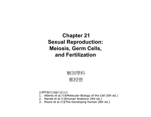
解 剖 學 科<br />
鄭 授 德<br />
此 PPT 檔 所 用 圖 片 取 自 於<br />
1. Alberts et al. 所 著 Molecular Biology of the Cell (5th ed.)<br />
2. Marieb et al. 所 著 Human Anatomy (4th ed.)<br />
3. Moore et al. 所 著 The Developing Human (8th ed.)
• Asexual reproduction<br />
– plants propagate vegetatively<br />
– Hydra can produce offspring by budding<br />
– offspring that are genetically identical to their parent<br />
• Sexual reproduction<br />
– mixes the genomes from two individuals
OVERVIEW OF SEXUAL REPRODUCTION<br />
• Diploid<br />
– each cell contains two sets of chromosomes,<br />
– one inherited from each parent<br />
• Haploid<br />
– each cell contains only one set of chromosomes<br />
– mixing the two genomes <strong>and</strong> restoring the diploid
<strong>Meiosis</strong><br />
– a diploid precursor cell gives rise to haploid progeny cells<br />
• haploid cells<br />
– produced by meiosis<br />
– develop into highly specialized gametes-eggs (or ova), sperm<br />
(or spermatozoa), pollen, or spores
<strong>Fertilization</strong><br />
• a haploid sperm fuses with a haploid<br />
egg to form a diploid cell (a fertilized<br />
egg, or zygote)<br />
• a new combination of chromosomes<br />
• the process of fertilization, in which an<br />
egg <strong>and</strong> a sperm fuse to form a new<br />
diploid organism
The Haploid Phase in Higher Eucaryotes<br />
Is Brief<br />
• diploid cells proliferate by mitotic cell division<br />
• haploid cells that form by meiosis do not<br />
proliferate<br />
• For all vertebrates<br />
– only the diploid cells proliferate<br />
– the haploid gametes exist only briefly for sexual<br />
fusion
• germ line (or germ cells)<br />
– include gametes <strong>and</strong> their specified diploid precursor cells<br />
• somatic cells<br />
– which form the rest of the body <strong>and</strong> ultimately leave no progeny
<strong>Meiosis</strong> Creates Genetic Diversity<br />
• Autosomes<br />
– common to all members of the species<br />
• Sex chromosomes<br />
– differently distributed according to the sex of the individual<br />
• homologous chromosomes, or homologs<br />
– two copies of each autosome, one from the mother <strong>and</strong> one from the<br />
father<br />
– physically pairing <strong>and</strong> undergoing genetic recombination
Crossing-over<br />
• the maternal <strong>and</strong> paternal versions of each chromosome have similar DNA<br />
sequences<br />
• they are not identical, <strong>and</strong> they undergo genetic recombination during<br />
meiosis<br />
• produce novel hybrid versions of each chromosome<br />
• each chromosome in a gamete contains a unique mixture of genetic<br />
information from both parents
Sexual Reproduction Gives Organisms a<br />
Competitive Advantage<br />
• resources spent on the machinery of sexual<br />
reproduction are large.<br />
• One advantage of sexual reproduction<br />
seems to be that the reshuffling of genes<br />
helps a species to survive in an<br />
unpredictably variable environment.<br />
• Another advantage of sexual reproduction<br />
seems to be that it can help eliminate<br />
deleterious genes from a population
MEIOSIS<br />
• haploid germ cells arise from a special kind of cell<br />
division in which the number of chromosomes is<br />
precisely halved<br />
• S phase is omitted; a single cell division produces two<br />
haploid cells directly<br />
• accurate chromosome segregation during meiosis
Gametes Are Produced by Two Meiotic<br />
Cell Divisions<br />
• the chromosomes have replicated their DNA (in meiotic S phase)<br />
• two copies are tightly bound together by cohesin complexes <br />
Sister chromatids<br />
• a single round of DNA replication is followed by two successive<br />
rounds of chromosome segregation
Duplicated Homologs (<strong>and</strong> Sex Chromosomes)<br />
Pair During Early Prophase I<br />
• Special mechanisms mediate these<br />
intimate interactions between homologs<br />
• Prolonged meiotic prophase (prophase I)<br />
• sister chromatids tightly glued together<br />
called pairing<br />
• forming a four-chromatid structure called<br />
a bivalent<br />
• breaks in chromatid double-str<strong>and</strong> DNA<br />
• a fragment of a maternal chromatid is<br />
exchanged for a corresponding fragment<br />
of a homologous paternal chromatid
• The ends of the chromosomes (the telomeres) are tightly<br />
bound to the inner surface of the nuclear envelope.<br />
• prevent chromosome entanglements<br />
• later still, disperse again
Pairing between sex chromosomes<br />
• A small region of homology between the X <strong>and</strong> the Y at one or both ends<br />
• only two types of sperm are normally produced<br />
• Sperms those containing one Y chromosome will give rise to male embryos<br />
• those containing one X chromosome will give rise to female embryos
Homolog Pairing Culminates in the<br />
Formation of a Synaptonemal Complex<br />
• The axial cores are about 400 nm apart<br />
• Programmed double-str<strong>and</strong> DNA breaks<br />
• Recombination complex assembles on a double-str<strong>and</strong> break<br />
• Presynaptic alignment of the homologs<br />
• During synapsis, the transverse filaments link the axial cores of a homolog<br />
pair as a synaptonemal complex, that bridges the gap of only 100 nm.
During dividing prophase I<br />
• Leptotene<br />
– homologs condense <strong>and</strong> pair, <strong>and</strong> genetic recombination begins<br />
• Zygotene<br />
– synaptonemal complex begins to assemble in local regions along the<br />
homologs<br />
• Pachytene<br />
– the homologs are synapsed along their entire lengths<br />
• Diplotene<br />
– disassembly of the synaptonemal complexes<br />
– concomitant condensation, shortening of chromosomes.
• Chiasmata (chiasma)<br />
– individual crossover events between nonsister chromatids<br />
• Diakinesis<br />
– the process of segregation<br />
– Prophase I ends; transition to metaphase I<br />
• proteins are important for crossing-over<br />
• Cohesin complexes<br />
– major components of the axial core of each homolog<br />
– play crucial parts in segregating
Homolog Segregation Depends on <strong>Meiosis</strong>-<br />
Specific, Kinetochore-Associated Proteins<br />
• Kinetochores<br />
– protein complexes associated with the centromeres<br />
• Kinetochores on the two sister chromatids attach to opposite poles of the<br />
spindle<br />
• Segregate into different daughter cells at anaphase<br />
• Arms of the sister chromatids of homologs separate at anaphase I
• Sisters stay glued together in the region of their centromeres until anaphase<br />
II<br />
• Shugoshins associated with kinetochores help ensure that sister<br />
kinetochores do not come apart at anaphase I<br />
• Proteolytic enzyme separase cleaves the cohesin complexes
• <strong>Meiosis</strong> II occurs without DNA replication.<br />
• Prophase II is brief<br />
• Metaphase II, anaphase II, <strong>and</strong> telophase II are in quick succession.<br />
• Four haploid nuclei produced<br />
• cytokinesis occurs<br />
• <strong>Meiosis</strong> is complete.
<strong>Meiosis</strong> Frequently Goes Wrong<br />
• female meiosis, which arrests for years after diplotene<br />
• meiosis I is completed only at ovulation<br />
• meiosis II only after the egg is fertilized.<br />
• such chromosome segregation errors during egg development are the<br />
commonest cause of both spontaneous abortion (miscarriage) <strong>and</strong> mental<br />
retardation in humans.<br />
• Nondisjunction<br />
– the result is that some of the haploid gametes produced lack a particular<br />
chromosome, while others have more than one copy of it.<br />
• Aneuploid<br />
– <strong>Cells</strong> with an abnormal number of chromosomes<br />
• Down syndrome<br />
– an extra copy of chromosome 21<br />
• Segregation errors during meiosis I increase greatly with advancing<br />
maternal age.
Normal<br />
Gametogenesis
Nondisjunction<br />
Homologs fail to<br />
separate; about 10% in<br />
human oocytes,<br />
inducing miscarriages or<br />
Down's syndrome
Nondisjunction
Genetic Reassortment<br />
• R<strong>and</strong>om distribution of maternal <strong>and</strong><br />
paternal homologs produce 2 n genetically<br />
different gametes.<br />
• 2 23 = 8.4 × 10 6
Crossing-Over Enhances Genetic Reassortment<br />
• Chromosomal crossing-over<br />
– DNA segments of homologous<br />
chromosomes are exchanged<br />
• On average, between two <strong>and</strong> three<br />
crossovers occur between each pair of<br />
human homologs.
Crossing-Over Is Highly Regulated<br />
• hold homologs together; properly segregated<br />
• double-str<strong>and</strong> breaks at “hot spots”<br />
• Crossovers operate before the synaptonemal complex assembles.<br />
• at least one crossover forms between the members of each homolog pair.<br />
• Crossover interference<br />
– presence of one crossover event inhibits another from forming close by
<strong>Meiosis</strong> Is Regulated Differently in<br />
Male <strong>and</strong> Female Mammals<br />
• <strong>Meiosis</strong> in a human male lasts for 24 days, compared<br />
with 12 days in the mouse.<br />
• In human females, it can last 40 years or more, because<br />
meiosis I arrests after diplotene.<br />
• Most of meiosis is spent in prophase I<br />
• Egg precursor cells (oocytes) begin meiosis in the fetal<br />
ovary but arrest after diplotene, after the synaptonemal<br />
complex has disassembled in meiosis I.<br />
• complete meiosis I<br />
– only after the female has become sexually mature<br />
– the oocyte is released from the ovary during ovulation<br />
• completes meiosis II<br />
– only if it is fertilized.
• It takes about 24 days for a human spermatocyte to<br />
complete meiosis.<br />
• About 20% of human eggs are aneuploid, compared with 3–<br />
4% of human sperm<br />
• Cell-cycle checkpoint mechanism arrests meiosis <strong>and</strong> leads<br />
to cell death by apoptosis.<br />
• The average number of new mutations contributed by<br />
fathers is larger than the number contributed by mothers.<br />
• by the end of meiosis, a mammalian egg is fully mature,<br />
whereas a sperm that has completed meiosis has only just<br />
begun its differentiation.
PRIMORDIAL GERM CELLS AND SEX<br />
DETERMINATION IN MAMMAL<br />
• Diploid primordial germ cells (PGCs) migrate to the<br />
developing gonads, which will form the ovaries in<br />
females <strong>and</strong> the testes in males.<br />
• Mitotic proliferation in the developing gonads<br />
• PGCs undergo meiosis <strong>and</strong> differentiate into mature<br />
haploid gametes - either eggs or sperm.
Signals from Neighbors Specify PGCs in<br />
Mammalian Embryos<br />
• germ cell determinants become PGCs<br />
• antibody that labels (in green) small granules (called P<br />
granules) that function as germ cell determinants.
• Totipotent – the potential to give rise to any of the cell types of the animal,<br />
including the germ cells<br />
• turning off the expression of a number of somatic cell genes <strong>and</strong> turning on<br />
the expression of genes involved in maintaining the special character of the<br />
germ cells.
Migration of mammalian PGCs<br />
• secreted signal proteins that help attract PGCs into the<br />
developing gonad at genital ridge are chemokines<br />
• embryonic germ (EG) cells resemble embryonic stem<br />
(ES) cells<br />
• The sex chromosomes in the somatic cells of the<br />
genital ridge determine which type of gonad the ridge<br />
becomes.<br />
• a single gene on the Y chromosome has an especially<br />
important role.
Male<br />
Female
The Sry Gene Directs the Developing<br />
Mammalian Gonad to Become a Testis<br />
• In egg-laying reptiles, the temperature of the incubating egg determines the<br />
sex of the offspring<br />
• The presence or absence of the Y chromosome determines the sex of the<br />
individual.<br />
• The Y chromosome directs the somatic cells of the genital ridge to develop<br />
into a testis.<br />
• the secreted signal protein Wnt4, for example, is required for normal<br />
mammalian ovary development.<br />
• The crucial gene on the Y chromosome is called Sry (sex-determining<br />
region of Y).<br />
• Sex-reversed mice cannot produce sperm, because they lack genes for<br />
sperm development.<br />
• Sry gene is introduced into the genome of an XX mouse zygote, the<br />
transgenic embryo produced develops as a male.<br />
• Sertoli cells are the main type of supporting cells in the testis.
Four ways Sertoli cells influencing other cells<br />
1. stimulate the PGCs entering<br />
meiosis <strong>and</strong> produce sperm.<br />
2. secrete anti-Müllerian hormone<br />
causing the Müllerian duct to<br />
regress<br />
3. stimulate endothelial <strong>and</strong> smooth<br />
muscle cells migrate into the<br />
developing gonad<br />
4. induce other somatic cells to<br />
become Leydig cells, which secrete<br />
the male sex hormone testosterone<br />
for developing Wolffian duct system,<br />
<strong>and</strong> male sexual identity
• Sry normally acts by inducing the<br />
expression of Sox9.<br />
• The Sox9 protein directly activates the<br />
gene encoding anti-Müllerian hormone.<br />
• In the absence of either Sry or Sox9, the<br />
genital ridge of an XY embryo develops<br />
into an ovary instead of a testis.<br />
• The supporting cells become follicle cells<br />
instead of Sertoli cells.<br />
• Other somatic cells become theca cells<br />
(instead of Leydig cells),<br />
• PGCs develop along a pathway that<br />
produces eggs rather than sperm.
• molecule retinoic acid produced by mesonephros adjacent to the developing<br />
gonad induces the proliferating germ-line cells to enter meiosis.<br />
• arrested after diplotene of prophase I, but still ovulate<br />
• Sertoli cells produce an enzyme that degrades retinoic acid, <strong>and</strong> prevent the<br />
germ-line cells to enter meiosis.<br />
• Much later to begin producing sperm.
Many Aspects of Sexual Reproduction<br />
Vary Greatly between Animal Species<br />
• C. elegans <strong>and</strong> Drosophila<br />
– by the ratio of X chromosomes to autosomes<br />
• C. elegans<br />
– transcriptional <strong>and</strong> translational controls on gene expression<br />
• Drosophila<br />
– on a cascade of regulated RNA splicing events,
EGGS<br />
• once activated, it can give rise to a complete new individual<br />
• Activation is usually the consequence of fertilization (fusion of a sperm with<br />
the egg).<br />
• Some lizards’ eggs become activated in the absence of sperm<br />
(parthenogenetically).<br />
• Mammals cannot; because of genomic imprinting (they require both<br />
maternal <strong>and</strong> paternal genetic contributions).<br />
• The cytoplasm of an egg can even reprogram a somatic cell nucleus so that<br />
the nucleus can direct the development of a new individual.<br />
• Reproductive cloning has been used to produce clones of various mammals.<br />
• Less than 5% of them develop to adulthood
An Egg Is Highly Specialized for<br />
Independent Development<br />
• Egg<br />
– giant single cell<br />
– cleaves into many smaller cells<br />
– diameter of about 0.1 mm in humans <strong>and</strong> sea urchins<br />
– 1–2 mm in frogs <strong>and</strong> fishes<br />
– many centimeters in birds <strong>and</strong> reptiles<br />
• A somatic cell has a diameter of only about 10–30<br />
micrometer
• Yolk<br />
– rich in lipids, proteins, <strong>and</strong> polysaccharides <strong>and</strong> yolk granules.<br />
• Egg coat<br />
– Extracellular matrix consisting largely of glycoproteins<br />
– Vitelline layer in eggs of sea urchins or chickens<br />
– Zona pellucida in mammalian eggs<br />
– acts as a species-specific barrier to sperm
• Cortical granules<br />
– specialized secretory vesicles in cortex of the egg cytoplasm.<br />
– exocytosis the contents of the granules alter the egg coat<br />
– so as to help prevent more than one sperm from fusing with the egg.<br />
• Some egg cytoplasmic components have a strikingly asymmetrical<br />
distribution to help establish the polarity of the embryo
Oogenesis<br />
Eggs Develop in Stages<br />
• differentiates into a mature egg (or ovum)<br />
• Primordial germ cells migrate to the forming gonad to become oogonia<br />
• proliferate by mitosis for a period before beginning meiosis I, at which point<br />
they are called primary oocytes.<br />
• oocytes arrest for a prolonged period in meiosis I<br />
• after completing meiosis I, they arrest again in metaphase II while awaiting<br />
fertilization
• before birth in mammals.<br />
• prior to the onset of meiosis I, the DNA replicates<br />
• start of prophase I crossing-over occurs between nonsister chromatids<br />
• arrested after diplotene of prophase I<br />
• primary oocytes synthesize a coat <strong>and</strong> cortical granules.<br />
• intensive biosynthetic activities<br />
• progress through meiosis I.<br />
• replicated homologous chromosomes segregate at anaphase I<br />
• a small polar body <strong>and</strong> a large secondary oocyte
• each chromosome is still composed of two sister chromatids held together<br />
at their centromeres.<br />
• At ovulation, the arrested secondary oocyte is released from the ovary,<br />
ready to be fertilized.<br />
• fertilization occurs, the cell completes meiosis, becoming a mature egg.<br />
• anaphase II, the large secondary oocyte divides to produce the mature egg<br />
(or ovum) <strong>and</strong> a second polar body<br />
• Because it is fertilized, it is also called a zygote.
Oocytes Use Special Mechanisms to<br />
Grow to Their Large Size
Extra gene copies in the cell<br />
• diploid chromosome set for RNA synthesis with 100 to<br />
500 copies of the ribosomal RNA genes<br />
• increased rate of protein synthesis<br />
From neighboring accessory cells<br />
• Yolk imported into the oocyte<br />
• In some invertebrates a common oogonium gives rise to<br />
one oocyte <strong>and</strong> 15 nurse cells<br />
• follicle cells surround each developing oocyte<br />
• gap-junction proteins (connexins) involved in connecting<br />
follicle cells to each other are different from those<br />
connecting follicle cells to the oocyte.
Most Human Oocytes Die Without Maturing<br />
• A single layer of follicle cells surrounds most of the primary oocytes in<br />
newborn girls.<br />
• Such an oocyte, together with its surrounding follicle cells, is called a<br />
primordial follicle<br />
• a small proportion of primordial follicles begin to grow to become developing<br />
follicles, in which multiple layers of follicle cells (now called granulosa cells)<br />
• Some of these developing follicles go on to acquire a fluid-filled cavity, or<br />
antrum, to become antral follicles.
• Follicle stimulating hormone (FSH) accelerates the growth of about 10–12<br />
antral follicles<br />
• Luteinizing hormone (LH) triggers ovulation<br />
• the dominant primary oocyte completes meiosis I<br />
• the resulting secondary oocyte arrests at metaphase II
Premordial follicle <strong>and</strong> Primary follicle<br />
• follicle cells secrete macromolecules<br />
• The communication between oocytes <strong>and</strong> their follicle cells
Secondary Follicle
Secondary Follicle
Oocyte in Follicle
Ovulated<br />
Oocyte
SPERM<br />
• Sperm (spermatozoon, plural spermatozoa) is often the smallest.<br />
• highly motile <strong>and</strong> streamlined for speed <strong>and</strong> efficiency in the task of<br />
fertilization.
Sperm Are Highly Adapted for Delivering<br />
Their DNA to an Egg<br />
• Head<br />
– highly condensed haploid nucleus<br />
– DNA in the nucleus is extremely tightly packed<br />
– chromosomes of many sperm lack the histones, but with protamines<br />
– Acrosomal vesicle - specialized secretory vesicle<br />
• Midpiece<br />
• Tail<br />
– Mitochondria - ATP is generated<br />
– strong flagellum propels the sperm<br />
– central axoneme emanates from a basal body<br />
– With two central singlet microtubules surrounded by nine evenly<br />
spaced microtubule doublets.<br />
– sliding of adjacent microtubule doublets past one another, driven by<br />
dynein motor proteins<br />
• Acrosome reaction<br />
– a sperm contacts the egg coat, the contents of the vesicle are<br />
released by exocytosis
Sperm Are Produced Continuously in the<br />
Mammalian Testis<br />
• Seminiferous tubules<br />
– epithelial lining of very long, tightly coiled<br />
tubes<br />
• Spermatogonia (singular,<br />
spermatogonium)<br />
– Immature germ cells<br />
– Stem cells - divide slowly by mitosis<br />
• Primary spermatocytes<br />
• <strong>Meiosis</strong> I<br />
• Secondary spermatocytes<br />
– 22 duplicated autosomal chromosomes<br />
– a duplicated X or Y chromosome<br />
• <strong>Meiosis</strong> II<br />
• Spermatids
• Epididymis<br />
– a coiled tube overlying the testis<br />
– where they are stored <strong>and</strong> undergo further maturation.<br />
• Capacitation<br />
– The stored sperm undergo further maturation in the female genital.
•Seminiferous Tubule
Sperm Develop as a Syncytium<br />
• no longer complete cytoplasmic division<br />
(cytokinesis)<br />
• connected by cytoplasmic bridges,<br />
forming a syncytium<br />
• individual sperm are released into the<br />
lumen of the seminiferous tubule.
FERTILIZATION<br />
• the binding of sperm to the egg coat (zona pellucida) induces the acrosome<br />
reaction<br />
• required for the sperm to burrow through the zona <strong>and</strong> fuse with the egg.<br />
• binding of the sperm to the egg plasma membrane<br />
• fusion with these membranes<br />
• sperm activates the egg<br />
• haploid nuclei of the two gametes come together
Ejaculated Sperm Become Capacitated in<br />
the Female Genital Tract<br />
• 300,000,000 or so human sperm ejaculated<br />
• only about 200 reach the site of fertilization<br />
• a sperm migrates through the layers of granulosa cells<br />
• cross the zona pellucida<br />
• bind to <strong>and</strong> fuse with the egg plasma membrane
Capacitation<br />
• sperm arrive in the oviduct.<br />
• takes about 5–6 hours in humans<br />
• changes in glycoproteins, lipids, <strong>and</strong> ion channels<br />
• change in the resting potential of this membrane<br />
• increase in cytosolic pH<br />
• tyrosine phosphorylation<br />
• unmasking of cell-surface receptors<br />
• greatly increases the motility of the flagellum<br />
• capable of undergoing the acrosome reaction.
Capacitated Sperm Bind to the Zona Pellucida<br />
<strong>and</strong> Undergo an Acrosome Reaction
<strong>Fertilization</strong>
Prevention of Polyspermy<br />
1. Change of membrane potential<br />
2. Formation of fertilization envelope<br />
A Model of <strong>Fertilization</strong><br />
• Strongylocentrotus purpuratus<br />
• Arbacia punctulata<br />
Ovulating Sea Urchin
TEM of the sea urchin egg<br />
• Cell membrane with microvilli<br />
• Coated with vitelline layer<br />
• Underlined with cortical<br />
granules <strong>and</strong> vesicles
Elevation of Vitelline Layer
Sperm Fusion Activates the Egg by<br />
Increasing Ca 2+ in the Cytosol<br />
• Albumin<br />
– extract cholesterol from the plasma membrane<br />
– increasing the ability of sperm cell membrane to fuse with the<br />
acrosome membrane during the acrosome reaction.<br />
• Ca 2+ <strong>and</strong> HCO 3<br />
–<br />
– activate adenylyl cyclase<br />
– produce cyclic AMP
Acrosomal Reaction
The Cortical Reaction Helps Ensure That<br />
Only One Sperm Fertilizes the Egg
Cortical Reaction
The Sperm Provides Centrioles as Well<br />
as Its Genome to the Zygote<br />
• Two haploid nuclei (pronuclei) have come together <strong>and</strong> combined their<br />
chromosomes into a single diploid nucleus<br />
• Centrosome duplicates<br />
• Organize the assembly of the first mitotic spindle in the zygote
<strong>Fertilization</strong>
(A) A meiotic spindle in a mature unfertilized<br />
secondary oocyte.<br />
(B) A fertilized egg extruding its second polar body<br />
about 5 hours after fusion with a sperm.<br />
The sperm head (left) has nucleated an array of<br />
microtubules.<br />
The egg <strong>and</strong> sperm pronuclei are still far apart.<br />
(C) The two pronuclei have come together.<br />
(D) By 16 hours after fusion with a sperm, the<br />
centrosome that entered the egg with the<br />
sperm has duplicated, <strong>and</strong> the daughter<br />
centrosomes have organized a bipolar mitotic<br />
spindle.<br />
The chromosomes of both pronuclei are<br />
aligned at the metaphase plate of the spindle.
IVF <strong>and</strong> ICSI Have Revolutionized the<br />
Treatment of Human Infertility<br />
• in vitro fertilization (IVF) 1978<br />
– multiple pregnancies occur in over 30% of cases<br />
– compared with about 2% in unassisted pregnancies.<br />
• intracytoplasmic sperm injection (ICSI) 1992<br />
– an egg is fertilized by injecting a single sperm into it<br />
– ICSI has a success rate of better than 50%
• Transferring the nucleus of a somatic cell from the animal to be cloned into<br />
an unfertilized egg that has had its own nucleus removed or destroyed.