The meninges in Cyclostomes, Selachians, and Teleosts ... - DWC
The meninges in Cyclostomes, Selachians, and Teleosts ... - DWC
The meninges in Cyclostomes, Selachians, and Teleosts ... - DWC

Create successful ePaper yourself
Turn your PDF publications into a flip-book with our unique Google optimized e-Paper software.
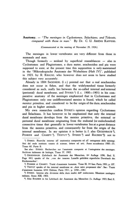
Anatomy. - "<strong>The</strong> <strong>men<strong>in</strong>ges</strong> <strong>in</strong> <strong>Cyclostomes</strong>. <strong>Selachians</strong>. <strong>and</strong> <strong>Teleosts</strong>.<br />
comp~red with those <strong>in</strong> man." By Dr. C. U . ARIËNS KAPPERS.<br />
(Communicated at the meet<strong>in</strong>g of November 29, 1924.)<br />
<strong>The</strong> <strong>men<strong>in</strong>ges</strong> <strong>in</strong> lower vertebrates are very different from those <strong>in</strong><br />
mammals <strong>and</strong> man.<br />
Though formerly - mislead by superficial resemblances - also <strong>in</strong><br />
<strong>Cyclostomes</strong> <strong>and</strong> Plagiostomes, a dura mater. arachnoidea <strong>and</strong> pia were<br />
supposed to exist, at the present time th is supposition is only ma<strong>in</strong>ta<strong>in</strong>ed<br />
<strong>in</strong> the "Mikroskopische Anatomie der Wirbeltiere (Heft IV)". published<br />
<strong>in</strong> 1923. by R. KRAusE, who however does not seem to have studied<br />
this subject very accurately.<br />
Already <strong>in</strong> 1884 SAGEMEHL (1. c.) po<strong>in</strong>ted out that a real arachnoidea<br />
does not occur <strong>in</strong> fishes, <strong>and</strong> that the widelymeshed tissue formerly<br />
considered as such, really lies between the so~called <strong>in</strong>ternal <strong>and</strong> external<br />
(periostal) dural membrane, <strong>and</strong> STERZI 1) (1. c. 1900-1901) <strong>in</strong> his com~<br />
parative anatomy of the <strong>men<strong>in</strong>ges</strong> emphasized that <strong>in</strong> <strong>Cyclostomes</strong> <strong>and</strong><br />
Plagiostomes only one undifferentiated men<strong>in</strong>x is found. which he ca lied<br />
men<strong>in</strong>x primitiva. <strong>and</strong> considered to be the orig<strong>in</strong> of the dura. arachnoidea<br />
<strong>and</strong> pia <strong>in</strong> higher animals.<br />
My own researches confirm STERzi's op<strong>in</strong>ion regard<strong>in</strong>g <strong>Cyclostomes</strong><br />
<strong>and</strong> <strong>Selachians</strong>. It has however to be emphasized that only the <strong>in</strong>ternal<br />
dural membrane develops from the men<strong>in</strong>x primitiva. the externalor<br />
periostal dural membrane orig<strong>in</strong>at<strong>in</strong>g from the endostal (or endochondral)<br />
connective tissue that generally <strong>in</strong> lower vertebrates lies at a great distance<br />
from the men<strong>in</strong>x primitiva. <strong>and</strong> consequently far from the orig<strong>in</strong> of the<br />
<strong>in</strong>ternal membrane. In my op<strong>in</strong>ion it is better (c. f. also GEGENBAUR 2).<br />
POIRIER <strong>and</strong> CHARPY 3). TESTUT 1). STERZI 5) <strong>and</strong> RAUBER 6)) not to<br />
1) STERZI. Ricerche <strong>in</strong>torno all ' anatomica comparata ed all' ontogenesi delle men<strong>in</strong>gi.<br />
Atti del reale <strong>in</strong>stituto veneto di seienze, lettere ed arti. Anno accademico 1900-01.<br />
Tomo 60. Parte 11,<br />
See also : STERZI. Recherches sur I'anatomie comparée et I'ontogénèse des <strong>men<strong>in</strong>ges</strong>,<br />
Archives italiennes de biologie, Tomo 37. 1902.<br />
2) GEGENBAUR. (Lehrbuch der Anatomie des Mensehen, 61e AuBage 1896, Bnd, 11,<br />
Page Hl) speaks of the "von der <strong>in</strong>neren Lamelle gebildete eigentliche Duralsack des<br />
Rückenmarkes" .<br />
3) POIRIER et CHARPY, Traité d'anatomie huma<strong>in</strong>e, Tome III. lier fase, Paris. 1901, p. 107.<br />
1) TESTUT speaks of the <strong>in</strong>ternal membrane as "dure mère propement dite", See: Traité<br />
d'anatomie huma<strong>in</strong>e, 61tme Edition, Paris 1911, p, 1050-1061.<br />
5) STERZI. Intorno alla divisione della dura madre delr endocranio, Monitore zoologico<br />
italiano. Anno XIII. 1902,<br />
6) Also RAUBER <strong>in</strong> his Lehrbuch der Anatomie des Mensehen (Ie Auftage 1903, Bnd, 11,
C. U. ARIËNS KAPPERS: "<strong>The</strong> meoioges io Cydostomes, <strong>Selachians</strong><br />
aod <strong>Teleosts</strong>, compared with those <strong>in</strong> man".<br />
perimen. tissue<br />
men. primo<br />
lig. lat.<br />
Fig. 1. Sp<strong>in</strong>al cord of Petromyzon <strong>in</strong> situ<br />
x x Space between men<strong>in</strong>x primitiva <strong>and</strong> cord. caused by retraction.<br />
mem. primo<br />
endochondr.<br />
peri men<strong>in</strong>gtissue<br />
vertebra<br />
retract.<br />
lig. lat.<br />
lig. lat.<br />
chorda men <strong>in</strong> x primo<br />
Fig. 2. Sp<strong>in</strong>al cord of Petromyzon <strong>in</strong> situ.<br />
x x x Spaces caused by retraction.<br />
Proceed<strong>in</strong>gs Royal Acad. Amsterdam. Vo\. XXVIII.
73<br />
eonsider the so-ealled externalor periostal membrane of the (sp<strong>in</strong>al) dura<br />
(which follows all the s<strong>in</strong>uosities of the bone) as a part of the dura<br />
proper. though it fuses with it <strong>in</strong> the eranial eavity <strong>in</strong> the adult (af ter an<br />
embryonie eondition <strong>in</strong> which it may be dist<strong>in</strong>guished from it).<br />
<strong>The</strong> dist<strong>in</strong>etion of aperiostal <strong>and</strong> an <strong>in</strong>ternal layer <strong>in</strong> the dura mater<br />
sp<strong>in</strong>alis only leads to eonfusion <strong>and</strong> to the idea of an ambiguous<br />
membrane. whieh ambiguity disappears by leav<strong>in</strong>g the so-ealled<br />
periostal dural membrane there. where it belongs af ter its nature <strong>and</strong><br />
orig<strong>in</strong>. viz. to the eonneetive tissue of the endoehondrium or endost <strong>and</strong><br />
not to the dura.<br />
Coneern<strong>in</strong>g the <strong>Cyclostomes</strong>. I found <strong>in</strong> Petromyzon fluv. (fig. l<strong>and</strong><br />
2) relations. as deseribed by STERZI.<br />
<strong>The</strong> medulla sp<strong>in</strong>alis is surrounded by a s<strong>in</strong>gle membrane <strong>in</strong> which<br />
no differentiation <strong>in</strong>to separate layers is visible. I eall this membrane.<br />
with STERZI. men<strong>in</strong>x primitiva. It is eont<strong>in</strong>uous with the sheath of the<br />
roots.<br />
This membrane. which shows strong lateral ligaments extend<strong>in</strong>g far<br />
laterally <strong>in</strong>to the perimen<strong>in</strong>geal tissue (fig. 1. 2) does not yet penetrate<br />
with septa <strong>in</strong>to the substanee of the sp<strong>in</strong>al ehord. so that the membrane<br />
easily detaehes (XX) from the eord. <strong>The</strong> nutrition of the sp<strong>in</strong>al eord<br />
has to pass everywhere through the superficial glious layer (Iimitans superficialis)<br />
I). th ere be<strong>in</strong>g no <strong>in</strong>tramedullary septa <strong>and</strong> <strong>in</strong>tramedullary vessels.<br />
Outside this men<strong>in</strong>x primitiva. which. as I shall show later on. also<br />
eonta<strong>in</strong>s the anlage of the dura mater (<strong>in</strong> the sense of the word mentioned<br />
above) lies a broad layer of large eells. which is to be eonsidered as a<br />
fill<strong>in</strong>g-tissue <strong>and</strong> reaehes as far as the endoehondrium of the vertebrae.<br />
This perimen<strong>in</strong>geal tissue eonsists of round <strong>and</strong> oval mueous eells.<br />
In some of my preparations spaces occur, that look like epidural s<strong>in</strong>usses. which also <strong>in</strong><br />
human embryos (v. GELDEREN l.c.) are found between the so-called <strong>in</strong>terior layer of the<br />
dura <strong>and</strong> the so-called periostal layer. I could not f<strong>in</strong>d however traces of blood here. <strong>The</strong>y<br />
are also more or less localspaces that do not spread over a great length. As I could<br />
not f<strong>in</strong>d any connection with the venae <strong>in</strong>vertebrales, they probably are retraction cavities<br />
(caused by the fixation).<br />
<strong>The</strong> relations <strong>in</strong> Plagiostomes do not show mueh differenee herewith.<br />
p. 337) speaks of "Lam<strong>in</strong>a <strong>in</strong>terna oder Dura sp<strong>in</strong>alis <strong>in</strong> engerem S<strong>in</strong>ne". A similar op<strong>in</strong>ion<br />
is given <strong>in</strong> CUNNINGHAM 's Textbook of Anatomy (3d Ed. 1909, page 600).<br />
I) Already <strong>in</strong> <strong>Selachians</strong>, men<strong>in</strong>geal septa with bloodvessels grow <strong>in</strong>to the sp<strong>in</strong>al cord,<br />
caus<strong>in</strong>g a doser relation between the nervous substance <strong>and</strong> the vascular system. As<br />
however the limitans gliosa superficialis grows at the same time with those septa, a real<br />
penetration of men<strong>in</strong>geal tissue <strong>in</strong>to the nervous substance itself does not occur. In fact<br />
the septalspaces are to be considered as the fissures <strong>in</strong> the forebra<strong>in</strong>, with th is difference<br />
that they are much smaller <strong>and</strong> for the greater part filled up with pia tissue, while the<br />
arachnoidal cavities <strong>in</strong> the bra<strong>in</strong> fissures are much wider <strong>and</strong> go further downwards.<br />
<strong>The</strong>y also resembie each other by the fact that the dura rema<strong>in</strong>s outside them, <strong>in</strong> the<br />
bra<strong>in</strong>, as weil as <strong>in</strong> the sp<strong>in</strong>al cord.
In Scyllium I also found only one men<strong>in</strong>x. to be called men<strong>in</strong>x primitiva.<br />
<strong>in</strong> which no differentiation <strong>in</strong> separate layers is visible. It conta<strong>in</strong>s small<br />
blood vessels. that penetrate with men<strong>in</strong>geal septa <strong>in</strong>to the sp<strong>in</strong>al cord.<br />
Moreover the sharks show the four sp<strong>in</strong>alligaments al ready described<br />
by STERZI : the rather strongly pronounced lateralligaments (fig. 3 <strong>and</strong> 4).<br />
<strong>and</strong> the th<strong>in</strong>ner. of ten scarcely developed. dorsal <strong>and</strong> ventral ligament.<br />
Only the lateral ligament extends for some distance through the perimen<strong>in</strong>geal<br />
tissue. <strong>The</strong> others are merely thicken<strong>in</strong>gs of the men<strong>in</strong>x primitiva.<br />
Besides this men<strong>in</strong>x primitiva which just as <strong>in</strong> <strong>Cyclostomes</strong>. cont<strong>in</strong>ues <strong>in</strong><br />
the rootsheath. a large amount of peri-men<strong>in</strong>geal tissue is found which<br />
reaches as far as the endochondrium <strong>and</strong> shows much more widely spread<br />
meshes than <strong>in</strong> <strong>Cyclostomes</strong>.<br />
Only here <strong>and</strong> th ere - especially <strong>in</strong> the neighbourhood of the endochondrium<br />
- it is a little more compact (fig. 4).<br />
Large th<strong>in</strong>walled ve<strong>in</strong>s (without muscular coat) are seen <strong>in</strong> the perimen<strong>in</strong>geal<br />
tissue. especially on the dorsal <strong>and</strong> lateral side. <strong>The</strong>re is no<br />
doubt as far as concerns their homology with the so-called epidural ve<strong>in</strong>s<br />
<strong>in</strong> mammals <strong>and</strong> man. though they are relatively much larger <strong>and</strong> therefore<br />
resembie s<strong>in</strong>usses. I)<br />
<strong>The</strong> relations <strong>in</strong> Ganoids (Acipenser <strong>and</strong> Polyodon) are similar to those<br />
<strong>in</strong> <strong>Selachians</strong>: so I shall not describe them aga<strong>in</strong>.<br />
My researches concern<strong>in</strong>g the relations <strong>in</strong> the most specialized group<br />
of fishes. the <strong>Teleosts</strong>. show that there are <strong>in</strong> this subdivision large<br />
differences <strong>in</strong> men<strong>in</strong>geal structure that might expla<strong>in</strong> the rather different<br />
descriptions present <strong>in</strong> litterature. if the chief <strong>and</strong> rather different descriptions<br />
those of SAGEMEHL <strong>and</strong> STERZI - were not both based<br />
partlyon the same material.<br />
STERZI (l.c. I. page 1142) exam<strong>in</strong>ed espeeially T<strong>in</strong>ea <strong>and</strong> further Cypr<strong>in</strong>ius. Esox.<br />
Barbus. Muraena. Anguilla. Rhombus. Solea <strong>and</strong> Labrax. <strong>and</strong> found everywhere I!nder<br />
the generally perimen<strong>in</strong>geal tissue only one men<strong>in</strong>x. which he ealls men<strong>in</strong>x primitiva. just<br />
as <strong>in</strong> <strong>Cyclostomes</strong> <strong>and</strong> Plagiostomes.<br />
He dist<strong>in</strong>guishes there<strong>in</strong> two layers. an exterior <strong>and</strong> <strong>in</strong>terior layer. <strong>The</strong> exterior lager<br />
is very th<strong>in</strong> <strong>and</strong> consists almost entirely of large Hattened eells. more or less pigmented.<br />
Further there are. star-shaped eells <strong>and</strong> some eells with long offshoots <strong>and</strong> eonneet<strong>in</strong>g<br />
ramifieations. Between them there are some pigmentcelIs. In th is layer also some elastic<br />
fibres are &een. He does not speak of a fibreus thicken<strong>in</strong>g of this exterior layer which<br />
might <strong>in</strong>dicate a dural development.<br />
<strong>The</strong> <strong>in</strong>terior layer is more developed. On this 2) (<strong>in</strong> this ?l layer. the bloodvessels oceur<br />
that supply the medulla. It consists of eonneetive tissue fibrils <strong>and</strong> elastic fibres. cross<strong>in</strong>g<br />
eaeh other <strong>in</strong> different direetions.<br />
He further mentions the ligaments <strong>and</strong> remarks that the sheath of the men<strong>in</strong>x eont<strong>in</strong>ues<br />
<strong>in</strong> the rootsheath.<br />
Coneern<strong>in</strong>g the perimen<strong>in</strong>geal space. he remarks that the latter is well developed dorsally<br />
I) Similar large epidural ve<strong>in</strong>s oceur <strong>in</strong> Carnivora. Edentates. Cetacea <strong>and</strong> Elephas.<br />
where the epidural spaee still prevails on the araehnoidea.<br />
2) <strong>The</strong> author (l.c. page 1143) says : "Su di esso poggiano i vasi."
C. U. ARI~NS KAPPERS I "<strong>The</strong> <strong>men<strong>in</strong>ges</strong> <strong>in</strong> <strong>Cyclostomes</strong>, <strong>Selachians</strong><br />
<strong>and</strong> <strong>Teleosts</strong>, compared with those of man".<br />
endochondr.<br />
perimen.<br />
tissue<br />
men.<br />
pro m.<br />
epidur.<br />
ve<strong>in</strong>'<br />
radix<br />
post.<br />
vertebra<br />
lig. lat.<br />
Fig. 3.<br />
art. vert. ant.<br />
Sp<strong>in</strong>al cord of Scyllium<br />
canicula <strong>in</strong><br />
situ.<br />
Epidur.<br />
men. primo ve<strong>in</strong> Endochondr.<br />
vertebra<br />
lig . lat.<br />
epidur.<br />
s<strong>in</strong>us<br />
men. primo<br />
rad. ant.<br />
rad. ant.<br />
perimen. tissue arteria vertebr. ant.<br />
Fig. 4. Sp<strong>in</strong>al cord of Scyllium canicula <strong>in</strong> situ.<br />
Proceed<strong>in</strong>gs Royal Acad. Amsterdam. Vol. XXVIII.
75<br />
<strong>and</strong> ventrally but may be reduced laterally to a narrow fissure. <strong>The</strong> perimen<strong>in</strong>geal tissue ')<br />
<strong>in</strong> this cavity consists of f<strong>in</strong>e trabecles between which lie large fatcells. Also many small<br />
vessels are found there. It has the function of a perimen<strong>in</strong>geal fill<strong>in</strong>g -tissue. Similar<br />
relations were found <strong>in</strong> the other bony-fishes which he exa m<strong>in</strong>ed.<br />
Quite different was the description given before him (1884) by SAGEMEHL2) who <strong>in</strong><br />
Siluroides <strong>and</strong> Cypr<strong>in</strong>oides (especially <strong>in</strong> Barbus <strong>and</strong> Perca) dist<strong>in</strong>guishes two <strong>men<strong>in</strong>ges</strong>,<br />
dist<strong>in</strong>ctly separated by a fissure, which he calls "pericerebraler Lymphraum" <strong>and</strong> which<br />
he considers to be the homologue of the subdural fissure <strong>in</strong> mammais.<br />
<strong>The</strong> membrane Iy<strong>in</strong>g <strong>in</strong>teriorly to this fissure is, accord<strong>in</strong>g to him. the orig<strong>in</strong> of both<br />
the pia <strong>and</strong> arachnoidea 3), although one cannot yet dist<strong>in</strong>guish there<strong>in</strong> those two<br />
membranes. <strong>The</strong> only dist<strong>in</strong>ction to be made <strong>in</strong> th is "vascular-membrane" ("Gefäszhaut"<br />
as he calls it) is that only the <strong>in</strong>terior layer of th is mem bra ne cont<strong>in</strong>ues <strong>in</strong>to the fissure<br />
media na anterior, while the exterior layer lies over it like a bridge. Thus far his description<br />
of the part Iy<strong>in</strong>g under the subdural fissure.<br />
Everyth<strong>in</strong>g outside the "pericerebrale Lymphra um " 4) SAGEMEHL considers as a dura<br />
mater <strong>in</strong> the old sense of the word, consider<strong>in</strong>g as such not only our fibrous dural<br />
membrane, but also our perimen<strong>in</strong>geal tissue <strong>and</strong> our periostal membrane (l.c. page 460-464).<br />
Resum<strong>in</strong>g we may say that there is a good deal of difference between<br />
the op<strong>in</strong>ions of SAGEMEHL <strong>and</strong> STERZJ. STERZI presumes that <strong>in</strong> fishes<br />
th ere is only one men<strong>in</strong>x which he calls men<strong>in</strong>x primitiva, which <strong>in</strong>cludes<br />
both the dura <strong>and</strong> the lepto-<strong>men<strong>in</strong>ges</strong> <strong>in</strong> an undifferentiated state. while<br />
SAGEMEHL dist<strong>in</strong>guishes a dural membrane <strong>and</strong> under it. separated by<br />
a fissure. a tissue which is the orig<strong>in</strong> of pia <strong>and</strong> arachnoidea (a men<strong>in</strong>x<br />
secundaria as STERZI caUs it <strong>in</strong> Reptiles <strong>and</strong> Birds). This men<strong>in</strong>x secundaria<br />
("Gefäszhaut" of SAGEMEHL) shows only <strong>in</strong> some places a differentiation<br />
<strong>in</strong> an <strong>in</strong>ner <strong>and</strong> outer membrane. which however has noth<strong>in</strong>g<br />
<strong>in</strong> common with the differentiation <strong>in</strong> arachnoidea <strong>and</strong> pia.<br />
<strong>The</strong> difference between these authors is the more strik<strong>in</strong>g as both. at<br />
least partly, exam<strong>in</strong>ed the same material (Barbus).<br />
Personal researches conv<strong>in</strong>ced me that the relations <strong>in</strong> <strong>Teleosts</strong> may<br />
be very different. I exam<strong>in</strong>ed a very small Teleost, Girard<strong>in</strong>us. <strong>and</strong><br />
compared it with a fish which may atta<strong>in</strong> a very considerable size.<br />
Lophius piscatorius. <strong>and</strong> found very different relations.<br />
In Girard<strong>in</strong>us no differentiation is visible <strong>in</strong> the men<strong>in</strong>geal tissue sur-<br />
') He (<strong>and</strong> also' SAGEMEHL) remarks that the perimen<strong>in</strong>geal tissue is mucous <strong>in</strong> Elasmobranchs<br />
<strong>and</strong> Ganoids. <strong>and</strong> adipose <strong>in</strong> <strong>Teleosts</strong> (l.c. page 1147). This is not always correct<br />
accord<strong>in</strong>g to my experience. An Acipenser sturio <strong>in</strong> my collection has for <strong>in</strong>stanee a large<br />
quantity of perimen<strong>in</strong>geal fat tissue, <strong>and</strong> I found mucous tissue <strong>in</strong> several <strong>Teleosts</strong>. It<br />
seems that both these tissues are most fit to serve as an buffer substance <strong>in</strong> a movable<br />
enclosure.<br />
2) SAGEMEHL. Beiträge zur vergleichenden Anatomie der Fische II. E<strong>in</strong>ige Bemerkungen<br />
über die Hirnhäute der Knochenfische. Morphologisches Jahrbuch Bnd. IX, page 457, 1884.<br />
3) For the <strong>Teleosts</strong> KRAUSE 's description is practically <strong>in</strong> accordance with SAGEMEHL.<br />
He, however, does not mention SAGEMEHL's name <strong>and</strong> conside'rs the <strong>in</strong>terior layer entirely<br />
as a pia mater, (l.c. page 647). That the <strong>in</strong>terior layer conta<strong>in</strong>s the orig<strong>in</strong> of both pia<br />
<strong>and</strong> arachnoidea, is not mentioned by KRAUSE.<br />
1) This word has noth<strong>in</strong>g to do with the so-called epicerebral space of HlS, which<br />
th is au thor abusively supposed to exist between membrana limitans gliae <strong>and</strong> the <strong>in</strong>tima piae.
76<br />
round<strong>in</strong>g the sp<strong>in</strong>al cord (<strong>and</strong> the bra<strong>in</strong>). So here with STERZI one may<br />
really speak of one men<strong>in</strong>x primitiva which also jo<strong>in</strong>s the periost. at least<br />
laterally where hárdly any perimen<strong>in</strong>geal space is seen between vertebra<br />
<strong>and</strong> men<strong>in</strong>x (fig. 5).<br />
Dorsally. where the space between the men <strong>in</strong> x <strong>and</strong> periost is wider.<br />
th ere occurs <strong>in</strong> this space a very th<strong>in</strong>. exceptionally widelymeshed perimen<strong>in</strong>geal<br />
tissue. <strong>in</strong> which. especially at its dorsal side. large ve<strong>in</strong>s appear.<br />
<strong>The</strong> same is found <strong>in</strong> the area of the oblongata <strong>and</strong> cranium. with this<br />
difference however. that there is a much larger quantity of perimen<strong>in</strong>geal<br />
tissue <strong>in</strong> the much larger cranial cavity. But neither here a differentiation<br />
is visible <strong>in</strong> the men<strong>in</strong>x primitiva. Consequently. with regard to th is<br />
anima!. STERZJ's description is correct.<br />
In Lophius piscatorius quite other relations were found. Here also a<br />
large quantity of widely meshed peri-men<strong>in</strong>geal tissue. <strong>The</strong> actual men<strong>in</strong>geal<br />
tissue Iy<strong>in</strong>g under it however shows a vere dist<strong>in</strong>ct differentiation.<br />
<strong>in</strong> two membranes (fig. 7 <strong>and</strong> 8).<br />
<strong>The</strong> outer part of the tissue Iy<strong>in</strong>g under the widely meshed perimen<strong>in</strong>geal<br />
mucous tissue forms a dense fibrous layer which <strong>in</strong> some places is larger<br />
than <strong>in</strong> others. but which may be seen everywhere as a dist<strong>in</strong>ct layer.<br />
If this layer we re separated from the underly<strong>in</strong>g men<strong>in</strong>geal tissue by<br />
a cont<strong>in</strong>uous split. one would be right <strong>in</strong> speak<strong>in</strong>g of a weil differentiated<br />
dura mater. Such a cont<strong>in</strong>uous split as described by SAGEMEHL<br />
<strong>and</strong> ca lied "pericerebraler Lymphraum" analogous to the subdural cavity<br />
<strong>in</strong> mammais. I cannot f<strong>in</strong>d <strong>in</strong> Lophius. <strong>The</strong> relations here are similar to<br />
those observed by VAN GELDEREN I) <strong>in</strong> early human embryos. This<br />
author found that the (<strong>in</strong>terior layer of the) ectomen<strong>in</strong>x has become a<br />
den ser tissue al ready <strong>in</strong> human embryos of 19.6 mmo (I. C. I page 2850).<br />
contrast<strong>in</strong>g dist<strong>in</strong>ctly with the leptomen<strong>in</strong>geal tissue Iy<strong>in</strong>g under it without<br />
be<strong>in</strong>g however separated from it by a fissure. which he even did not<br />
yet f<strong>in</strong>d <strong>in</strong> a stage of 25-30 mm .. but only saw. occurr<strong>in</strong>g first as local<br />
dehiscentions. <strong>in</strong> an embryo of 35-40 mmo<br />
<strong>The</strong> same condition I found <strong>in</strong> Lophius. where I could not perceive<br />
a cont<strong>in</strong>uous split. but only local dehiscentions between the dural<br />
membrane <strong>and</strong> the tissue of the men<strong>in</strong>x secunda ria Iy<strong>in</strong>g under it.<br />
(see fig. 8).<br />
Yet I do not hesitate to consider the fibrous exterior membrane of<br />
the men<strong>in</strong>x primitiva <strong>in</strong> Lophius as dural tissue. as the fibrous condensation<br />
proves that it is develop<strong>in</strong>g <strong>in</strong>to the direction of the strongly fibrous<br />
dura mater <strong>and</strong> not <strong>in</strong>to the direction of the arachnoidea. which is becom<strong>in</strong>g<br />
much more widelymeshed. If there were a cont<strong>in</strong>uous fissure. then<br />
it would not be correct to speak of an exterior dural membrane of the<br />
I) V . GELDEREN. De ontwikkel<strong>in</strong>g der s<strong>in</strong>us durae mat ris bij den mensch.<br />
Ned. Tijdsch. V . Geneeskunde. Vol. 68. 1924. Iste Helft. NO. 25. Pag. 2850. <strong>and</strong> Vol. 58<br />
of the Anatom. AnzeiÇJer. 1924. "Zur vergleichenden Anatomie der S<strong>in</strong>us durae matris" .
C. U. ARl~NS KAPPERS: "<strong>The</strong> <strong>men<strong>in</strong>ges</strong> <strong>in</strong> <strong>Cyclostomes</strong>, <strong>Selachians</strong><br />
<strong>and</strong> <strong>Teleosts</strong>, compared with those <strong>in</strong> man".<br />
epidur.<br />
s<strong>in</strong>us<br />
vertebra<br />
men. primo<br />
Fig. S.<br />
Sp<strong>in</strong>al cord of Girard<strong>in</strong>us (cervical).<br />
In situ.<br />
skull men. primo perimen. tissue<br />
Fig. 6.<br />
Frontal part of the midbra<strong>in</strong> of Girard<strong>in</strong>us <strong>in</strong> the skull.<br />
Proceed<strong>in</strong>gs Royal Acad. Amsterdam. Vol. XXVIII.
c. U. ARI~NS KAPPERS: "<strong>The</strong> <strong>men<strong>in</strong>ges</strong> <strong>in</strong> <strong>Cyclostomes</strong>, <strong>Selachians</strong><br />
<strong>and</strong> Tele08ts, compared with those <strong>in</strong> man".<br />
supramed. sp<strong>in</strong>. gang!. cells<br />
perim.<br />
tissue<br />
dural<br />
layer<br />
leptomen<strong>in</strong>x (men. secunda ria)<br />
Fig. 7. Cervical cord of Lophius <strong>in</strong> the <strong>men<strong>in</strong>ges</strong>.<br />
dural<br />
layer<br />
r"'HNIIIftt"-<br />
fissure<br />
Fig. 8.<br />
medulla sp<strong>in</strong>alis men. secundo (a)<br />
Enlarged photograph of the <strong>men<strong>in</strong>ges</strong> <strong>in</strong> Loph i uS:'<br />
Proceed<strong>in</strong>gs Royal Acad. Amsterdam. Vol. XXVIII.
77<br />
men<strong>in</strong>x primitiva. but of a rea I dura mater <strong>and</strong> a men<strong>in</strong>x secundaria. as<br />
they occur <strong>in</strong> Reptiles.<br />
Concern<strong>in</strong>g the condition of the <strong>in</strong>ner layer of the men<strong>in</strong>x. its much<br />
more widely meshed character is strik<strong>in</strong>g (fig. 8-10). In many places<br />
we may dist<strong>in</strong>guish <strong>in</strong> it an exterior layer a from an <strong>in</strong>terior layer b<br />
(see fig. 7-9). In the former. Iy<strong>in</strong>g directly underneath the mesothelial<br />
layer by which it is covered. the cells of ten st<strong>and</strong> almost perpendicularly<br />
(like palisades) on the external layer of flat cells (fig. 8 : a). while the<br />
meshes of the <strong>in</strong>terior part are much less regular. Another difference is<br />
that only the <strong>in</strong>terior layer follows the fissures <strong>and</strong> the septa <strong>and</strong> moreover<br />
it conta<strong>in</strong>s more small bloodvessels. runn<strong>in</strong>g <strong>in</strong> that part that lies<br />
immediately on the limitans gliae.<br />
Though there is here a fairly widely meshed tissue. especially <strong>in</strong> the palisadepart.<br />
accord<strong>in</strong>g to my op<strong>in</strong>ion we may not compare this with the<br />
trabecular tissue of the arachnoidea s<strong>in</strong>ce rea I "trabecles". that is to say<br />
fibrillar threads of connective tissue covered with mesothelial cells. do not<br />
occur here. <strong>The</strong> pseudo-trabecles are ramifications of s<strong>in</strong>gle cells <strong>and</strong><br />
consequently might be ca lied monocellular trabecles similarly as those<br />
occurr<strong>in</strong>g <strong>in</strong> the widely meshed reticular tissue of Iymph gl<strong>and</strong>s. Moreover<br />
<strong>in</strong> rea I arachnoid tissue the meshes are much wider <strong>and</strong> the trabecles<br />
far less numerous.<br />
Another argument pleads for this. In mammals the arachnoidea has<br />
very wide spa ces just at the dorsal side of the oblongata on the choroid<br />
of the fourth ventricIe (cisterna posterior cerebelli) <strong>and</strong> communicat<strong>in</strong>g with<br />
the ventricIe by means of the foramen Magendi (where this occurs). In<br />
Lophius. however. the widely meshed leptomen<strong>in</strong>geal tissue surround<strong>in</strong>g the<br />
whole surface area of the sp<strong>in</strong>al cord (fig. 7) on all sides. <strong>in</strong> the area<br />
of the calamus (fig. 9) is dorsally a Iittle less developed. <strong>and</strong> on the<br />
choroid roof still less so. be<strong>in</strong>g especially developed at the lateral <strong>and</strong><br />
ventral sides of the oblongata I) (see fig. 10).<br />
This seems to be <strong>in</strong> favor of my op<strong>in</strong>ion that this tissue does not yet<br />
perform an important function as a receptaculum of ventricular fluid.<br />
runn<strong>in</strong>g <strong>in</strong>to it <strong>in</strong> higher animals <strong>and</strong> form<strong>in</strong>g most of the liquor ce rebrosp<strong>in</strong>alis<br />
externus. but here performs chiefly the same function as widely<br />
meshed reticular connective tissue does <strong>in</strong> other places. f. i. <strong>in</strong> the<br />
<strong>in</strong>test<strong>in</strong>a <strong>and</strong> Iymph gl<strong>and</strong>s.<br />
That th is differentiation occurred <strong>in</strong> Lophius <strong>and</strong> not <strong>in</strong> Girard<strong>in</strong>us<br />
may perhaps be partly due to the much larger space of the vertebral<br />
canal <strong>in</strong> the latter. In larger fishes the skull <strong>and</strong> vertebral canal <strong>in</strong>crease<br />
much more than the nervous system itself. <strong>and</strong> ow<strong>in</strong>g to that the tissue<br />
Iy<strong>in</strong>g between them also <strong>in</strong>creases considerably. That th is <strong>in</strong>crease which<br />
is very obvious <strong>in</strong> the perimen<strong>in</strong>geal tissue <strong>in</strong> Lophius. does not only<br />
I) Ontogenetically WEED (Anat. Rec . Vol. 10. 1916. p. 479) found the men<strong>in</strong>geal<br />
differentiation also occurr<strong>in</strong>g nrst <strong>in</strong> the basal parts.
78<br />
concern the perimen<strong>in</strong>gnal tissue (as happ~ns <strong>in</strong> the cranial cavity of<br />
Girard<strong>in</strong>us. which also is much larger than the vertebral canal of this<br />
animal. fig. 6) but <strong>in</strong> Lophius also holds good for the men<strong>in</strong>geal tissue<br />
itself. po<strong>in</strong>ts however to a higher differentiation I) to a stage immediately<br />
preced<strong>in</strong>g an arachnoidal development.<br />
Thusfar the results of my microscopic research on the men<strong>in</strong>g es <strong>in</strong><br />
fishes. where the large quantity of perimen<strong>in</strong>geal. mucous or adipose tissue<br />
st<strong>and</strong>s prom<strong>in</strong>ent : its quality of a buffer tissue be<strong>in</strong>g of great use<br />
to the large flexibility of fishes 2). We know that traces of a th <strong>in</strong> perimen<strong>in</strong>geal<br />
adipose tissue still occur <strong>in</strong> man. <strong>in</strong> the spa ce between the<br />
actual dural membrane <strong>and</strong> the endost of the vertebrae. while it disappears<br />
<strong>in</strong> the cranial cavity. which is much less subject to changes<br />
<strong>in</strong> form 3).<br />
<strong>The</strong> comparison of the relations between fishes <strong>and</strong> man. however. asks<br />
for a further explanation as far as concerns the development of the<br />
arachnoidal spa ces <strong>and</strong> the liquor cerebro-sp<strong>in</strong>alis extern us.<br />
<strong>The</strong>re is no doubt that the lowest vertebrates as <strong>Cyclostomes</strong>. Plagiostomes<br />
<strong>and</strong> Ganoids do not have actual arachnoidal cavities <strong>and</strong><br />
consequenty no liquor cerebro-sp<strong>in</strong>alis externus which <strong>in</strong> mamma Is fills<br />
the subarachnoidal cavities <strong>and</strong> whose total volume <strong>in</strong> man considerably<br />
surpasses the volume of the liquor cerebro-sp<strong>in</strong>alis <strong>in</strong>ternus (ventricular<br />
fluid) .<br />
Together with the want of liquor cerebro-sp<strong>in</strong>alis externus. we see<br />
the strik<strong>in</strong>g fact that the liquor cerebro-sp<strong>in</strong>alis <strong>in</strong>ternus - the vent ricular<br />
liquor _ . is not seldom very richly developed <strong>in</strong> lower fishes.<br />
This relative large volume of liquor cerebro-sp<strong>in</strong>alis <strong>in</strong>tern us. is not<br />
only proved by the wide ventricles <strong>in</strong> Plagiostomes (especially sharks)<br />
<strong>and</strong> <strong>Cyclostomes</strong>. but also by the fact that. wh ere these ventricles are<br />
covered at the surface by a choroidal membrane. this membrane generally<br />
bulges outward considerably. as is shown e.g. <strong>in</strong> the fourth ventricle<br />
<strong>and</strong> the roof of the midbra<strong>in</strong> of Petromyzon (fig. 11). Also other primitive<br />
fishes - f. i. Ceratodus (v. D. HORST) - have similar protrud<strong>in</strong>g<br />
choroid membranes. <strong>and</strong> with some fishes (Lepidosteus <strong>and</strong> Amia). the<br />
choroid roof of the third ventricle (the so-called parencephalon) even<br />
evag<strong>in</strong>ates <strong>in</strong> such a degree to all sides that choroidal sacks are formed<br />
filled with liquor <strong>in</strong>ternus. extend<strong>in</strong>g outside alongside the bra<strong>in</strong>wall. far<br />
frontally as weIl as caudally 4) (fig. 12).<br />
I) Girard<strong>in</strong>us belongs to the Haplomi. that are considered to st<strong>and</strong> at a lower level<br />
than the group of the Pediculati. to which Lophius belongs.<br />
2) <strong>The</strong> fact that the lIexions made by fishes <strong>in</strong> swimm<strong>in</strong>g are chieHy lateral may perhaps<br />
expla<strong>in</strong> the preponderance <strong>in</strong> the size <strong>and</strong> development of the lateral ligaments.<br />
3) Compare also POIRIER et CHARPY. I.c. page 107.<br />
1) ARIË-NS KAPPERS. Untersuchungen über das Gehirn der Knochganoïden. Amia calva<br />
und Lepidosteus osseus. Abh<strong>and</strong>lungen der Senckenbergischen Naturf. Gesellschaft. Frankfurt<br />
a/Ma<strong>in</strong>. 1907.
C. u. ARrnNS KAPPERS: "<strong>The</strong> men<strong>in</strong>g es <strong>in</strong> Cycl08tomes. <strong>Selachians</strong><br />
<strong>and</strong> <strong>Teleosts</strong>. compared with those <strong>in</strong> man".<br />
choroid<br />
leptomen.<br />
dural layer<br />
dural layer<br />
leptomen<strong>in</strong>x<br />
b<br />
a<br />
Fig. 9.<br />
Lophius. Oblongata on the level of the Calamus.<br />
durallayer<br />
leptom.<br />
choroid<br />
durallayer<br />
leptomen<strong>in</strong>x<br />
Fig. 10.<br />
Men<strong>in</strong>ges of Lophius immediately beh<strong>in</strong>d the cerebellum.<br />
Proceed<strong>in</strong>gs Royal Acad. Amsterdam. Vol. XXVIII.
C. U. ARlt~NS KAPPERS: "<strong>The</strong> <strong>men<strong>in</strong>ges</strong> <strong>in</strong> <strong>Cyclostomes</strong>, <strong>Selachians</strong><br />
<strong>and</strong> <strong>Teleosts</strong>, compared with those <strong>in</strong> man".<br />
tecturn<br />
calam.<br />
cerebelI.<br />
F ig. 11.<br />
Fourth ventricIe with high choroidal roof <strong>in</strong> Petromyzon<br />
f1uviatilis ; sagitta l.<br />
Fig. 12. Cross section through the fronta l part of the thalamus with<br />
large recessus dorsalis. laterales <strong>and</strong> ventra les of the choroid of the<br />
third ventricIe <strong>in</strong> Lepidosteus osse us.<br />
Proceed<strong>in</strong>gs Royal Acad. Amsterdam. Vol. XXVIII.
79<br />
li is evident by all th is that the liquor <strong>in</strong>ternus has a relatively large<br />
volume <strong>in</strong> many lower animais. <strong>in</strong> strong contrast to the absence of<br />
arachnoidal cavities <strong>and</strong> liquor extern us.<br />
It is <strong>in</strong>terest<strong>in</strong>g that <strong>in</strong> higher animais. especially mammals -<br />
wh ere the arachnoidal cavities with their liquor externus develop very<br />
strongly. 1) <strong>and</strong> at last surpass the 'volume of the ventricular liquor -<br />
the choroid~membranes do no more ude as sacks. but (with few<br />
exceptions) 2) grow <strong>in</strong>wards <strong>in</strong>to the ventricles as ventricular dra<strong>in</strong><strong>in</strong>g<br />
organs.<br />
In my h<strong>and</strong>book of comparative bra<strong>in</strong>~anatomy (part II. page 820)<br />
1 already stated that the co<strong>in</strong>cidence of the accumulation arachnoidal<br />
liquor on one h<strong>and</strong>. <strong>and</strong> the grow<strong>in</strong>g of the choroid membranes <strong>in</strong>to<br />
the ventricles on the other (secret<strong>in</strong>g liquor <strong>in</strong>to <strong>and</strong> at the same time<br />
dra<strong>in</strong><strong>in</strong>g the ventricles) is not accidental. It is sure that most of<br />
the liquor externus does not orig<strong>in</strong>ate at the place where it later<br />
occurs. but - certa<strong>in</strong>ly for the greater part - orig<strong>in</strong>ates from the<br />
ventricular fluid. which diffuses through the choroid~membranes (with or<br />
- <strong>in</strong> most mammals - without assistance of foram<strong>in</strong>a of LUSCHKA or<br />
MAGENDI 3)).<br />
This orig<strong>in</strong> of Iiquor externus certa<strong>in</strong>ly is the most important one. though it may be<br />
added that <strong>in</strong> some places a slight diffusion of ventricular Iiquor takes place through the<br />
ependyma of the ventricles. <strong>and</strong> arrives <strong>in</strong>to the Virchow-Rob<strong>in</strong> spaces round the bra<strong>in</strong>vessels<br />
<strong>and</strong> so <strong>in</strong> the arachnoidal cavities.<br />
In connection with this it is <strong>in</strong>terest<strong>in</strong>g that Dr. FREDERIKSE <strong>in</strong> the Institute for Bra<strong>in</strong>research<br />
could prove the existence of so-called .. Kittsubstanz" (as also occurs between<br />
choroid cells <strong>and</strong> between <strong>in</strong>test<strong>in</strong>al epithelium) between the ependyma cells of the ventricles<br />
<strong>in</strong> the Iizard.<br />
In view of the fact that the liquor arachnoidalis orig<strong>in</strong>ates certa<strong>in</strong>ly for<br />
the larger part by the diffusion of ventricular fluid through the choroid.<br />
it is not strange that the formation of the arachnoidal sacks <strong>in</strong> mammals<br />
arises at the same time with a more dra<strong>in</strong><strong>in</strong>g action <strong>and</strong> <strong>in</strong>version of the<br />
choroid.<br />
Of both choroidal functions. viz. the secretion of liquor <strong>in</strong>to the ven~<br />
tricIe 'I) at one side. <strong>and</strong> at the other the dra<strong>in</strong><strong>in</strong>g of ventricular fluid.<br />
1) Smaller arachnoidal cavities occur already <strong>in</strong> birds. as HANSEN PRUSS could show<br />
by <strong>in</strong>jections. Journ. of Comp. Neur. Vol. 36. 1923.<br />
2) <strong>The</strong> recessus laterales of the oblongata.<br />
3) <strong>The</strong>se foram<strong>in</strong>a are seen for the first time <strong>in</strong> mammals <strong>and</strong> do not occur <strong>in</strong> all<br />
mammals. <strong>The</strong>y are even sometimes (but rarely) fail<strong>in</strong>g <strong>in</strong> man.<br />
'I) <strong>The</strong> ependyma also takes some part here <strong>in</strong>. at least <strong>in</strong> some places (f.i. the <strong>in</strong>fundibulum,<br />
see my book, Vol. 11. page 821. fig . 437, <strong>and</strong> page 853, fig . 455 A <strong>and</strong> B) <strong>and</strong><br />
the communications of WICLOCKI <strong>and</strong> PUTNAM . Note on the anatomy of the areae<br />
postremae. Anat. Record Vol. 19. 1920 <strong>and</strong> : Further observations on the anatomy <strong>and</strong><br />
physiology of the a reae postremae; Anat. Record. Vol. 27, 1924.
80<br />
the first mentioned function occurs first 1). <strong>and</strong> this expla<strong>in</strong>s as weil the<br />
strong protrusion of the choroidal sacks <strong>in</strong> lower fishes. as the absence<br />
of proper arachnoidal cavities <strong>in</strong> these animals.<br />
1) That th is process repeats itself <strong>in</strong> the same order <strong>in</strong> embryologie development is<br />
shown by the researches of LEW IS WEED. who proved that while the ventrlcular liquid<br />
appears <strong>in</strong> embryos already <strong>in</strong> the first stage of ventricular development. the liquor extemus<br />
is found <strong>in</strong> the arachnoidal cavities of the pig for the first time <strong>in</strong> an embryo of 14 mMo<br />
See his researches : Development of the cerebro-sp<strong>in</strong>al spaces <strong>in</strong> pig <strong>and</strong> man: Contributions<br />
to embryology. published by the Carnegie Institution. Vol. V. 1917.