the entire issue - McGill Science Undergraduate Research Journal ...
the entire issue - McGill Science Undergraduate Research Journal ...
the entire issue - McGill Science Undergraduate Research Journal ...

You also want an ePaper? Increase the reach of your titles
YUMPU automatically turns print PDFs into web optimized ePapers that Google loves.
Adrian J. Ebsary, Chelsea M. Nimmo, Michael J. Wong and Jason C. Young<br />
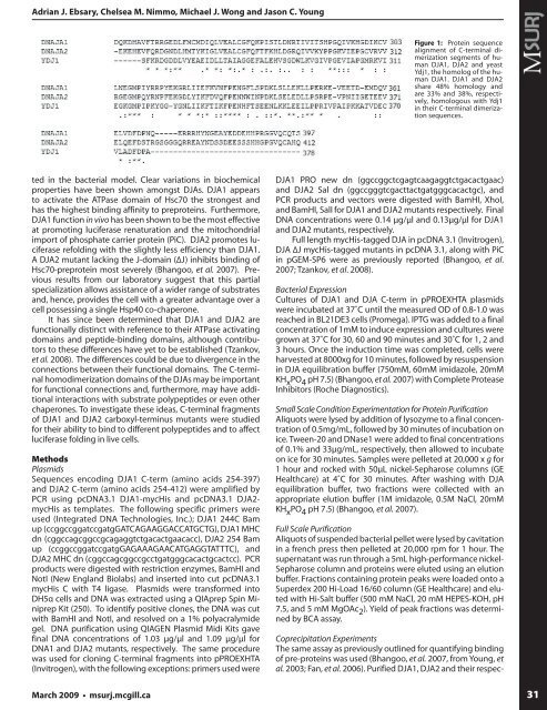
Figure 1: Protein sequence<br />
alignment of C-terminal dimerization<br />
segments of human<br />
DJA1, DJA2 and yeast<br />
Ydj1, <strong>the</strong> homolog of <strong>the</strong> human<br />
DJA1. DJA1 and DJA2<br />
share 48% homology and<br />
are 33% and 38%, respectively,<br />
homologous with Ydj1<br />
in <strong>the</strong>ir C-terminal dimerization<br />
sequences.<br />
Coprecipitation Experiments<br />
The same assay as previously outlined for quantifying binding<br />
of pre-proteins was used (Bhangoo, et al. 2007, from Young, et<br />
al. 2003; Fan, et al. 2006). Purified DJA1, DJA2 and <strong>the</strong>ir respected<br />
in <strong>the</strong> bacterial model. Clear variations in biochemical<br />
properties have been shown amongst DJAs. DJA1 appears<br />
to activate <strong>the</strong> ATPase domain of Hsc70 <strong>the</strong> strongest and<br />
has <strong>the</strong> highest binding affinity to preproteins. Fur<strong>the</strong>rmore,<br />
DJA1 function in vivo has been shown to be <strong>the</strong> most effective<br />
at promoting luciferase renaturation and <strong>the</strong> mitochondrial<br />
import of phosphate carrier protein (PiC). DJA2 promotes luciferase<br />
refolding with <strong>the</strong> slightly less efficiency than DJA1.<br />
A DJA2 mutant lacking <strong>the</strong> J-domain (ΔJ) inhibits binding of<br />
Hsc70-preprotein most severely (Bhangoo, et al. 2007). Previous<br />
results from our laboratory suggest that this partial<br />
specialization allows assistance of a wider range of substrates<br />
and, hence, provides <strong>the</strong> cell with a greater advantage over a<br />
cell possessing a single Hsp40 co-chaperone.<br />
It has since been determined that DJA1 and DJA2 are<br />
functionally distinct with reference to <strong>the</strong>ir ATPase activating<br />
domains and peptide-binding domains, although contributors<br />
to <strong>the</strong>se differences have yet to be established (Tzankov,<br />
et al. 2008). The differences could be due to divergence in <strong>the</strong><br />
connections between <strong>the</strong>ir functional domains. The C-terminal<br />
homodimerization domains of <strong>the</strong> DJAs may be important<br />
for functional connections and, fur<strong>the</strong>rmore, may have additional<br />
interactions with substrate polypeptides or even o<strong>the</strong>r<br />
chaperones. To investigate <strong>the</strong>se ideas, C-terminal fragments<br />
of DJA1 and DJA2 carboxyl-terminus mutants were studied<br />
for <strong>the</strong>ir ability to bind to different polypeptides and to affect<br />
luciferase folding in live cells.<br />
Methods<br />
Plasmids<br />
Sequences encoding DJA1 C-term (amino acids 254-397)<br />
and DJA2 C-term (amino acids 254-412) were amplified by<br />
PCR using pcDNA3.1 DJA1-mycHis and pcDNA3.1 DJA2-<br />
mycHis as templates. The following specific primers were<br />
used (Integrated DNA Technologies, Inc.); DJA1 244C Bam<br />
up (ccggccggatccgatgGATCAGAAGGACCATGCTG), DJA1 MHC<br />
dn (cggccagcggccgcagaggtctgacactgaacacc), DJA2 254 Bam<br />
up (ccggccggatccgatgGAGAAAGAACATGAGGTATTTC), and<br />
DJA2 MHC dn (cggccagcggccgcctgatgggcacactgcactcc). PCR<br />
products were digested with restriction enzymes, BamHI and<br />
NotI (New England Biolabs) and inserted into cut pcDNA3.1<br />
mycHis C with T4 ligase. Plasmids were transformed into<br />
DH5α cells and DNA was extracted using a QIAprep Spin Miniprep<br />
Kit (250). To identify positive clones, <strong>the</strong> DNA was cut<br />
with BamHI and NotI, and resolved on a 1% polyacralymide<br />
gel. DNA purification using QIAGEN Plasmid Midi Kits gave<br />
final DNA concentrations of 1.03 μg/μl and 1.09 μg/μl for<br />
DNA1 and DJA2 mutants, respectively. The same procedure<br />
was used for cloning C-terminal fragments into pPROEXHTA<br />
(Invitrogen), with <strong>the</strong> following exceptions: primers used were<br />
DJA1 PRO new dn (ggccggctcgagtcaagaggtctgacactgaac)<br />
and DJA2 Sal dn (ggccgggtcgacttactgatgggcacactgc), and<br />
PCR products and vectors were digested with BamHI, XhoI,<br />
and BamHI, SalI for DJA1 and DJA2 mutants respectively. Final<br />
DNA concentrations were 0.14 μg/μl and 0.13μg/μl for DJA1<br />
and DJA2 mutants, respectively.<br />
Full length mycHis-tagged DJA in pcDNA 3.1 (Invitrogen),<br />
DJA ΔJ mycHis-tagged mutants in pcDNA 3.1, along with PiC<br />
in pGEM-SP6 were as previously reported (Bhangoo, et al.<br />
2007; Tzankov, et al. 2008).<br />
Bacterial Expression<br />
Cultures of DJA1 and DJA C-term in pPROEXHTA plasmids<br />
were incubated at 37˚C until <strong>the</strong> measured OD of 0.8-1.0 was<br />
reached in BL21DE3 cells (Promega). IPTG was added to a final<br />
concentration of 1mM to induce expression and cultures were<br />
grown at 37˚C for 30, 60 and 90 minutes and 30˚C for 1, 2 and<br />
3 hours. Once <strong>the</strong> induction time was completed, cells were<br />
harvested at 8000xg for 10 minutes, followed by resuspension<br />
in DJA equilibration buffer (750mM, 60mM imidazole, 20mM<br />
KH x PO 4 pH 7.5) (Bhangoo, et al. 2007) with Complete Protease<br />
Inhibitors (Roche Diagnostics).<br />
Small Scale Condition Experimentation for Protein Purification<br />
Aliquots were lysed by addition of lysozyme to a final concentration<br />
of 0.5mg/mL, followed by 30 minutes of incubation on<br />
ice. Tween-20 and DNase1 were added to final concentrations<br />
of 0.1% and 33μg/mL, respectively, <strong>the</strong>n allowed to incubate<br />
on ice for 30 minutes. Samples were pelleted at 20,000 x g for<br />
1 hour and rocked with 50μL nickel-Sepharose columns (GE<br />
Healthcare) at 4˚C for 30 minutes. After washing with DJA<br />
equilibration buffer, two fractions were collected with an<br />
appropriate elution buffer (1M imidazole, 0.5M NaCl, 20mM<br />
KH x PO 4 pH 7.5) (Bhangoo, et al. 2007).<br />
Full Scale Purification<br />
Aliquots of suspended bacterial pellet were lysed by cavitation<br />
in a french press <strong>the</strong>n pelleted at 20,000 rpm for 1 hour. The<br />
supernatant was run through a 5mL high-performance nickel-<br />
Sepharose column and proteins were eluted using an elution<br />
buffer. Fractions containing protein peaks were loaded onto a<br />
Superdex 200 Hi-Load 16/60 column (GE Healthcare) and eluted<br />
with Hi-Salt buffer (500 mM NaCl, 20 mM HEPES-KOH, pH<br />
7.5, and 5 mM MgOAc 2 ). Yield of peak fractions was determined<br />
by BCA assay.<br />
March 2009 • msurj.mcgill.ca<br />
31