the entire issue - McGill Science Undergraduate Research Journal ...
the entire issue - McGill Science Undergraduate Research Journal ...
the entire issue - McGill Science Undergraduate Research Journal ...

You also want an ePaper? Increase the reach of your titles
YUMPU automatically turns print PDFs into web optimized ePapers that Google loves.
Adrian J. Ebsary, Chelsea M. Nimmo, Michael J. Wong and Jason C. Young<br />
Results<br />
Isolation of DJA1 and DJA2 C-terminal Mutants<br />
The predicted C-terminal homodimerization domains of DJA1<br />
and DJA2 (A1-C and A2-C, respectively) are well conserved, although<br />
not identical (Figure 1). To study <strong>the</strong>ir possible functions<br />
and properties, protein fragments of <strong>the</strong> C-termini of DJA1 and<br />
DJA2 fused to N-terminal His-tags were first expressed in E. coli,<br />
<strong>the</strong>n purified.<br />
To determine <strong>the</strong> optimal conditions for bacterial expression,<br />
induction at 37°C and 30°C was tested at several time<br />
points. At each temperature/time point, <strong>the</strong> bacteria expressing<br />
A1-C and A2-C were lysed and single step purifications<br />
on nickel-Sepharose were carried out. SDS-PAGE gel analysis<br />
of pellet, flow-through, wash and two eluates was conducted<br />
for both C-terminal proteins. Expression profiles were selected<br />
for magnitude of <strong>the</strong> expected product band and significance<br />
of contaminating agents in eluates. In most conditions,<br />
<strong>the</strong> proteins appeared to express well, although <strong>the</strong>re were<br />
differences in amount and number of contaminants after <strong>the</strong><br />
trial purification. For A1-C and A2-C, it was determined that<br />
<strong>the</strong> optimal conditions for large scale purification were 30˚C<br />
for 2 hours and 37˚C for 1 hour, respectively.<br />
The A1-C and A2-C proteins were next purified on a larger<br />
scale to produce amounts sufficient for in vitro experiments.<br />
After expressing <strong>the</strong> proteins under <strong>the</strong> conditions described<br />
above, a first purification step using a nickel-Sepharose column<br />
was performed. As expected, <strong>the</strong> proteins eluted in a narrow<br />
peak and appeared relatively pure. These fractions were pooled<br />
and loaded on a Superdex 200 gel filtration column, producing<br />
broad elution profiles for both A1-C and A2-C. Some<br />
aggregated material eluted in <strong>the</strong> void volume, but one distinctive<br />
peak in <strong>the</strong> separation range of <strong>the</strong> column was indicated<br />
by <strong>the</strong> UV absorbance profiles. Interestingly, a smaller earlier<br />
peak was observed for both A1-C and A2-C upon gel filtration<br />
(Figure 2, and data not shown), suggesting possible formation<br />
of tetramers. Because SDS-PAGE gel analysis showed that both<br />
peaks contained <strong>the</strong> correct proteins, fractions combining<br />
both peaks were pooled and concentrated. Final concentra-<br />
tions of A1-C and A2-C were determined to be 1.22mg/mL and<br />
0.803mg/mL, respectively, when concentrated to 2mL.<br />
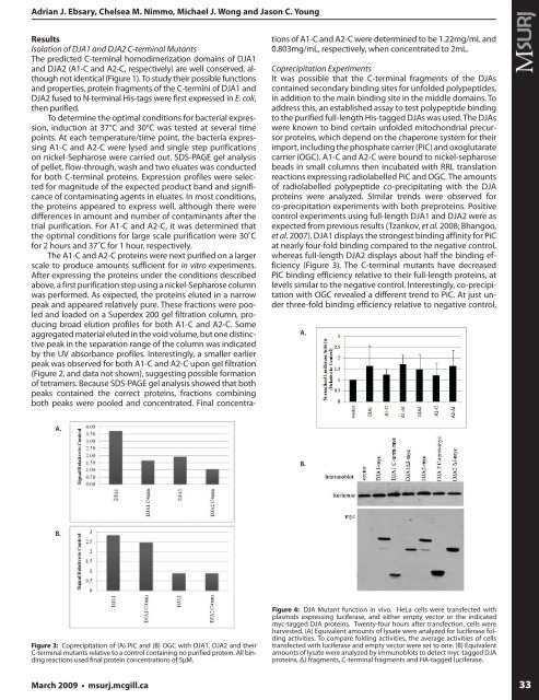
Coprecipitation Experiments<br />
It was possible that <strong>the</strong> C-terminal fragments of <strong>the</strong> DJAs<br />
contained secondary binding sites for unfolded polypeptides,<br />
in addition to <strong>the</strong> main binding site in <strong>the</strong> middle domains. To<br />
address this, an established assay to test polypeptide binding<br />
to <strong>the</strong> purified full-length His-tagged DJAs was used. The DJAs<br />
were known to bind certain unfolded mitochondrial precursor<br />
proteins, which depend on <strong>the</strong> chaperone system for <strong>the</strong>ir<br />
import, including <strong>the</strong> phosphate carrier (PiC) and oxoglutarate<br />
carrier (OGC). A1-C and A2-C were bound to nickel-sepharose<br />
beads in small columns <strong>the</strong>n incubated with RRL translation<br />
reactions expressing radiolabelled PiC and OGC. The amounts<br />
of radiolabelled polypeptide co-precipitating with <strong>the</strong> DJA<br />
proteins were analyzed. Similar trends were observed for<br />
co-precipitation experiments with both preproteins. Positive<br />
control experiments using full-length DJA1 and DJA2 were as<br />
expected from previous results (Tzankov, et al. 2008; Bhangoo,<br />
et al. 2007). DJA1 displays <strong>the</strong> strongest binding affinity for PiC<br />
at nearly four-fold binding compared to <strong>the</strong> negative control,<br />
whereas full-length DJA2 displays about half <strong>the</strong> binding efficiency<br />
(Figure 3). The C-terminal mutants have decreased<br />
PiC binding efficiency relative to <strong>the</strong>ir full-length proteins, at<br />
levels similar to <strong>the</strong> negative control. Interestingly, co-precipitation<br />
with OGC revealed a different trend to PiC. At just under<br />
three-fold binding efficiency relative to negative control,<br />
A.<br />
A.<br />
B.<br />
B.<br />
Figure 3: Coprecipitation of (A) PiC and (B) OGC with DJA1, DJA2 and <strong>the</strong>ir<br />
C-terminal mutants relative to a control containing no purified protein. All binding<br />
reactions used final protein concentrations of 5μM.<br />
Figure 4: DJA Mutant function in vivo. HeLa cells were transfected with<br />
plasmids expressing luciferase, and ei<strong>the</strong>r empty vector or <strong>the</strong> indicated<br />
myc-tagged DJA proteins. Twenty-four hours after transfection, cells were<br />
harvested. (A) Equivalent amounts of lysate were analyzed for luciferase folding<br />
activities. To compare folding activities, <strong>the</strong> average activities of cells<br />
transfected with luciferase and empty vector were set to one. (B) Equivalent<br />
amounts of lysate were analyzed by immunoblots to detect myc-tagged DJA<br />
proteins, ΔJ fragments, C-terminal fragments and HA-tagged luciferase.<br />
March 2009 • msurj.mcgill.ca<br />
33