the entire issue - McGill Science Undergraduate Research Journal ...
the entire issue - McGill Science Undergraduate Research Journal ...
the entire issue - McGill Science Undergraduate Research Journal ...

Create successful ePaper yourself
Turn your PDF publications into a flip-book with our unique Google optimized e-Paper software.
<strong>McGill</strong> <strong>Science</strong> <strong>Undergraduate</strong> <strong>Research</strong> <strong>Journal</strong><br />
About mSURJ<br />
Despite <strong>the</strong> importance of effective communication in science, most students<br />
do not participate directly in <strong>the</strong> publication process as part of <strong>the</strong>ir undergraduate<br />
education. The <strong>McGill</strong> <strong>Science</strong> <strong>Undergraduate</strong> <strong>Research</strong> <strong>Journal</strong> (mSURJ)<br />
addresses this need by providing undergraduates with <strong>the</strong> opportunity to share<br />
<strong>the</strong>ir achievements with a diverse research community. Our competitive selection<br />
process unites student researchers, student editors, graduate students, faculty<br />
supervisors and reviewers in producing peer-reviewed academic articles.<br />
As such, mSURJ is a training experience for students to present a clear, engaging<br />
account of <strong>the</strong>ir work.<br />
The editorial board is composed of undergraduate students from various disciplines<br />
united under <strong>the</strong> mandate to conduct a formal editorial evaluation of all<br />
submissions. Our editors are involved in <strong>the</strong> solicitation of research articles, <strong>the</strong><br />
coordination of <strong>the</strong> peer-review process and ensuring that all submissions comply<br />
with mSURJ guidelines.<br />
Our guidelines describe <strong>the</strong> journal framework and provide authors, editors and<br />
<strong>the</strong> public with all relevant information to get involved. Please visit our website<br />
at msurj.mcgill.ca or contact us at mcgillsurj@gmail.com for any o<strong>the</strong>r questions<br />
or concerns.<br />
ON THE COVER<br />
Designed by DEBBIE GELTNER<br />
In recognition of <strong>the</strong> 200th anniversary of Darwin’s birthday and <strong>the</strong> 150th anniversary<br />
of his seminal treatise on evolution, The Origin of Species, our cover depicts<br />
a variety of organisms from three of <strong>the</strong> taxonomic kingdoms as proposed by<br />
Prof. Robert H. Whittaker in 1969. On <strong>the</strong> back cover is a dodecahedron, a shape<br />
made up of 12 pentagons and one of <strong>the</strong> five Platonic Solids that has preoccupied<br />
notables such as Plato and Kepler. The background image is a photo taken<br />
by <strong>the</strong> Hubble Telescope of <strong>the</strong> M 78 reflection nebula found in <strong>the</strong> Orion constellation<br />
complex.
<strong>McGill</strong> <strong>Science</strong> <strong>Undergraduate</strong><br />
<strong>Research</strong> <strong>Journal</strong><br />
805 Sherbrooke St. West Room 1B21<br />
Montréal, Québec, H3A 2K6 Canada<br />
Phone: (514) 398-6979<br />
Fax: (514) 398-6766<br />
Email: mcgillsurj@gmail.com<br />
Web site: msurj.mcgill.ca<br />
Volume 4, Issue 1<br />
March 2009<br />
The 2008-2009 MSURJ committee<br />
EDITOR-IN-CHIEF<br />
MANAGING EDITOR<br />
Adrian J. Ebsary<br />
Sushmita Shivkumar<br />
Marzieh Ghiasi<br />
EDITORIAL BOARD MEMBERS<br />
Shaima Al-Khabouri<br />
Eric Eckbo<br />
Xin Feng<br />
Daniel Friedlander<br />
Maya Kaczorowski<br />
Pinky Langat<br />
Nadine Shatilla<br />
Vimarsha Swami<br />
Daniel Ting<br />
Emily Weizel<br />
Marnie Wilson<br />
Lisa Zhang<br />
ASSISTANT EDITOR<br />
LAYOUT DESIGN<br />
GRAPHIC DESIGN<br />
Neil Issar<br />
Ca<strong>the</strong>rine Godin, godin.ca<strong>the</strong>rine@videotron.ca<br />
Debbie Geltner, debgelts@gmail.com<br />
3
CONTENTS<br />
7<br />
9<br />
11<br />
15<br />
Acknowledgements<br />
Feature<br />
HM: A Legacy in Neuroscience<br />
ERIC ECKBO<br />
Uncovering fluctuations in atmospheric<br />
transmission using<br />
<strong>the</strong> VERITAS pointing monitors<br />
ILYA FEIGE<br />
Feature<br />
Culture and <strong>the</strong> Aging Brain<br />
BHAIRAVI BALRAM<br />
20 Investigating <strong>the</strong> role of heme<br />
oxygenase-1 in β-thalassemia<br />
pathophysiology<br />
ARIELLE MENDEL<br />
25 Case Study<br />
How taxonomic revisions affect <strong>the</strong><br />
interpretation of specimen identification<br />
in biological field data<br />
KATHERINE HUEBNER<br />
30 DJA1 and DJA2 Carboxyl-Terminal<br />
Fragments and <strong>the</strong>ir Role in Peptide<br />
Binding and Luciferase Refolding<br />
36<br />
39<br />
ADRIAN J. EBSARY, CHELSEA NIMMO,<br />
MICHAEL J. WONG, JASON C. YOUNG<br />
Feature<br />
The Evolution of Algorithms to Find<br />
Prime Numbers<br />
MAYA KACZOROWSKI<br />
Review<br />
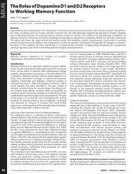
The Roles of Dopamine D1 and<br />
D2 Receptors in Working Memory<br />
Function<br />
JOHN T. P. LIGGINS<br />
* Color version of graphics is available online at http://msurj.mcgill.ca<br />
5
Acknowledgements<br />
This <strong>issue</strong> of mSURJ would not have been possible without <strong>the</strong> generous support of <strong>the</strong> following<br />
donors from <strong>the</strong> <strong>McGill</strong> community:<br />
• Faculty of <strong>Science</strong><br />
• Faculty of Medicine<br />
• Department of Earth and Planetary <strong>Science</strong>s<br />
• <strong>Science</strong> <strong>Undergraduate</strong> Society (SUS)<br />
• <strong>McGill</strong> School of Environment<br />
• <strong>McGill</strong> Psychology Students Association (MPSA)<br />
• Anatomy and Cell Biology Student Society (MACSS)<br />
• Biochemistry <strong>Undergraduate</strong> Society<br />
• <strong>McGill</strong> Biochemistry <strong>Undergraduate</strong> Society (BUGS)<br />
We would like to acknowledge Mr. Victor Chisholm, <strong>Undergraduate</strong> <strong>Research</strong> Officer, for his advice<br />
and support.<br />
The editorial board would also like to extend its gratitude to <strong>the</strong> professors, post-doctoral fellows<br />
and students who participated in our peer-review for <strong>the</strong>ir time and expertise. The strength of our<br />
editorial board relies on <strong>the</strong>ir valuable input.<br />
Finally, we wish to thank <strong>the</strong> student contributors for <strong>the</strong>ir tireless efforts and dedication to <strong>the</strong><br />
scientific process.<br />
7
HM: A Legacy in Neuroscience<br />
Eric Eckbo<br />
Department of Psychology, <strong>McGill</strong> University, Stewart Biology Building, 1205 Dr. Penfield Avenue, Montreal, Quebec, Canada, H3A 1B1<br />
Article submitted: January 4, 2009 - Article accepted: February 5, 2009<br />
“The problem of neurology is to understand man himself.”<br />
Walking by <strong>the</strong> Montreal Neurological Institute (MNI)<br />
and Hospital, one might give brief regard to <strong>the</strong>se words<br />
coined by Wilder Penfield and prominently displayed on <strong>the</strong><br />
edifice of <strong>the</strong> building. It is unlikely, however, that most passersby<br />
would have an accurate grasp of <strong>the</strong> groundbreaking<br />
work that has been undertaken by scientists at this institute<br />
– research that has delved into <strong>the</strong> inner workings of <strong>the</strong> human<br />
mind. During <strong>the</strong> 1930s, Penfield developed <strong>the</strong> surgical<br />
treatment of epilepsy by means of temporal lobe resection,<br />
and this procedure became a standard among many neurosurgical<br />
centres (Greenblatt, Dagi, et al. 1997). The MNI is also<br />
where Brenda Milner began her groundbreaking work in <strong>the</strong><br />
field of neuropsychology, most notably with a patient who<br />
until recently was simply known as HM.<br />
Nowadays, it is a rare occurrence that one can attribute<br />
significant advances in science to a single individual, especially<br />
one with no post-secondary education or formal training.<br />
However, no neuroscience textbook would be complete<br />
without mention of HM, a patient suffering from severe nonlocalized<br />
epilepsy and perhaps <strong>the</strong> most studied individual in<br />
<strong>the</strong> field (Corkin, 1984). H.M. was born in 1926, and at <strong>the</strong> age<br />
of 7 was involved in a bicycle accident that left him unconscious<br />
for 5 minutes. In addition, he suffered a laceration to<br />
<strong>the</strong> left supra-orbital region of his head (Corkin, 1984). At <strong>the</strong><br />
age of 10, HM began exhibiting minor seizures, during which<br />
he would cross his arms and legs, open his mouth, close his<br />
eyes, and generally exhibit a lack of responsiveness. By <strong>the</strong><br />
time he was 16, <strong>the</strong> seizures had progressed to major attacks.<br />
These general convulsions included tongue-biting, urinary<br />
incontinence, loss of consciousness, and ensuing drowsiness<br />
(Scoville and Milner, 1957). It is believed that <strong>the</strong> bicycle incident<br />
was linked to <strong>the</strong> onset of epilepsy, though <strong>the</strong> basic<br />
radiological studies and physical examinations available at<br />
<strong>the</strong> time showed normal findings (Scoville and Milner, 1957).<br />
This may, however, have only been a reflection of <strong>the</strong> state<br />
of imaging technology at <strong>the</strong> time and it is unclear whe<strong>the</strong>r<br />
modern neuroimaging techniques would have identified<br />
abnormalities. HM’s family history shows a presence of epilepsy,<br />
which may be indicative of an unrelated causative factor<br />
(Corkin, 2002). None<strong>the</strong>less, <strong>the</strong> etiology of HM’s disorder<br />
still remains inconclusive.<br />
In 1953, at <strong>the</strong> age of 27, HM underwent an experimental<br />
procedure in order to alleviate <strong>the</strong> severe epilepsy that<br />
had remained largely unresponsive to anticonvulsive drug<br />
<strong>the</strong>rapy. At this point, he was having 10 petit mal seizures a<br />
day and at least 1 major seizure per week (Corkin, 1984). Electroencephalographic<br />
recordings indicated diffuse abnormalities;<br />
hence, <strong>the</strong> decision was made to surgically remove <strong>the</strong><br />
medial temporal lobe structures, which were known to have<br />
epileptogenic qualities (Scoville and Milner, 1957). The bilateral<br />
medial temporal lobe resection extended 8cm posteriorly<br />
from <strong>the</strong> temporal tip, including <strong>the</strong> amygdaloid complex,<br />
temporal pole, and a large part of <strong>the</strong> hippocampal formation<br />
(Scoville and Milner, 1957). During <strong>the</strong> surgery, HM was fully<br />
conscious and talking (Corkin, 1984). The surgery succeeded<br />
in abating <strong>the</strong> severity of HM’s seizures and he recovered<br />
from <strong>the</strong> surgery without any complications. However, <strong>the</strong>re<br />
was one completely unexpected side effect of <strong>the</strong> surgery:<br />
severe anterograde amnesia, characterized by a loss of ability<br />
to formulate new memories (Scoville and Milner, 1957).<br />
The beginning of <strong>the</strong> nineteenth century marked <strong>the</strong> onset<br />
of <strong>the</strong> memory debate in <strong>the</strong> neuroscience community.<br />
Specifically, scientists began to question where memory is stored<br />
and <strong>the</strong> extent of its localization in <strong>the</strong> brain (Greenblatt,<br />
Dagi, et al. 1997). In <strong>the</strong> early half of <strong>the</strong> twentieth century,<br />
Karl Lashley, a prominent neuropsychologist, conducted studies<br />
in rats that led to his <strong>the</strong>ory of mass action. Removal of<br />
cortical areas of rat brains did not show any evidence of memory<br />
storage localization, but ra<strong>the</strong>r demonstrated that <strong>the</strong><br />
extent of memory deficit is proportional to <strong>the</strong> amount of cortical<br />
t<strong>issue</strong> removed (Greenblatt, Dagi, et al. 1997). Opponents<br />
of this <strong>the</strong>ory, notably Donald Hebb, proposed an alternate<br />
view. Hebb <strong>the</strong>orized that “assemblies” of cells work toge<strong>the</strong>r<br />
to represent information and that <strong>the</strong>se complexes are widely<br />
distributed. In <strong>the</strong> event of a localized lesion, <strong>the</strong> distributional<br />
nature and significant number of interconnected cells would<br />
ensure continued functioning (Milner, Squire, et al. 1998).<br />
After performing <strong>the</strong> procedure on H.M. and recognizing<br />
<strong>the</strong> unexpected amnesic syndrome, William Scoville invited<br />
Brenda Milner to Connecticut to systematically evaluate HM’s<br />
condition using neuropsychological methods. In 1957, Milner<br />
and Scoville published what would soon become a groundbreaking<br />
paper on memory. The surgeon and neuropsychologist<br />
described <strong>the</strong> results of testing of HM and nine o<strong>the</strong>r<br />
patients, who had been treated for psychosis using neurosurgical<br />
methods similar to those performed on HM (Scoville and<br />
Milner, 1957). HM was a unique case to consider since he did<br />
not suffer from psychosis, and his surgery was “frankly experimental”<br />
(Scoville and Milner, 1957). At <strong>the</strong> onset of extensive<br />
testing in 1955, memory deficits were immediately apparent<br />
– HM gave <strong>the</strong> date as March 1953 and his age as 27 (Scoville<br />
and Milner, 1957). Milner and Scoville (1957) also noted that<br />
“he reverted constantly to boyhood events and seemed scarcely<br />
to realize that he had had an operation.” HM’s IQ on <strong>the</strong><br />
Wechsler-Bellevue Intelligence Scale actually improved from a<br />
preoperative score of 104 to a postoperative 112 due to <strong>the</strong> reduction<br />
in seizures (Scoville and Milner, 1957). In contrast, his<br />
score was determined to be 67 on <strong>the</strong> Wechsler Memory Scale,<br />
far below average for someone of his intellectual capacity (Scoville<br />
and Milner, 1957). A battery of tests confirmed Milner’s<br />
suspicions: HM suffered from a complete loss of memory for<br />
all events occurring after <strong>the</strong> surgery, and a partial retrograde<br />
amnesia for three years preceding <strong>the</strong> surgery (Milner, Corkin,<br />
et al. 1968). Studies performed later in 1985 showed that this<br />
retrograde amnesia extended to include a period of 11 years<br />
prior to surgery (Corkin, 2002). Memories of his early life events<br />
and his pre-surgical personality remained unaffected.<br />
Fur<strong>the</strong>r testing also demonstrated <strong>the</strong> pervasiveness of<br />
HM’s memory disorder. He was severely impaired regardless<br />
of <strong>the</strong> type of memory test, <strong>the</strong> nature of <strong>the</strong> stimulus, or <strong>the</strong><br />
sensory modality through which <strong>the</strong> test was delivered (Milner,<br />
Corkin, et al. 1968; Corkin, 2002). He was unable to successfully<br />
acquire long-term episodic memory (events in a spatial/<br />
time context) or semantic memory (general knowledge and<br />
factual information); however, he had functional short-term<br />
memory (Milner, Corkin, et al. 1968; Corkin, 2002). HM was<br />
readily able to register new information within his immediate<br />
March 2009 • msurj.mcgill.ca 9
HM: A Legacy in Neuroscience<br />
memory span, but failed recall tests as soon as <strong>the</strong> information<br />
exceeded that time-span or his attention was diverted (Scoville<br />
and Milner, 1957). The evidence was progressively mounting<br />
for <strong>the</strong> role of <strong>the</strong> medial temporal lobe as a consolidation<br />
centre of long-term memory separate from short-term and<br />
working memory (Squire and Zolamorgan, 1991).<br />
Since <strong>the</strong> surgery, HM has been examined and tested in<br />
a variety of o<strong>the</strong>r psychological domains. Milner was <strong>the</strong> first<br />
to demonstrate H.M’s preserved learning capabilities in <strong>the</strong><br />
form of motor learning tasks (Milner, 1970). This finding was<br />
achieved using a mirror-drawing task, in which HM exhibited<br />
learning without being able to recall any of <strong>the</strong> sessions in<br />
which he engaged in <strong>the</strong> learning (Milner, 1970). Additional<br />
studies have shown that HM withheld preserved residual<br />
learning capacities, such as perceptual learning and priming<br />
repetition (O'Kane, Kensinger, et al. 2004). Subsequent advancements<br />
in radiological imaging technology have allowed researchers<br />
to use magnetic resonance imaging to confirm <strong>the</strong><br />
true extent of temporal lobe damage that HM had acquired.<br />
Corkin et al. (1997) found that <strong>the</strong> temporal lobe lesions were<br />
bilaterally symmetrical and included <strong>the</strong> amygdaloid complex,<br />
most of <strong>the</strong> entorhinal cortex, and approximately half of <strong>the</strong><br />
hippocampal formation. The parahippocampal cortex was largely<br />
spared. The MRI results indicated that <strong>the</strong> extent of <strong>the</strong><br />
lesions was less than Scoville estimated at <strong>the</strong> time of surgery<br />
(Corkin, Amaral, et al. 1997). Additional abnormalities included<br />
atrophy of <strong>the</strong> cerebellum and shrunken mammillary bodies.<br />
As <strong>the</strong> authors concluded, “<strong>the</strong>se findings reinforce <strong>the</strong> view<br />
that lesions of <strong>the</strong> hippocampal formation and adjacent cortical<br />
structures can produce global and enduring amnesia.”<br />
HM has greatly advanced our understanding of <strong>the</strong> human<br />
brain and cognition. Studies conducted on him, along<br />
with <strong>the</strong> o<strong>the</strong>r patients given bilateral hippocampal zone excisions<br />
for psychosis treatment, have provided conclusive evidence<br />
that <strong>the</strong> medial temporal lobes are crucial regions for<br />
memory encoding. Given this evidence, HM has helped ensure<br />
that no o<strong>the</strong>r patient has had a bilateral resection from this<br />
critical area of <strong>the</strong> brain. The extent of HM’s impairments due<br />
to <strong>the</strong> surgery prompted his surgeon, William Scoville, to campaign<br />
against continuation of <strong>the</strong> procedure (Corkin, 2002).<br />
After <strong>the</strong> surgery, HM was cared for by his mo<strong>the</strong>r. Although<br />
quiet in social situations, he appeared at ease and<br />
still enjoyed puns and semantic ambiguities (Corkin, 2002).<br />
He would often apologize when interacting with o<strong>the</strong>r people<br />
for his apparent lack of manners, such as forgetting <strong>the</strong><br />
names of individuals he was just introduced to (Milner, Corkin,<br />
et al. 1968). In 1980, HM moved into a nursing home due<br />
to <strong>the</strong> ailing health of his caregivers at home and was reported<br />
to have participated in <strong>the</strong> daily activities such as games,<br />
crafts, and poetry (Corkin, 1984). In <strong>the</strong> early evening hours of<br />
December 1, 2008, HM died as a result of heart failure at <strong>the</strong><br />
nursing home he had lived in for decades.<br />
It is essential that we not forget <strong>the</strong> humanity behind <strong>the</strong><br />
person; many of <strong>the</strong> articles describing his psychological testing<br />
are also interspersed with anecdotal tales speaking to<br />
his continued sense of humour and social graces. While HM<br />
died completely unaware of his monumental contributions<br />
to <strong>the</strong> scientific community, even after his death, his brain<br />
will be preserved for fur<strong>the</strong>r study. In this age of cutting edge<br />
technology and research it is easy to forget <strong>the</strong> humble roots<br />
of neuroscience. The loss of HM serves as a reminder of a time<br />
not very long ago when this field was in its infancy.<br />
“Every day is alone in itself, whatever enjoyment I’ve had,<br />
and whatever sorrow I’ve had”<br />
- Henry Gustav Molaison, aka “HM” (1926 – 2008)<br />
References<br />
1.<br />
2.<br />
3.<br />
4.<br />
5.<br />
6.<br />
7.<br />
8.<br />
9.<br />
Corkin, S. 1984. Lasting Consequences of Bilateral Medial<br />
Temporal Lobectomy - Clinical Course and Experimental<br />
Findings in HM. Seminars in Neurology. 4(2):249-259.<br />
Corkin, S. 2002. What's new with <strong>the</strong> amnesic patient<br />
H.M.? Nat Rev Neurosci 3(2):153-160.<br />
Corkin, S., Amaral, D.G., et al. 1997. H. M.'s medial temporal<br />
lobe lesion: findings from magnetic resonance imaging.<br />
J Neurosci. 17(10):3964-79.<br />
Greenblatt, S.H., Dagi, T.F., et al. 1997. A History of Neurosurgery.<br />
New York, NY, USA: Thieme.<br />
Milner, B. 1970. Memory and <strong>the</strong> medial temporal regions<br />
of <strong>the</strong> brain. Biology of Memory. New York, NY,<br />
USA: Academic Press, Inc. pp29-50.<br />
Milner, B., Corkin, S., et al. 1968. Fur<strong>the</strong>r Analysis of Hippocampal<br />
Amnesic Syndrome - 14-Year Follow-up Study<br />
of HM. Neuropsychologia. 6(3):215-230.<br />
Milner, B., Squire, L.R., et al. 1998. Cognitive Neuroscience<br />
and <strong>the</strong> Study of Memory. Neuron. 20(3):445-468.<br />
O'Kane, G., Kensinger, E.A. et al. 2004. Evidence for semantic<br />
learning in profound amnesia: An investigation<br />
with patient H.M. Hippocampus. 14(4):417-425.<br />
Scoville, W.B. and Milner, B. 1957. Loss of recent memory<br />
after bilateral hippocampal lesions. J Neurol Neurosurg<br />
Psychiatry. 20(1):11-21.<br />
10. Squire, L. R. and Zolamorgan, S. 1991. The Medial Temporal-Lobe<br />
Memory System. <strong>Science</strong>. 253(5026):1380-1386<br />
10 MSURJ • Volume 4, Issue 1
Uncovering fluctuations in atmospheric transmission<br />
using <strong>the</strong> VERITAS pointing monitors<br />
Ilya Feige<br />
Department of Physics, <strong>McGill</strong> University, 3600 rue University, Montréal, QC, Canada H3A 2T8<br />
Article submitted: December 28, 2008 - Arlicle accepted: February 5, 2009<br />
Abstract<br />
Our project encompasses <strong>the</strong> creation of software that provides nightly estimates of atmospheric transmission and tracks its long-term<br />
fluctuations for <strong>the</strong> VERITAS telescopes. Using archived image files taken from <strong>the</strong> pointing monitors of <strong>the</strong> VERITAS telescope, we wrote<br />
software that selects stars of appropriate brightness and quantifies <strong>the</strong>ir intensity. We <strong>the</strong>n plotted <strong>the</strong> star intensity as a function of <strong>the</strong><br />
secant of <strong>the</strong> telescope angle from <strong>the</strong> zenith and observed a linear relationship. The ratio of this slope divided by its intercept has a<br />
value that is independent of <strong>the</strong> stars chosen and is proportional to <strong>the</strong> length of attenuation of light travelling through <strong>the</strong> atmosphere.<br />
By analyzing nightly data for all four telescopes, one can measure <strong>the</strong> magnitude of <strong>the</strong> fluctuations in atmospheric transmission<br />
using <strong>the</strong> ratio of <strong>the</strong> slope over <strong>the</strong> intercept. This method will allow improvements in <strong>the</strong> quality of <strong>the</strong> measurements taken by <strong>the</strong><br />
VERITAS telescopes, by giving VERITAS control over <strong>the</strong> effects of <strong>the</strong> atmosphere on <strong>the</strong>ir star intensity data.<br />
Keywords: Gamma rays, Cherenkov Radiation, Atmospheric<br />
transmissions, VERITAS.<br />
Glossary<br />
Telescope zenith angle: Angle between <strong>the</strong> vertical and <strong>the</strong><br />
direction in which <strong>the</strong> telescope is pointing.<br />
CCD camera: A charge-coupled device camera measures <strong>the</strong><br />
motion of electric charges created by <strong>the</strong> passing of a photon,<br />
thus forming an image of <strong>the</strong> source of photons.<br />
Flux: Amount that flows through a unit area per unit time.<br />
Attenuation length: Characteristic distance required for a<br />
signal to reach of its initial strength.<br />
Flat fielding: Calibrating <strong>the</strong> array of pixels in a CCD camera<br />
so that all pixels respond equally to excitation.<br />
Introduction<br />
The Very Energetic Radiation Imaging Telescope Array System<br />
(VERITAS) is an array of four telescopes, each 12 m in<br />
diameter and located at <strong>the</strong> base of Mt. Hopkins near Tucson,<br />
Arizona. A collaboration of more than 100 scientists<br />
from institutions in Canada, <strong>the</strong> U.S., Ireland and England<br />
operates VERITAS to find and measure astrophysical sources<br />
of gamma rays with energies in excess of 100 GeV. The<br />
VERITAS base camp is shown in Figure 1.<br />
Very high energy gamma-ray astronomy relies on <strong>the</strong><br />
measurement of Cherenkov radiation in <strong>the</strong> atmosphere.<br />
Gamma rays coming from space collide with <strong>the</strong> nuclei of<br />
air molecules in <strong>the</strong> earth’s atmosphere roughly 10 to 20 km<br />
above ground level, producing an “air shower” of secondary<br />
particles—mostly electrons and positrons—that hurtle<br />
toward <strong>the</strong> ground. In this process, <strong>the</strong> <strong>entire</strong> energy of <strong>the</strong><br />
gamma-ray is converted to <strong>the</strong> mass and kinetic energy of<br />
<strong>the</strong> secondary particles. Although <strong>the</strong> secondary particles<br />
travel slower than <strong>the</strong> speed of light in vacuum, <strong>the</strong>y travel<br />
faster than <strong>the</strong> speed of light in air due to <strong>the</strong>ir higher<br />
energies. This results in an electromagnetic shock wave<br />
comparable to a sonic boom. The shock wave comes in <strong>the</strong><br />
form of bluish light called Cherenkov radiation. Cherenkov<br />
radiation is emitted by all <strong>the</strong> charged secondary particles<br />
and propagates to <strong>the</strong> ground where it can be measured by<br />
optical devices (Hanna, 2007). Using optical telescopes and<br />
extremely fast cameras 1 , it is possible to measure <strong>the</strong> Cherenkov<br />
radiation and generate an image of <strong>the</strong> air shower. Typically,<br />
a thin, tubular shape is observed that can be used to<br />
trace backwards to <strong>the</strong> origin of <strong>the</strong> gamma ray (Figure 2).<br />
March 2009 • msurj.mcgill.ca<br />
The image generated from a single telescope is not sufficient<br />
to determine <strong>the</strong> origin of a gamma-ray induced air<br />
shower, since <strong>the</strong>re is no indication where <strong>the</strong> signal originated<br />
from along <strong>the</strong> long axis. However, multiple telescopes<br />
positioned in an array allow researchers to view <strong>the</strong> air<br />
shower from several perspectives, resulting in images with<br />
different orientations (Figure 3). By superimposing images<br />
of <strong>the</strong> same air shower, <strong>the</strong> source of <strong>the</strong> gamma rays<br />
can be determined by <strong>the</strong> intersection of <strong>the</strong> lines drawn<br />
through <strong>the</strong> long axis of each image.<br />
M.K. Daniel (2007) has shown that <strong>the</strong> atmospheric regions<br />
surveyed by VERITAS display substantial changes in<br />
atmospheric transmission both daily and annually. Fluctuations<br />
in atmospheric transmission alter <strong>the</strong> levels of scattering<br />
or absorption of Cherenkov radiation, which affects telescopic<br />
measurements. To improve gamma-ray detection<br />
from <strong>the</strong> ground, we hope to quantify <strong>the</strong> effect of atmospheric<br />
transmission from archived images taken by <strong>the</strong> VE-<br />
RITAS pointing monitor, a CCD camera directed towards <strong>the</strong><br />
telescope’s field of view. From <strong>the</strong>se images, it is possible to<br />
determine <strong>the</strong> telescope zenith angle and <strong>the</strong> intensity of<br />
<strong>the</strong> star that is being examined. By relating <strong>the</strong>se two values,<br />
we can calculate <strong>the</strong> amount of atmosphere that a star's light<br />
will have to penetrate before reaching <strong>the</strong> telescope.<br />
Since <strong>the</strong> amount of atmosphere that <strong>the</strong> Cherenkov light<br />
must travel through increases with <strong>the</strong> zenith angle, we expect<br />
<strong>the</strong> extinction curve to display a decrease in intensity<br />
as a function of <strong>the</strong> telescope's angle from <strong>the</strong> zenith.<br />
Methods<br />
Analysis Procedure<br />
Our software finds individual stars in images that are<br />
taken from <strong>the</strong> telescope's pointing monitors every fifteen<br />
minutes. The software <strong>the</strong>n filters those stars and<br />
selects <strong>the</strong> fifteen brightest stars from <strong>the</strong> image, excluding<br />
stars that have pixel values above <strong>the</strong> highest value<br />
that <strong>the</strong> camera can measure (Figure 4). For each star<br />
that is chosen, a histogram is created of all <strong>the</strong> pixels<br />
that contribute to its background noise. Each of <strong>the</strong>se<br />
histograms is used to measure <strong>the</strong> mean pixel noise in<br />
<strong>the</strong> neighbourhood of that star.<br />
The mean pixel noise is subtracted from <strong>the</strong> sum of all<br />
1. The VERITAS cameras contain 499 photo-multiplier tubes (PMT) that are digitised at a rate<br />
of 500 mega-samples/sec (Swordy and Brocious, 2007). Each PMT is capable of measuring a<br />
single photon and can determine its arrival time to within a few billionths of a second.<br />
* Corresponding author. E-mail: ilya.feige@mail.mcgill.ca<br />
11
Uncovering fluctuations in atmospheric transmission using <strong>the</strong> VERITAS pointing monitors<br />
Figure 1: The VERI-<br />
TAS telescope array,<br />
located at <strong>the</strong> base<br />
of Mt.Hopkins, Tucson,<br />
Arizona<br />
<strong>the</strong> pixel values within a given radius to obtain <strong>the</strong> intensity<br />
of <strong>the</strong> star. To determine <strong>the</strong> most accurate radius to use, <strong>the</strong><br />
intensity is plotted as a function of <strong>the</strong> radius. The radius that<br />
demonstrates <strong>the</strong> least change in intensity will best describe<br />
<strong>the</strong> star’s actual intensity. The error in <strong>the</strong> star's intensity can<br />
be computed using <strong>the</strong> error in <strong>the</strong> radius.<br />
The software tracks <strong>the</strong> intensity of each star through<br />
many images and plots its intensity values as a function of<br />
<strong>the</strong> secant of <strong>the</strong> telescope zenith angle. Theoretically, it<br />
can be shown that <strong>the</strong> flux of light from a star, Φ (1), follows<br />
<strong>the</strong> relation:<br />
Eqation (1): where θ is <strong>the</strong> telescope's angle from <strong>the</strong> zenith, h is <strong>the</strong> height of <strong>the</strong><br />
atmosphere, lo is <strong>the</strong> attenuation length of light in <strong>the</strong> atmosphere and Φ is <strong>the</strong> flux<br />
as measured above <strong>the</strong> atmosphere (Hanna, D. 2008). Thus, a plot of star intensity<br />
versus <strong>the</strong> secant of <strong>the</strong> telescope zenith angle should be linear (Figure 5).<br />
Results<br />
From Figure 5, we see that <strong>the</strong> intensity of <strong>the</strong> stars in <strong>the</strong><br />
pointing monitor images follows a linear relation when<br />
plotted against <strong>the</strong> secant of <strong>the</strong> telescope’s angle from <strong>the</strong><br />
zenith (Equation 1). Moreover, <strong>the</strong> ratio of <strong>the</strong> slope over<br />
<strong>the</strong> intercept of this relation, which is - h/ l 0 , is independent<br />
of <strong>the</strong> stars used (Figure 6). In Figure 7 all of <strong>the</strong> , - h/<br />
l 0 values that were extracted from each night’s images are<br />
plotted with <strong>the</strong>ir date (yy/mm/dd) on <strong>the</strong> x-axis. These are<br />
<strong>the</strong> weighted sums of each value from all four telescopes.<br />
The few points with very large errors in Figure 7 are cases<br />
where too few stars were found to fit <strong>the</strong> plots of intensity<br />
versus secant of <strong>the</strong> telescope zenith angle meaningfully,<br />
so only a few of <strong>the</strong>se plots were considered in <strong>the</strong> averaging<br />
of data. Figure 7 clearly shows significant fluctuations<br />
in - h/ l 0 which implicates daily and monthly fluctuations in<br />
<strong>the</strong> atmosphere, as supported by previous research (Daniel,<br />
2007). Due to <strong>the</strong> large variation in intensity values as<br />
a function of <strong>the</strong> secant of <strong>the</strong> telescope zenith angle, we<br />
cannot be certain that <strong>the</strong> variations in - h/ l 0 , are strictly<br />
from fluctuations in atmospheric transmission.<br />
Figure 2: An air shower produced by an incoming gamma ray colliding with<br />
<strong>the</strong> nucleus of a molecule in <strong>the</strong> atmosphere. (Courtesy of <strong>the</strong> VERITAS Education<br />
Website, veritas.adlerplanetarium.org)<br />
Figure 3: Stereo imaging of a gamma ray induced air shower with multiple telescopes.<br />
The turquoise colour represents <strong>the</strong> Cherenkov radiation. (Courtesy of <strong>the</strong><br />
VERITAS Education Website, veritas.adlerplanetarium.org)<br />
12 MSURJ • Volume 4, Issue 1
Ilya Feige<br />
Discussion<br />
The systematic error in <strong>the</strong> intensity<br />
In <strong>the</strong> course of our research, we discovered that <strong>the</strong> pointing<br />
monitors capture images every two seconds and store<br />
<strong>the</strong> star's peak pixel value and its coordinates in a database<br />
for <strong>the</strong> 30 brightest stars in <strong>the</strong> image. In Figure 8, we see<br />
<strong>the</strong> peak pixel value of one star for four hours as a function<br />
of time in minutes. The blue triangles represent times<br />
when <strong>the</strong> telescope changed positions with respect to <strong>the</strong><br />
star to get a sample of its background in a process called<br />
wobbling. We see that when <strong>the</strong> star changes positions in<br />
<strong>the</strong> image, its peak pixel value jumps significantly. This is<br />
emphasized by <strong>the</strong> plot in Figure 9, where we see a particular<br />
star's intensity versus secant of <strong>the</strong> zenith angle plot<br />
compared to <strong>the</strong> same star sampled every two seconds in<br />
<strong>the</strong> database. The jumps in intensity follow a pattern that<br />
is characteristic of a star moving in <strong>the</strong> image of a non-flat-<br />
Figure 6: The ratio of <strong>the</strong> slope over intercept, - h/ l 0 , of linear fits as that in Figure<br />
5. The blue dashed line represents <strong>the</strong> best fit of <strong>the</strong> constant that we expect.<br />
Figure 4: A two dimensional histogram of a star in an image taken by <strong>the</strong><br />
pointing monitors, along with a fitted Gaussian function. Each bin in <strong>the</strong> histogram<br />
corresponds to a pixel in <strong>the</strong> image.<br />
Figure 7: The ratio of <strong>the</strong> slope over <strong>the</strong> intercept, - h/ l 0 , weighted for each<br />
night by fitting a constant to all <strong>the</strong> values obtained from that night (as in<br />
Figure 6) and <strong>the</strong>n fur<strong>the</strong>r averaged over all four telescopes.<br />
Figure 5: The upper plot in this figure is <strong>the</strong> star intensity as a function of sec<br />
θ, where θ is <strong>the</strong> zenith angle of <strong>the</strong> telescope, for a star in a group of 17 images.<br />
The smaller plot below is <strong>the</strong> <strong>entire</strong> image noise as a function of sec θ.<br />
Figure 8: The peak pixel value from <strong>the</strong> database, plotted against time for a single<br />
star tracked for four hours. The blue triangles represent times when <strong>the</strong> telescope wobbled,<br />
which corresponds to <strong>the</strong> star making a jump to a new location in <strong>the</strong> image.<br />
March 2009 • msurj.mcgill.ca<br />
13
Uncovering fluctuations in atmospheric transmission using <strong>the</strong> VERITAS pointing monitors<br />
fielded CCD camera (Roper Scientific, 2006) and provides<br />
a reason for <strong>the</strong> large spread in intensities. As a result of<br />
our research, measures are being taken to flat-field <strong>the</strong> CCD<br />
cameras so that this process of measuring <strong>the</strong> atmospheric<br />
fluctuations may be fur<strong>the</strong>r studied.<br />
Conclusions<br />
By analyzing <strong>the</strong> pixel intensity for <strong>the</strong> stars chosen by our<br />
software for <strong>the</strong> VERITAS telescope array, we have found<br />
that <strong>the</strong> relation between <strong>the</strong> intensity and <strong>the</strong> secant of<br />
<strong>the</strong> telescope zenith angle is linear. We have also found<br />
that <strong>the</strong> ratio of <strong>the</strong> slope over <strong>the</strong> intercept of this relation<br />
is independent of <strong>the</strong> star used to measure this data.<br />
Since we have shown that <strong>the</strong> fluctuations of star intensity<br />
values are due to wobbling, this suggests that our method<br />
of measuring <strong>the</strong> atmospheric effects on <strong>the</strong> telescope<br />
data can quantify atmospheric transmission. Remaining<br />
work includes flat-fielding <strong>the</strong> VERITAS pointing monitors<br />
and implementing our software at <strong>the</strong> level of <strong>the</strong> base<br />
camp. This would allow more sophisticated values that<br />
our software extracts from <strong>the</strong> images to be stored at two<br />
second intervals in <strong>the</strong> database by <strong>the</strong> pointing monitors,<br />
including each star's pixel coordinates and relative<br />
and absolute intensities. In <strong>the</strong> long term, this would provide<br />
VERITAS scientists with a robust and precise method<br />
of quantifying and correcting for atmospheric effects on<br />
<strong>the</strong>ir gamma-ray measurements.<br />
Acknowledgements<br />
I would like to thank <strong>the</strong> following people for <strong>the</strong> success<br />
of this project:<br />
Prof. David Hanna, Mr. Andrew McCann, Mr. Michael Mc-<br />
Cutcheon, Ms. Mary Bautista, Prof. Gil Holder and Mr.<br />
Charles Rabideau.<br />
References<br />
1.<br />
2.<br />
3.<br />
4.<br />
5.<br />
Daniel, M.K. 2007. Application of radiosonde data to<br />
VERITAS simulations. 30th International Cosmic Ray<br />
Conference.<br />
Hanna, D. 2008. Photometry with <strong>the</strong> VERITAS pointing<br />
monitors. VERITAS internal note.<br />
Hanna, D. 2007. VERITAS telescopes celebrate first light.<br />
Cern courier July/August.<br />
Roper Scientific GmbH, Germany. 2006. Flat Field Correction.(le<br />
site web n’est pas une reference <strong>entire</strong> et doit<br />
etre lier a la reference #4) . http://www.roperscientific.<br />
de/tflatfield.html<br />
Swordy, S.P., Brocious. D. 2007. New High-Energy Telescope<br />
Array Marks First Light. VERITAS press release.<br />
Figure 9: The top plot is <strong>the</strong> extinction curve for a star tracked through <strong>the</strong> pointing monitor images. Below is <strong>the</strong> peak pixel value of <strong>the</strong> same star<br />
shown above but sampled every two seconds from <strong>the</strong> database values.<br />
14 MSURJ • Volume 4, Issue 1
Culture and <strong>the</strong> Aging Brain<br />
Bhairavi Balram*<br />
Department of Neurology and Neuroscience, <strong>McGill</strong> University, Montreal Neurological Institute, 3801 University Street, Montreal, Quebec, Canada, H3A 2B4<br />
Article submitted: January 18, 2009 - Article accepted: February 5, 2009<br />
Glossary<br />
Working memory: Refers to <strong>the</strong> structures and processes in <strong>the</strong><br />
brain used to temporarily store and manipulate information.<br />
Neurocognitive system: A term used to describe <strong>the</strong> particular<br />
brain areas, neural pathways and networks responsible<br />
for specific behaviour and thought processes.<br />
Cognitive pragmatics: A term used to describe knowledge<br />
that has been acquired through a lifetime. It differs for people<br />
who are from different backgrounds and raised in different<br />
environments<br />
Binding process: The ability to connect a particular object to<br />
its background<br />
<strong>Research</strong> in <strong>the</strong> cognitive neuroscience of aging has revealed<br />
several significant changes that alter <strong>the</strong> functioning of <strong>the</strong><br />
brain over an individual’s lifetime. Particularly, some of <strong>the</strong>se<br />
discoveries have contributed to our understanding of agerelated<br />
degenerative diseases and cognitive decline. Most<br />
of <strong>the</strong>se studies have, however, been conducted in Western<br />
(North American and Western European) populations, casting<br />
doubt on <strong>the</strong> universality of <strong>the</strong>se findings. Cross-cultural investigations<br />
allow for <strong>the</strong> distinction between changes in <strong>the</strong><br />
brain that are a consequence of aging and those changes that<br />
are due to <strong>the</strong> impact of life experiences. <strong>Research</strong> suggests<br />
that different cultures place emphasis on distinct aspects of<br />
information and use different strategies for processing this<br />
information (Nisbett and Masuda, 2003). Therefore, performing<br />
cognitive studies taking into account cultural context<br />
may provide an effective avenue for differentiating between<br />
age-related neural changes that persist across cultures and<br />
those that are driven by culture specific life experiences.<br />
For <strong>the</strong> purposes for this paper, culture is defined as behavioural<br />
patterns, beliefs and experiences shared by individuals<br />
from a similar geographic region. This paper describes different<br />
cognitive processes, such as perception and working memory,<br />
and examines studies that reveal cultural differences in <strong>the</strong>se<br />
cognitive processes. Fur<strong>the</strong>rmore, this paper provides a glimpse<br />
of <strong>the</strong> interplay between experience (through culture) and<br />
neurobiology (through aging) that moulds <strong>the</strong> neurocognitive<br />
system. Neuroimaging techniques such as functional Magnetic<br />
Resonance Imaging (fMRI) have allowed scientists to visualize<br />
<strong>the</strong> differences between an aging brain 1 and a young adult’s<br />
brain, revealing that, relative to <strong>the</strong> younger brain, <strong>the</strong> former is<br />
constantly changing and adapting to its diminishing efficiency<br />
(Reuter-Lorenz, et al,2005). <strong>Research</strong> on neurocognitive aspects<br />
specific to culture should ideally distinguish whe<strong>the</strong>r <strong>the</strong> continual<br />
adaptations occurring in <strong>the</strong> brain of <strong>the</strong> aging adult follows<br />
an intrinsic neurobiological design or whe<strong>the</strong>r <strong>the</strong> brain is<br />
responding to life experiences that alter its circuitry.<br />
An effective way to understand <strong>the</strong> impact of culture on<br />
behaviour and <strong>the</strong> organization of neural pathways is to compare<br />
cultures that are hypo<strong>the</strong>sized to be different in some<br />
fundamental neurocognitive process (Norenzayan, 1999). We<br />
will focus primarily on comparisons between East Asian and<br />
Western cultures, mostly since <strong>the</strong> majority of current research<br />
into culture and its effects on neurobiology compare <strong>the</strong>se<br />
two ethnic groups. This is a particularly relevant comparison<br />
because <strong>the</strong>re are documented differences in <strong>the</strong> techniques<br />
<strong>the</strong>se populations use to process information. These differences<br />
March 2009 • msurj.mcgill.ca<br />
include categorization, reliance on rules, and <strong>the</strong> use of logic<br />
(Norenzayan, 1999). While it has been suggested that <strong>the</strong> differences<br />
in cognitive processes can be attributed to differences<br />
in perception as well as differences in what aspects of <strong>the</strong> environment<br />
receive more attention, it has been demonstrated that<br />
East Asians and Westerners do in fact engage different networks<br />
in <strong>the</strong> ventral visual cortex, a section specialized for processing<br />
different elements of a scene (Norenzayan, 1999). East Asians<br />
place more emphasis on <strong>the</strong> contextual relationships between<br />
objects, whereas Westerners tend to give undivided attention<br />
to individual focal objects (Chua et al., 2005). It is believed that<br />
<strong>the</strong> differences in perception, cognition, and attention can be<br />
attributed to differences in social structures and practices. The<br />
notion that East Asians emphasize <strong>the</strong> role of social relations<br />
and harmony could be explained by <strong>the</strong> fact that agriculture<br />
has played an important role in East Asia for a greater period of<br />
time than in Western culture. Agricultural settings encouraged<br />
cooperation between farmers, because it was essential for sustained<br />
crop production (Nakamura, 1964). The economy in ancient<br />
Greece 2 was quite different from that of East Asia; <strong>the</strong> land<br />
did not lend itself to agriculture due to <strong>the</strong> mountainous terrain.<br />
Common Greek occupations, such as hunting, fishing, and domestic<br />
gardening, did not require extensive social collaboration.<br />
The individual nature of <strong>the</strong> professions rendered minimal<br />
need for interaction between individuals, depending <strong>entire</strong>ly on<br />
personal skill. Consequently, attention could be maintained on<br />
a focal object. Essentially, Western societies were motivated by<br />
individual successes, while East Asian cultures gave more value<br />
to <strong>the</strong> prosperity of <strong>the</strong> community. These different values may<br />
have been perpetuated throughout many generations and are<br />
now reflected in <strong>the</strong> way East Asians and Westerners perceive<br />
<strong>the</strong>ir environment, making it plausible to explore <strong>the</strong> divergence<br />
in <strong>the</strong>ir cognitive and perceptual processes (Nakamura, 1964).<br />
<strong>Research</strong> on <strong>the</strong> behavioural aspects of aging indicates a<br />
decline in <strong>the</strong> efficiency of basic cognitive processes, such as<br />
speed of thought, working memory, and long term memory; in<br />
contrast, knowledge 3 is preserved and in some cases increases<br />
(Park, et al, 2002). In order to investigate <strong>the</strong> joint impact of<br />
culture and aging on cognition it is important to make a distinction<br />
between <strong>the</strong> different domains of cognition. Park, et<br />
al. (1999) proposed two domains of cognition – basic cognitive<br />
hardware or mechanics, such as speed and working memory,<br />
and cognitive software or pragmatics, comparable to acquired<br />
knowledge. Using <strong>the</strong>se definitions, one might expect that<br />
differences in <strong>the</strong> basic cognitive processes (hardware) seen in<br />
young adults would be minimized in older adults since age-related<br />
reductions in capacity limit <strong>the</strong> flexibility of mental operations.<br />
This would render experimental results increasingly<br />
similar across cultures as <strong>the</strong> age of <strong>the</strong> participant increases.<br />
<strong>Research</strong> on old and young Americans and Chinese has supported<br />
this model. An experiment by Hedden, et al. (2002) studied<br />
<strong>the</strong> backward digit span, which assesses participants’ ability<br />
to manipulate a series of numbers in working memory and<br />
<strong>the</strong>n repeat <strong>the</strong> numbers back in <strong>the</strong> reverse order in which<br />
<strong>the</strong>y were originally presented. <strong>Research</strong>ers found a larger<br />
cultural difference between younger adults than older adults.<br />
1. The aging brain refers to older adults above <strong>the</strong> age of 65.<br />
2. Ancient Greece refers to <strong>the</strong> period of Greek History lasting from 1100 BC to 146 BC.<br />
2. Knowledge refers to information acquired throughout a lifetime.<br />
* Corresponding author. E-mail: bhairavi.balram@mail.mcgill.ca<br />
15
Culture and <strong>the</strong> Aging Brain<br />
Moreover, in a study related to memory, in which subjects recalled<br />
<strong>the</strong> identity of <strong>the</strong> person presenting facts in a video, no<br />
cultural differences were observed; however, large differences<br />
between age groups were reported (Chua, et al. 2006). Park,<br />
et al. (1999) conducted an additional study in which multiple<br />
measures of speed and working memories of young and old<br />
Americans and Chinese were collected. The results from this<br />
study indicated that <strong>the</strong>re were larger differences associated<br />
with age compared to culture.<br />
Conversely, one might expect that culture would magnify<br />
<strong>the</strong> effects of aging on cognitive pragmatics. Cognitive pragmatics<br />
is based on acquired knowledge and <strong>the</strong> assumption<br />
that older people have more experience within a culture than<br />
younger people. This <strong>the</strong>ory is supported by <strong>the</strong> findings of a<br />
study conducted by Yoon, et al. (2004). To assess differences<br />
in categorical association, groups of young and old American<br />
and Chinese were provided with <strong>the</strong> names of 105 categories –<br />
for example, kitchen appliances, fruit, and seasons of <strong>the</strong> year.<br />
They were <strong>the</strong>n asked to provide five examples <strong>the</strong>y associated<br />
with each category. The results indicated that of <strong>the</strong> 105 categories,<br />
only examples from 13 were <strong>the</strong> same for participants<br />
from both cultures. Namely, <strong>the</strong>re was a greater similarity in<br />
<strong>the</strong> examples that were recalled between individuals from <strong>the</strong><br />
same cultural category, regardless of age group.<br />
Given <strong>the</strong> above results, <strong>the</strong> relatively modest impact of<br />
culture and <strong>the</strong> stronger effects of age suggest that biological<br />
changes are primarily responsible for <strong>the</strong> age-related differences<br />
in resource-demanding, strategic functions like working memory<br />
and speed of thought. In contrast, <strong>the</strong> impact of culture<br />
assumes a far greater role in <strong>the</strong> development of knowledgebased<br />
structures. With regard to <strong>the</strong> two domains of cognitive<br />
processes, <strong>the</strong>se results reveal <strong>the</strong> dichotomy between <strong>the</strong> impact<br />
of biological aging and culture on <strong>the</strong> consistency and flexibility,<br />
respectively, of cognitive structures in aging brains.<br />
We have, so far, discussed behavioural studies that have demonstrated<br />
<strong>the</strong> relative invariance in cognitive behaviour as a<br />
function of culture. Now, we look at neuroimaging studies that<br />
reveal <strong>the</strong> impact of culture on <strong>the</strong> neural structure of aging<br />
brains. Despite <strong>the</strong> relative invariance of basic cognitive behaviours<br />
as a function of culture, <strong>the</strong> underlying neuroscience of<br />
aging and culture is quite different. There is well-documented<br />
research suggesting that <strong>the</strong> aging brain undergoes constant<br />
structural and cellular changes. At <strong>the</strong> cellular level, <strong>the</strong>re is<br />
a decrease in dopamine receptors with age 4 (Backman, et al.<br />
2000), and <strong>the</strong>re is evidence that a decrease in <strong>the</strong>se receptors<br />
is predictive of a decline in cognitive processes (Volkow, et al.<br />
1998). Neuroimaging techniques have greatly advanced our<br />
knowledge of <strong>the</strong> structural changes that occur in <strong>the</strong> brain<br />
and have supported <strong>the</strong>ories speculating this correlation. Given<br />
<strong>the</strong> decline of multiple cognitive systems as a function of<br />
age, one might expect that neural activity would systematically<br />
decrease, paralleling <strong>the</strong> behavioural decline. However,<br />
functional neuroimaging suggests that <strong>the</strong> aging brain is a dynamic<br />
system. When behavioural performance on a working or<br />
long-term memory task is equivalent in young and old adults,<br />
<strong>the</strong> parts of <strong>the</strong> brain activated during those behaviours differ.<br />
To elaborate, when performing <strong>the</strong> same task: (a) neural activation<br />
is distributed across more brain sites and structures in old<br />
adults compared to young adults 5 , (b) older adults frequently<br />
engage <strong>the</strong> same region in both hemispheres of <strong>the</strong> brain for<br />
tasks whereas younger adults activate only one hemisphere,<br />
and (c) sometimes older adults show a greater level of activation<br />
than younger adults in <strong>the</strong> same neural regions (Reuter-<br />
Lorenz, et al. 2005). Studies have demonstrated that prefrontal<br />
activity during cognitive processes, such as episodic memory,<br />
semantic memory, working memory and perception, tends to<br />
be less lateralized 6 in older adults than in young adults. This<br />
age-related increase in <strong>the</strong> use of both hemispheres for a given<br />
Figure 1: The four quartet conditions: four repeated objects and scenes (old object, old scene); four novel scenes with a repeated object (old object, new scene);<br />
four novel objects within a repeated scene (new object, old scene); and four novel objects with four novel scenes (new object, new scene).<br />
4. Changes in <strong>the</strong> neurotransmitter mechanisms, including Dopamine, have been associated<br />
with normal aging. It is believed that <strong>the</strong> loss of Dopamine receptors causes alterations<br />
in frontal lobe processing and is responsible for age-related cognitive decline.<br />
5. In this experiment, young adult participants were between <strong>the</strong> ages of 30 and 35 years.<br />
6. When performing a specific task, lateralization refers to <strong>the</strong> localization of brain func-<br />
16 tion to a particular hemisphere, right or left, of <strong>the</strong> brain.<br />
MSURJ • Volume 4, Issue 1
Bhairavi Balram<br />
Figure 2: Mean magnitude of adaptation in young East Asians, young Westerners,<br />
elderly East Asians, and elderly Westerners. (A) Responses in hippocampal<br />
(left panel) and parahippocampal (right panel) binding regions. (B) Responses in<br />
left and right parahippocampal areas engaged in background processing. (C) Responses<br />
in left and right lateral occipital complex engaged in object processing.<br />
Standard error bars are shown.<br />
task indicates <strong>the</strong> possibility that bilateral activation compensates<br />
for neural decline (Reuter-Lorenz, 2000).<br />
Supporting evidence that <strong>the</strong> brain responds to <strong>the</strong> challenges<br />
of neurobiological aging by reorganizing, neuroimaging<br />
findings also suggest that neural structures may change<br />
as a result of prolonged exposure to stimuli. A recent study<br />
conducted by Maguire et al. (2000) has provided evidence<br />
that taxicab drivers have more posterior hippocampal volume<br />
than non taxi drivers and more importantly, within <strong>the</strong><br />
group of taxi drivers, <strong>the</strong> subjects with more experience in<br />
<strong>the</strong> profession have a larger hippocampal volume than subjects<br />
who have been taxi drivers for a lesser number of years.<br />
Following <strong>the</strong> notion that environment and experience<br />
may shape cognition and neural organization, we can hypo<strong>the</strong>size<br />
that differences in cultural values and customs could<br />
affect <strong>the</strong> development of neural activation patterns. In order<br />
to analyze this concept, a functional magnetic resonance<br />
imaging study (fMRI) was conducted by Goh, et al. (2007) utilizing<br />
<strong>the</strong>ories from previous studies that revealed differences<br />
in visual processing among East Asians and Westerners<br />
(Chua, et al. 2005). The study investigated how culture might<br />
interact with age differences in visual processing of objects<br />
and backgrounds, as well as <strong>the</strong> contextual binding of objects<br />
to backgrounds. In order to examine this, 37 young and old<br />
East Asians and 38 young and old Americans were presented<br />
with a series of four pictures in which ei<strong>the</strong>r <strong>the</strong> central object<br />
or <strong>the</strong> background of <strong>the</strong> picture varied, (Figure 1,).<br />
The attenuation of <strong>the</strong> blood oxygen level dependent<br />
(BOLD) signal 7 that occurred with repetition of different elements<br />
of <strong>the</strong> pictures was measured. These measurements<br />
helped examine how specialized areas within <strong>the</strong> ventral<br />
visual cortex adapt to <strong>the</strong> repetition of different elements<br />
of a scene. That is, as subjects viewed <strong>the</strong> same images repeatedly,<br />
<strong>the</strong> object processing areas of <strong>the</strong> brain or <strong>the</strong> background<br />
processing regions of <strong>the</strong> brain adapted to <strong>the</strong> repetition.<br />
As regions adapted, <strong>the</strong>y required less energy, and<br />
March 2009 • msurj.mcgill.ca<br />
Figure 3: Object processing regions decline with age, but do so disproportionately<br />
in elderly East Asians.<br />
thus <strong>the</strong> BOLD signal in <strong>the</strong> fMRI attenuated. This method, effectively,<br />
dissociated <strong>the</strong> object processing regions, <strong>the</strong> background<br />
processing regions and binding processing regions<br />
of <strong>the</strong> brain. The pattern of results showed that adaptation in<br />
<strong>the</strong> parahippocampal gyrus is equivalent in all four groups<br />
(young and old Americans; young and old East Asians) for<br />
<strong>the</strong> processing of background information. The results also<br />
revealed that older adults in both cultures exhibited diminished<br />
binding process (Figure 2). The paper thus suggested<br />
that <strong>the</strong>se findings support <strong>the</strong> <strong>the</strong>ory that biological mechanisms,<br />
ra<strong>the</strong>r than cultural experience, are responsible for decreasing<br />
<strong>the</strong> ability of elderly people to engage <strong>the</strong>ir medial<br />
temporal lobe structures in binding processes to <strong>the</strong> same<br />
extent as younger adults.<br />
The most noteworthy finding of <strong>the</strong> study by Goh, et al.<br />
(2007) is that object processing regions decline with age, but<br />
<strong>the</strong>y do so disproportionately in elderly East Asians (Figure 3).<br />
The young East Asians and Westerners show relatively similar<br />
engagement of <strong>the</strong> lateral occipital cortex, but <strong>the</strong> elderly<br />
East Asians show a larger deficit of <strong>the</strong>se object-processing<br />
structures than do <strong>the</strong> elderly Westerners. These findings<br />
suggest that <strong>the</strong> cultural differences in neural response are<br />
magnified over <strong>the</strong> lifespan of an individual. This data, combined<br />
with behavioural data revealing that East Asians show<br />
more eye fixations on backgrounds than on objects (Chua, et<br />
al. 2005), may indicate that after a lifetime of culturally biased<br />
information processing, <strong>the</strong> neural circuitry for looking at<br />
scenes may be sculpted in a way specific to each culture.<br />
<strong>Research</strong> into <strong>the</strong> cultural neuroscience of aging has<br />
great potential to reveal <strong>the</strong> relative contributions of experience<br />
and biology to <strong>the</strong> process of aging. Cultural brain<br />
research may hold <strong>the</strong> key to <strong>the</strong> “use it or lose it” hypo<strong>the</strong>sis<br />
– <strong>the</strong> concept that neurocognitive health is maintained<br />
by sustained intellectual engagement throughout a lifetime<br />
(Hultsch, et al. 1999). If we can identify structures that are engaged<br />
more by East Asians than Westerners, perhaps it can<br />
be postulated that <strong>the</strong>se structures will maintain volume<br />
and function better in <strong>the</strong> culture that uses <strong>the</strong>m more. Similarly,<br />
if certain patterns of neural recruitment are shown to<br />
7. Almost all fMRI research uses Blood-oxygen-level-dependent or BOLD as <strong>the</strong><br />
method to determine regions of brain activity. Neurons do not have internal reserves<br />
of energy in <strong>the</strong> form of glucose or oxygen. Thus, blood needs to release oxygen at a<br />
greater rate to active neurons than to inactive neurons. The difference in magnetic<br />
susceptibility between oxygenated or deoxygenated blood leads to magnetic signal<br />
variation which can be detected using an MRI scanner.<br />
17
Culture and <strong>the</strong> Aging Brain<br />
be universal with age across cultures, we could hypo<strong>the</strong>size<br />
that such recruitment patterns are a result of biological aging<br />
ra<strong>the</strong>r than experience.<br />
Among <strong>the</strong> comparison studies that have been conducted<br />
on Western and East Asian populations, in this article we<br />
focus specifically on how <strong>the</strong>y perceive <strong>the</strong>ir environment. The<br />
argument has long been made that Westerners, while viewing<br />
scenes, focus on <strong>the</strong> object independent of <strong>the</strong> context. In<br />
contrast, subjects from East Asian cultures are more inclined to<br />
attend to <strong>the</strong> context and to <strong>the</strong> relationship between objects<br />
and <strong>the</strong> environment. Recent findings by Masuda and Nisbett<br />
(2006) support this speculation. The authors presented Americans<br />
and Japanese subjects with two animated vignettes<br />
of scenes (e.g. a Japanese and American city) that differed in<br />
various small details. Some of <strong>the</strong> changes were made in <strong>the</strong><br />
attributes of <strong>the</strong> salient, focal objects (such as changes <strong>the</strong><br />
tires or <strong>the</strong> hub caps) and o<strong>the</strong>r changes were made in <strong>the</strong><br />
field or context (such as <strong>the</strong> relationship between <strong>the</strong> cars and<br />
<strong>the</strong> buildings), including <strong>the</strong> background objects (such as <strong>the</strong><br />
buildings) and location of objects. Americans detected more<br />
changes in <strong>the</strong> focal objects whereas Japanese detected more<br />
changes in <strong>the</strong> field and relationships between objects. The<br />
findings reveal subtle yet qualitatively different styles of attending<br />
to information in <strong>the</strong> environment.<br />
Figure 4: Japanese City<br />
Figure 5: American City<br />
Using <strong>the</strong> results from this study, that <strong>the</strong>re is a cultural<br />
difference in <strong>the</strong> viewing of scenes, we hypo<strong>the</strong>sized that<br />
<strong>the</strong>re must be a cultural difference in <strong>the</strong> way people navigate<br />
through <strong>the</strong>ir environment. To move in <strong>the</strong> environment,<br />
humans adopt different navigational strategies, which use<br />
different parts of <strong>the</strong> brain. To reach a target location, one<br />
may use a “spatial memory strategy” by learning <strong>the</strong> relationships<br />
between landmarks in <strong>the</strong> environment. This strategy<br />
is based on <strong>the</strong> formation of a cognitive map and allows<br />
a target to be reached in a direct path from any given location.<br />
This type of spatial strategy has been shown to depend<br />
on <strong>the</strong> hippocampus. Alternatively, one can navigate through<br />
<strong>the</strong> environment without knowledge of <strong>the</strong> relationship<br />
between landmarks, but instead by using a series of left and<br />
right turns. Successful repetition of this non-spatial strategy<br />
leads to a “response strategy” known to involve <strong>the</strong> caudate<br />
nucleus, a form of implicit memory or habit, (Berthoz, et al.<br />
2001). Given <strong>the</strong> fact that we have identified two different navigational<br />
strategies that are dependent on different areas of<br />
<strong>the</strong> brain (spatial memory dependent on <strong>the</strong> hippocampus<br />
and response learning dependent on <strong>the</strong> caudate nucleus),<br />
and given what we know about cultural differences in <strong>the</strong><br />
population regarding scenes, it can be hypo<strong>the</strong>sized that we<br />
will find cross-cultural differences in navigation strategies. If<br />
a certain population is more attentive to <strong>the</strong> background, we<br />
expect <strong>the</strong>m to exhibit a spatial memory strategy, i.e., using<br />
<strong>the</strong> relationships between landmarks in <strong>the</strong> environment to<br />
find a specific destination during navigation. Conversely, we<br />
expect that populations that are hypo<strong>the</strong>sized to pay more<br />
attention to focal objects employ <strong>the</strong> o<strong>the</strong>r navigational strategy,<br />
i.e. a response learning strategy, by using a series of right<br />
and left turns from a given start position.<br />
Cultural differences in navigation imply that a particular<br />
brain system is used preferentially over ano<strong>the</strong>r.<br />
Results from <strong>the</strong>se navigation studies have implications<br />
for neurological and psychiatric disorders, such as Alzheimer’s<br />
dementia, as well as for successful aging. During <strong>the</strong><br />
course of normal healthy and unhealthy aging, <strong>the</strong>re is an<br />
overall decrease in hippocampal volume which has been<br />
associated with cognitive decline (Moffat, et al. 2006), but<br />
in patients with Alzheimer's dementia, <strong>the</strong> atrophy is more<br />
extreme and precedes <strong>the</strong> onset of dementia (Csernansky,<br />
et al. 2006). As such, a population (Japanese) using spatial<br />
memory strategy to a greater extent may have greater grey<br />
matter in <strong>the</strong> hippocampus, thus reducing <strong>the</strong> risk for dementia.<br />
Coincidently, <strong>the</strong> incidence rate of Alzheimer’s disease<br />
is significantly lower in non-western countries, such<br />
as Japan, than in North America. The Honolulu-Asia aging<br />
study revealed that <strong>the</strong> prevalence rate of Alzheimer’s disease<br />
among Hawaiian Japanese-Americans men is similar<br />
to that seen for North Americans in general, while far exceeding<br />
rates typically seen in Japan. They thus concluded<br />
that Japanese males who immigrate to <strong>the</strong> island of Hawaii<br />
are more likely to have Alzheimer’s disease than Japanese<br />
men of <strong>the</strong> same age living in Japan, suggesting influences<br />
o<strong>the</strong>r than race. (White, et al.1996)<br />
This article began by providing a glimpse into <strong>the</strong> interplay<br />
between experience (through culture) and neurobiology<br />
(through aging) that moulds <strong>the</strong> neurocognitive system. In<br />
order to fur<strong>the</strong>r elucidate <strong>the</strong> cultural differences in cognitive<br />
processes, comparison studies between East Asians and Westerners<br />
were discussed. It was established that <strong>the</strong>re are fundamental<br />
differences in <strong>the</strong> way East Asians and Westerners<br />
process information, specifically <strong>the</strong> way <strong>the</strong>y perceive <strong>the</strong>ir<br />
18 MSURJ • Volume 4, Issue 1
Bhairavi Balram<br />
environment. Studies by Masuda, et al. showed that East Asians<br />
pay more attention to <strong>the</strong> relationships between <strong>the</strong> object<br />
and <strong>the</strong> background, whereas Westerners pay more attention<br />
to focal objects. Using this knowledge, we hypo<strong>the</strong>sized that if<br />
<strong>the</strong>re is a cultural difference in way people view scenes, <strong>the</strong>n<br />
<strong>the</strong>re should be a difference in <strong>the</strong> strategies <strong>the</strong>y employ<br />
to navigate through <strong>the</strong>ir environment. Thus, by examining<br />
studies of cultural differences in navigation and memory, we<br />
can fur<strong>the</strong>r understand <strong>the</strong> factors involved in degenerative<br />
diseases such as, Alzheimer’s disease. The world’s aging population<br />
is now living longer emphasizing <strong>the</strong> effects that age-releated<br />
diseases affect <strong>the</strong>ir lives. It is imperative that scientists<br />
study this novel population to expand our knowledge of age<br />
related brain changes. Ultimately, findings from age-related<br />
brain research will help us elucidate <strong>the</strong> factors involved in reducing<br />
<strong>the</strong> risks of degenerative diseases and provide answers<br />
to a fast growing geriatric population.<br />
References<br />
1.<br />
2.<br />
3.<br />
Backman, L., Ginovart, N., Dixon, R., Wahlin, T.B.R,<br />
Wahlin, A., Halldin, D. and Farde, L. 2000. Age-related<br />
cognitive deficits mediated by changes in<br />
<strong>the</strong> striatal dopamine system. Am J Psychiatry.<br />
157:635-637.<br />
Berthoz, A. 2001. Neural basis of spatial orientation<br />
and memory of routes: Topokinetic memory or topokines<strong>the</strong>sic<br />
memory. Rev Neurol. 157:779-789.<br />
Boduroglu, A., Luo, T., Park, D.C. 2002. Metamemory<br />
assessments and age-related stereotypes: a comparison<br />
of American and Chinese cultures. Poster presentation<br />
at <strong>the</strong> Ninth Cognitive Aging Conference,<br />
Atlanta, Georgia.<br />
March 2009 • msurj.mcgill.ca<br />
19
Investigation of <strong>the</strong> role of heme oxygenase-1 in<br />
β-thalassemia pathophysiology<br />
Arielle Mendel 1 , Daniel Santos 2 and Prem Ponka 1,2<br />
1.Department of Physiology, <strong>McGill</strong> University, 3655 Promenade Sir William Osler, Montréal, Québec, H3G 1Y6<br />
2.Lady Davis Institute for Medical <strong>Research</strong>, 3755 Côte Ste-Ca<strong>the</strong>rine Road, Montreal, Quebec H3T 1E2<br />
Article submitted: January 18, 2009 - Article accepted: February 5, 2009<br />
Abstract<br />
β -thalassemia is an inherited disorder characterized by impaired hemoglobin syn<strong>the</strong>sis in developing erythrocytes due to unbalanced<br />
globin chain production, resulting in severe oxidative damage to cells. Heme oxygenase-1 (HO-1) has been shown to have antioxidant<br />
benefits and is upregulated in response to oxidative stress; thus increased HO-1 levels could serve as an indicator of β-thalassemia<br />
pathophysiology. To test this hypo<strong>the</strong>sis, we created a cell culture model of β-thalassemia by decreasing β globin expression in murine<br />
erythroleukemic (MEL) cells using short interfering RNA (siRNA). An increase in HO-1 protein expression, along with a decrease in heme<br />
levels was observed in cells where β globin reduction was sustained for 96 hours. Fur<strong>the</strong>r investigation is needed to determine <strong>the</strong> role<br />
that HO-1 plays in normal erythroid development and confirm <strong>the</strong> observed relationship between β globin reduction and increased<br />
HO-1 expression, however, our results provide evidence that this method serves as an effective and novel cell model of β thalassemia.<br />
Keywords<br />
Erythroid cell, hemoglobin, heme oxygenase, β-thalassemia.<br />
Glossary<br />
Erythroid cell: cell undergoing differentiation to become an<br />
erythrocyte (red blood cell)<br />
Hemoglobin: <strong>the</strong> hemoprotein responsible for binding and<br />
carrying oxygen in erythrocytes<br />
Heme oxygenase: rate-limiting enzyme in <strong>the</strong> catalytic<br />
breakdown of heme, a component of hemoglobin<br />
β thalassemia: erythrocyte pathology involving reduced β<br />
globin chain syn<strong>the</strong>sis and impaired production of functional<br />
hemoglobin.<br />
Introduction<br />
Adult hemoglobin is primarily composed of a single heme moiety<br />
in a complex with two α globin and two β globin protein<br />
chains (Higgs, 1993). The expression levels of each component<br />
are tightly controlled in <strong>the</strong> developing erythrocyte to prevent<br />
accumulation of excess heme or globin (Ponka, 1999; Higgs,<br />
1993). Heme is produced in <strong>the</strong> mitochondria of aerobic cells<br />
as a complex of iron (II) sitting at <strong>the</strong> centre of a macrocyclic<br />
compound, protoporphyrin IX. Specifically, heme production<br />
in erythroid cells is limited by <strong>the</strong> uptake of exogenous iron<br />
via transferrin receptors. Iron regulates heme biosyn<strong>the</strong>sis by<br />
controlling <strong>the</strong> translation of erythroid-specific aminolevulinic<br />
acid synthase (ALA-S2), <strong>the</strong> first enzyme to act in <strong>the</strong> production<br />
of protoporphyrin IX.<br />
During <strong>the</strong> production of hemoglobin, heme regulates<br />
globin at <strong>the</strong> transcriptional and translational levels and has<br />
been found to promote β-globin transcription by blocking <strong>the</strong><br />
activity of <strong>the</strong> transcriptional repressor Bach1 during hemoglobin<br />
syn<strong>the</strong>sis (Ponka, 1999; Tahara, et al. 2004). Additionally,<br />
heme upregulates NF-E2, an erythroid-specific transcription<br />
factor involved in globin gene activation (Ponka, 1999), and is<br />
required to inactivate <strong>the</strong> heme regulated inhibitor (HRI), which<br />
inhibits proteins involved in <strong>the</strong> initiation of globin translation<br />
by phosphorylation (Ponka, 1999; Chen, 2007). These mechanisms<br />
ensure that globin is produced proportionally to <strong>the</strong><br />
intracellular concentration of heme.<br />
Heme transcription and translation must be tightly maintained<br />
in all cells, since increased syn<strong>the</strong>sis of heme above<br />
homeostatic levels can be highly toxic (Graca-Souza, et al.<br />
2005). Heme oxygenase (HO) is a ubiquitous microsomal enzyme<br />
that is responsible for catalyzing <strong>the</strong> rate-limiting step<br />
in heme catabolism: <strong>the</strong> release of iron, carbon monoxide,<br />
and biliverdin (Ponka, 1999). Iron is <strong>the</strong>n recycled within <strong>the</strong><br />
body for subsequent hemoglobin syn<strong>the</strong>sis in erythroid cells.<br />
While 85% of organismal heme syn<strong>the</strong>sis occurs in immature<br />
erythroid cells, most heme catabolism takes place in splenic<br />
macrophages by <strong>the</strong> HO-1 isozyme (32 000 kDa) during <strong>the</strong><br />
recycling of senescent erythrocytes (Ponka, 1999; Abraham,<br />
et al. 2008). The identification of multiple transcriptional inducers<br />
and response elements suggests a complexity that could<br />
allow multiple HO-1 activation pathways, each unique to different<br />
cell types, although it is unclear what role HO-1 plays<br />
in <strong>the</strong> developing erythrocyte. The heme molecule has been<br />
demonstrated to increase HO-1 expression through interaction<br />
with Bach1 (Alam, et al. 2005), which would suggest that<br />
increased heme levels would result in increased heme degradation.<br />
However, heme must accumulate in high concentrations<br />
during erythroid development in order to produce hemoglobin,<br />
suggesting that <strong>the</strong> cell must have a regulatory mechanism<br />
that prevents heme degradation by HO-1. Fur<strong>the</strong>rmore, it<br />
is possible that HO-1 could be active in <strong>the</strong> erythroid cell under<br />
pathophysiological conditions relating to hemoglobin syn<strong>the</strong>sis,<br />
such as β thalassemia.<br />
β thalassemia is characterized by decreased syn<strong>the</strong>sis of β<br />
globin chains during erythroid differentiation, usually due to a<br />
mutation in <strong>the</strong> β globin gene that may impair transcription,<br />
mRNA processing, or translation. As a result, balanced hemoglobin<br />
syn<strong>the</strong>sis and overall erythropoeisis are hampered, producing<br />
microcytic and hypochromic erythrocytes (Urbinati, et<br />
al. 2006). When excess hemoglobin is present in its non-tetrameric<br />
form, it becomes unstable and <strong>the</strong> α-chain precipitates<br />
on membrane structural proteins. The α hemoglobin chain<br />
also reacts with oxygen to form peroxyl radicals (ROO˙), which<br />
can lead to significant oxidative damage (Urbinati, et al. 2006;<br />
Xie, et al. 2007; Sassa, 2004). Degradation of excess α-chains<br />
promotes autoxidation of hemoglobin to me<strong>the</strong>moglobin<br />
(MetHb) that contains Fe 3 + instead of Fe 2 + and thus cannot<br />
carry oxygen (Nagababu, et al. 2008). Finally, <strong>the</strong> dissociation<br />
of heme to free heme and iron, followed by <strong>the</strong>ir reaction with<br />
hydrogen peroxide, generates a cascade of oxidative reactions<br />
that destabilize cell membranes (Nagababu, et al. 2008; Graca-<br />
Souza, et al. 2005). About 365 000 infants are born each year with<br />
βthalassemia worldwide and <strong>the</strong>se patients will exhibit many<br />
chronic symptoms throughout <strong>the</strong>ir lives, including iron over-<br />
20 MSURJ • Volume 4, Issue 1
Arielle Mendel, Daniel Santos and Prem Ponka<br />
load from repeated transfusions (Urbinati, et al. 2006), making<br />
this disease of significant interest to health science researchers.<br />
HO-1 also has great physiological importance as an antioxidant<br />
(Ponka, 1999; Abraham, et al. 2008). Several cellular<br />
signaling cascades are thought to increase HO-1 expression in<br />
response to oxidative stress (Maines, 2005). As well, stress response<br />
elements (StREs) within <strong>the</strong> HO-1 gene promoter have<br />
been identified (Poss, et al. 1997). Biliverdin, a product of heme<br />
degradation, has been proposed to mediate this StRE activity<br />
because it is an antioxidant (Ponka, 1999). Also, since heme<br />
has <strong>the</strong> potential to generate reactive oxygen species (ROS),<br />
its removal may be one way in which HO-1 prevents oxidative<br />
stress. However, it has recently been suggested that <strong>the</strong><br />
anti-stress functions of <strong>the</strong> enzyme are independent of cellular<br />
heme catabolism, and that oxidative protection is conferred<br />
through a different mechanism (Sheftel, et al. 2007). Since HO-1<br />
is upregulated in several cell types in response to oxidative<br />
stress, this begs <strong>the</strong> question of whe<strong>the</strong>r HO-1 plays any role in<br />
<strong>the</strong> pathophysiology of β thalassemia, where oxidative damage<br />
is incurred through <strong>the</strong> mechanisms described previously.<br />
Our research seeks to clarify whe<strong>the</strong>r HO-1 is upregulated in<br />
<strong>the</strong> β thalassemic erythroid cell in response to <strong>the</strong> destructive<br />
effects of α-Hb, metHb, free heme and iron that accumulate<br />
due to ineffective hemoglobin syn<strong>the</strong>sis.<br />
The cell culture models for βthalassemia to date have<br />
been achieved through <strong>the</strong> entrapment of heme-containing<br />
α-hemoglobin chains in erythrocytes, simulating a<br />
βthalassemic phenotype through unbalanced globin levels<br />
and partially syn<strong>the</strong>sized hemoglobin (Scott, et al. 1990; Szuber,<br />
et al. 2008). Our model mimics <strong>the</strong> pathophysiological<br />
conditions of βthalassemia by promoting <strong>the</strong> degradation of<br />
β globin mRNA through <strong>the</strong> mammalian RNAi system (Shi,<br />
2003), using short interfering RNA (siRNA) that has sequence<br />
homology to β globin mRNA. siRNA methods have previously<br />
been used in MEL cells and mouse models to decrease α and<br />
β globin levels (Voon, et al. 2008; Xie, et al. 2007 ). This technique<br />
should decrease β globin protein expression to simulate<br />
β+thalassemia, <strong>the</strong> heterozygous phenotype, by promoting<br />
<strong>the</strong> unbalanced expression of α and β globin and preventing<br />
proper Hb syn<strong>the</strong>sis. Murine erythroleukemic (MEL) cells have<br />
been frequently used as a model for studying developing<br />
erythrocytes (Sheftel, et al. 2007; Voon, et al. 2008), and thus we<br />
used this cell line in our model of β thalassemia. We measured<br />
<strong>the</strong> expression levels of HO-1, β globin and heme in assessing<br />
this model. Decreased levels of β globin were expected due<br />
to siRNA-mediated downregulation. Converesely, HO-1 levels<br />
were expected as a result of upregulated transcription in response<br />
to oxidative stress or due to activation from heme that<br />
is not bound in a hemoglobin tetramer, so-called “uncommitted<br />
heme”. Cellular heme will be measured as it is both an activator<br />
and <strong>the</strong> substrate of HO-1.<br />
.<br />
Materials and Methods<br />
Cell culture and Transfection<br />
MEL cells were cultured in Dulbecco’s Modified Eagle Medium<br />
(DMEM) with 10% Fetal Bovine Serum, to which penicillin<br />
(100 units/mL of medium) and streptomycin (100 μg/mL of<br />
medium) were added (all from Invitrogen). All experiments<br />
were conducted in uninduced MEL cells (control cells) as well<br />
as cells treated with 2% DMSO (Me 2 SO 2 ) to promote differentiation<br />
and hemoglobinization (Friend, et al. 1971). Cells were<br />
plated with 0.5x10 6 cells per well in 2 mL of DMEM with 10%<br />
serum and without antibiotics. 100 pmol of Hbb1 (β-globin)<br />
siRNA was transfected into MEL cells, using 5 μl of Lipofectami-<br />
ne TM transfecting reagent (Invitrogen), according to <strong>the</strong> manufacturer’s<br />
instructions. DMEM medium without serum or antibiotics<br />
was used for <strong>the</strong> transfection. MEL cells treated with<br />
Lipofectamine TM without siRNA served as negative controls.<br />
Controls containing ‘scramble’ or redundant siRNA sequences<br />
were omitted from <strong>the</strong> experiments. Cells were kept in a 37° C<br />
incubator until <strong>the</strong>y were harvested.<br />
In an initial experiment, cells were ei<strong>the</strong>r treated with<br />
DMSO 48 hours prior to transfection and harvested after 48<br />
hours, or treated 24 hours after transfection and harvested<br />
after 48 hours after DMSO treatment. Also for this experiment<br />
only, cells were transfected with ei<strong>the</strong>r 100pmol or 200pmol of<br />
siRNA, using 6 μl of transfecting reagent for <strong>the</strong> latter amount.<br />
In all subsequent experiments, DMSO treatment was carried<br />
out six hours after transfection.<br />
In <strong>the</strong> second experiment, cells were harvested at 48, 72<br />
and 96 hours to check whe<strong>the</strong>r <strong>the</strong> transient effects of siRNA<br />
attenuated over time.<br />
The third set of experiments consisted of harvesting at<br />
48 and 96 hours after transfection. The 96-hour cells were retransfected<br />
after 48 hours, at which point DMSO was added<br />
to <strong>the</strong> cells previously treated with DMSO, to maintain a 2%<br />
concentration®.<br />
siRNA sequence<br />
The following sequence was used as a short interfering RNA to decrease<br />
cellular β globin mRNA levels using <strong>the</strong> RNAi system. The<br />
sequence was obtained from Thermo Scientific Dharmacon .<br />
5’-GGGCAGGCTGCTGGTTGTCTAC-3’<br />
Western blot<br />
Cells were harvested and lysed using Munro lysis buffer, a<br />
solution of 10mM Hepes (pH 7.6), 3mM MgCl 2 , 40 mM KCl,<br />
5% glycerol, and 0.2% NP-40). Protein was determined with<br />
a Bradford assay, using a protein assay dye reagent (BioRad),<br />
and spectrophotometry absorbance of 540nm. Samples<br />
were boiled for 7 minutes prior to loading to disrupt <strong>the</strong><br />
hemoglobin structure. Protein was separated using a 15%<br />
SDS-polyacrylamide gel at 40 mA current for 10 minutes, followed<br />
by 60mA for <strong>the</strong> rest of <strong>the</strong> separation. Gel was <strong>the</strong>n<br />
transferred to a nitrocellulose blotting membrane (BioTrace TM )<br />
at 200 mA for two hours at 4°C. Prestained molecular weight<br />
markers (Fermentas TM ) were used to estimate <strong>the</strong> molecular<br />
mass of proteins. Membranes were cut into strips according<br />
to <strong>the</strong> positions of β actin, HO-1 and globin protein in order<br />
to separately treat sections with <strong>the</strong>ir respective primary antibodies,<br />
and were washed in primary antibody solutions at 4°C<br />
overnight. Rabbit primary antibodies against β-actin and HO-1<br />
(StressGen®) were used in a dilution of 1:5000. Globin rabbit<br />
primary antibody was used in a 1:10000 dilution to detect β<br />
globin protein (MP Biomedicals, Inc). Membranes were treated<br />
for two hours at room temperature with rabbit secondary antibody<br />
in a dilution of 1:20,000. The western blot was developed<br />
using HyBlot CL TM autoradiography film (Denville Scientific<br />
Inc). HO-1 and globin protein levels were normalized according<br />
to corresponding β-actin levels in each sample.<br />
Heme measurement<br />
MEL cells were transfected and treated with DMSO as described<br />
above. A solution of 0.1M citric acid and 0.1M sodium<br />
phosphate in a ratio of 1:2 was combined with hydrogen peroxide<br />
(1 μL/mL of solution) and 0.5g of o-phenylenediamine<br />
dihydrochloride (Sigma) per 25mL of solution. Experimental<br />
cells were harvested after 48 hours, 50 μl of each sample was<br />
March 2009 • msurj.mcgill.ca<br />
21
Investigation of <strong>the</strong> role of heme oxygenase-1 in β-thalassemia pathophysiology<br />
collected and lysed by adding 125 μL of <strong>the</strong> NP-40 Munro Lysis<br />
buffer <strong>the</strong>n incubating with cell lysates at 37° C for 5 minutes.<br />
Heme reacts with this solution to promote a colour change<br />
that is proportional to <strong>the</strong> level of heme in <strong>the</strong> lysates. 25 μL of<br />
8 M sulphuric acid was added to <strong>the</strong> samples to stop <strong>the</strong> reaction.<br />
Absorbance was measured at 492 nm for all lysates.<br />
Figure 1: A) Western blot of β globin (Hbb1) and β actin levels in MEL cells treated<br />
with DMSO 24 hours prior to transfection with Hbb1 siRNA, harvested 48 hours after<br />
transfection. Figure represents <strong>the</strong> results of one experiment. B) Plot of β globin protein<br />
levels in pre-treated MEL cells normalized to β actin expression. C) Western blot of β<br />
globin (Hbb1) and β actin levels in MEL cells treated with DMSO 24 hours after transfection<br />
with Hbb1 siRNA, harvested 96 hours after transfection. D) Plot of β globin protein<br />
levels in MEL cells treated with DMSO 24 hours after transfection, normalized to β actin<br />
expression. NOTE: “Treatment 1” refers to 100 pmol of Hbb1-siRNA and 5μl of Lipofectamine<br />
transfection reagent. “Treatment 2” refers to cells treated with 200 pmol of Hbb1<br />
siRNA and 6 μl of Lipofectamine transfection reagent (see methods).<br />
Results<br />
Protein expression<br />
A single, initial experiment was conducted to determine when<br />
differentiation should be induced with DMSO relative to siRNA<br />
transfection. Figure 1 shows <strong>the</strong> results of two separate gels,<br />
<strong>the</strong> first (A, B) represents globin expression in cells pre-treated<br />
with DMSO, and <strong>the</strong> second (C, D) represents globin expression<br />
in cells that were induced to differentiate after transfection.<br />
Note that in both cases, <strong>the</strong> variation in globin expression<br />
levels in cells that have not been treated with DMSO suggests<br />
<strong>the</strong>y have not significantly exceeded <strong>the</strong> basal level of MEL cell<br />
expression. A small reduction in β globin levels was observed<br />
in pre-treated cells using 200 pmol of siRNA, but this was not<br />
<strong>the</strong> case for cells treated with 100 pmol of siRNA. If cells are<br />
signalled to hemoglobinize before transfected with β globin<br />
siRNA, large amounts of <strong>the</strong> tetrameric globin protein would<br />
have been produced before <strong>the</strong> RNAi system could knock down<br />
β globin mRNA levels. However, a large reduction in β globin<br />
expression was observed when cells were transfected before<br />
DMSO treatment, using 100pmol and 200pmol of siRNA. Subsequent<br />
experiments used 100 pmol of siRNA exclusively.<br />
For <strong>the</strong> following set of experiments, we used a timecourse<br />
approach to determine <strong>the</strong> length of time that <strong>the</strong> transient<br />
activity of <strong>the</strong> siRNA can sustain a reduction in β globin.<br />
We also examined levels of HO-1 expression during <strong>the</strong> period<br />
following β globin reduction. Protein levels from cells that<br />
were harvested at three different time points were measured<br />
using a western blot. Figure 2 depicts cells that were treated<br />
with DMSO six hours after transfection. A notable reduction in<br />
globin was found in siRNA-treated cells that were harvested<br />
after 48 hours, compared with mock-transfected, DMSOtreated<br />
cells. In cells transfected after 72 hours and 96 hours,<br />
no reduction was observed, and globin levels progressively in-<br />
Figure 2: Cells treated with DMSO 6 hours after transfection, and harvested<br />
at ei<strong>the</strong>r 48, 72, or 96 hours. For each time point, cells were ei<strong>the</strong>r not transfected<br />
(DMSO), mock-transfected (DMSO-), or transfected with Hbb1 siRNA<br />
(DMSO siRNA). Control cells that were not treated with DMSO are not shown.<br />
Figure represents <strong>the</strong> results of one experiment. A) Western blot of HO-1 and<br />
β globin with β actin as a loading reference. Wells correspond to <strong>the</strong> plots<br />
labelled in B). B) Plot comparing β globin with HO-1 levels, normalized to β<br />
actin. The Y-axis uses arbitrary units.<br />
Figure 3: Non-transfected (DMSO), mock-transfected (DMSO-) and transfected<br />
cells (DMSO siRNA), harvested after 96 hours when re-transfected<br />
48 hours after initial transfection. Controls untreated with DMSO are not<br />
shown. Figure represents <strong>the</strong> results of a single experiment. A) Western blot<br />
of HO-1 and β globin with β actin as a loading reference. B) Plot comparing<br />
HO-1 and β globin protein levels, normalized to corresponding β actin. The<br />
Y-axis uses arbitrary units.<br />
22 MSURJ • Volume 4, Issue 1
Arielle Mendel, Daniel Santos and Prem Ponka<br />
creased. It appears that <strong>the</strong> knock-down effects of <strong>the</strong> siRNA<br />
are attenuated after about 48 hours, likely because globin production<br />
ensues due to MEL cell differentiation.<br />
The amount of HO-1 did not increase in <strong>the</strong> same cells<br />
that showed a reduction in β globin levels. In Figure 2 B),<br />
HO-1 levels are slightly increased in <strong>the</strong> siRNA-treated cells<br />
compared to mock-transfected cells in <strong>the</strong> 48-hour and 72-<br />
hour group. However, no conclusions can be drawn from this<br />
experiment regarding <strong>the</strong> activity of HO-1 in response to β<br />
thalassemia-type conditions because globin levels were not<br />
sufficiently decreased in <strong>the</strong> 72-hour and 96-hour cells. These<br />
results do suggest that HO-1 levels do not change over time<br />
in normally differentiating MEL cells.<br />
In order to maintain decreased levels of β globin mRNA,<br />
and to provide time for α-hemoglobin chains to accumulate<br />
within <strong>the</strong> erythrocytes, during <strong>the</strong> next set of experiments,<br />
cells were transfected a second time with siRNA 48 hours after<br />
<strong>the</strong> initial transfection, <strong>the</strong>n harvested after 96 hours. Figure 3<br />
shows a reduction in <strong>the</strong> globin levels of <strong>the</strong> transfected cells<br />
was observed when compared with mock-transfected cells.<br />
A concomitant increase in <strong>the</strong> levels of HO-1 was observed<br />
in transfected cells compared to mock-transfected and nontransfected<br />
controls treated with DMSO.<br />
Discussion<br />
Our siRNA approach to developing a β-thalassemic cell model<br />
represents a novel tool for investigating <strong>the</strong> pathology of this<br />
acquired disease. We conclude that in order to sustain reduced<br />
β-globin protein levels, repeated transfection is required due<br />
to <strong>the</strong> transient knock down effect of siRNA. DMSO-treated<br />
MEL cells have a limited lifespan, thus harvesting must take<br />
place no later than 96 hours. Although results from all western<br />
blots showed decreased globin levels in cells transfected<br />
with siRNA, it is necessary to quantify <strong>the</strong> amounts of α and β<br />
globin separately, to clarify whe<strong>the</strong>r globin chain syn<strong>the</strong>sis is<br />
sufficiently unbalanced to mimic a β-thalassemic phenotype.<br />
Fur<strong>the</strong>r experiments should also include non-specific siRNA<br />
sequences to serve as additional controls.<br />
Heme oxygenase-1 expression did not substantially<br />
increase in knock-down cells that were harvested 48 hours<br />
after transfection with Hbb1 siRNA (Figure 2). However, after<br />
96 hours, cells showed reduced globin levels due to siRNA<br />
treatment as well as an observable increase in HO-1 protein<br />
levels (Figure 3). Despite <strong>the</strong> fact that HO-1 protein concen-<br />
trations only displayed visible increases when <strong>the</strong>y were harvested<br />
after 48 hours this does not rule out <strong>the</strong> possibility that<br />
<strong>the</strong> enzyme’s activity may be increasing within <strong>the</strong> cell in response<br />
to pathophysiological changes, even if HO-1 syn<strong>the</strong>sis<br />
is not yet being transcriptionally activated.<br />
Heme levels in β globin knock-down cells were distinctly<br />
lower than those of DMSO-treated controls (Figure 4). In <strong>the</strong><br />
MEL cell model, siRNA-mediated reduction of β globin mRNA<br />
is <strong>the</strong>oretically hampering balanced hemoglobin syn<strong>the</strong>sis<br />
and may explain <strong>the</strong> decrease in heme levels. Current understanding<br />
of hemoglobin syn<strong>the</strong>sis places suggests heme is<br />
a regulator of globin expression (Ponka 1999), but it is possible<br />
that <strong>the</strong> regulatory pathway is more complex than current<br />
research would suggest. As well, decreases in β-globin<br />
levels may have unknown cis or trans effects on o<strong>the</strong>r proteins<br />
involved in heme syn<strong>the</strong>sis. One way to fur<strong>the</strong>r investigate<br />
<strong>the</strong>se <strong>issue</strong>s would be to culture transfected cells with<br />
59Fe 2 -transferrin and measure 59 Fe incorporation into cells<br />
and cellular heme (Sheftel, et al. 2007). Previous results in our<br />
laboratory demonstrate that “uncommitted” heme inhibits<br />
iron acquisition from transferrin, as well as its incorporation<br />
into heme (Ponka, et al. 1988). Hence, it is conceivable that<br />
heme associated with excess β-globin chains could inhibit<br />
iron acquisition from transferrin and consequently, heme<br />
biosyn<strong>the</strong>sis. Ano<strong>the</strong>r possibility is that HO-1 catalyzes <strong>the</strong><br />
degradation of any heme proteins that are not successfully<br />
incorporated into hemoglobin tetramers. This would explain<br />
<strong>the</strong> observed increase in HO-1 expression, which could also<br />
be initiated by unincorporated heme.<br />
Finally, it is important to note that <strong>the</strong> physiological role<br />
of HO-1 in <strong>the</strong> developing erythrocyte is not yet fully understood.<br />
Current knowledge suggests that <strong>the</strong>re must be a<br />
control mechanism to prevent HO-1 from degrading heme<br />
before it reaches a concentration sufficient for hemoglobin<br />
syn<strong>the</strong>sis. Therefore, if heme is not degraded by HO-1 during<br />
normal cellular differentiation, unbalanced hemoglobin syn<strong>the</strong>sis<br />
could result in substantial degradation of unincorporated<br />
heme by HO-1.<br />
The role that heme oxygenase plays during erythroid differentiation<br />
is still a onditions provides considerable possibilities<br />
for future study. Key questions include how HO-1 activity<br />
during impaired erythroid development affects <strong>the</strong> characteristics<br />
and lifespan of β-thalassemic red blood cells that enter<br />
circulation. HO-1’s ability to degrade excess heme and exert<br />
Figure 4: Plot of heme levels in cells harvested 48 hours after transfection, normalized to protein levels of corresponding cell lysates. Non-transfected (DMSO), mocktransfected<br />
(DMSO-), or transfected cells (DMSO siRNA), as well as corresponding control cells left untreated with DMSO are shown. The Y-axis depicts arbitrary units of<br />
absorbance. Figure represents <strong>the</strong> results of a single experiment.<br />
March 2009 • msurj.mcgill.ca<br />
23
Investigation of <strong>the</strong> role of heme oxygenase-1 in β-thalassemia pathophysiology<br />
a protective effect by reducing free radical generation and<br />
oxidative stress is ano<strong>the</strong>r possible area of investigation. The<br />
Hmox1 promoter exhibits a (GT) n repeat polymorphism, which<br />
affects <strong>the</strong> degree to which it is induced in different individuals<br />
(Exner et al. 2004). Future research could investigate <strong>the</strong>se<br />
polymorphisms in conjunction with <strong>the</strong> differing severities of<br />
β-thalassemic phenotypes that exist across a variety of ethnicities;<br />
it is possible that this disease phenotype could be affected<br />
by <strong>the</strong> ability to upregulate heme oxygenase-1.<br />
Acknowledgements<br />
I thank Dr. Ponka and Daniel Santos, and <strong>the</strong> rest of <strong>the</strong> Ponka<br />
laboratory at <strong>the</strong> Lady Davis Institute for <strong>the</strong>ir instruction and<br />
supervision.<br />
References<br />
1.<br />
2.<br />
3.<br />
4.<br />
5.<br />
6.<br />
Abraham, N. G. and Kappas, A. 2008. Pharmacological and clinical<br />
aspects of heme oxygenase. Pharmacol Rev. 60(1):79-127.<br />
Alam, J. and Cook, J.L. 2005. Heme oxygenase-1 gene<br />
regulation: transcription factors and transduction pathways.<br />
In Heme Oxygenase, edited by L. E. Otterbein and B.<br />
S. Zuckerbraun. New York: Nova <strong>Science</strong> Publishers, Inc.<br />
Chen, J. J. 2007. Regulation of protein syn<strong>the</strong>sis by <strong>the</strong><br />
heme-regulated eIF2alpha kinase: relevance to anemias.<br />
Blood 109 (7):2693-9.<br />
Exner, M., E. Minar, O. Wagner, and M. Schillinger. 2004.<br />
The role of heme oxygenase-1 promoter polymorphisms<br />
in human disease. Free Radic Biol Med 37 (8): 1097-104.<br />
Friend, C., W. Scher, J. G. Holland, and T. Sato. 1971.<br />
Hemoglobin syn<strong>the</strong>sis in murine virus-induced leukemic<br />
cells in vitro: stimulation of erythroid differentiation by<br />
dimethyl sulfoxide. Proc Natl Acad Sci U S A 68 (2):378-82.<br />
Graca-Souza, AV, G Paiva-Silva, and MF Oliveira. 2005.<br />
Hematophagy and heme toxicity: what can we learn<br />
from natural-born vampires? In Heme Oxygenase, edited<br />
by L. E. Otterbein and B. S. Zuckerbraun. New York: Nova<br />
<strong>Science</strong> Publishers, Inc.<br />
7. Higgs, D. R. 1993. The thalassaemia syndromes. Q J Med<br />
86 (9):559-64.<br />
8.<br />
9.<br />
Maines, M. D. 2005. The heme oxygenase system: update<br />
2005. Antioxid Redox Signal 7 (11-12):1761-6.<br />
Nagababu, E., M. E. Fabry, R. L. Nagel, and J. M. Rifkind. 2008.<br />
Heme degradation and oxidative stress in murine models<br />
for hemoglobinopathies: thalassemia, sickle cell disease<br />
and hemoglobin C disease. Blood Cells Mol Dis 41 (1):60-6.<br />
10. Ponka, P. 1999. Cell biology of heme. Am J Med Sci 318<br />
(4):241-56.<br />
11. Ponka, P., H. M. Schulman, and J Martinez-Mendellin.<br />
1988. Haem inhiits iro uptake subsequent to<br />
endocytosis of transferrin in reticulocytes. Biochem<br />
J 251 (1): 105-9.<br />
12. Poss, K. D., and S. Tonegawa. 1997. Reduced stress defense<br />
in heme oxygenase 1-deficient cells. Proc Natl Acad<br />
Sci U S A 94 (20):10925-30.<br />
13. Sassa, S. 2004. Why heme needs to be degraded to iron,<br />
biliverdin IXalpha, and carbon monoxide? Antioxid Redox<br />
Signal 6 (5):819-24.<br />
14. Scott, M. D., P. Rouyer-Fessard, B. H. Lubin, and Y. Beuzard.<br />
1990. Entrapment of purified alpha-hemoglobin chains<br />
in normal erythrocytes. A model for β thalassemia. J Biol<br />
Chem 265 (29):17953-9.<br />
15. Sheftel, A. D., S. F. Kim, and P. Ponka. 2007. Non-heme induction<br />
of heme oxygenase-1 does not alter cellular iron<br />
metabolism. J Biol Chem 282 (14):10480-6.<br />
16. Shi, Y. 2003. Mammalian RNAi for <strong>the</strong> masses. Trends<br />
Genet 19 (1):9-12.<br />
17. Szuber, N., J. L. Buss, S. Soe-Lin, H. Felfly, M. Trudel, and P.<br />
Ponka. 2008. Alternative treatment paradigm for thalassemia<br />
using iron chelators. Exp Hematol 36 (7):773-85.<br />
18. Tahara, T., J. Sun, K. Nakanishi, M. Yamamoto, H.<br />
Mori, T. Saito, H. Fujita, K. Igarashi, and S. Taketani.<br />
2004. Heme positively regulates <strong>the</strong> expression of<br />
beta-globin at <strong>the</strong> locus control region via <strong>the</strong> transcriptional<br />
factor Bach1 in erythroid cells. J Biol<br />
Chem 279 (7):5480-7.<br />
19. Urbinati, F., C. Madigan, and P. Malik. 2006. Pathophysiology<br />
and <strong>the</strong>rapy for haemoglobinopathies. Part II: thalassaemias.<br />
Expert Rev Mol Med 8 (10):1-26.<br />
20. Voon, H. P., H. Wardan, and J. Vadolas. 2008. siRNA-mediated<br />
reduction of alpha-globin results in phenotypic<br />
improvements in beta-thalassemic cells. Haematologica<br />
93 (8):1238-42.<br />
21. Xie, S. Y., Z. R. Ren, J. Z. Zhang, X. B. Guo, Q. X. Wang, S. Wang,<br />
D. Lin, X. L. Gong, W. Li, S. Z. Huang, F. Zeng, and Y. T. Zeng.<br />
2007. Restoration of <strong>the</strong> balanced alpha/beta-globin gene<br />
expression in beta654-thalassemia mice using combined<br />
RNAi and antisense RNA approach. Hum Mol Genet 16<br />
(21):2616-25.<br />
24 MSURJ • Volume 4, Issue 1
How taxonomic revisions affect <strong>the</strong> interpretation<br />
of specimen identification in biological field data<br />
Ka<strong>the</strong>rine Huebner*<br />
Department Of Biology, Stewart Biology Building, 1205, Dr. Penfield, Montreal, Quebec, H3A 1B1<br />
Article submitted: November 13, 2008 - Article accepted February 9, 2009<br />
Abstract<br />
Cumulative revisions in taxonomy of organisms over time can create difficulties for researchers in numerous scientific fields,<br />
including conservation biology. This paper compares <strong>the</strong> taxonomic names of <strong>the</strong> Collembola species named in Ivan P. Vtorov’s<br />
1993 paper entitled Feral Pig Removal: Effects on Soil Microarthropods in a Hawaiian Rain Forest, with <strong>the</strong> more modern Collembola<br />
checklists. In comparing Vtorov’s original graphs with recreated graphs, this study finds that out of <strong>the</strong> sixteen species Vtorov<br />
collected, three had changed generic names, two had changed species names, and one was absent from modern Collembola<br />
checklists. Five taxa were identified only up to genus, making it impossible to evaluate <strong>the</strong>m in comparison with modern checklists.<br />
Only six species matched current moneclature of soil microarthropods. Not only were <strong>the</strong>re taxonomic reclassifications, but<br />
also differences in <strong>the</strong> descriptions of species’ ecological status indicating whe<strong>the</strong>r <strong>the</strong> species was endemic or adventive. None<br />
of <strong>the</strong> three species described as endemic in Vtorov’s study were listed as such in current checklists. Additionally, Vtorov did not<br />
deposit voucher specimens, so morphological comparisons or re-identification of species named in his study are impossible.<br />
Inconsistencies due to changes in nomenclature and <strong>the</strong> species’ ecological status or lack of physical documentation can, as<br />
shown here, be detrimental to researchers examining data from older studies.<br />
Keywords:<br />
Taxonomic revisions, Collembola, Microarthropods, Feral<br />
Pig, Sus scrofa.<br />
Glossary<br />
Dichotomous key: A tool used to classify organisms into<br />
respective taxa by following a series of choices; each choice<br />
presents alternative sets of descriptive morphological traits.<br />
The key eventually leads to <strong>the</strong> taxon name of <strong>the</strong> characterized<br />
organism.<br />
Collembola: Minute, wingless insects that live in soil communities<br />
and primarily feed on detritus; common name is springtails.<br />
Endemic species: A species with <strong>the</strong> ecological state of<br />
being unique to a particular geographic location, such as a<br />
specific island, habitat type, nation, or o<strong>the</strong>r defined zone.<br />
Adventive species: These species have been introduced to<br />
a new habitat or environment that is outside of <strong>the</strong>ir native<br />
geographic range; <strong>the</strong>y are nei<strong>the</strong>r native nor fully established<br />
in <strong>the</strong> new habitat or environment<br />
Soil Microarthropods: A group denoting small invertebrates<br />
that are less than two mm in length; <strong>the</strong>y are found<br />
in <strong>the</strong> phylum arthropoda; <strong>the</strong> most well known groups are<br />
<strong>the</strong> mites (Acari) and springtails (Collembola).<br />
Cosmopolitan species: A plant or animal species that is<br />
found almost anywhere in <strong>the</strong> world.<br />
Nearctic: Of, relating to, or denoting a zoogeographical<br />
region comprising North America as far south as nor<strong>the</strong>rn<br />
Mexico, toge<strong>the</strong>r with Greenland.<br />
Holarctic: Of, relating to, or denoting a zoogeographical<br />
region comprising <strong>the</strong> Nearctic and Palearctic regions<br />
(Europe, parts of N. Africa and Asia) combined. The two<br />
continents have been linked intermittently by <strong>the</strong> Bering<br />
land bridge, and <strong>the</strong>ir fauna are closely related.<br />
Voucher specimens: Any specimen that is retained as a reference<br />
for future morphological comparison. These should<br />
ideally be in a publicly accessible scientific reference collection.<br />
A type specimen is a particular voucher specimen that<br />
serves as a basis for <strong>the</strong> taxonomic description of a species<br />
through physical comparisons.<br />
Biomass: The total mass of organisms in a given area or<br />
volume.<br />
March 2009 • msurj.mcgill.ca<br />
Introduction<br />
Taxonomy is considered a science in its own respect, but it is<br />
also an essential tool for applied or experimental studies in<br />
many fields of biology, especially in microbiology and ecology.<br />
All biologists are involved in <strong>the</strong> identification of <strong>the</strong><br />
particular organisms <strong>the</strong>y are studying, but taxonomists produce<br />
or alter <strong>the</strong> classification of organisms as needed and<br />
provide tools by which fellow scientists can identify specimens.<br />
The number of described organisms has continually<br />
increased since <strong>the</strong> time of Linnaeus due to <strong>the</strong> discovery of<br />
new species. This growth requires methods for identification<br />
of organisms to be practical, easy, and flexible in order to<br />
efficiently adapt to changes in how organisms are classified.<br />
In addition, novel and efficient organization of data and<br />
infrastructure will have to be implemented in order for<br />
researchers to have <strong>the</strong> tools necessary to access this growing<br />
body of taxonomic data.<br />
Brief Overview of Ivan P. Vtorov’s Study<br />
Vtorov’s study was carried out in <strong>the</strong> Hawaii Volcanoes<br />
National Park (HAVO), Hawaii. The feral pig (Sus scrofa) is an<br />
invasive species that is problematic for Hawaiian ecosystems<br />
because it creates wallows and compacts <strong>the</strong> soil when foraging<br />
for food. The resulting higher soil density is known to<br />
negatively affect populations of endemic Collembola. The<br />
composition of Microarthropod communities can be used to<br />
indicate <strong>the</strong> quality of soil health (Straalen and Krivolutsky,<br />
1996). Vtorov sampled and classified various soil microarthropod<br />
insects, but specifically focused on <strong>the</strong> presence of<br />
endemic Collembolan species as environmental indicators of<br />
forest succession following <strong>the</strong> removal of feral pigs. Results<br />
indicated that with ecological recovery of <strong>the</strong> sampling area,<br />
<strong>the</strong> total density of soil microarthropods nearly doubled, <strong>the</strong><br />
biomass rose by 2.5 times and <strong>the</strong> number of species of<br />
Collembola doubled. The endemic population of Collembola<br />
increased with a restoration of populations within seven<br />
years while <strong>the</strong> number of adventive species of Collembola<br />
decreased. (Vtorov, 1993).<br />
* Corresponding author. E-mail: ka<strong>the</strong>rine.huebner@mail.mcgill.ca<br />
25
How taxonomic revisions affect <strong>the</strong> interpretation of specimen identification in biological field data<br />
Table 1: Species lists and<br />
taxonomic comparisons from<br />
2007 using current Hawaiian<br />
microarthropod references,<br />
Bellinger, Christiansen, 1992<br />
& <strong>the</strong> Microarthropod Checklist<br />
4th ed., 2002. 1<br />
Here, we examine <strong>the</strong> consequences of taxonomic revisions as<br />
<strong>the</strong>y pertain to <strong>the</strong> interpretation of ecological data. We reevaluate<br />
Vtorov’s study by comparing his conclusions to those<br />
made interpreting his data with current taxonomic references.<br />
Methods<br />
In order to evaluate <strong>the</strong> effects taxonomic revisions have on<br />
present day research, we utilized a number of methods.<br />
Ideally, voucher specimens would have been obtained and<br />
compared to museum specimens and modern taxonomic<br />
checklists. Unfortunately, Vtorov did not leave any voucher<br />
specimens so morphological comparisons of species named<br />
by Vtorov were not possible. Since we were unable to employ<br />
this method, our analysis was restricted to using Vtorov’s<br />
data and comparing this with modern checklists.<br />
First, we compared Vtorov’s listed specimen inventories<br />
to modern references. Then we fur<strong>the</strong>r examined all discrepancies<br />
between <strong>the</strong>m to determine if <strong>the</strong>y were due to possible<br />
taxonomic reclassifications, changes in species ecological<br />
status, or misidentifications by Vtorov. We also calculated <strong>the</strong><br />
total number of organisms of each Collembola species named<br />
in Vtorov’s study, reanalyzed <strong>the</strong> data graphically, and compared<br />
<strong>the</strong> results of <strong>the</strong> recreated graphs to those of his original<br />
graphs. This was done in order to determine if <strong>the</strong>re were differences<br />
in <strong>the</strong> ecological status or taxonomic classifications<br />
of organisms named in Vtorov’s study and to see if <strong>the</strong>se differences<br />
affected <strong>the</strong> interpretation of his results.<br />
The most recent taxonomic references for Hawaiian Collembola<br />
species were used to evaluate <strong>the</strong> species lists and<br />
taxonomic classifications named in Vtorov’s study. These included<br />
Bellinger and Christiansen’s (1992) taxonomic dichotomous<br />
key and <strong>the</strong> Microarthropod Checklist 4th ed. (2002).<br />
Vtorov’s references included Bellinger & Christiansen (1989),<br />
a reference which is no longer used in <strong>the</strong> field of taxonomy<br />
because it is outdated. Vtorov’s original data from his Table<br />
1 (not seen here) entitled “Numbers and biomass of microarthropods<br />
under ohi’a trees and tree ferns at fenced sites<br />
free of pigs for 0, 2-4, and 7 years” and Table 3 (not seen here)<br />
entitled, “Restoration of springtail poulations (in percent of<br />
numbers) under ohi’a trees and tree ferns at fenced sites free<br />
from pigs for 0, 2-4, and 7 years” were used to calculate <strong>the</strong> total<br />
number of each Collembola species examined in his study.<br />
These results were tabulated and graphs were reconstructed<br />
using Microsoft Excel.<br />
In his data, Vtorov listed <strong>the</strong> number of individuals of<br />
each Collembola species per square meter, <strong>the</strong> percentage of<br />
each Collembola species in <strong>the</strong> total population and <strong>the</strong> average<br />
number of Collembola species per square meter. We used<br />
<strong>the</strong>se values to graph Vtorov’s original data and fur<strong>the</strong>r analyse<br />
any reclassification and incompatibilities in <strong>the</strong> description<br />
of ecological species status in light of modern Collembola<br />
taxonomic references. Vtorov did not define which species<br />
he classified as cosmopolitan leading to some ambiguity in<br />
classification (Figure 1). To compensate for this, species from<br />
Vtorov’s data were analyzed graphically in two groups of ecological<br />
species status. The classification of “Cosmopolitan” was<br />
ei<strong>the</strong>r considered alone or it was considered to comprise all<br />
species listed as not endemic, i.e., all species listed as nearctic,<br />
cosmopolitan, pacific, holarctic, and tropical.<br />
The discrepancies revealed by <strong>the</strong> reconstructed graphs<br />
showed how changes in taxonomic names for many of<br />
<strong>the</strong> Collembola species were significant enough to change<br />
Vtorov’s original results. If we had access to a history of <strong>the</strong><br />
revisions of <strong>the</strong> name of each Collembola species since 1993,<br />
it would have been possible to clarify <strong>the</strong> reasons for <strong>the</strong> differences<br />
between <strong>the</strong> recreated graphs and Vtorov’s original<br />
graphs. Unfortunately, we did not have access to precise records<br />
of Hawaiian Collembola taxonomic historical literature<br />
because no databases or libraries contain clear, detailed, or<br />
accessible historic records of Collembola reclassifications to<br />
our knowledge. However, by analyzing graphs with various<br />
combinations of ecological species status, it was possible to<br />
determine if <strong>the</strong> discrepancies were a result of ambiguous<br />
groupings of ecological species status or because of recent<br />
taxonomic revisions. This comprehensive analysis and reconstruction<br />
of data allowed careful consideration of how taxonomic<br />
incompatibilities between Vtorov’s data and present day<br />
checklists might affect research on Collembola using more<br />
modern resources.<br />
Results<br />
Many taxonomic differences were found between Vtorov’s<br />
species lists and <strong>the</strong> current Hawaiian microarthropod references.<br />
Several of <strong>the</strong> sixteen Collembola species reported in<br />
1993 have ei<strong>the</strong>r disappeared from <strong>the</strong> checklist altoge<strong>the</strong>r,<br />
have had changes in <strong>the</strong>ir name or have seen changes in<br />
ecological status. The status of a species can be listed as<br />
endemic to Hawaii or adventive, having been introduced to<br />
Hawaii. Lepidocyrtus inornatus, Salina maculata, and<br />
Isotomiella sp. were described as endemic in Vtorov’s species<br />
list while <strong>the</strong>y are listed as adventive in <strong>the</strong> most recent literature.<br />
Here it should be noted that <strong>the</strong> standard procedure<br />
for naming specimens whose genus is known but <strong>the</strong> species<br />
identity cannot be determined is to name <strong>the</strong>m “sp.” following<br />
<strong>the</strong> genus name; for example, an unknown species of <strong>the</strong><br />
genus Salina would be labelled Salina sp. Vtorov had a total<br />
of five specimens that were identified in this manner, making<br />
it difficult to analyze this data. Isotomiella sp. is likely an<br />
1. According to Bellinger and Christiansen (1992), <strong>the</strong> family, Isotomidae, has only<br />
26<br />
one species listed under <strong>the</strong> genera, Cryptopigus (Cryptopigus <strong>the</strong>rmophilis).<br />
Thermophilis is <strong>the</strong> most common Collembola near vents in Hawaii Volcanoes<br />
National Park. It is found in litter soil and bird nests from sea level to 4000 ft.<br />
MSURJ • Volume 4, Issue 1
Ka<strong>the</strong>rine Huebner<br />
Figure 1: Changes in cosmopolitan and<br />
endemic Collembola species in fenced<br />
areas where feral pigs had been removed<br />
7 years previously, 2-4 years previously,<br />
and where pigs are still present<br />
(0 years) (Vtorov, 1993).<br />
Figure 2: Changes in cosmopolitan and<br />
endemic Collembola species in fenced<br />
areas where feral pigs had been removed<br />
0, 2-4, and 7 years previously. In this<br />
graph, all species listed as non-endemic<br />
by Vtorov (1993) i.e., all species listed as<br />
nearctic, cosmopolitan, pacific, holarctic<br />
and tropical, were combined, considered<br />
as cosmopolitan and were compared to<br />
endemic species.<br />
adventive species because all species in this genus are listed<br />
as adventive according to Microarthropod Checklist 4th ed.<br />
(2002), although <strong>the</strong>re is no way of being certain. There were<br />
additional changes in nomeclature and incompatibilities<br />
between Vtorov’s taxonomic assignments and <strong>the</strong> presently<br />
accepted names of <strong>the</strong> organisms studied by Vtorov. Using a<br />
dichotomous key to identify <strong>the</strong> possible modern species<br />
names that were closest to Vtorov’s names, Protaphorura<br />
cryptopyga was most likely identified as Allaphorura cryptopyga<br />
by Vtorov (Arthropod sp. Checklist 4th ed., 2002),<br />
Cryptopigus caecus was most likely identified as Cryptopigus<br />
<strong>the</strong>rmophilis, Homidia sauteri was most likely identified as<br />
Entomobrya (subgenus homidia) sauteri, Salina maculata was<br />
most likely identified as Salina celebensis, Mesaphorura sp.<br />
was most likely identified as Tullbergia (subgenus mesaphorura)<br />
sp. and Parisotoma dichaeta was not present in <strong>the</strong> modern<br />
literature (Table 1). There was no way of knowing if <strong>the</strong>se<br />
discrepancies were a result of reclassification or misidentifications<br />
by Vtorov as no voucher specimens were retained for<br />
comparison.<br />
For faster and more accurate identification, Vtorov (1993)<br />
used a tiered approach to <strong>the</strong> taxonomic resolution of <strong>the</strong><br />
species he named in his study. He identified his specimens of<br />
interest (Collembola) to <strong>the</strong> level of species while identifying<br />
less relevant specimens to a broader taxonomic grouping<br />
such as family or order. For example, he identified mite superfamilies<br />
(Oribatida, Acaridida, Gamasida, and Prostimata) and<br />
grouped several of <strong>the</strong> microarthropod specimens into even<br />
less specific classifications such as nematodes, enchytraenia<br />
worms, earthworms, butterfly larvae, molluscs, and protura,<br />
with Collembola being <strong>the</strong> only group classified to <strong>the</strong> level<br />
of genus and species.<br />
Out of <strong>the</strong> sixteen Collembola species collected by Vtorov,<br />
six matched modern nomenclature according to <strong>the</strong> current<br />
Microarthropod Checklist 4th ed., (2002). Three species had<br />
changed genus names, two had changed species name, and<br />
one was absent from <strong>the</strong> Microarthropod Checklist 4th ed.<br />
(2002) altoge<strong>the</strong>r (Table 1).<br />
In Figure 1, Vtorov examined changes in <strong>the</strong> number of<br />
cosmopolitan and endemic Collembola species in fenced areas,<br />
where feral pigs had been removed 7, 2-4 and 0 years prior to<br />
sampling. His results showed an overall decrease in cosmopolitan<br />
species and an overall increase in endemic species over<br />
time after <strong>the</strong> removal of feral pigs. In Figure 2, all species that<br />
were not endemic were considered to be cosmopolitan, i.e., all<br />
those species listed as nearctic, cosmopolitan, pacific, holarctic<br />
and tropical. Both adventive and endemic species populations<br />
increased with time after removal of feral pigs. Although this<br />
was similar to <strong>the</strong> original findings (Figure 1), we found a higher<br />
number of adventive species than Vtorov originally found.<br />
When we graphed <strong>the</strong> two species that Vtorov listed as cosmopolitan<br />
in comparison to endemic species, we observed an overall<br />
increase in both cosmopolitan species and endemic species<br />
(Figure 3), which was in accordance with his original findings (Figure<br />
1). In order to clarify <strong>the</strong> trend seen in Figure 3, we created<br />
ano<strong>the</strong>r graph with a scale of higher resolution showing only<br />
March 2009 • msurj.mcgill.ca<br />
27
How taxonomic revisions affect <strong>the</strong> interpretation of specimen identification in biological field data<br />
Figure 3: Changes in Cosmopolitan and<br />
endemic Collembola species in fenced<br />
areas where feral pigs had been removed<br />
0, 2-4, and 7 years previously. In this<br />
graph, all species listed as cosmopolitan<br />
(only) were combined and were compared<br />
to endemic species.<br />
Figure 4: Changes in cosmopolitan Collembola<br />
species in fenced areas where feral<br />
pigs had been removed 0, 2-4, and 7 years<br />
previously. The trend in endemic species<br />
numbers were removed to show <strong>the</strong> trend<br />
in cosmopolitan species. All species listed<br />
as cosmopolitan (only) were combined and<br />
considered at a scale that demonstrates <strong>the</strong><br />
increasing trend more clearly.<br />
Figure 5: Vtorov’s data integrated with<br />
species status obtained from modern day<br />
checklists. Changes in adventive (in this case<br />
all non-endemic species were grouped toge<strong>the</strong>r)<br />
and endemic Collembola species in<br />
fenced areas where feral pigs had been removed<br />
0, 2-4, and 7 years previously. None of<br />
<strong>the</strong> species that Vtorov (1993) recorded were<br />
listed as endemic in <strong>the</strong> current checklists<br />
<strong>the</strong> effects on <strong>the</strong> cosmopolitan species. Here, it was clear that<br />
<strong>the</strong> number of cosmopolitan species increased overall with<br />
time after removal of feral pigs; data taken 0 years and 7 years<br />
after removal of feral pigs showed almost identical numbers<br />
of cosmopolitan species; <strong>the</strong>re was a sharp decline in <strong>the</strong> 2-4<br />
years after removal of feral pigs followed by ano<strong>the</strong>r sharp increase<br />
from 2-4 years to 7 years (Figure 4).<br />
A comparison of Vtorov’s species lists with modern checklists<br />
showed no species listed as endemic in modern checklists,<br />
and <strong>the</strong> species that he listed as adventive showed a<br />
gradual increase in population over time after removal of pigs<br />
from <strong>the</strong> area (Figure 5).<br />
Discussion<br />
The discrepancies found in this case study include differences<br />
in Collembola taxonomic classification, ecological species<br />
status and <strong>the</strong> conflicting results between Vtorov’s data and<br />
<strong>the</strong> revised graphs, all of which can be explained by a number<br />
of factors. Changes in taxonomic nomenclature and<br />
ecological status designations of <strong>the</strong> Collembola species<br />
28 MSURJ • Volume 4, Issue 1
Ka<strong>the</strong>rine Huebner<br />
since 1993 had <strong>the</strong> largest effect on <strong>the</strong> discrepancies found.<br />
In addition, Vtorov did not clearly specify which species were<br />
placed in <strong>the</strong> cosmopolitan group, which complicated <strong>the</strong><br />
interpretation of his data. The degree to which Vtorov’s<br />
graphs differ from <strong>the</strong> graphs recreated in this study can be<br />
explained by <strong>the</strong> modern techniques of graphing employed.<br />
For example, <strong>the</strong>re could have been different scaling or<br />
rounding off to <strong>the</strong> nearest decimal point of numbers in <strong>the</strong><br />
Microsoft Excel program, or perhaps errors introduced by<br />
manual drafting of figures in <strong>the</strong> original study compared to<br />
more accurate computerized drafting.<br />
The largest discrepancies are primarily due to <strong>the</strong> adoption<br />
of new taxonomic nomenclature and reclassifications since<br />
1993. This finding emphasizes <strong>the</strong> point that undocumented<br />
or inaccessible knowledge of revisions in taxonomic names accumulate<br />
over time, making interpretation of <strong>the</strong> original data<br />
a challenge for modern research.<br />
Though we particularly focused upon changes in taxonomy<br />
of Collembola, <strong>the</strong> changes in ecological status of species<br />
are also apparent in Vtorov’s study, as all <strong>the</strong> species Vtorov listed<br />
as endemic are considered adventive in modern checklists<br />
(Table 1). The status of a species as adventive or endemic is of<br />
particular interest to scientists in this area of <strong>the</strong> world as an<br />
island of Hawaii’s size, isolation and fragile ecosystem renders<br />
it highly susceptible to ecological invasion by alien plants and<br />
animals (Canfield and Loope, 2000).<br />
These findings also underline <strong>the</strong> importance of permanent<br />
physical documentation of specimens in taxonomic<br />
research. Voucher specimens are collections of organisms<br />
that are kept as confirmation of <strong>the</strong> identification of species;<br />
unfortunately, <strong>the</strong>re are no supplementary data or voucher<br />
specimens available from Vtorov’s study to enable fur<strong>the</strong>r investigation<br />
of <strong>the</strong> discussed taxonomic discrepancies. There<br />
is no way of telling, for example, if Cryptopigus caecus (Vtorov,<br />
1993) is similar or even identical to <strong>the</strong> reclassified Cryptopigus<br />
<strong>the</strong>rmophilus (Nishida, 2002). As primary repositories for voucher<br />
specimens, museums are associated with many kinds of<br />
biological research and play an important role in documenting<br />
biological diversity through voucher specimen databases and<br />
catalogues (Wheeler, 2004). If Vtorov had retained voucher<br />
specimens, <strong>the</strong>se could have been compared to museum specimens<br />
in conjunction with modern checklists to determine<br />
<strong>the</strong>ir correct classification. While descriptions, drawings and<br />
photographs can supplement identifications, <strong>the</strong> actual preserved<br />
specimens are essential to compare <strong>the</strong> original species<br />
with those identified using more modern techniques.<br />
Here, we highlight one situation in which discrepancies<br />
between original identification and modern nomenclature may<br />
have been created by taxonomic revisions introduced since <strong>the</strong><br />
original research. Difficulties, in differentiating between technical<br />
errors and actual revisions as <strong>the</strong> cause of <strong>the</strong>se discrepancies,<br />
are compounded by <strong>the</strong> lack of voucher specimens to properly<br />
compare species originally identified by Vtorov. Our findings underscore<br />
<strong>the</strong> need for centralization of taxonomic revisions and<br />
nomenclature as well as <strong>the</strong> need for <strong>the</strong> retention of preserved<br />
voucher specimens for future comparison.<br />
Fortunately, <strong>the</strong>re are current and ongoing endeavours to<br />
reduce any discrepancies caused by taxonomic revision. The centralization<br />
of taxonomic information, including current and past<br />
knowledge of revisions in taxonomic names, will make <strong>the</strong> interpretation<br />
of original data less of a challenge for modern research.<br />
Rapid and efficient access to large amounts of taxonomic<br />
data, including past and present name revisions, is now possible<br />
through online resources and databases. Not all taxonomic groups<br />
are available through extensive and complete online databases<br />
yet, but ongoing endeavours and collaboration by museums and<br />
specialized researchers are working towards improving existing<br />
databases and <strong>the</strong> creation of new ones. Such databases will hopefully<br />
enable researchers to easily and accurately refer to scientific<br />
literature containing recently revised taxonomy.<br />
Acknowledgements<br />
We are grateful to Hawaii Volcanoes National Park, and to D.<br />
Foote and M. Montgomery for <strong>the</strong>ir guidance at <strong>the</strong> USGS<br />
Pacific Island Ecosystems <strong>Research</strong> Center, Kilauea Field<br />
Station. We are also grateful to G. Fussmann of <strong>McGill</strong><br />
University for his comments on <strong>the</strong> manuscript.<br />
References<br />
1.<br />
Agosti, D., Johnson, N.F. 2002. Taxonomists need better<br />
access to published data. Nature 417:222.<br />
2. Anon. 2002. Genomics and taxonomy for all. Nature. 417: 573.<br />
3. Atkins, et al. 2003. Revolutionizing science and engineering<br />
through cyber infrastructure. Report of <strong>the</strong> National <strong>Science</strong><br />
Foundation blue ribbon advisory panel on cyber infrastructure.<br />
Arlington, VA: US National <strong>Science</strong> Foundation.<br />
4.<br />
5.<br />
6.<br />
7.<br />
8.<br />
9.<br />
Bellinger, Christiansen. 1992. Insects of Hawaii, Vol. 15,<br />
Collembola. University of Hawaii Press, HI,<br />
Brooks, T.M., Da Fonseca, G.A.B., Rodriques, A.S.L. 2004.<br />
Protected areas and species. Cons. Biol. 18: 616-618.<br />
Canfield, J., Loope, L. Hawaii: Model for Addressing Invasive<br />
Species. People, Land and Water, Issue: August 2000. URL:<br />
http://www.usgs.gov/invasive_species/plw/hawaii01.html<br />
DeSalle, R., Amato, G. 2004. The expansion of conservation<br />
genetics. Nat. Rev. Gen. 5: 702-712.<br />
Van Straalen, N., Krivolutsky, D. (eds). 1996. Bioindicator Systems<br />
for Soil pollution, NATO ASI. Series 2: Environment-Vol 16.<br />
Vtorov, I. 1993. Feral Pig Removal: Effects on Soil Microarthropods<br />
in a Hawaiian Rain Forest. <strong>Journal</strong> of Wildlife<br />
Management. 57(4):875-80.<br />
10. Wheeler, D.Q. 2004. Taxonomic triage and <strong>the</strong> poverty of<br />
phylogeny. Philosophical Transactions: Biological <strong>Science</strong>s.<br />
359(1444):571-583.<br />
11. Woese, C.R., Kandler, O., Wheelis, M.L. 1990. Towards a<br />
natural system of organisms: proposal for <strong>the</strong> domains archaea,<br />
bacteria and eucarya. Proc. Nat’l Academy of <strong>Science</strong><br />
USA. 87 (12):4576-9.<br />
March 2009 • msurj.mcgill.ca<br />
29
DJA1 and DJA2 Carboxyl-Terminal Fragments and <strong>the</strong>ir<br />
Role in Peptide Binding and Luciferase Refolding<br />
Adrian J. Ebsary*, Chelsea M. Nimmo, Michael J. Wong and Jason C. Young<br />
Department of Biochemistry, Francesco Bellini Life <strong>Science</strong>s Building, 3649 promenade Sir-William-Osler, Montreal, PQ, Canada H3G 0B1<br />
Article submitted: : January 10th, 2009 - Article accepted: February 5th, 2009<br />
Abstract<br />
DJA1 and DJA2 are Hsp40-family co-chaperone proteins that regulate <strong>the</strong> activity of <strong>the</strong> chaperone Hsc70/Hsp70. The<br />
N-terminal J domains of DJA1 and DJA2 promote ATP hydrolysis and polypeptide binding by Hsc70. Both co-chaperones are<br />
also thought to bind unfolded polypeptides in <strong>the</strong>ir central to C-terminal regions. Yet, <strong>the</strong> two are functionally distinct, possibly<br />
due to divergence in <strong>the</strong> connections between <strong>the</strong>ir functional domains. Here, we constructed C-terminal fragments of<br />
DJA1 (A1-C, residues 254-397) and DJA2 (A2-C, residues 254-412), expected to contain <strong>the</strong> homodimerization site but not <strong>the</strong><br />
main polypeptide-binding region. The fragments were expressed in E. coli under optimized conditions, and purified.<br />
Co-precipitation experiments with <strong>the</strong> pure fragments suggested that A1-C retained partial binding to model polypeptides,<br />
while A2-C showed less binding. The fragments were <strong>the</strong>n transfected into HeLa cells, in which overexpression of full-length<br />
DJA1 and DJA2 had previously been found to increase <strong>the</strong> folding of a co-transfected model protein. Both A1-C and A2-C<br />
increased folding moderately, less than <strong>the</strong> full-length co-chaperones, but notably <strong>the</strong> fragments did not inhibit folding. These<br />
results suggest that <strong>the</strong>se C-terminal domains may provide a second site for polypeptide binding, contributing to <strong>the</strong> substrate<br />
specificity or overall activity of <strong>the</strong> co-chaperones.<br />
Glossary<br />
Hsp70: Heat shock protein 70kDA; a chaperone<br />
DJA: DnaJ domain-containing co-chaperone of Hsp70<br />
A1/2-C: Protein fragment of corresponding DJA protein containing<br />
only <strong>the</strong> C-terminal domain<br />
NEF: Nucleotide Exchange Factor<br />
Keywords<br />
Chaperone, DnaJ co-chaperones, Hsp40, Hsp70, DJA1, DJA2,<br />
DNAJA1, DNAJA2<br />
Introduction<br />
Molecular chaperones are considered key players in a variety<br />
of events related to protein metabolism. Chaperones display<br />
increased expression under conditions of cellular stress and<br />
are believed to mediate folding and prevent aggregation of<br />
proteins. They also play important roles in protein translation,<br />
translocation and degradation.<br />
One of <strong>the</strong> most important chaperones, Heat Shock Protein<br />
70 (Hsp70), is a ubiquitous chaperone that appears to<br />
be particularly important in genital development (Dix, et al.<br />
1996). Hsp70 chaperones bind to hydrophobic regions of unfolded<br />
polypeptides and prevent <strong>the</strong> formation of non-productive<br />
aggregates while promoting polypeptide folding.<br />
Although <strong>the</strong> exact mechanism by which it promotes folding<br />
is still under contention, it is believed that Hsp70 acts as a molecular<br />
wrench to pull protein segments apart during folding<br />
to allow <strong>the</strong> polypeptide to re-nature <strong>the</strong> hydrophobic region<br />
that is affected (Goloubinoff, et al. 2007). A cycle of ATP binding,<br />
hydrolysis and nucleotide exchange controls this refolding<br />
activity (Qiu, et al. 2006). When bound to ATP, Hsp70 has<br />
a low affinity for unfolded polypeptides, but hydrolysis of ATP<br />
to ADP leaves <strong>the</strong> Hsp70:ADP complex in a state competent<br />
for polypeptide binding. Previous work suggests that nucleotide<br />
exchange factors (NEFs) are responsible for triggering<br />
Hsp70 dissociation from ADP (Cyr, et al. 2008), while <strong>the</strong> J-domain<br />
of co-chaperones from <strong>the</strong> DnaJ/Hsp40 family appears<br />
to stimulate hydrolysis of ATP (Tzankov, et al. 2008). The first<br />
biochemical and genetic evidence to support this mechanism<br />
of ATPase regulation by DnaJ proteins was identified in Escherichia<br />
coli, however, much of our understanding of J-protein<br />
function comes from work performed on Ydj1, <strong>the</strong> DJA1 equivalent<br />
in Saccharomyces cerevisiae.<br />
Between lower and higher eukaryotes, <strong>the</strong>re is significant<br />
sequence conservation in both <strong>the</strong> J-domain and <strong>the</strong> C-terminal<br />
domain (Figure 1) (Cheetham, et al. 1998). Crystal structures of<br />
Ydj1 segments suggest that <strong>the</strong> C-terminal domain of DnaJ proteins<br />
is involved in dimerization (Wu, et al. 2005) and may also<br />
play a role in binding short peptide sequences (Li, et al. 2003).<br />
As well, research on Ydj1’s functional domains has proven valuable<br />
in elucidating <strong>the</strong> diverse spectrum of roles that <strong>the</strong> human<br />
DnaJ homologs play within <strong>the</strong> cell (Ramos, et al. 2008).<br />
One structural modification that is shared between DNA-<br />
JA1, DNAJA2 and Ydj1 is a farnesylation motif. Located at <strong>the</strong><br />
C-terminus, <strong>the</strong> farnesyl group is covalently attached to a cysteine<br />
residue by a thioester bond and mimicks <strong>the</strong> hydrophobic<br />
qualities of a lipid due to its highly aliphatic, conjugated<br />
structure (Wright and Philips, 2006). Although <strong>the</strong> significance<br />
of this modification in Hsp40 homologs is still not clear,<br />
farnesylation is often used to target proteins to membranes. A<br />
recent paper by Flom, et al. suggests that disruption of farnesyltransferases<br />
disrupts Ydj1 interaction with Hsp90 chaperones,<br />
however, this data has yet to be confirmed in mammalian<br />
cells (Flom, et al. 2008).<br />
More than 20 DnaJ proteins have been reported in humans<br />
and <strong>the</strong>se can be divided into three groups based on<br />
<strong>the</strong> presence or absence of certain functional domains. Type<br />
I Hsp40 proteins have <strong>the</strong> same structural design as DnaJ; a J-<br />
domain located at <strong>the</strong> amino-terminus, followed by a linker region<br />
rich in glycine and phenylalanine, a zinc-finger region and<br />
a carboxyl-terminal homodimerization domain. Type II proteins<br />
lack <strong>the</strong> zinc-finger region, while Type III proteins consist<br />
only of a J-domain. In bacteria, DnaJ is <strong>the</strong> only J-domain cochaperone<br />
of DnaK, <strong>the</strong> Hsp40 homolog. In humans, however,<br />
<strong>the</strong>re are three cytosolic Type I co-chaperones, DJA1, DJA2<br />
and DJA4. DJA4 and <strong>the</strong> Type II co-chaperones appear to have<br />
ei<strong>the</strong>r specialized functions or increased levels of syn<strong>the</strong>sis under<br />
stress conditions, whereas DJA1 and DJA2 are expressed<br />
constitutively in all cells (Terada, et al. 2000). Accordingly, DJA1<br />
and DJA2 appear to be <strong>the</strong> main regulators of Hsp70.<br />
Recent work from this laboratory suggests a more complex<br />
Hsp70/Hsp40 relationship than <strong>the</strong> DnaJ/DnaK presen-<br />
30 * Corresponding author. E-mail: Adrian.ebsary@mail.mcgill.ca<br />
MSURJ • Volume 4, Issue 1
Adrian J. Ebsary, Chelsea M. Nimmo, Michael J. Wong and Jason C. Young<br />
Figure 1: Protein sequence<br />
alignment of C-terminal dimerization<br />
segments of human<br />
DJA1, DJA2 and yeast<br />
Ydj1, <strong>the</strong> homolog of <strong>the</strong> human<br />
DJA1. DJA1 and DJA2<br />
share 48% homology and<br />
are 33% and 38%, respectively,<br />
homologous with Ydj1<br />
in <strong>the</strong>ir C-terminal dimerization<br />
sequences.<br />
Coprecipitation Experiments<br />
The same assay as previously outlined for quantifying binding<br />
of pre-proteins was used (Bhangoo, et al. 2007, from Young, et<br />
al. 2003; Fan, et al. 2006). Purified DJA1, DJA2 and <strong>the</strong>ir respected<br />
in <strong>the</strong> bacterial model. Clear variations in biochemical<br />
properties have been shown amongst DJAs. DJA1 appears<br />
to activate <strong>the</strong> ATPase domain of Hsc70 <strong>the</strong> strongest and<br />
has <strong>the</strong> highest binding affinity to preproteins. Fur<strong>the</strong>rmore,<br />
DJA1 function in vivo has been shown to be <strong>the</strong> most effective<br />
at promoting luciferase renaturation and <strong>the</strong> mitochondrial<br />
import of phosphate carrier protein (PiC). DJA2 promotes luciferase<br />
refolding with <strong>the</strong> slightly less efficiency than DJA1.<br />
A DJA2 mutant lacking <strong>the</strong> J-domain (ΔJ) inhibits binding of<br />
Hsc70-preprotein most severely (Bhangoo, et al. 2007). Previous<br />
results from our laboratory suggest that this partial<br />
specialization allows assistance of a wider range of substrates<br />
and, hence, provides <strong>the</strong> cell with a greater advantage over a<br />
cell possessing a single Hsp40 co-chaperone.<br />
It has since been determined that DJA1 and DJA2 are<br />
functionally distinct with reference to <strong>the</strong>ir ATPase activating<br />
domains and peptide-binding domains, although contributors<br />
to <strong>the</strong>se differences have yet to be established (Tzankov,<br />
et al. 2008). The differences could be due to divergence in <strong>the</strong><br />
connections between <strong>the</strong>ir functional domains. The C-terminal<br />
homodimerization domains of <strong>the</strong> DJAs may be important<br />
for functional connections and, fur<strong>the</strong>rmore, may have additional<br />
interactions with substrate polypeptides or even o<strong>the</strong>r<br />
chaperones. To investigate <strong>the</strong>se ideas, C-terminal fragments<br />
of DJA1 and DJA2 carboxyl-terminus mutants were studied<br />
for <strong>the</strong>ir ability to bind to different polypeptides and to affect<br />
luciferase folding in live cells.<br />
Methods<br />
Plasmids<br />
Sequences encoding DJA1 C-term (amino acids 254-397)<br />
and DJA2 C-term (amino acids 254-412) were amplified by<br />
PCR using pcDNA3.1 DJA1-mycHis and pcDNA3.1 DJA2-<br />
mycHis as templates. The following specific primers were<br />
used (Integrated DNA Technologies, Inc.); DJA1 244C Bam<br />
up (ccggccggatccgatgGATCAGAAGGACCATGCTG), DJA1 MHC<br />
dn (cggccagcggccgcagaggtctgacactgaacacc), DJA2 254 Bam<br />
up (ccggccggatccgatgGAGAAAGAACATGAGGTATTTC), and<br />
DJA2 MHC dn (cggccagcggccgcctgatgggcacactgcactcc). PCR<br />
products were digested with restriction enzymes, BamHI and<br />
NotI (New England Biolabs) and inserted into cut pcDNA3.1<br />
mycHis C with T4 ligase. Plasmids were transformed into<br />
DH5α cells and DNA was extracted using a QIAprep Spin Miniprep<br />
Kit (250). To identify positive clones, <strong>the</strong> DNA was cut<br />
with BamHI and NotI, and resolved on a 1% polyacralymide<br />
gel. DNA purification using QIAGEN Plasmid Midi Kits gave<br />
final DNA concentrations of 1.03 μg/μl and 1.09 μg/μl for<br />
DNA1 and DJA2 mutants, respectively. The same procedure<br />
was used for cloning C-terminal fragments into pPROEXHTA<br />
(Invitrogen), with <strong>the</strong> following exceptions: primers used were<br />
DJA1 PRO new dn (ggccggctcgagtcaagaggtctgacactgaac)<br />
and DJA2 Sal dn (ggccgggtcgacttactgatgggcacactgc), and<br />
PCR products and vectors were digested with BamHI, XhoI,<br />
and BamHI, SalI for DJA1 and DJA2 mutants respectively. Final<br />
DNA concentrations were 0.14 μg/μl and 0.13μg/μl for DJA1<br />
and DJA2 mutants, respectively.<br />
Full length mycHis-tagged DJA in pcDNA 3.1 (Invitrogen),<br />
DJA ΔJ mycHis-tagged mutants in pcDNA 3.1, along with PiC<br />
in pGEM-SP6 were as previously reported (Bhangoo, et al.<br />
2007; Tzankov, et al. 2008).<br />
Bacterial Expression<br />
Cultures of DJA1 and DJA C-term in pPROEXHTA plasmids<br />
were incubated at 37˚C until <strong>the</strong> measured OD of 0.8-1.0 was<br />
reached in BL21DE3 cells (Promega). IPTG was added to a final<br />
concentration of 1mM to induce expression and cultures were<br />
grown at 37˚C for 30, 60 and 90 minutes and 30˚C for 1, 2 and<br />
3 hours. Once <strong>the</strong> induction time was completed, cells were<br />
harvested at 8000xg for 10 minutes, followed by resuspension<br />
in DJA equilibration buffer (750mM, 60mM imidazole, 20mM<br />
KH x PO 4 pH 7.5) (Bhangoo, et al. 2007) with Complete Protease<br />
Inhibitors (Roche Diagnostics).<br />
Small Scale Condition Experimentation for Protein Purification<br />
Aliquots were lysed by addition of lysozyme to a final concentration<br />
of 0.5mg/mL, followed by 30 minutes of incubation on<br />
ice. Tween-20 and DNase1 were added to final concentrations<br />
of 0.1% and 33μg/mL, respectively, <strong>the</strong>n allowed to incubate<br />
on ice for 30 minutes. Samples were pelleted at 20,000 x g for<br />
1 hour and rocked with 50μL nickel-Sepharose columns (GE<br />
Healthcare) at 4˚C for 30 minutes. After washing with DJA<br />
equilibration buffer, two fractions were collected with an<br />
appropriate elution buffer (1M imidazole, 0.5M NaCl, 20mM<br />
KH x PO 4 pH 7.5) (Bhangoo, et al. 2007).<br />
Full Scale Purification<br />
Aliquots of suspended bacterial pellet were lysed by cavitation<br />
in a french press <strong>the</strong>n pelleted at 20,000 rpm for 1 hour. The<br />
supernatant was run through a 5mL high-performance nickel-<br />
Sepharose column and proteins were eluted using an elution<br />
buffer. Fractions containing protein peaks were loaded onto a<br />
Superdex 200 Hi-Load 16/60 column (GE Healthcare) and eluted<br />
with Hi-Salt buffer (500 mM NaCl, 20 mM HEPES-KOH, pH<br />
7.5, and 5 mM MgOAc 2 ). Yield of peak fractions was determined<br />
by BCA assay.<br />
March 2009 • msurj.mcgill.ca<br />
31
DJA1 and DJA2 Carboxyl-Terminal Fragments and <strong>the</strong>ir Role in Peptide Binding and Luciferase Refolding<br />
tive C-terminal mutants at final concentrations of 5μM were<br />
bound to nickel-Sepharose in buffer H (500mM NaCl, 20mM<br />
HEPES-KOH at pH 7.5, 5mM MgOAc 2 ) for 30 minutes at 4˚C.<br />
Rabbit reticulocyte lysate expression systems were used to<br />
translate <strong>the</strong> phosphate carrier (PiC) and oxoglutarate carrier<br />
(OGC) (Palmisano, A. et al. 1998) with SP6 polymerase, diluted<br />
1:20 into buffer GTI (20mM imidazole, 0.1% Triton X-100,<br />
500mM NaCl, 20mM HEPES-KOH pH 7.5, 5mM MgOAc 2 ) with<br />
2mg/mL ovalbumin. Protein reactions were terminated with<br />
0.1U/μL apyrase after 2 minutes at room temperature. DJAprotein<br />
complexes were recovered on beads for 30 minutes<br />
at 4˚C <strong>the</strong>n washed with buffer GTI. All washing steps were<br />
conducted with Buffer G (100mM KOAc, 20mM HEPES-KOH<br />
pH 7.5, 5mM MgOAc 2 ). Complexes were eluted using Laemmli<br />
loading buffer and 50mM EDTA, <strong>the</strong>n analyzed on 12%<br />
SDS-PAGE and Typhoon Phosphimager (GE Healthcare).<br />
Construct Transfection and Luciferase Assays<br />
HeLa cells were maintained in DMEM containing 4.5 g/l glucose,<br />
36 mg/l pyruvate, 2mM glutamine, and 10% fetal bovine<br />
serum (Invitrogen). Cells were grown to a density of 2.5 x 10 5<br />
cells/ml, and co-transfected using 2 μl of Lipofectamine 2000<br />
PLUS (Invitrogen) in twelve-well plates with ei<strong>the</strong>r 1.6 μg of<br />
DJA C-term mycHis, DJA ΔJ mycHis, <strong>the</strong> full length mycHis<br />
DJA, or vector alone, and 0.4 μg of HA-tagged luciferase. All<br />
of <strong>the</strong>se DNA samples were cloned in pcDNA 3.1. Twenty-four<br />
hours after transfection, cells were harvested with 0.4mL 1X<br />
PBS, centrifuged at 600 x g for five minutes and lysed using<br />
80 μL of 1% Triton-X 100 in 1X PBS. Lysates were left at 4°C<br />
for four minutes and cleared by centrifugation at 20,000 x g<br />
for five minutes. Lysates were assayed for luciferase activity<br />
using luciferase reagent (Promega) in a 1:5 dilution and RLU/<br />
sec measurements were recorded using a SIRIUS Luminometer<br />
V3.2. Equal amounts of lysate were resolved by 12% SDS-<br />
PAGE and analyzed with immunoblots using α-HA (Upstate<br />
Cell Signaling Solutions) and 9E10 α-c-Myc (Covance) as primary<br />
antibodies, and α-mouse-HRP (Stressgen Bioreagents)<br />
as secondary antibody. Signals were observed on HyBlot CL<br />
autoradiography film using ECL reagent (GE Healthcare) with<br />
various exposure times, ranging from three seconds to three<br />
minutes. Protein concentration was assayed using BCA reagents<br />
(Thermo Scientific) and 10 μL of cell lysate in a 1:500<br />
dilution. Optical Density (OD) measurements at 562 nm were<br />
taken using a Beckman Coulter DU 730 UV/Vis Spectrophotometer;<br />
<strong>the</strong>se readings were used to calculate <strong>the</strong> normalized<br />
luciferase activity.<br />
Figure 2: (A) The UV profile of DJA1 C-term<br />
elution fractions from Superdex Size-<br />
Exclusion chromatography column and<br />
SDS-PAGE analysis of fraction content<br />
from fractions (B) 3-10 and (C) 11-17.<br />
32 MSURJ • Volume 4, Issue 1
Adrian J. Ebsary, Chelsea M. Nimmo, Michael J. Wong and Jason C. Young<br />
Results<br />
Isolation of DJA1 and DJA2 C-terminal Mutants<br />
The predicted C-terminal homodimerization domains of DJA1<br />
and DJA2 (A1-C and A2-C, respectively) are well conserved, although<br />
not identical (Figure 1). To study <strong>the</strong>ir possible functions<br />
and properties, protein fragments of <strong>the</strong> C-termini of DJA1 and<br />
DJA2 fused to N-terminal His-tags were first expressed in E. coli,<br />
<strong>the</strong>n purified.<br />
To determine <strong>the</strong> optimal conditions for bacterial expression,<br />
induction at 37°C and 30°C was tested at several time<br />
points. At each temperature/time point, <strong>the</strong> bacteria expressing<br />
A1-C and A2-C were lysed and single step purifications<br />
on nickel-Sepharose were carried out. SDS-PAGE gel analysis<br />
of pellet, flow-through, wash and two eluates was conducted<br />
for both C-terminal proteins. Expression profiles were selected<br />
for magnitude of <strong>the</strong> expected product band and significance<br />
of contaminating agents in eluates. In most conditions,<br />
<strong>the</strong> proteins appeared to express well, although <strong>the</strong>re were<br />
differences in amount and number of contaminants after <strong>the</strong><br />
trial purification. For A1-C and A2-C, it was determined that<br />
<strong>the</strong> optimal conditions for large scale purification were 30˚C<br />
for 2 hours and 37˚C for 1 hour, respectively.<br />
The A1-C and A2-C proteins were next purified on a larger<br />
scale to produce amounts sufficient for in vitro experiments.<br />
After expressing <strong>the</strong> proteins under <strong>the</strong> conditions described<br />
above, a first purification step using a nickel-Sepharose column<br />
was performed. As expected, <strong>the</strong> proteins eluted in a narrow<br />
peak and appeared relatively pure. These fractions were pooled<br />
and loaded on a Superdex 200 gel filtration column, producing<br />
broad elution profiles for both A1-C and A2-C. Some<br />
aggregated material eluted in <strong>the</strong> void volume, but one distinctive<br />
peak in <strong>the</strong> separation range of <strong>the</strong> column was indicated<br />
by <strong>the</strong> UV absorbance profiles. Interestingly, a smaller earlier<br />
peak was observed for both A1-C and A2-C upon gel filtration<br />
(Figure 2, and data not shown), suggesting possible formation<br />
of tetramers. Because SDS-PAGE gel analysis showed that both<br />
peaks contained <strong>the</strong> correct proteins, fractions combining<br />
both peaks were pooled and concentrated. Final concentra-<br />
tions of A1-C and A2-C were determined to be 1.22mg/mL and<br />
0.803mg/mL, respectively, when concentrated to 2mL.<br />
Coprecipitation Experiments<br />
It was possible that <strong>the</strong> C-terminal fragments of <strong>the</strong> DJAs<br />
contained secondary binding sites for unfolded polypeptides,<br />
in addition to <strong>the</strong> main binding site in <strong>the</strong> middle domains. To<br />
address this, an established assay to test polypeptide binding<br />
to <strong>the</strong> purified full-length His-tagged DJAs was used. The DJAs<br />
were known to bind certain unfolded mitochondrial precursor<br />
proteins, which depend on <strong>the</strong> chaperone system for <strong>the</strong>ir<br />
import, including <strong>the</strong> phosphate carrier (PiC) and oxoglutarate<br />
carrier (OGC). A1-C and A2-C were bound to nickel-sepharose<br />
beads in small columns <strong>the</strong>n incubated with RRL translation<br />
reactions expressing radiolabelled PiC and OGC. The amounts<br />
of radiolabelled polypeptide co-precipitating with <strong>the</strong> DJA<br />
proteins were analyzed. Similar trends were observed for<br />
co-precipitation experiments with both preproteins. Positive<br />
control experiments using full-length DJA1 and DJA2 were as<br />
expected from previous results (Tzankov, et al. 2008; Bhangoo,<br />
et al. 2007). DJA1 displays <strong>the</strong> strongest binding affinity for PiC<br />
at nearly four-fold binding compared to <strong>the</strong> negative control,<br />
whereas full-length DJA2 displays about half <strong>the</strong> binding efficiency<br />
(Figure 3). The C-terminal mutants have decreased<br />
PiC binding efficiency relative to <strong>the</strong>ir full-length proteins, at<br />
levels similar to <strong>the</strong> negative control. Interestingly, co-precipitation<br />
with OGC revealed a different trend to PiC. At just under<br />
three-fold binding efficiency relative to negative control,<br />
A.<br />
A.<br />
B.<br />
B.<br />
Figure 3: Coprecipitation of (A) PiC and (B) OGC with DJA1, DJA2 and <strong>the</strong>ir<br />
C-terminal mutants relative to a control containing no purified protein. All binding<br />
reactions used final protein concentrations of 5μM.<br />
Figure 4: DJA Mutant function in vivo. HeLa cells were transfected with<br />
plasmids expressing luciferase, and ei<strong>the</strong>r empty vector or <strong>the</strong> indicated<br />
myc-tagged DJA proteins. Twenty-four hours after transfection, cells were<br />
harvested. (A) Equivalent amounts of lysate were analyzed for luciferase folding<br />
activities. To compare folding activities, <strong>the</strong> average activities of cells<br />
transfected with luciferase and empty vector were set to one. (B) Equivalent<br />
amounts of lysate were analyzed by immunoblots to detect myc-tagged DJA<br />
proteins, ΔJ fragments, C-terminal fragments and HA-tagged luciferase.<br />
March 2009 • msurj.mcgill.ca<br />
33
DJA1 and DJA2 Carboxyl-Terminal Fragments and <strong>the</strong>ir Role in Peptide Binding and Luciferase Refolding<br />
A.<br />
D.<br />
Figure 5: Model of DJA and fragment structure in vivo and in vitro. (A) Full<br />
length DJA1 and DJA2 are present as homodimers with three distinct domains;<br />
<strong>the</strong> J-domain, <strong>the</strong> middle domain and <strong>the</strong> C-terminal domain. When<br />
transfected in cells, it forms heterodimers with (B) <strong>the</strong> ΔJ fragments and (D)<br />
<strong>the</strong> C-terminal fragments. Our paper and previous research suggest that<br />
when purified, <strong>the</strong> homodimers formed by (C) <strong>the</strong> ΔJ DJAs and (E) <strong>the</strong> C-terminal<br />
fragments may both have <strong>the</strong> ability to bind substrate.<br />
DJA1 exhibits lesser affinity for OGC relative to PiC, but both<br />
DJA1 and <strong>the</strong> A1-C C-terminal fragment appear to be significantly<br />
better than DJA2 and A2-C at binding OGC.<br />
DJA Mutant Function in vivo<br />
Previous experiments have demonstrated <strong>the</strong> ability of DJA<br />
co-chaperones to promote polypeptide folding in <strong>the</strong> native<br />
cellular environment (Bhangoo, et al. 2007). In this study, we<br />
examined <strong>the</strong> effects of overexpressing DJA1 and DJA2 as well<br />
as <strong>the</strong>ir C-terminal fragments on <strong>the</strong> folding of firefly luciferase<br />
in HeLa cells. When <strong>the</strong> wild type DJAs were overexpressed<br />
<strong>the</strong>y both appeared to display an increased activity relative to<br />
<strong>the</strong> control (Figure 4). Wild type DJA1 displayed an average<br />
relative normalized luciferase activity of 1.63, somewhat less<br />
than has been reported by a previous publication from this<br />
laboratory (Bhangoo, et al. 2007). Also, DJA1 was shown to<br />
be more effective at promoting luciferase activity than DJA2<br />
(Bhangoo, et al. 2007; Tzankov, et al. 2008) and DJA2 produced<br />
a 1.47 fold increase over <strong>the</strong> control. Interestingly, overexpression<br />
of <strong>the</strong> DJA C-term mutants resulted in similar increases in<br />
activity. The DJA1 C-term mutant produced a relative luciferase<br />
activity of 1.25, while <strong>the</strong> DJA2 C-term mutant produced<br />
a result of 1.21 relative to control. As a fur<strong>the</strong>r comparison,<br />
mutants of DJA1 and DJA2 lacking <strong>the</strong> N-terminal J domains,<br />
but having complete central domains, were also tested in <strong>the</strong><br />
HeLa cells. These fragments, A1-ΔJ and A2-ΔJ, also promoted<br />
luciferase activity at 1.71 and 1.63 fold over <strong>the</strong> control. Overall,<br />
it appears that <strong>the</strong> C-term DJA mutant co-chaperones are<br />
capable of promoting polypeptide folding in vivo.<br />
B.<br />
E.<br />
C.<br />
Discussion<br />
In this paper, we have explored <strong>the</strong> roles of DJA1 and DJA2<br />
both in vitro and in vivo by comparing <strong>the</strong>m to peptide fragments<br />
containing only carboxy-terminal segments. Although<br />
it may seem that stimulation of Hsp70 with a DnaJ protein<br />
lacking its J-domain would be impossible, our research<br />
suggests that C-terminal DJA1 and DJA2 still retain some of<br />
<strong>the</strong> stimulation potential of <strong>the</strong>ir full-length proteins. We<br />
have also outlined an effective purification method for <strong>the</strong><br />
C-terminal DJA fragments based on methods outlined by<br />
Bhangoo, et al.<br />
The DJA1 and DJA2 C-terminal mutants have been<br />
shown to display decreased binding affinity for peptide substrates<br />
when compared to <strong>the</strong> full-length proteins. This is an<br />
interesting result because previous studies on Ydj1, <strong>the</strong> yeast<br />
homolog of DJA1, suggest that <strong>the</strong> cysteine rich region and<br />
<strong>the</strong> G/F regions may also contribute to some binding specificity<br />
(Yan, et al. 1999). This implies that <strong>the</strong> C-terminal portions<br />
must have at least some partial capacity to bind substrate<br />
and are, <strong>the</strong>refore, likely to determine peptide specificity. Although<br />
fur<strong>the</strong>r research needs to be done before drawing any<br />
definite conclusions, <strong>the</strong> differences between DJA1 and DJA2<br />
binding to PiC and OGC support <strong>the</strong> <strong>the</strong>ory that DnaJ homologues<br />
may carry peptide specificities. Fur<strong>the</strong>r studies should<br />
aim to determine what specific sequence within <strong>the</strong> C-terminus<br />
is responsible for specificity, and whe<strong>the</strong>r this specificity can<br />
be controlled by selectively mutating residues. Being able to<br />
control Hsp70 binding partners in cells could be an invaluable<br />
tool for <strong>the</strong>rapeutics targeting neurodegenerative diseases.<br />
The transfected DJA C-terminal mutants appear to have<br />
reduced refolding of firefly luciferase when compared to <strong>the</strong><br />
full-length DJA proteins in HeLa cells, but <strong>the</strong>y still increase<br />
luciferase activity. They do not act as dominant negatives by<br />
inhibiting luciferase folding, as might be expected. This could<br />
be explained if heterodimers of <strong>the</strong> DJAs form upon overexpression<br />
of <strong>the</strong> C-terminal fragments, and <strong>the</strong>se proteins having<br />
a single J domain and central region are sufficient for cochaperone<br />
activity (Figure 5). This is supported by <strong>the</strong> results<br />
presented here and by Tzankov, et al., suggesting that a heterodimer<br />
of a DJA and its fragment lacking a J-domain are sufficient<br />
for stimulating activity of Hsp70 in luciferase refolding.<br />
We interpret <strong>the</strong>se results as evidence that DJA1 and DJA2 do<br />
not homodimerize out of any functional necessity, since this<br />
structural feature has shown to be unnecessary for stimulating<br />
Hsp70 activity. Instead, <strong>the</strong> DJAs form dimers to increase<br />
<strong>the</strong>ir ability to activate Hsp70 ATPase function. However, as<br />
evident from <strong>the</strong> error present in Figure 4, <strong>the</strong>se conclusions<br />
should only be used as guidelines for future research. We will<br />
need to elucidate whe<strong>the</strong>r heterodimers can form between<br />
DJA proteins and o<strong>the</strong>r proteins by increasing <strong>the</strong> reproducibility<br />
of <strong>the</strong> in vivo data.<br />
We have also outlined <strong>the</strong> experimental basics necessary<br />
to implement <strong>the</strong> C-terminal DJA1 and DJA2 proteins as experimental<br />
models for co-chaperone research. As researchers<br />
continue to deconstruct <strong>the</strong> still poorly understood physical<br />
framework of DnaJ proteins, creating a library of mutant and<br />
fragmented proteins will ensure <strong>the</strong>y have <strong>the</strong> tools necessary<br />
to experiment effectively. Although fur<strong>the</strong>r research is<br />
required to clearly elucidate <strong>the</strong> potential of <strong>the</strong> C-terminal<br />
fragments as co-chaperones, we have presented a clear description<br />
of <strong>the</strong>ir isolation and methods for investigating <strong>the</strong>ir<br />
peptide binding and luciferase refolding activity.<br />
References<br />
1.<br />
2.<br />
3.<br />
Bhangoo, M.K., Tzankov, S., Fan, A.C., Dejgaard, K., Thomas,<br />
D.Y. and Young, J.C. 2007. Multiple 40-kDa Heat-<br />
Shock Protein Chaperones Function in Tom70-dependent<br />
Mitochondrial Import. Mol Biol Cell. 18(9): 3414-28.<br />
Cheetham, M.E. and Caplan, A.J. 1998. Structure, function<br />
and evolution of DnaJ: conservation and adaptation of<br />
chaperone function. Cell Stress Chaperones. 3(1):28-36.<br />
Cyr, D.M., Langer, T. and Douglas, M.G. 1994. DnaJ-like<br />
proteins: molecular chaperones and specific regulators<br />
of Hsp70. TIBS Reviews. 19(4):176-81.<br />
34 MSURJ • Volume 4, Issue 1
Adrian J. Ebsary, Chelsea M. Nimmo, Michael J. Wong and Jason C. Young<br />
4.<br />
5.<br />
6.<br />
7.<br />
8.<br />
Cyr, D.M. 2008. Swapping Nucleotides, Tuning Hsp70.<br />
Cell. 133(6):945-947.<br />
Dix, D.J., Allen, J.W., Collins, B.W., Mori, C., Nakamura, N.,<br />
Poorman-Allen, P., Goulding, E.H. and Eddy, E.M. 1996.<br />
Targeted gene disruption of Hsp70-2 results in failed<br />
meiosis, germ cell apoptosis, and male infertility. Proc<br />
Natl Acad Sci. 93(8) 3264-8.<br />
Goloubinoff, P. and De Los Rios, P. 2007. The mechanism<br />
of Hsp70 chaperones: (entropic) pulling <strong>the</strong> models toge<strong>the</strong>r.<br />
Trends Biochem Sci. 32(8):372-80.<br />
Fan, A.C., Bhangoo, M.K. and Young, J.C. 2006. Hsp90<br />
functions in <strong>the</strong> targeting and outer membrane translocation<br />
steps of Tom70-mediated mitochondrial import. J<br />
Biol Chem. 281(22):33313-24.<br />
Flom, G.A., Lemieszek, M., Fortunato, E.A. and Johnson, J.L.<br />
2008. Farnesylation of Ydj1 is Required for In Vivo Interaction<br />
with Hsp90 Client Proteins. Mol Biol Cell. 19(12):5249-58.<br />
12. Qiu, X.B., Shao, Y.M., Miao, S. and Wang, L. 2006.<br />
The diversity of <strong>the</strong> DnaJ/Hsp40 family, <strong>the</strong> crucial<br />
partners for Hsp70 chaperones. Cell Mol Life Sci.<br />
63(22):2560-70.<br />
13. Ramos, C.H., Oliveira, C.L., Fan, C.Y., Torriani, I.L. and Cyr,<br />
D.M. 2008. Conserved central domains control <strong>the</strong> quaternary<br />
structure of type I and type II Hsp40 molecular<br />
chaperones. J Mol Biol. 383(1):155-66.<br />
14. Terada, K. and Mori, M. 2000. Human DnaJ homologs dj2<br />
and dj3, and bag-1 are positive cochaperones of hsc70. J<br />
Biol Chem. 275(32):24728-34.<br />
15. Tzankov, S., Wong, M.J., Shi, K., Nassif, C. and Young, J.C.<br />
2008. Functional Divergence between Co-chaperones of<br />
Hsc70. J Biol Chem. 283:27100-9.<br />
16. Wright, L.P. and Philips, M.R. 2006. Thematic review series:<br />
lipid post-translational modifications. CAAX modification<br />
and membrane targeting of Ras. J Mol Biol. 356:802-11.<br />
9.<br />
Hartl, F.U. and Hayer-Hartl, M. 2002. Molecular Chaperones<br />
in <strong>the</strong> Cytosol: From Nascent Chain to Folded Protein.<br />
<strong>Science</strong>. 295(5561):1852-8.<br />
10. Li, J., Qian, X. and Sha, B. 2003. The crystal structure of <strong>the</strong><br />
yeast Hsp40 Ydj1 complexed with its peptide substrate.<br />
Structure. 11(12):1475-83.<br />
11. Palmisano, A., Zara, V., Hönlinger, A., Vozza, A., Dekker,<br />
P.J., Pfanner, N. and Palmieri, F. 1998. Targeting and assembly<br />
of <strong>the</strong> oxoglutarate carrier: general principles for<br />
biogenesis of carrier proteins of <strong>the</strong> mitochondrial inner<br />
membrane. Biochem J. 333(Pt.1):151-8.<br />
17. Wu, Y., Li, J., Jin, Z., Fu, Z. and Sha, B. 2005. The crystal<br />
structure of <strong>the</strong> C-terminal fragment of yeast Hsp40 Ydj1<br />
reveals novel dimerization motif for Hsp40. J Mol Biol.<br />
346(4):1005-11.<br />
18. Yan, W. and Craig E.A. 1999. The glycine-phenylalaninerich<br />
region determines <strong>the</strong> specificity of <strong>the</strong> yeast Hsp40<br />
Sis1. Mol Cell Biol. 19(11):7751-8.<br />
19. Young, J.C., Agashe, V.R., Siegers, K. and Hartl,<br />
F.U. 2004. Pathways of Chaperone-Mediated Protein<br />
Folding in <strong>the</strong> Cytosol. Nat Rev Mol Cell Biol.<br />
5(10):781-791.<br />
March 2009 • msurj.mcgill.ca<br />
35
The Evolution of Algorithms to find Prime Numbers<br />
Maya Kaczorowski<br />
Department of Ma<strong>the</strong>matics and Statistics, <strong>McGill</strong> University, Burnside Hall, 1005 805, Sherbrooke Street West, Montreal, Quebec, Canada, H3A 2K6<br />
Received: January 4, 2009 - Accepted: February 5, 2009<br />
Prime numbers are what is left when you have taken all <strong>the</strong> patterns away. I think<br />
prime numbers are like life. They are very logical but you could never work out<br />
<strong>the</strong> rules, even if you spent all your time thinking about <strong>the</strong>m.<br />
- Mark Haddon, The Curious Incident of <strong>the</strong> Dog in <strong>the</strong> Night-time<br />
Introduction<br />
Prime numbers, and <strong>the</strong> deterministic formulas used to find <strong>the</strong>m,<br />
have garnered considerable attention from ma<strong>the</strong>maticians, professionals<br />
and amateurs alike. A prime number is a positive integer,<br />
excluding 1, whose only divisors are 1 and itself. For example, 23 is<br />
a prime number as it can only be divided by 1 and 23. A number<br />
that is not prime is called a composite number.<br />
While prime numbers under 100 are fairly abundant,<br />
<strong>the</strong>y become less frequent and difficult to find in a systematic<br />
manner as <strong>the</strong> digits in <strong>the</strong> number increase since <strong>the</strong>y do not<br />
appear to follow a predictable distribution. So why do researchers<br />
keep studying <strong>the</strong>m? For over 150 years, ma<strong>the</strong>maticians<br />
have attempted to uncover a deterministic formula to identify<br />
prime numbers. If such a formula existed, all numbers could<br />
be factored relatively quickly using computers. Paradoxically,<br />
much of electronic data today is encrypted by taking advantage<br />
of <strong>the</strong> fact that it is difficult and time consuming for a computer<br />
program to factor a large composite number. A formula<br />
to find all prime numbers would be a significant breakthrough<br />
in ma<strong>the</strong>matics, but severely detrimental to data security.<br />
A simple algorithm to find prime numbers:<br />
The Sieve of Eratos<strong>the</strong>nes<br />
As early as 200 BCE, in <strong>the</strong>ir efforts to determine <strong>the</strong> first few<br />
prime numbers, Greek ma<strong>the</strong>maticians developed an algorithm<br />
requiring relatively easy calculations. All integer numbers<br />
greater than 1 can be uniquely factored as a product of prime<br />
numbers; this is <strong>the</strong> Fundamental Theorem of Arithmetic (Andrews,<br />
1994). Consequently, it is a corollary that any composite<br />
number must have at least one factor smaller than or equal to<br />
its square root. For example, consider <strong>the</strong> factorization of 118:<br />
118=2•59, and <strong>the</strong> factor 2 is less than 10.86=√118.<br />
Since all composite numbers have prime factors smaller<br />
than or equal to <strong>the</strong>ir square roots, it follows that prime numbers,<br />
which cannot be factored, do not. This idea prompted <strong>the</strong><br />
Ancient Greek ma<strong>the</strong>matician Eratos<strong>the</strong>nes to conceive of <strong>the</strong><br />
Sieve of Eratos<strong>the</strong>nes to find small prime numbers (Ore, 1988).<br />
In order to find prime numbers less than 100, for instance, Eratos<strong>the</strong>nes<br />
would remove all factors of 2; <strong>the</strong>n all factors of 3;<br />
<strong>the</strong>n since 4 is not a prime, having already been removed as<br />
factors of 2, remove all factors of 5; etc. as seen in Figure 1.<br />
The Sieve of Eratos<strong>the</strong>nes is an example of a deterministic<br />
algorithm used to unearth all prime numbers, but is only practical<br />
for “small” prime numbers, those less than 10,000,000 (Ore,<br />
1988). Beyond that boundary, it is too resource-consuming for<br />
a computer to perform such a calculation.<br />
The sporadic, but never-ending, primes:<br />
<strong>the</strong> Prime Number Theorem and <strong>the</strong> Infinity of Primes<br />
Even though <strong>the</strong> Sieve of Eratos<strong>the</strong>nes offers an effective<br />
algorithm for finding small prime numbers, it gives little insight<br />
into <strong>the</strong> distribution of prime numbers. Carl Friedrich Gauss<br />
was <strong>the</strong> first to notice <strong>the</strong> only clear distributive property of<br />
prime numbers: <strong>the</strong>y get scarcer as numbers get larger. Among<br />
<strong>the</strong> first 10 integers, 40% are prime; among <strong>the</strong> first 100, 1 in 4<br />
is prime. This pattern continues, such that in <strong>the</strong> first 100,000<br />
integers, 1 in 10.4 is prime (Peterson, 1996). In fact, Gauss wrote<br />
that “this frequency is on <strong>the</strong> average inversely proportional to<br />
<strong>the</strong> [natural] logarithm” (Tschinkel, 2006), so <strong>the</strong> approximate<br />
number of primes below a number n follows Equation(1):<br />
Equation 1: Gauss’ equation of <strong>the</strong> distribution of prime numbers<br />
The French ma<strong>the</strong>matician Adrien Marie Legendre independently<br />
developed a similar equation just a few years later.<br />
The result is known as <strong>the</strong> Prime Number Theorem, which<br />
while giving no definitive equation to find prime numbers, provides<br />
an approximation of <strong>the</strong> distance between prime numbers<br />
within any given interval. In fact, it states that <strong>the</strong> average<br />
distance between two consecutive primes near some number<br />
n is close to <strong>the</strong> natural logarithm of n (Peterson, 1996). For<br />
example, since ln(1000)=6.91, near 1000, approximately every<br />
seventh number should be prime.<br />
As <strong>the</strong> density of prime numbers decreases, it might be expected<br />
that eventually prime numbers get so scarce that <strong>the</strong>re<br />
exists a single largest prime number. However, <strong>the</strong> infinitude<br />
of prime numbers has been known since 300 BCE when it was<br />
established in Euclid’s Elements. Euclid’s proof hinges on <strong>the</strong><br />
Fundamental Theorem of Arithmetic: if <strong>the</strong>re is a single largest<br />
prime number, <strong>the</strong>re would be a finite set of prime numbers.<br />
This <strong>the</strong>orem implies that all composite numbers could <strong>the</strong>n<br />
be factored into <strong>the</strong>se prime numbers. However, Euclid found a<br />
number that could not be divided by any of <strong>the</strong>se prime numbers,<br />
thus necessitating <strong>the</strong> existence of ano<strong>the</strong>r prime number<br />
(Ore, 1988). By induction, prime numbers are thus infinite.<br />
For a concise proof and examples, see Figure 2.<br />
Despite providing insight into <strong>the</strong> distribution of prime numbers<br />
over <strong>the</strong> real number line, <strong>the</strong> Prime Number Theorem did not<br />
contribute to creating a definitive formula to find prime numbers.<br />
Finding a faster algorithm: Euler's formula, <strong>the</strong> Riemann<br />
hypo<strong>the</strong>sis, and a polynomial-time algorithm<br />
Although <strong>the</strong> Sieve of Eratos<strong>the</strong>nes is a foolproof method to find prime<br />
numbers, this primitive algorithm is very time consuming, and ma<strong>the</strong>maticians<br />
have devoted <strong>the</strong>ir efforts to finding a faster method.<br />
Leonhard Euler spent many years working on a deterministic<br />
formula for finding prime numbers and eventually developed <strong>the</strong><br />
equation seen in Equation 2. However, this equation only works<br />
for restricted inputs and does not determine consecutive prime<br />
numbers, meaning that its use as a test of primality is limited.<br />
Equation 2: Euler’s prime generating function<br />
36 MSURJ • Volume 4, Issue 1
Maya Kaczorowski<br />
Figure 1: The Sieve of Eratos<strong>the</strong>nes<br />
Figure 2: Short proof of <strong>the</strong> infinitude of prime numbers<br />
Euler also developed <strong>the</strong> zeta function, relating a sum<br />
of fractions to a product of prime numbers, shown in Figure<br />
3. Bernhard Riemann extended it into what is now known<br />
as <strong>the</strong> Riemann Zeta function (Ivic, 2003). In 1859 Riemann<br />
published his results and hypo<strong>the</strong>sized that a function that<br />
has a zero root uniquely defines a prime number. Recently,<br />
in 2004, Gourdon and Sebah verified <strong>the</strong> Riemann hypo<strong>the</strong>sis<br />
for <strong>the</strong> first 10 trillion zeroes (Crandall and Pomerance,<br />
2005); never<strong>the</strong>less, <strong>the</strong> hypo<strong>the</strong>sis remains unproven.<br />
The discovery of prime numbers is simplified by computer<br />
programs, where <strong>the</strong> main challenge is finding a more<br />
rapid algorithm. Computer algorithms are usually compared<br />
using runtime analysis, which determines <strong>the</strong> worse<br />
case runtime given an input of length n. The runtime of a<br />
program is a function of <strong>the</strong> length of <strong>the</strong> input, and can be<br />
a polynomial, logarithmic or exponential equation. As <strong>the</strong><br />
input size increases, an exponential runtime will always be<br />
longer than a polynomial runtime, which in turn will be longer<br />
than a logarithmic runtime.<br />
The Sieve of Eratos<strong>the</strong>nes is an exponential algorithm to<br />
find prime numbers, which renders it ineffective in finding<br />
exceptionally large prime numbers. Since Eratos<strong>the</strong>nes, all<br />
deterministic algorithms to find primes have been exponential,<br />
so it was remarkable when a relatively simple deterministic<br />
polynomial algorithm was finally discovered in 2002 by<br />
Agrawal, Kayal and Saxena (AKS). The AKS algorithm, based<br />
on Fermat’s Little Theorem and o<strong>the</strong>r proven ma<strong>the</strong>matical<br />
assumptions, is an improvement but may still have an extremely<br />
long runtime, rendering it impractical. The search for an<br />
expeditious yet deterministic formula to find prime numbers<br />
is still underway, and <strong>the</strong>re is no doubt that computers will<br />
continue to provide ma<strong>the</strong>maticians with <strong>the</strong> ability to make<br />
fur<strong>the</strong>r improvements.<br />
Figure 1: The Riemann-Zeta function<br />
The use of prime numbers in Cryptography and <strong>the</strong> consequences<br />
of a deterministic formula for finding prime numbers<br />
Cryptography, <strong>the</strong> science of encrypting and decrypting messages<br />
for transmission between a sender and an intended recipient,<br />
may at first glance seem unrelated to <strong>the</strong> discovery of<br />
prime numbers. Beginning in Roman times, information was<br />
frequently encrypted using a private-key, meaning that <strong>the</strong><br />
sender and <strong>the</strong> recipient had to define a decryption codec that<br />
would allow <strong>the</strong> recipient to decode <strong>the</strong> message. This practice<br />
became impractical as technology evolved, since <strong>the</strong>re was often<br />
no secure way to communicate <strong>the</strong> private-key, especially<br />
over considerable distances. To deal with <strong>the</strong>se challenges,<br />
public-key cryptography was developed. Presently, <strong>the</strong> most<br />
common encryption methods in use rely on <strong>the</strong> difficulty of<br />
efficiently finding prime numbers.<br />
Whereas prime numbers are known up to several thousands<br />
of digits, it is much harder to factor composite numbers<br />
with several thousands of digits, especially if <strong>the</strong>y are composed<br />
of large prime numbers; a deterministic exponential<br />
time algorithm could take centuries to factor <strong>the</strong> composite<br />
number. This is <strong>the</strong> basis of public-key cryptography using<br />
<strong>the</strong> RSA encryption method created in 1978 by Rivest, Shamir<br />
and Adleman. Two large prime numbers p and q decide a large<br />
composite number N=pq as well as <strong>the</strong> encryption key e<br />
using an equation. From p, q and e, <strong>the</strong> decryption key d is determined<br />
by <strong>the</strong> same equation. An individual who wants to<br />
receive information securely makes public N and e, allowing<br />
anyone to send <strong>the</strong>m <strong>the</strong> information. If <strong>the</strong> information is<br />
intercepted, <strong>the</strong>n knowledge of d, which can only be determined<br />
if p, q and e are known, is required for decryption. This<br />
means that in order to decrypt <strong>the</strong> intercepted information,<br />
<strong>the</strong> individual, who only knows N and e, must factor N back<br />
into p and q, which is a restrictively time consuming process.<br />
On <strong>the</strong> o<strong>the</strong>r hand, if prime numbers could be found quickly,<br />
<strong>the</strong>n composite numbers could be factored much more<br />
swiftly, and <strong>the</strong> RSA method would fail, rendering electronic<br />
public-key encryption insecure.<br />
A similar threat to secure encryption is quantum computing,<br />
which is based on <strong>the</strong> principle that quantum properties could<br />
be used to represent data and perform operations as a traditional<br />
computer does. If quantum computing evolves beyond <strong>the</strong> experimental<br />
stages where it currently is, it presents <strong>the</strong> possibility of performing<br />
computations in record time. The same algorithms could<br />
be used, but would be executed much faster. For example, while it<br />
may take centuries to break an RSA code using a traditional computer,<br />
a quantum computer could take just seconds or minutes.<br />
March 2009 • msurj.mcgill.ca<br />
37
The Evolution of Algorithms to find Prime Numbers<br />
Conclusion<br />
The field of prime numbers is ever changing, with new prime<br />
numbers discovered every few years. Early ma<strong>the</strong>maticians<br />
dealt with <strong>the</strong> distribution and infinitude of prime numbers,<br />
whereas modern ma<strong>the</strong>maticians aspire to find a deterministic<br />
formula to identify all prime numbers. Algorithms that<br />
quickly generate small prime numbers already exist, and <strong>the</strong><br />
recent AKS polynomial time algorithm will even allow large<br />
prime numbers to be found quickly. Although finding prime<br />
numbers with ease would be a seminal accomplishment in<br />
ma<strong>the</strong>matics, it would also create new challenges for <strong>the</strong><br />
safety of electronic data encryption. Are <strong>the</strong> benefits of a<br />
deterministic prime generator to ma<strong>the</strong>matics worth <strong>the</strong><br />
destruction of <strong>the</strong> most common form of data encryption in<br />
computer science? Only time will tell.<br />
References<br />
1.<br />
2.<br />
Agrawal. Manindra. Kayal. Neeraj. Saxena. Nitin.<br />
2004. PRIMES is in P. Annals of Ma<strong>the</strong>matics 160:<br />
781-793.<br />
Andrews, G. E. 1994. Number Theory. Mineola, NY: Courier<br />
Dover Publications.<br />
3.<br />
4.<br />
5.<br />
6.<br />
7.<br />
8.<br />
9.<br />
Crandall, R. E. and Pomerance, C. 2005. Prime Numbers.<br />
New York, NY: Springer.<br />
Haddon, M. 2003. The Curious Incident of <strong>the</strong> Dog in <strong>the</strong><br />
Night-time. London: Vintage Random House.<br />
Ivic, A. 2003. The Riemann Zeta-Function. Mineola, NY:<br />
Courier Dover Publications.<br />
Ore, O. 1988. Number Theory and Its History. Mineola,<br />
NY: Courier Dover Publications.<br />
Peterson, I. 1996. Prime Theorem of <strong>the</strong> Century. Math-<br />
Trek, 1 (41). December<br />
14, 2008. http://www.maa.org/mathland/mathland_12_23.html<br />
Rivest, R.L. Shamir, A. Adleman, L. 1978. A Method for Obtaining<br />
Digital Signatures and Public-Key Cryptosystems.<br />
Communications of <strong>the</strong> ACM 21(2):120-126.<br />
10. Tschinkel, Y. 2006. About <strong>the</strong> cover: On <strong>the</strong> distribution<br />
of primes – Gauss’ tables. Bulletin of <strong>the</strong> American Ma<strong>the</strong>matical<br />
Society 43:89-91.<br />
38 MSURJ • Volume 4, Issue 1
The Roles of Dopamine D1 and D2 Receptors<br />
in Working Memory Function<br />
John T. P. Liggins*<br />
Department of Psychiatry, <strong>McGill</strong> University, 1033 Pine Avenue West, Montreal, Quebec, Canada H3A 1A1<br />
Received: January 11th, 2009 - Accepted: February 9th, 2009<br />
Abstract<br />
Dopamine has been implicated in <strong>the</strong> modulation of working memory via its interactions with circuits located in <strong>the</strong> prefrontal<br />
cortex of rodents and non-human primates. However, <strong>the</strong> role that pathways triggered by dopamine receptor subtypes<br />
play in affecting processes of working memory remains unclear. In humans, <strong>the</strong> evidence for dopaminergic modulation of<br />
working memory is controversial and <strong>the</strong> neurological substrates for dopamine’s modulatory effects are not fully understood.<br />
This paper will review <strong>the</strong> major animal and human studies that implicate synaptic dopaminergic transmission in working<br />
memory function and will outline a new framework to clarify <strong>the</strong> specific contribution of dopamine D2 receptors to <strong>the</strong> performance<br />
of this cognitive function. Specifically, it is proposed that activation of hippocampal dopamine D2 receptors by<br />
chemical agonists could result in <strong>the</strong> enhancement of spatial working memory.<br />
Keywords<br />
Working memory, Dopamine, D1 receptor, D2 receptor,<br />
Hippocampus, Dorsolateral prefrontal cortex.<br />
Introduction<br />
Working memory is an important cognitive process mediated<br />
by circuits involving <strong>the</strong> prefrontal cortex (PFC) and posterior<br />
cortical areas. In patients with schizophrenia, altered<br />
synaptic dopaminergic transmission in <strong>the</strong> dorsolateral PFC<br />
is related to deficient working memory (Abi-Dargham, et al.<br />
2002). Since this deficit is thought to underlie <strong>the</strong> extensive<br />
cognitive impairment and negative symptoms present in<br />
schizophrenia (Green, 1996; Abi-Dargham and Moore, 2003),<br />
much research has been devoted in <strong>the</strong> past couple of<br />
decades to determining <strong>the</strong> neurobiology of working memory.<br />
Despite <strong>the</strong>se efforts, however, <strong>the</strong>re are presently no<br />
evidence-based treatments available to ameliorate <strong>the</strong> highly<br />
debilitative cognitive symptoms of schizophrenia. The goal<br />
of this paper is to review <strong>the</strong> roles of dopamine D1 and D2<br />
receptors in working memory and to highlight a potential<br />
new direction for research in humans that could fur<strong>the</strong>r<br />
improve our understanding of <strong>the</strong> neurobiological underpinnings<br />
of this important cognitive function.<br />
Overview of <strong>the</strong> Prefrontal Cortex and Working Memory<br />
The prefrontal cortex is a collection of distinct cortical areas<br />
located anterior to <strong>the</strong> frontal eye fields in <strong>the</strong> frontal lobe. In<br />
humans it is larger relative to lower mammals such as monkeys,<br />
cats, dogs and squirrels. This anatomical difference is thought to<br />
account for some of <strong>the</strong> pronounced differences in cognitive<br />
ability that are observed between species (Squire, et al. 2003).<br />
The PFC can be separated into three major subdivisions based<br />
on general differences in cytoarchitecture and connectivity:<br />
ventromedial, ventrolateral and dorsolateral (Fellows, 2004; see<br />
Figure 1). The PFC is <strong>the</strong> substrate for <strong>the</strong> sophisticated cognitive<br />
processing that serves to organize and guide complex behaviour.<br />
Specific processes governed by <strong>the</strong> PFC include decision<br />
making, working memory, planning, emotional regulation and<br />
reward processing. Collectively, <strong>the</strong>se processes are called<br />
executive cognitive functions.<br />
The ventrolateral and dorsolateral areas of <strong>the</strong> PFC, in unison<br />
with posterior cortical areas, such as <strong>the</strong> posterior parietal<br />
cortex, are involved in working memory. While it is widely<br />
accepted that <strong>the</strong>se broad cortical areas are critically involved<br />
in working memory, <strong>the</strong>re has been less agreement about<br />
<strong>the</strong> precise contributions of each area to this cognitive func-<br />
tion. In a seminal paper in 1988, Goldman-Rakic outlined a<br />
domain-specific <strong>the</strong>ory of working memory, hypo<strong>the</strong>sizing<br />
that <strong>the</strong> dorsal PFC processes spatial working memory information<br />
and <strong>the</strong> ventral PFC processes non-spatial working<br />
memory information. More recently, Petrides (1995, 1996)<br />
put forth a process-specific model or two-stage hypo<strong>the</strong>sis<br />
of working memory function. In this model, information<br />
becomes progressively processed along a pathway leading<br />
from <strong>the</strong> ventral lateral to dorsal lateral PFC. Specifically, <strong>the</strong><br />
mid-ventral lateral PFC retrieves task-relevant information<br />
from posterior cortical association areas and transmits it to<br />
<strong>the</strong> mid-dorsal lateral PFC which is involved in <strong>the</strong> monitoring<br />
of information. Manipulation of task-relevant information,<br />
ano<strong>the</strong>r important facet of working memory, is conducted<br />
in <strong>the</strong> posterior parietal cortex (Champod and<br />
Petrides, 2007). Emerging neurophysiological evidence generally<br />
supports <strong>the</strong> domain-specific <strong>the</strong>ory over <strong>the</strong> processspecific<br />
model (for a review, see Tanji and Hoshi, 2008).<br />
Dopamine Receptors<br />
The mesocortical dopamine system projects from <strong>the</strong> ventral<br />
tegmental area of <strong>the</strong> midbrain to various areas of <strong>the</strong> PFC,<br />
including <strong>the</strong> orbitofrontal, medial, dorsolateral and cingulate<br />
regions (Abi-Dargham and Moore, 2003). Dopamine<br />
receptors are found on both pre and post-synaptic neurons<br />
(Siegel, et al. 2006). There are five different types of dopamine<br />
receptors, typically classified into D1-like receptors (D1 and<br />
D5) and D2-like receptors (D2, D3 and D4) (Siegel, et al. 2006).<br />
D1-like receptors cause increases in cyclic AMP concentrations,<br />
whereas D2-like receptors cause decreases in cyclic<br />
AMP concentrations and calcium channel activity and<br />
increases in potassium channel activity (Siegel, et al. 2006).<br />
The five different dopamine receptors show unique patterns<br />
of regional distribution in <strong>the</strong> human brain (for a review, see<br />
Joyce and Meader-Woodruff, 1997; Abi-Dargham and Moore,<br />
2003). D1 receptors are present in high densities in cortical<br />
areas, including <strong>the</strong> PFC, and <strong>the</strong> striatum. D2 receptors are<br />
present in high densities in <strong>the</strong> striatum, but at very low densities<br />
in <strong>the</strong> PFC. D3 receptors are particularly dense in <strong>the</strong><br />
ventral striatum while D4 receptors are located in <strong>the</strong> PFC<br />
and hippocampus. D5 receptors are present in <strong>the</strong> hippocampus<br />
and entorhinal cortex.<br />
Animal Studies: How is dopamine involved in working memory?<br />
Studies conducted in rodents and non-human primates suggest<br />
that dopamine exerts its modulatory effects on working<br />
39 * Corresponding author. E-mail: john.liggins@mail.mcgill.ca<br />
MSURJ • Volume 4, Issue 1
John T. P. Liggins<br />
Figure 1: Subdivisions of <strong>the</strong> prefrontal cortex (PFC).The PFC is composed of all regions anterior to <strong>the</strong> frontal eye fields, namely <strong>the</strong> ventromedial, ventrolateral<br />
and dorsolateral frontal regions and <strong>the</strong> frontal poles. Reproduced with permission from Fellows (2004).<br />
memory primarily via <strong>the</strong> D1 receptor in <strong>the</strong> PFC. A seminal<br />
study by Brozoski, et al. (1979) showed that selective depletion<br />
of dopamine in <strong>the</strong> dorsolateral PFC of rhesus monkeys<br />
significantly impaired <strong>the</strong>ir reaction on a delayed spatial<br />
alternation task. In this task, one of two wells was baited with<br />
food. After selecting <strong>the</strong> food, <strong>the</strong> subject waited over a period<br />
of delay ranging from 7 to 60 seconds until <strong>the</strong> next trial commenced.<br />
Then, <strong>the</strong> subject had to select <strong>the</strong> well that was not<br />
baited in <strong>the</strong> previous trial, i.e., it had to alternate its selection of<br />
wells on successive trials. In <strong>the</strong> same experiment, Brozoski, et al.<br />
(1979) also ablated <strong>the</strong> dorsolateral PFC of one subject and<br />
observed a decrease in performance on <strong>the</strong> delayed spatial<br />
alternation task that was similar in magnitude to <strong>the</strong> dopaminedepleted<br />
subjects. Specifically, <strong>the</strong> performance of both subject<br />
types on this task following a 20 second delay fell to between<br />
40% - 60% accuracy after <strong>the</strong> experimental manipulation compared<br />
to between 70% - 100% accuracy prior to <strong>the</strong> experimental<br />
manipulation. Notably, <strong>the</strong> impairment induced by dopamine<br />
depletion was subsequently reversed by administration<br />
of levodopa (<strong>the</strong> immediate precursor to dopamine in <strong>the</strong> syn<strong>the</strong>tic<br />
pathway) or apomorphine (a combined D1 and D2 receptor<br />
agonist). However, Brozoski, et al. (1979) posited that <strong>the</strong><br />
location of action for <strong>the</strong> levodopa-induced reversal of impairment<br />
was not confined to <strong>the</strong> dorsolateral PFC because <strong>the</strong><br />
drug was administered systemically via an intraperitoneal injection.<br />
None<strong>the</strong>less, this study suggested that dopamine receptors<br />
in <strong>the</strong> PFC could play an important role in <strong>the</strong> modulation<br />
of working memory and spurred fur<strong>the</strong>r research into <strong>the</strong><br />
mechanisms underlying this relationship.<br />
Subsequent studies conducted in rodents and non-human<br />
primates sought to clarify <strong>the</strong> behavioral effects of altering<br />
synaptic transmission at different dopamine receptors<br />
and to determine <strong>the</strong> cellular mechanisms underlying <strong>the</strong>se<br />
effects. The results of several behavioral studies involving<br />
pharmacological manipulation of dopaminergic transmission<br />
suggested that D1 receptors in <strong>the</strong> PFC may critically influence<br />
performance accuracy on various working memory tasks<br />
(Amico, et al. 2007; Cai and Arnsten, 1997; Gonzalez-Burgos et<br />
al., 2005; Henze, et al. 2000; Kobori, et al. 2006), though D2 receptors<br />
can also play a role under some drug and task conditions<br />
(Druzin, et al. 2000; Karl, et al. 2006). Moreover, <strong>the</strong>re may<br />
be an optimal range of D1 receptor stimulation in <strong>the</strong> PFC that<br />
underlies this effect, such that working memory performance<br />
and synaptic dopamine concentrations in <strong>the</strong> PFC follow an<br />
inverted-U function (Kroner, et al. 2007; Vijayraghavan, et al.<br />
2007; Williams and Goldman-Rakic, 1995; Zahrt, et al. 1997).<br />
In two classic studies by Sawaguchi and Goldman-Rakic<br />
(1991, 1994), monkeys were tested with an oculomotor de-<br />
layed response task after receiving intra-cerebral injections of<br />
ei<strong>the</strong>r D1 and D2 receptor antagonists or saline control injections.<br />
In this task, subjects fixated <strong>the</strong>ir gaze on a central spot<br />
of a screen and remained fixated on this spot while a visual<br />
stimulus was presented somewhere else on <strong>the</strong> screen, within<br />
<strong>the</strong>ir peripheral visual field. After a delay period of 1.5 to 6 seconds,<br />
<strong>the</strong> stimulus at <strong>the</strong> fixation point was turned off, instructing<br />
subjects to shift <strong>the</strong>ir gaze (make a saccade) to <strong>the</strong><br />
point on <strong>the</strong> screen where <strong>the</strong> visual stimulus was previously<br />
located. Compared to injections of saline and D2 receptor antagonists,<br />
various D1 receptor antagonists induced a delay and<br />
dose-dependent impairment in performance accuracy on <strong>the</strong><br />
occulomotor delayed response task. Non-specific effects of<br />
<strong>the</strong> antagonists on motor function and visual perception were<br />
controlled for by using a separate oculomotor task that required<br />
<strong>the</strong> monkeys to make a sensory-guided saccade ra<strong>the</strong>r than<br />
a memory-guided saccade. Arnsten, et al. (1994) complemented<br />
<strong>the</strong>se results by demonstrating that monkeys were impaired<br />
on a delayed response task after systemic administration<br />
of a D1 receptor antagonist, while <strong>the</strong>ir performance improved<br />
after systemic administration of a D1 receptor agonist. In each<br />
trial of this task, a well was baited with food while <strong>the</strong> monkey<br />
watched. Then, all of <strong>the</strong> wells (two to four) were covered with<br />
plaques. For <strong>the</strong> delay period (0 to 40 seconds), an opaque<br />
screen was lowered. After <strong>the</strong> delay period, <strong>the</strong> monkey had<br />
to recall which well had been previously baited with food and<br />
select it. In ano<strong>the</strong>r study performed on monkeys, Castner, et<br />
al. (2000) effectively reversed working memory impairments<br />
on a delayed response task by selectively stimulating D1 receptors.<br />
Previously, it has been demonstrated that D1 receptors<br />
are down-regulated in <strong>the</strong> PFC of primates after chronic<br />
administration of antipsychotic drugs that antagonize D2 receptors<br />
(Lidow and Goldman-Rakic, 1994). In order to replicate<br />
this effect at <strong>the</strong> D1 receptor, Castner, et al. (2000) chronically<br />
administered <strong>the</strong> potent D2 receptor antagonist haloperidol<br />
to monkeys. After a period of one year on this regimen, <strong>the</strong><br />
monkeys were given a D1 agonist with or without haloperidol<br />
and <strong>the</strong>ir performance was assessed on a delayed response<br />
task. The number of correct responses on this task increased<br />
significantly over time in monkeys given <strong>the</strong> D1 agonist, effectively<br />
reversing <strong>the</strong> performance decline observed during and<br />
after long periods of haloperidol administration. Considered<br />
toge<strong>the</strong>r, <strong>the</strong>se studies suggest an important role for D1 receptors<br />
in PFC working memory processes.<br />
The modulatory influence of dopamine on working memory is<br />
not limited to <strong>the</strong> dorsolateral region of <strong>the</strong> PFC. A study conducted<br />
by Seamans, et al. (1998) showed that dopaminergic<br />
modulation of <strong>the</strong> pre-limbic region of <strong>the</strong> PFC in <strong>the</strong> rodent<br />
March 2009 • msurj.mcgill.ca<br />
40
The Roles of Dopamine D1 and D2 Receptors in Working Memory Function<br />
brain could also affect spatial working memory performance.<br />
They used delayed and non-delayed versions of <strong>the</strong> spatial winshift<br />
task to assess spatial working memory after injection of D1<br />
or D2 antagonists via a cannula into <strong>the</strong> pre-limbic region of<br />
rodents. In this task, <strong>the</strong> animal is placed in an eight-armed maze<br />
(spatial aspect). In a training session, <strong>the</strong> animal obtains food in<br />
four out of eight arms of <strong>the</strong> maze, while <strong>the</strong> o<strong>the</strong>r four out of<br />
eight arms are blocked by barriers. In <strong>the</strong> delayed version, <strong>the</strong><br />
animal is placed back in <strong>the</strong> maze after a delay of 30 minutes and<br />
must now locate food in <strong>the</strong> four out of eight arms that were<br />
previously blocked. In <strong>the</strong> non-delayed version, <strong>the</strong> animal must<br />
immediately locate <strong>the</strong> food in <strong>the</strong> same manner as described<br />
above. After obtaining food in one arm of a maze, <strong>the</strong> animal<br />
must switch to a different arm to obtain ano<strong>the</strong>r piece of food<br />
(win-shift aspect). Half of <strong>the</strong> arms are baited with food, while <strong>the</strong><br />
o<strong>the</strong>r half are empty. After infusions of a D1 receptor antagonist<br />
into <strong>the</strong> pre-limbic region of <strong>the</strong> PFC, rodents were significantly<br />
impaired in <strong>the</strong> delayed version of <strong>the</strong> spatial win-shift task, but<br />
not in <strong>the</strong> non-delayed version. Interestingly, injection of a D2<br />
receptor antagonist did not affect performance of this task relative<br />
to <strong>the</strong> baseline. These results fur<strong>the</strong>r support <strong>the</strong> <strong>the</strong>ory that<br />
D1 receptors have a greater influence on <strong>the</strong> functioning of<br />
working memory in <strong>the</strong> PFC than D2 receptors.<br />
While D1 receptors may contribute significantly to<br />
<strong>the</strong> dopaminergic modulation of working memory, recent<br />
studies posit that dopamine also exerts its effects through<br />
o<strong>the</strong>r receptors. Von Huben, et al. (2006) found evidence<br />
for D2 receptor modulation of working memory in monkeys<br />
after systemic administration of raclopride (a D2 antagonist)<br />
and SCH23390 (a D1 antagonist). In this study, accuracy<br />
on a self-ordered spatial search task was significantly<br />
decreased in monkeys administered raclopride compared<br />
to those administered SCH23390. In this task, two, three or<br />
four coloured rectangles were presented randomly in different<br />
locations on a dark screen. The subject had to select<br />
each of <strong>the</strong> coloured rectangles once, without reselecting<br />
any of <strong>the</strong> coloured rectangles. It is important to note,<br />
however, that <strong>the</strong> negative linear association observed<br />
between D1 agonist administration and accuracy on <strong>the</strong><br />
self-ordered spatial search task was not statistically significant.<br />
Although this contradicts o<strong>the</strong>r statistically-substantiated<br />
studies that demonstrate a preferential modulatory<br />
effect of D1 receptor agents over D2 receptor agents when<br />
administered systemically (Arnsten, et al. 1994; Castner,<br />
et al. 2000), it does raise questions concerning relative influences<br />
of D1 and D2 receptors.<br />
Human Studies: Are D2 receptors less important than D1<br />
receptors for working memory?<br />
Studies in humans involving <strong>the</strong> selective modulation of D1<br />
and D2 receptors also exhibit a spectrum of results. Several<br />
papers have demonstrated facilitation of working memory following<br />
administration of D2 receptor agonists (Kimberg, et al.<br />
1997, 2001; Luciana, et al. 1992, 1998; Luciana and Collins,<br />
1997) while o<strong>the</strong>rs show impairment of working memory after<br />
administration of D2 receptor antagonists (Luciana and Collins,<br />
1997; Mehta, et al. 2004). In addition, some studies have<br />
reported no effect of D2 receptor modulation on working<br />
memory (Kimberg, et al. 2001; Muller, et al. 1998). Ellis, et al.<br />
(2005) also published data suggesting that combined stimulation<br />
of D1 and D2 receptors in dopamine-depleted participants<br />
results in an impairment of working memory. Thus, <strong>the</strong><br />
relative contributions of D1 and D2 receptors to working<br />
memory function in humans remains unclear.<br />
Work by Muller, et al. (1998) suggests that D2 receptors<br />
may play a less significant role than D1 receptors in modulating<br />
working memory in humans. In this study, a pharmacological<br />
subtraction paradigm was used to compare <strong>the</strong><br />
relative contributions of D1 and D2 receptors on working<br />
memory since a chemical agonist with specificity for D1<br />
receptors in humans has yet to be discovered. Participants<br />
were tested on two separate days after administration of<br />
comparable doses of pergolide (a mixed D1 and D2 receptor<br />
agonist) or bromocriptine (a D2 agonist). It is a “subtraction<br />
paradigm” because <strong>the</strong> effect of stimulating D1 receptors is<br />
examined indirectly by subtracting <strong>the</strong> effect of a mixed D1<br />
and D2 receptor agonist (pergolide) from <strong>the</strong> effect of a D2<br />
agonist (bromocriptine). Working memory was tested using<br />
a visuospatial delayed matching task in which participants<br />
had to remember <strong>the</strong> location of seven points on a screen,<br />
over a delay period of 2, 8 or 16 seconds, and <strong>the</strong>n determine<br />
if a newly presented pattern matched <strong>the</strong> previously<br />
viewed pattern. The results showed that working memory<br />
performance was improved by administration of pergolide,<br />
but not bromocriptine. Since <strong>the</strong> drugs were administered<br />
at comparable doses in an attempt to provide a similar level<br />
of D2 receptor stimulation, <strong>the</strong> authors concluded that only<br />
D1 receptor stimulation was responsible for <strong>the</strong> observed<br />
performance enhancement on <strong>the</strong> working memory task.<br />
Conceivably, it is possible that stimulation of both D1 and<br />
D2 receptors resulted in working memory improvement because<br />
of a common action on a downstream molecule. In <strong>the</strong><br />
case of <strong>the</strong> D2 receptor stimulation alone, this downstream<br />
molecule may not have been adequately affected. However,<br />
this is not a likely explanation for <strong>the</strong> observed effect in <strong>the</strong><br />
study by Muller, et al.(1998), given that D1 and D2 receptors<br />
generally have different effects on downstream molecules,<br />
such as cyclic AMP (Siegel, et al. 2006).<br />
While it is likely that D1 receptors also prominently modulate<br />
working memory in humans, evidence has emerged that<br />
suggests D2 receptors could play a specialized role in facilitating<br />
spatial aspects of working memory. In a series of studies<br />
using <strong>the</strong> D2 receptor agonist bromocriptine in healthy human<br />
volunteers, Luciana, et al. (1992, 1997, 1998) observed ameliorated<br />
performance on a visuospatial delayed response task<br />
and no effect on <strong>the</strong> visuospatial non-delayed response task or<br />
o<strong>the</strong>r tasks assessing attention, arousal, and motor and perceptual<br />
functioning. At <strong>the</strong> beginning of each trial of <strong>the</strong> visuospatial<br />
delayed response task, <strong>the</strong> participant initially fixated <strong>the</strong>ir<br />
eyes on a central point of a computer screen. A black circle subsequently<br />
appeared somewhere on <strong>the</strong> screen in <strong>the</strong>ir peripheral<br />
vision. After a delay period of 500, 4000 or 8000 milliseconds,<br />
participants were prompted to touch <strong>the</strong> screen with a pen to<br />
indicate where <strong>the</strong> black circle was situated prior to <strong>the</strong> delay.<br />
In <strong>the</strong> visuospatial non-delayed response task, however, participants<br />
selected <strong>the</strong> location of <strong>the</strong> black circle immediately<br />
after it was presented instead of after a delay period. Specifically,<br />
Luciana and Collins (1997) showed that <strong>the</strong> D2 antagonist<br />
haloperidol impaired performance on this task more severely<br />
when subjects were asked to indicate <strong>the</strong> location of <strong>the</strong> circle<br />
after a short delay compared to those who were asked to recall<br />
<strong>the</strong> location of <strong>the</strong> circle immediately. Moreover, it was observed<br />
in <strong>the</strong> same study that antagonism and agonism of <strong>the</strong> D2<br />
receptor modulated performance of memory tasks requiring<br />
spatial coordination but did not alter performance of working<br />
memory tasks lacking a spatial component. Thus, it is possible<br />
that D2 receptors could play a specialized role in controlling <strong>the</strong><br />
spatial components of working memory.<br />
41 MSURJ • Volume 4, Issue 1
John T. P. Liggins<br />
Hippocampal D2 Receptors: A potential substrate for <strong>the</strong><br />
facilitation of spatial working memory?<br />
The juxtaposition of studies by Luciana, et al. (1992, 1997,<br />
1998) and Muller, et al. (1998) serves to highlight <strong>the</strong> continuing<br />
controversy surrounding <strong>the</strong> involvement of D2 receptors<br />
in working memory. Various explanations have been<br />
offered to account for <strong>the</strong>se contradictory results, including differences<br />
in <strong>the</strong> working memory tasks used across studies (Von<br />
Huben, et al. 2006), D2 receptor involvement in <strong>the</strong> focusing of<br />
attention via a striatal mechanism (Mehta, et al. 2004), D2 receptor<br />
facilitation of goal-directed behaviour (Luciana, et al. 1992)<br />
and D2 receptor contributions to <strong>the</strong> alteration of PFC inputs<br />
(Abi-Dargham, et al. 2003). None of <strong>the</strong>se possible explanations<br />
can be discarded because all studies in humans, and some studies<br />
in animals, have used systemic administration as a means of<br />
targeting drugs to dopamine receptors, resulting in indiscriminate<br />
distribution of <strong>the</strong>se drugs to <strong>the</strong> <strong>entire</strong> brain. Still, <strong>the</strong>se<br />
<strong>the</strong>ories do not adequately account for <strong>the</strong> selective facilitation<br />
of spatial working memory by drugs modulating D2 receptors<br />
observed in both monkey and human studies (Luciana, et al.<br />
1992, 1997, 1998; Mehta, et al. 2004; Von Huben, et al. 2006).<br />
While animal studies overwhelmingly suggest that D1 receptors<br />
ra<strong>the</strong>r than D2 receptors critically modulate working memory<br />
processes subserved by <strong>the</strong> PFC, findings from human studies<br />
suggest that D2 receptors outside <strong>the</strong> PFC could potentially<br />
contribute to this cognitive process. Based on this proposition,<br />
and emerging evidence from animal studies and recent human<br />
neuroimaging and post-mortem studies, an alternative framework<br />
for <strong>the</strong> influence of D2 receptors outside of <strong>the</strong> PFC on<br />
working memory function will be outlined.<br />
Post-mortem studies in humans have shown <strong>the</strong> precise<br />
anatomical location and relative distribution of D2 receptors<br />
in <strong>the</strong> brain. Using radioactively labeled D2 receptor agonist<br />
[3H]CV 205-502 and <strong>the</strong> D2 receptor antagonist [3H]Spiroperidol<br />
to label D2 receptors in post-mortem human brain t<strong>issue</strong><br />
slices, Camps, et al. (1989) found moderate densities of D2 receptors<br />
in areas CA1 and CA3 of <strong>the</strong> hippocampus. Kohler, et<br />
al. (1991) used <strong>the</strong> radioactive D2 receptor ligand 125I-NCQ<br />
298 and found that <strong>the</strong> highest densities of D2 receptors in <strong>the</strong><br />
human brain t<strong>issue</strong> samples were located in <strong>the</strong> outer layers of<br />
<strong>the</strong> presubiculum and hilus of <strong>the</strong> dentate gyrus, in <strong>the</strong> hippocampus.<br />
Comparatively, Cortes, et al. (1989) showed that high<br />
densities of D1 receptors were located primarily in <strong>the</strong> striatum,<br />
while intermediate densities were located in <strong>the</strong> cerebral<br />
cortex, amygdala, mammillary bodies and area CA1 of <strong>the</strong> hippocampus<br />
of post-mortem human brain t<strong>issue</strong> slices. It can be<br />
inferred from <strong>the</strong>se anatomical findings that moderate levels<br />
of D2 receptors are present in different regions of <strong>the</strong> hippocampus<br />
and directly adjacent brain t<strong>issue</strong> as well. Additionally,<br />
higher densities of D1 receptors relative to D2 receptors were<br />
found in <strong>the</strong> PFC. This evidence supports <strong>the</strong> proposition that<br />
D2 receptor modulating agents could exert <strong>the</strong>ir effects on<br />
spatial working memory outside of <strong>the</strong> PFC, specifically in <strong>the</strong><br />
hippocampus.<br />
Previous findings in animals have similarly suggested that<br />
dopaminergic transmission plays an important role in <strong>the</strong> function<br />
of normal spatial working memory (Beatty and Rush, 1983;<br />
Oades, 1981; Bushnell and Levin, 1993; Kim and Levin, 1996;<br />
Korz and Frey, 2007; Lopes Aguiar, et al. 2008; Simon, et al. 1986;<br />
Wilkerson and Levin, 1999; Wisman, et al. 2008), most likely<br />
through a pathway located in <strong>the</strong> hippocampus. In two separate<br />
studies with rats, Packard and White (1989, 1991) found that<br />
<strong>the</strong> D2 receptor agonist LY 171555 facilitated spatial working<br />
memory performance as assessed by <strong>the</strong> win-shift paradigm.<br />
Although it was found that intra-hippocampal injection of <strong>the</strong><br />
D1 agonist SKF-38393 also facilitated spatial working memory<br />
performance in rats (Packard and White, 1991), this could have<br />
been due to <strong>the</strong> different drug administration methods used<br />
in <strong>the</strong> two studies. In <strong>the</strong> first study (Packard and White, 1989),<br />
rats received subcutaneous injections of each drug into <strong>the</strong><br />
systemic circulation, while in <strong>the</strong> second study (Packard and<br />
White, 1991), rats received direct injections into <strong>the</strong> dorsal hippocampus.<br />
Previous research has indicated that <strong>the</strong> effect of<br />
D2 receptor modulation on spatial working memory is more<br />
robust when experimental manipulations target <strong>the</strong> ventral<br />
hippocampus specifically, ra<strong>the</strong>r than in combination with<br />
o<strong>the</strong>r areas (Wilkerson and Levin, 1999). This could account for<br />
<strong>the</strong> increased facilitation of spatial working memory that was<br />
observed after subcutaneous injection of a D2 receptor agonist<br />
compared to direct injections into <strong>the</strong> dorsal hippocampus.<br />
Recent human neuroimaging studies fur<strong>the</strong>r support <strong>the</strong><br />
proposition that D2 receptors in <strong>the</strong> hippocampus could modulate<br />
cognitive functions subserved by <strong>the</strong> PFC. For example, Takahashi,<br />
et al. (2007, 2008) found a positive correlation between<br />
performance on tasks assessing memory, verbal fluency and<br />
executive cognitive function and D2 receptor binding in <strong>the</strong> hippocampus.<br />
The authors suggest that D2 receptors in <strong>the</strong> hippocampus<br />
influence memory functions subserved by this structure<br />
as well as o<strong>the</strong>r cognitive functions executed in <strong>the</strong> PFC. It should<br />
be noted, however, that <strong>the</strong> task used in <strong>the</strong>se studies to assess<br />
“frontal lobe function” (<strong>the</strong> Wisconsin Card Sorting Task) does not<br />
explicitly test working memory function. Additionally, <strong>the</strong> authors<br />
highlight several specific neurochemical and neurophysiological<br />
processes initiated by D2 receptors in <strong>the</strong> hippocampus and suggest<br />
that <strong>the</strong>y are at least partly responsible for influencing <strong>the</strong><br />
dopamine receptor-cognitive function relationships demonstrated<br />
in <strong>the</strong>ir studies. These findings suggest that hippocampal D2<br />
receptors influence circuits between this structure and <strong>the</strong> PFC<br />
and this could have an effect on executive cognitive functions,<br />
including working memory. The present framework extends this<br />
notion by suggesting a specific construct that is modulated by D2<br />
receptors in <strong>the</strong> hippocampus, namely spatial working memory.<br />
Smialowski and Bijak (1989) proposed that facilitation of spatial<br />
working memory is a direct result of changes to levels of excitatory<br />
and inhibitory stimulation and to long-term potentiation<br />
of hippocampal CA1 neurons. This hypo<strong>the</strong>sis was derived from<br />
electrophysiological studies with rat hippocampal t<strong>issue</strong> slices<br />
that demonstrated a modulatory effect of D2 receptor agonists<br />
and antagonists on <strong>the</strong> firing rates of CA1 pyramidal neurons.<br />
Specifically, it was shown that D2 receptor agonists and antagonists<br />
caused increases and decreases, respectively, in CA1 hippocampal<br />
neuron firing rates in <strong>the</strong> rat brain. Conversely, <strong>the</strong>y found<br />
that agonist-activation of <strong>the</strong> D1 receptor in <strong>the</strong> same hippocampal<br />
slices decreased <strong>the</strong> rate of CA1 neuronal firing. These results<br />
suggest that stimulation of D2 receptors in <strong>the</strong> hippocampus facilitates<br />
excitation of CA1 pyramidal neurons, while D1 receptor<br />
stimulation elicits <strong>the</strong> opposite response. A more recent study by<br />
Hammad and Wagner (2006) suggests that drugs that stimulate<br />
<strong>the</strong> D2-like family of receptors could increase <strong>the</strong> firing rates of CA1<br />
pyramidal neurons by decreasing <strong>the</strong> effects of <strong>the</strong>ir local inhibitory<br />
signals. O<strong>the</strong>r studies have also demonstrated that activation<br />
of D2 receptors in CA1 neurons of <strong>the</strong> hippocampus increases effects<br />
associated with long term potentiation (Swant and Wagner,<br />
2006; Thompson, et al. 2005). While <strong>the</strong>se findings point to a direct<br />
role for hippocampal D2 receptors in modulating spatial working<br />
memory functions, it is possible that <strong>the</strong>se are indirect effects that<br />
are actually due to <strong>the</strong> increased acetylcholine release associated<br />
with D2 receptor stimulation (Imperato, et al. 1993; Umegaki, et<br />
March 2009 • msurj.mcgill.ca<br />
42
The Roles of Dopamine D1 and D2 Receptors in Working Memory Function<br />
al. 2001). None<strong>the</strong>less, extrapolating <strong>the</strong>se findings to humans<br />
suggests that hippocampal D2 receptor activation by chemical<br />
agonists is at least partly responsible for <strong>the</strong> facilitation of spatial<br />
working memory seen in humans.<br />
Conclusion<br />
More work is needed to specify <strong>the</strong> nature of <strong>the</strong> relationship<br />
between hippocampal D2 receptors and components of working<br />
memory in humans. Specifically, future neuroimaging studies<br />
need to determine if hippocampal D2 receptor binding correlates<br />
with performance on tasks that explicitly test spatial and<br />
non-spatial working memory, such as <strong>the</strong> visuospatial delayed<br />
response task and <strong>the</strong> n-back task, respectively. If, contrary to<br />
expectations, a dissociation between <strong>the</strong>se two constructs is<br />
not observed, it will be necessary to conduct subsequent functional<br />
and receptor neuroimaging studies in humans with tasks<br />
that separate working memory into putative component processes,<br />
such as retrieval, monitoring and manipulation. This<br />
would provide insight into <strong>the</strong> specific role that D2 receptors<br />
play in affecting specific component processes related to working<br />
memory. Additionally, <strong>the</strong> development of a clinically-viable<br />
D1 agonist would allow for a direct comparison of its effects on<br />
working memory to those initiated by a D2 agonist, such as<br />
bromocriptine. This approach could be used in unison with<br />
functional and receptor neuroimaging designs to enhance our<br />
understanding of <strong>the</strong> respective contributions of <strong>the</strong>se receptor<br />
subtypes to working memory in humans. Fur<strong>the</strong>rmore, it will be<br />
necessary to elucidate <strong>the</strong> specific neurochemical and neurophysiological<br />
mechanisms that underpin <strong>the</strong> suspected relationship<br />
between hippocampal D2 receptors and spatial working<br />
memory.<br />
Most research into <strong>the</strong> effect of modulating synaptic dopamine<br />
transmission on working memory has focused on <strong>the</strong> roles<br />
of D1 and D2 receptors in <strong>the</strong> PFC. While evidence in rodents and<br />
non-human primates has overwhelmingly pointed to D1 receptors<br />
as <strong>the</strong> most important receptors in <strong>the</strong> modulation of PFCassociated<br />
working memory functions, evidence from studies<br />
in humans is less conclusive. This paper has reviewed <strong>the</strong> major<br />
findings in <strong>the</strong> animal and human literature and has proposed<br />
a framework that implicates synaptic dopamine transmission<br />
at hippocampal D2 receptors in <strong>the</strong> modulation of spatial working<br />
memory. A fuller understanding of <strong>the</strong> neurochemical and<br />
neurophysiological mechanisms that underlie working memory<br />
could one day lead to clinically-relevant advances in <strong>the</strong> treatment<br />
of <strong>the</strong> debilitating cognitive impairments associated with<br />
neuropsychiatric diseases such as schizophrenia.<br />
References<br />
1.<br />
2.<br />
Abi-Dargham, A., Moore, H. 2003. Prefrontal DA transmission<br />
at D1 receptors and <strong>the</strong>pathology of schizophrenia.<br />
Neuroscientist. 9(5):404-416.<br />
Abi-Dargham, A., Mawlawi, O., Lombardo, I., Gil, R., Martinez,<br />
D., Huang, Y., et al. 2002. Prefrontal dopamine D1<br />
receptors and working memory in schizophrenia. <strong>Journal</strong><br />
of<br />
3. Neuroscience. 22(9):3708-3719.<br />
4.<br />
Amico, F., Spowart-Manning, L., Anwyl, R., and Rowan, M.<br />
J. 2007. Performance- and task-dependent effects of <strong>the</strong><br />
dopamine D1/D5 receptor agonist SKF 38393 on learning<br />
and memory in <strong>the</strong> rat. European <strong>Journal</strong> of Pharmacology.<br />
577(1-3):71-77.<br />
5.<br />
6.<br />
7.<br />
8.<br />
9.<br />
Arnsten, A. F. 1997. Catecholamine regulation of <strong>the</strong> prefrontal<br />
cortex. <strong>Journal</strong> of Psychopharmacology. 11(2):151-162.<br />
Arnsten, A. F., Cai, J. X., Murphy, B. L., Goldman-Rakic, P.<br />
S. 1994. Dopamine D1 receptor mechanisms in <strong>the</strong> cognitive<br />
performance of young adult and aged monkeys.<br />
Psychopharmacology. 116(2):143-151.<br />
Beatty, W. W., Rush, J. R. 1983. Spatial working memory<br />
in rats: effects of monoaminergic antagonists. Pharmacology,<br />
Biochemistry & Behavior. 18(1):7-12.<br />
Brozoski, T. J., Brown, R. M., Rosvold, H. E., Goldman, P. S.<br />
1979. Cognitive deficit caused by regional depletion of<br />
dopamine in prefrontal cortex of rhesus monkey. <strong>Science</strong>.<br />
205(4409):929-932.<br />
Bushnell, P. J., Levin, E. D. 1993. Effects of dopaminergic<br />
drugs on working and reference memory in rats. Pharmacology,<br />
Biochemistry & Behavior. 45(4):765-776.<br />
10. Cai, J. X., Arnsten, A. F. 1997. Dose-dependent effects of <strong>the</strong><br />
dopamine D1 receptor agonists A77636 or SKF81297 on<br />
spatial working memory in aged monkeys. <strong>Journal</strong> of Pharmacology<br />
& Experimental Therapeutics. 283(1):183-189.<br />
11. Camperi, M., Rugai, N. 2000. Modeling dopamine modulation<br />
of delay-period activity in prefrontal cortex during<br />
working memory processes. Neurocomputing: An International<br />
<strong>Journal</strong>. 32-33:651-657.<br />
12. Camps, M., Cortes, R., Gueye, B., Probst, A., Palacios, J. M.<br />
1989. Dopamine receptors in human brain: autoradiographic<br />
distribution of D2 sites. Neuroscience, 28(2), 275-290.<br />
13. Castner, S. A., Williams, G. V., Goldman-Rakic, P. S. 2000.<br />
Reversal of antipsychotic-induced working memory deficits<br />
by short-term dopamine D1 receptor stimulation.<br />
<strong>Science</strong>. 287(5460): 2020-2022.<br />
14. Champod, A. S., Petrides, M. 2007. Dissociable roles of<br />
<strong>the</strong> posterior parietal and <strong>the</strong> prefrontal cortex in manipulation<br />
and monitoring processes. Proceedings of <strong>the</strong><br />
National Academy of <strong>Science</strong>s of <strong>the</strong> United States of America.<br />
104(37):14837-14842.<br />
15. Cortes, R., Gueye, B., Pazos, A., Probst, A., Palacios, J. M.<br />
1989. Dopamine receptors in human brain: autoradiographic<br />
distribution of D1 sites. Neuroscience. 28(2):263-273.<br />
16. Druzin, M. Y., Kurzina, N. P., Malinina, E. P., Kozlov, A. P.<br />
2000. The effects of local application of D2 selective dopaminergic<br />
drugs into <strong>the</strong> medial prefrontal cortex of rats in<br />
a delayed spatial choice task. Behavioural Brain <strong>Research</strong>.<br />
109(1):99-111.<br />
17. Ellis, K. A., Mehta, M. A., Wesnes, K. A., Armstrong, S., Nathan,<br />
P. J. 2005. Combined D1/D2 receptor stimulation under<br />
conditions of dopamine depletion impairs spatial working<br />
memory performance in humans. Psychopharmacology.<br />
181(4):771-780.<br />
18. Fellows, L. K. 2004. The cognitive neuroscience of human<br />
decision making: a review and conceptual framework.<br />
Behav Cogn Neurosci Rev. 3(3):159-172.<br />
43 MSURJ • Volume 4, Issue 1
John T. P. Liggins<br />
19. Goldman-Rakic, P. S. 1988. Topography of cognition: parallel<br />
distributed networks in primate association cortex.<br />
Annu Rev Neurosci. 11:137-156.<br />
20. Gonzalez-Burgos, G., Kroener, S., Seamans, J. K., Lewis,<br />
D. A., Barrionuevo, G. 2005. Dopaminergic modulation<br />
of short-term synaptic plasticity in fast-spiking interneurons<br />
of primate dorsolateral prefrontal cortex. <strong>Journal</strong> of<br />
Neurophysiology. 94(6):4168-4177.<br />
21. Green, M. F. 1996. What are <strong>the</strong> functional consequences<br />
of neurocognitive deficits in schizophrenia?. American<br />
<strong>Journal</strong> of Psychiatry. 153(3):321-330.<br />
22. Hammad, H., Wagner, J. J. 2006. Dopamine-mediated<br />
disinhibition in <strong>the</strong> CA1 region of rat hippocampus via<br />
D3 receptor activation. <strong>Journal</strong> of Pharmacology & Experimental<br />
Therapeutic. 316(1):113-120.<br />
23. Henze, D. A., Gonzalez-Burgos, G. R., Urban, N. N., Lewis,<br />
D. A., Barrionuevo, G. 2000. Dopamine increases excitability<br />
of pyramidal neurons in primate prefrontal cortex.<br />
<strong>Journal</strong> of Neurophysiology. 84(6):2799-2809.<br />
24. Imperato, A., Obinu, M. C., Gessa, G. L. 1993. Stimulation<br />
of both dopamine D1 and D2 receptors facilitates<br />
in vivo acetylcholine release in <strong>the</strong> hippocampus. Brain<br />
<strong>Research</strong>. 618(2):341-345.<br />
25. Joyce, J. N., Meador-Woodruff, J. H. 1997. Linking <strong>the</strong> family of<br />
D2 receptors to neuronal circuits in human brain: insights into<br />
schizophrenia. Neuropsychopharmacology. 16(6):375-384.<br />
26. Karl, T., Duffy, L., O’Brien, E., Matsumoto, I., Dedova, I. 2006.<br />
Behavioural effects of chronic haloperidol and risperidone<br />
treatment in rats. Behavioural Brain <strong>Research</strong>. 171(2):286-294.<br />
27. Kim, J. S., Levin, E. D. 1996. Nicotinic, muscarinic and dopaminergic<br />
actions in <strong>the</strong> ventral hippocampus and <strong>the</strong><br />
nucleus accumbens: effects on spatial working memory<br />
in rats. Brain <strong>Research</strong>. 725(2):231-240.<br />
28. Kimberg, D. Y., Aguirre, G. K., Lease, J., D’Esposito, M.<br />
2001. Cortical effects of bromocriptine, a D-2 dopamine<br />
receptor agonist, in human subjects, revealed by fMRI.<br />
Human Brain Mapping. 12(4):246-257.<br />
29. Kimberg, D. Y., D’Esposito, M., Farah, M. J. 1997. Effects of<br />
bromocriptine on human subjects depend on working<br />
memory capacity. Neuroreport. 8(16):3581-3585.<br />
30. Kobori, N., Dash, P. K. 2006. Reversal of Brain Injury-Induced<br />
Prefrontal Glutamic Acid Decarboxylase Expression<br />
and Working Memory Deficits by D-sub-1 Receptor Antagonism.<br />
<strong>Journal</strong> of Neuroscience. 26(16):4236-4246.<br />
31. Kohler, C., Ericson, H., Hogberg, T., Halldin, C., Chan-Palay,<br />
V. 1991. Dopamine D2 receptors in <strong>the</strong> rat, monkey<br />
and <strong>the</strong> post-mortem human hippocampus. An autoradiographic<br />
study using <strong>the</strong> novel D2-selective ligand<br />
125I-NCQ 298. Neuroscience Letters. 125(1):12-14.<br />
32. Korz, V., Frey, J. U. 2007. Hormonal and monoamine signaling<br />
during reinforcement of hippocampal long-term potentiation<br />
and memory retrieval. Learning & Memory. 14(3):160-166.<br />
33. Kroner, S., Krimer, L. S., Lewis, D. A., Barrionuevo, G. 2007.<br />
Dopamine increases inhibition in <strong>the</strong> monkey dorsolateral<br />
prefrontal cortex through cell type-specific modulation<br />
of interneurons. Cerebral Cortex. 17(5):1020-1032.<br />
34. Lidow, M.S., Goldman-Rakic, P.S. 1994. A common action<br />
of clozapine, haloperidol, and remoxipride on D1- and<br />
D2-dopaminergic receptors in <strong>the</strong> primate cerebral cortex.<br />
Proceedings of <strong>the</strong> National Academy of <strong>Science</strong>s of <strong>the</strong><br />
United States of America. 91(10):4353-4356.<br />
35. Lopes Aguiar, C., Romcy-Pereira, R. N., Escorsim Szawka,<br />
R., Galvis-Alonso, O. Y., Anselmo-Franci, J. A., Pereira<br />
Leite, J. 2008. Muscarinic acetylcholine neurotransmission<br />
enhances <strong>the</strong> late-phase of long-term potentiation<br />
in <strong>the</strong> hippocampal-prefrontal cortex pathway of rats in<br />
vivo: a possible involvement of monoaminergic systems.<br />
Neuroscience. 153(4):1309-1319.<br />
36. Luciana, M., Collins, P. F. 1997. Dopaminergic modulation of<br />
working memory for spatial but not object cues in normal<br />
humans. <strong>Journal</strong> of Cognitive Neuroscience. 9(3):330-347.<br />
37. Luciana, M., Collins, P. F., Depue, R. A. 1998. Opposing roles for<br />
dopamine and serotonin in <strong>the</strong> modulation of human spatial<br />
working memory functions. Cerebral Cortex. 8(3):218-226.<br />
38. Luciana, M., Depue, R. A., Arbisi, P., Leon, A. 1992. Facilitation<br />
of working memory in humans by a D-sub-2 dopamine receptor<br />
agonist. <strong>Journal</strong> of Cognitive Neuroscience. 4(1):58-68.<br />
39. Mehta, M. A., Manes, F. F., Magnolfi, G., Sahakian, B. J.,<br />
Robbins, T. W. 2004. Impaired set-shifting and dissociable<br />
effects on tests of spatial working memory following<br />
<strong>the</strong> dopamine D2 receptor antagonist sulpiride in human<br />
volunteers. Psychopharmacology. 176(3-4):331-342.<br />
40. Muller, U., von Cramon, D. Y., Pollmann, S. 1998. D1- versus<br />
D2-receptor modulation of visuospatial working memory<br />
in humans. <strong>Journal</strong> of Neuroscience. 18(7):2720-2728.<br />
41. Oades, R. D. 1981. Impairments of search behaviour in<br />
rats after haloperidol treatment, hippocampal or neocortical<br />
damage suggest a mesocorticolimbic role in cognition.<br />
Biological Psychology. 12(1):77-85.<br />
42. Packard, M. G., White, N. M. 1989. Memory facilitation<br />
produced by dopamine agonists: role of receptor subtype<br />
and mnemonic requirements. Pharmacology, Biochemistry<br />
& Behavior. 33(3):511-518.<br />
43. Packard, M. G., White, N. M. 1991. Dissociation of hippocampus<br />
and caudate nucleus memory systems by posttraining<br />
intracerebral injection of dopamine agonists.<br />
Behavioral Neuroscience. 105(2):295-306.<br />
44. Petrides, M. 1995. Impairments on nonspatial self-ordered<br />
and externally ordered working memory tasks<br />
after lesions of <strong>the</strong> mid-dorsal part of <strong>the</strong> lateral frontal<br />
cortex in <strong>the</strong> monkey. J Neurosci. 15(1 Pt 1):359-375.<br />
45. Petrides, M. 1996. Specialized systems for <strong>the</strong> processing<br />
of mnemonic information within <strong>the</strong> primate frontal<br />
cortex. Philos Trans R Soc Lond B Biol Sci. 351(1346):1455-<br />
1461; discussion 1461-1452.<br />
March 2009 • msurj.mcgill.ca<br />
44
The Roles of Dopamine D1 and D2 Receptors in Working Memory Function<br />
46. Sawaguchi, T., Goldman-Rakic, P. S. 1991. D1 dopamine<br />
receptors in prefrontal cortex: involvement in working<br />
memory. <strong>Science</strong>. 251(4996):947-950.<br />
47. Sawaguchi, T., Goldman-Rakic, P. S. 1994. The role of D1-<br />
dopamine receptor in working memory: local injections<br />
of dopamine antagonists into <strong>the</strong> prefrontal cortex of<br />
rhesus monkeys performing an oculomotor delayed-response<br />
task. <strong>Journal</strong> of Neurophysiology. 71(2):515-528.<br />
48. Seamans, J. K., Floresco, S. B., Phillips, A. G. 1998. D1 receptor<br />
modulation of hippocampal-prefrontal cortical<br />
circuits integrating spatial memory with executive functions<br />
in <strong>the</strong> rat. <strong>Journal</strong> of Neuroscience. 18(4):1613-1621.<br />
49. Siegel, G.J., Albers, R.W., Brady, S.T., Price, D.L. (Eds.). 2006. Basic<br />
Neurochemistry. Burlington, MA: Elsevier Academic Press.<br />
50. Simon, H., Taghzouti, K., Le Moal, M. 1986. Deficits in spatial-memory<br />
tasks following lesions of septal dopaminergic<br />
terminals in <strong>the</strong> rat. Behavioural Brain <strong>Research</strong>. 19(1):7-16.<br />
51. Smialowski, A., Bijak, M. 1987. Excitatory and inhibitory action<br />
of dopamine on hippocampal neurons in vitro. Involvement<br />
of D2 and D1 receptors. Neuroscience. 23(1):95-101.<br />
52. Squire, L.R., Bloom, F.E., McConnell, S.K., Roberts, J.L.,<br />
Spitzer, N.C., Zigmond, M.J. (eds.). 2003. Fundamental<br />
Neuroscience. San Diego, CA: Elsevier Academic Press.<br />
53. Swant, J., Wagner, J. J. 2006. Dopamine transporter blockade<br />
increases LTP in <strong>the</strong> CA1 region of <strong>the</strong> rat hippocampus<br />
via activation of <strong>the</strong> D3 dopamine receptor.<br />
Learning & Memory. 13(2):161-167.<br />
54. Takahashi, H., Kato, M., Hayashi, M., Okubo, Y., Takano,<br />
A., Ito, H., et al. 2007. Memory and frontal lobe functions;<br />
possible relations with dopamine D2 receptors in <strong>the</strong><br />
hippocampus. Neuroimage. 34(4):1643-1649.<br />
55. Tanji, J., Hoshi, E. 2008. Role of <strong>the</strong> lateral prefrontal cortex<br />
in executive behavioral control. Physiol Rev. 88(1):37-57.<br />
56. Thompson, A. M., Swant, J.,Wagner, J. J. 2005. Cocaineinduced<br />
modulation of long-term potentiation in <strong>the</strong><br />
CA1 region of rat hippocampus. Neuropharmacology.<br />
49(2):185-194.<br />
57. Umegaki, H., Munoz, J., Meyer, R. C., Spangler, E. L., Yoshimura,<br />
J., Ikari, H., et al. 2001. Involvement of dopamine<br />
D(2) receptors in complex maze learning and acetylcholine<br />
release in ventral hippocampus of rats. Neuroscience.<br />
103(1):27-33.<br />
58. Vijayraghavan, S., Wang, M., Birnbaum, S. G.,<br />
Williams, G. V., Arnsten, A. F. 2007. Inverted-U dopamine<br />
D1 receptor actions on prefrontal neurons<br />
engaged in working memory. Nature Neuroscience.<br />
10(3):376-384.<br />
59. Von Huben, S. N., Davis, S. A., Lay, C. C., Katner, S. N.,<br />
Crean, R. D., Taffe, M. A. 2006. Differential contributions<br />
of dopaminergic D-sub-1- and D-sub-2-like receptors to<br />
cognitive function in rhesus monkeys. Psychopharmacology.<br />
188(4):586-596.<br />
60. Wilkerson, A., Levin, E. D. 1999. Ventral hippocampal dopamine<br />
D1 and D2 systems and spatial working memory<br />
in rats. Neuroscience. 89(3):743-749.<br />
61. Williams, G. V., Goldman-Rakic, P. S. 1995. Modulation of<br />
memory fields by dopamine D1 receptors in prefrontal<br />
cortex. Nature. 376(6541):572-575.<br />
62. Wisman, L. A. B., Sahin, G., Maingay, M., Leanza, G., Kirik,<br />
D. 2008. Functional convergence of dopaminergic<br />
and cholinergic input is critical for hippocampus-dependent<br />
working memory. <strong>Journal</strong> of Neuroscience.<br />
28(31):7797-7807.<br />
63. Zahrt, J., Taylor, J. R., Ma<strong>the</strong>w, R. G., Arnsten, A. F. 1997.<br />
Supranormal stimulation of D1 dopamine receptors<br />
in <strong>the</strong> rodent prefrontal cortex impairs spatial working<br />
memory performance. <strong>Journal</strong> of Neuroscience.<br />
17(21):8528-8535.<br />
45 MSURJ • Volume 4, Issue 1