HCMC_P_049062 - Hennepin County Medical Center
HCMC_P_049062 - Hennepin County Medical Center
HCMC_P_049062 - Hennepin County Medical Center

Create successful ePaper yourself
Turn your PDF publications into a flip-book with our unique Google optimized e-Paper software.
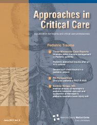
Case Reports<br />
Figure One. The C-MAC videolaryngoscope. The camera is fixed<br />
in the handle, with the lens within the blade. Images are projected<br />
onto a video screen that allows others besides the intubator to<br />
view the anatomy during the intubation procedure. Its usefulness<br />
is demonstrated in a case of difficult boogie placement, seen at<br />
http://www.hqmeded.com/video/40879557<br />
invaginated scolex is clearly seen inside of the cystic cavity.<br />
T = 124 minutes: She began to cough against the<br />
vent and was, therefore, paralyzed with vecuronium.<br />
An ABG revealed a mild acidosis with a pH of 7.19.<br />
Her EKG showed sinus tachycardia with lateral<br />
t wave inversions, concerning for demand ischemia.<br />
T = 164 minutes: The patient was taken to CT<br />
scanner for a CT of the neck with IV contrast to rule<br />
out Ludwig’s angina. Her CT showed a massively<br />
swollen tongue filling the airway and protruding from<br />
the mouth, with significant narrowing of the airway.<br />
There was only mild swelling of the sublingual and<br />
submandibular areas, suggesting that angioedema<br />
was more likely the cause of swelling than a soft<br />
tissue abscess.<br />
T = 184 minutes: She was transferred to the MICU<br />
with a secure airway.<br />
The patient’s benazepril was discontinued. Serial<br />
troponins were followed, given her abnormal ECG<br />
post -intubation and the witnessed episodes of<br />
bigeminy and ventricular tachycardia during<br />
intubation attempts. Her troponin peaked at 0.193<br />
ng/mL. Transthoracic echocardiogram showed a<br />
normal left ventricular ejection fraction, but she was<br />
noted to have an area of hypokinesis in the mid<br />
portion of the intraventricular septum, concerning for<br />
atypical stress cardiomyopathy.<br />
On hospital day 2, the patient had significant<br />
improvement in her tongue swelling. Given her<br />
extremely difficult intubation in the emergency<br />
department, she was extubated over an exchange<br />
catheter with anesthesia at the bedside. She<br />
tolerated extubation well. She was transferred to the<br />
medicine floor and discharged the next day. She was<br />
instructed to discard her amlodipine- benazepril<br />
combination pills at home, and given a new<br />
prescription for amlodipine alone. She was advised to<br />
avoid ACE inhibitors and an allergy alert was placed<br />
in her chart.<br />
Discussion<br />
Two important airway adjuncts were used in the<br />
management of this difficult airway. The C-MAC<br />
videolaryngoscope (Figure One) uses a modified<br />
Macintosh laryngoscope blade with an integrated<br />
video camera, which is directed toward the blade tip.<br />
More details of its functions and an example of its<br />
usefulness are illustrated in a video that shows<br />
difficulty passing a boogie. See http://www.hqmeded.<br />
com/video/40879557. The video screen image allows<br />
the attending physician to visualize the airway as the<br />
resident is intubating (see video). It also allows for<br />
recording of the intubation to facilitate image review<br />
and teaching opportunities. Studies have demonstrated<br />
a greater proportion of successful intubations and a<br />
greater percentage of Cormack-Lehane grade I or II<br />
views when compared with direct laryngoscopy.<br />
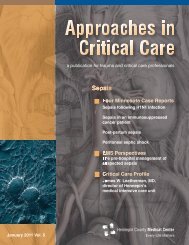
Figure Two. The intubating laryngeal mask airway (ILMA). The<br />
procedure for its placement can be viewed at http://www.hqmeded.<br />
com/video/13164204.<br />
The ILMA (Figure Two) is a supraglottic airway device<br />
that is useful for difficult airway management. The<br />
Approaches in Critical Care | January 2013 | 7