Thoracic Disk Herniation With Paraparesis Treated With ... - Healio
Thoracic Disk Herniation With Paraparesis Treated With ... - Healio
Thoracic Disk Herniation With Paraparesis Treated With ... - Healio

You also want an ePaper? Increase the reach of your titles
YUMPU automatically turns print PDFs into web optimized ePapers that Google loves.
<strong>Thoracic</strong> <strong>Disk</strong> <strong>Herniation</strong> with <strong>Paraparesis</strong> | Ueda et al<br />
A<br />
herniated thoracic intervertebral<br />
disk causing spinal cord compression<br />
with paraparesis is rare<br />
in children. The symptoms of thoracic disk<br />
herniation usually subside spontaneously,<br />
and conservative treatment is sufficient.<br />
Surgical treatment should be reserved for<br />
significant neurologic problems. Methods<br />
of operative treatment for thoracic disk<br />
herniation in children include decompressive<br />
laminectomy, 1-4 posterior diskectomy,<br />
5,6 and laminoplasty. 7 In the current<br />
study, a 14-year-old girl presented with<br />
an intervertebral disk herniation at T5-T6<br />
that was causing paraparesis. To the authors’<br />
knowledge, this is the first report on<br />
this disease that was treated by transthoracic<br />
microdiskectomy without fusion.<br />
Case Report<br />
A previously healthy 14-year-old girl<br />
presented with a 4-month history of diffuse<br />
back pain and sudden-onset paraparesis<br />
after running up stairs. She reported<br />
listlessness and numbness of the bilateral<br />
lower extremities and had lost the ability<br />
to ambulate. She reported no history of<br />
trauma, fever, weight loss, or other constitutional<br />
symptoms, including skeletal<br />
dysplasia. On examination, she had tenderness<br />
on percussion of the spinous processes<br />
of the middle thoracic spine. Motor<br />
strength was 4/5 in both legs. Bilateral<br />
tendon reflexes of the lower extremities<br />
were hyperactive, and knee and ankle clonus<br />
were positive. Hypoesthesia existed at<br />
T6 and below. No bowel or bladder dysfunction<br />
existed. Chemistry, hematology<br />
tests, and urinalysis were normal.<br />
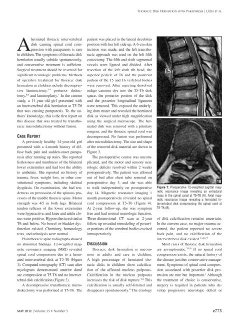
Plain thoracic spine radiographs showed<br />
no abnormal findings. T2-weighted magnetic<br />
resonance imaging (MRI) revealed<br />
spinal cord compression due to a herniated<br />
intervertebral disk at T5-T6 (Figure<br />
1). Computed tomography (CT) scan after<br />
myelogram demonstrated anterior dural<br />
sac compression at T5-T6 and no intervertebral<br />
disk calcification (Figure 2).<br />
A decompressive transthoracic microdiskectomy<br />
was performed at T5-T6. The<br />
patient was placed in the lateral decubitus<br />
position with her left side up. A 6-cm skin<br />
incision was made, and the left transthoracic<br />
approach was used on the left fifth<br />
costectomy. The fifth and sixth segmental<br />
vessels were ligated and divided. After<br />
resection of the left sixth rib head, the<br />
superior pedicle of T6 and the posterior<br />
portion of the T5 and T6 vertebral bodies<br />
were removed. After injecting dissolved<br />
indigo carmine dye into the T5-T6 disk<br />
space, the posterior portion of the disk<br />
and the posterior longitudinal ligament<br />
were removed. This exposed the underlying<br />
dura mater and revealed the herniated<br />
disk as viewed under high magnification<br />
using the surgical microscope. The herniated<br />
disk was removed with a pituitary<br />
rongeur, and the thoracic spinal cord was<br />
decompressed. No fusion was performed<br />
after microdiskectomy. The size and shape<br />
of the removed disk material are shown in<br />
Figure 3.<br />
The postoperative course was uncomplicated,<br />
and the motor and sensory neurologic<br />
deficits resolved within 2 weeks<br />
postoperatively. The patient was allowed<br />
out of bed after chest tube removal on<br />
postoperative day 3, and she was able<br />
to walk independently on postoperative<br />
day 14. Magnetic resonance imaging 1<br />
month postoperatively revealed no spinal<br />
cord compression at T5-T6 (Figure 4).<br />
At 2-year follow-up, she was symptom<br />
free and had normal neurologic function.<br />
Three-dimensional CT scan at 2-year<br />
follow-up revealed remodeling of posterior<br />
portions of the vertebral bodies excised<br />
intraoperatively.<br />
Discussion<br />
<strong>Thoracic</strong> disk herniation is uncommon<br />
in adults and rare in children.<br />
A high percentage of herniated thoracic<br />
disks in children show calcification<br />
of the affected nucleus pulposus.<br />
Calcification in the nucleus pulposus<br />
increases the risk of disk rupture. 2,8 This<br />
calcification is usually self-limited and<br />
disappears spontaneously. 4 The etiology<br />
1A<br />
1B<br />
Figure 1: Preoperative T2-weighted sagittal magnetic<br />
resonance image revealing an extradural<br />
mass in the spinal canal at T5-T6 (A). Axial magnetic<br />
resonance image revealing a herniated intervertebral<br />
disk compressing the spinal cord at<br />
T5-T6 (B).<br />
of disk calcification remains uncertain.<br />
In the current case, no major trauma occurred,<br />
the patient reported no severe<br />
back pain, and no calcification of the<br />
intervertebral disk existed. 1-4,6,7<br />
Most cases of thoracic disk herniation<br />
are asymptomatic. 3,8,9 If no spinal cord<br />
compression exists, the natural history of<br />
the disease justifies conservative management.<br />
Symptoms of spinal cord compression<br />
associated with posterior disk protrusion<br />
are rare but important. 3 Although<br />
the treatment of choice is conservative,<br />
surgery is required in patients who develop<br />
progressive neurologic deficit or<br />
MAY 2012 | Volume 35 • Number 5<br />
e775