Full text-PDF - Histology and Histopathology
Full text-PDF - Histology and Histopathology
Full text-PDF - Histology and Histopathology

Create successful ePaper yourself
Turn your PDF publications into a flip-book with our unique Google optimized e-Paper software.
928<br />
Effects of high protein diet on liver<br />
vertebrates in the absence of food intake (Kraus-<br />
Friedmann, 1984). The usage of this metabolic pathway<br />
increases in HP diets <strong>and</strong> remains constant on lack of<br />
long-term food intake (Silva <strong>and</strong> Migliorini, 1990;<br />
Sartori et al., 1995). HP diets promise that as long as<br />
carbohydrates are restricted, some weight will be lost;<br />
despite no limitation in food consumption. High protein<br />
<strong>and</strong> high lipid diets start metabolic ketosis, <strong>and</strong> these<br />
diets are charming because they lead to rapid loss of<br />
weight. The weight loss in the short term was reported to<br />
be a result of the diuretic effect that was caused by low<br />
carbohydrate intake <strong>and</strong> it was proved that this low<br />
calorie intake might cause lack of appetite as long as the<br />
same diet was kept up (Jama, 1973; Denke, 2001). Some<br />
high protein, very low carbohydrate, weight-loss diets<br />
induce ketosis <strong>and</strong> when carbohydrate intake or<br />
utilization is insufficient to provide glucose to the cells,<br />
ketone bodies that are formed from fatty acids are used<br />
as an energy source. An increase in ketones can disturb<br />
the body’s acid-base balance, causing metabolic acidosis<br />
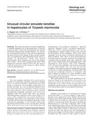
Fig. 5. Light micrograph of the histological view of livers in HP (20).<br />
Observe the minimal quantities of glycogen deposits in the hepotocyte<br />
cytoplasm (a) around the central vein (cv). They correspond to the pinkpurple<br />
stained areas. The nucleus of hepotocyte (b) <strong>and</strong> sinusoids (c).<br />
PAS, x 200<br />
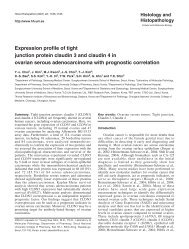
Fig. 6. Light micrograph of the histological view of livers in HP (20).<br />
Observe the similarity of the image as in figure 5. The glycogen deposits<br />
were removed <strong>and</strong> only minimal quantities of certain carbohydrate were<br />
left (a). PAS stain after diastase digestion, x 200<br />
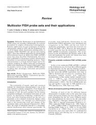
Fig. 7. Light micrograph of the histological view of livers in HP group<br />
(30). Glycogen deposits decreased to a minumum (a), showing a<br />
remarkable increase of regenerative activity of hepotoctes (b),<br />
erythrocytes (c), brownish deposits within hepotocytes (d) <strong>and</strong> cenral<br />
vein (cv). PAS, x 600<br />
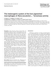
Fig. 8. Light micrograph of the histological view of livers in HP group<br />
(15). Observe the little amount of glycogen deposits (a) around the<br />
central vein (cv). Binucleate liver cells (b), little width sinusoids (c). PAS,<br />
x 400